Anthropological identification and from the I-II Claudius ... · Anthropological identification and...
28
Anthropological identification and paleopathological study of a skeleton, probably pertaining to the Roman doctor from the I-II century b.C., Tiberius Claudius Apollinaris (Tarragona, Spain) DOMINGO CAMPILLO (") FRANCESC TARRATS (""') SUMMARY Aim of the study. Histoncal new. Study of the individual in tomb n.O 2 at «Roben d'Agui- lo» street (Tarragona). Anthropological study. Anthropological summary. Pathology study. Pathologic conclusions. Comparative study between the cranium and the head of the bust. Si- milar studies. Bibliography. I RESUMEN Hemos realizado el estudio antropológico de un esqueleto procedente de una tumba ro- mana del siglo I-II a.c., con la finalidad de constatar si la morfología del cráneo se correspon- día con la de un busto intimamente relacionado con su tumba, que por una serie de circuns- tancias arqueológicas, parecía corresponder a un médico de época romana. Depués del estudio antropométrico, craneográfico y patológico, llegamos a la conclusión de que los restos exhumados eran compatibles con la morfologia del busto, con lo que se rea- firma el criterio arqueológico de que probablemente, se trata del médico tarraconense Tibe- rius Claudius Apollinaris.. - Fecha de aceptación: 15 de mayo de 1991. (*) Professor of the Autonomic University of Barcelona and Head of the Laboratory of Pa- leoanthropology and Paleopathology of the Archeological Museum of Barcelona. (*') Director of the National Archeological Museum of Tarragona. DYNAMIS Acta Hispanica ad Medicinae S~ientiarurnque Historiam Illustrandam. Vol. '1 1, 1991, pp. 387-41.4. ISSN: 021 1-9536
Transcript of Anthropological identification and from the I-II Claudius ... · Anthropological identification and...
Anthropological identification and paleopathological study of a skeleton, probably pertaining to the Roman doctor from the I-II century b.C., Tiberius Claudius Apollinaris (Tarragona, Spain)
DOMINGO CAMPILLO (") FRANCESC TARRATS (""')
SUMMARY
Aim of the study. Histoncal new. Study of the individual in tomb n.O 2 at «Roben d'Agui- lo» street (Tarragona). Anthropological study. Anthropological summary. Pathology study. Pathologic conclusions. Comparative study between the cranium and the head of the bust. Si- milar studies. Bibliography.
I RESUMEN
Hemos realizado el estudio antropológico de un esqueleto procedente de una tumba ro- mana del siglo I-II a.c. , con la finalidad de constatar si la morfología del cráneo se correspon- día con la de un busto intimamente relacionado con su tumba, que por una serie de circuns- tancias arqueológicas, parecía corresponder a un médico de época romana.
Depués del estudio antropométrico, craneográfico y patológico, llegamos a la conclusión de que los restos exhumados eran compatibles con la morfologia del busto, con lo que se rea- firma el criterio arqueológico de que probablemente, se trata del médico tarraconense Tibe- rius Claudius Apollinaris..
-
Fecha de aceptación: 15 de mayo de 1991.
( * ) Professor of the Autonomic University of Barcelona and Head of the Laboratory of Pa- leoanthropology and Paleopathology of the Archeological Museum of Barcelona.
(*') Director of the National Archeological Museum of Tarragona. DYNAMIS Acta Hispanica ad Medicinae S~ientiarurnque Historiam Illustrandam. Vol. '1 1, 1991, pp. 387-41.4. ISSN: 021 1-9536
388 DOMINGO CAMPILLO y FRANCESC TARRATS
AIM OF THE STUDY
In view of the archeolgical findings and of some skeletal remains which appeared to be correlated between each other, some doubts arose as to whether the bust corresponded to the individual mentioned on the funeral plate and to the skeleton deposited inside the sarcophagus. With the aim of attempting to clarify this problem, we proposed an anthropological study of the skeleton, with the aim of verifying whether the craneal morphology and the individuals's constitution were compatible with the morphological cha- racteristics of the person represented in the bust. Our study has been com- pleted with the paleopathological investigation which could also have had a certain influence on the individual's physical aspect.
In view of the results obtained, we consider it of interest to present our study, as there are few similar paleopathological papers in the literature.
HISTORICAL NEWS
In 17 13, Iosep Boy, an engineer from the city of Tarragona, Spain, reco- vered a funeral inscription dedicated to Tib(erius) Cl(audius) Apollinaris artis medicin(a)e doctiss(imus). This gravestone was located by the author of this compilation set into one of the walls of the city recinct, aithough it no longer exists, constituting the first historical testimony of the practice of me- dical science in Tarragona. Later, the archeological excavations which have been carried out in the ancient city of Tarraco, especially since the middle of the XIX century have led to the recuperation of several bronze instruments related with the said profession. But the inscription is still the only persona- lised source of information in relation to this activity.
The site at which Boy located the inscription clearly indicates that the piece was transported to be reused as construction material in the reforms which were introduced into the walled recinct and which came from a ne- cropolis of the roman city, almost without doubt close to the point at which the gravestone was reused.
Thanks to the picture and to the descripion that the engineer Boy lega- ted the chronology of the piece has been determined fairly precisely. Gkza Alfoldy situates it around the I or I1 century b.C. It corresponds to a funeral inscription belonging to the Hight Empire. Present-day knowledge of the roman city of Tarraco leads us to deduce that this gravestone could only
Identification of a skeleton 389
have been extracted from the necropolis existing near the old Via Augusta, relatively close to the section of wall where we believe it was later re-used ac- cording to the data provided by Boy.
In the year 1978, during a series of emergency archeological excavations performed in this sector of the necropolis, as a consequence of the Robert D'Aguiló Street urbanization, amongst other remains a stonc sarcophagus containing human remains accompanied by a rich aray of associated funeral objects composed of: 16 glass ointment flasks, 1 small glass bottle, 1 bone box, 1 flattened marble stone (((coticula))), 1 bronze needle, 1 bronze qua- drangular shaped mirror, remains of a bone comb, bronze frameworks and other elements of the same metal were found, possibly belonging to a woo- den box, 1 bronze coin (dupondi) from the Tarraco mirit, an issue from the era of Tiberio.
Some of the elements of the complex funeral objects (bone box, coticula and the bronze needle especially) appear to be related with a person linked to the practice of medicine.
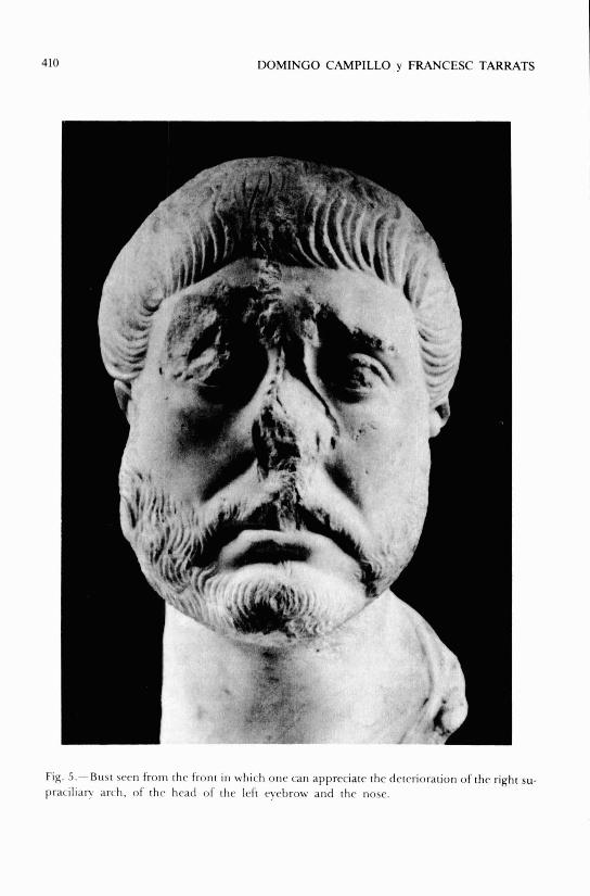
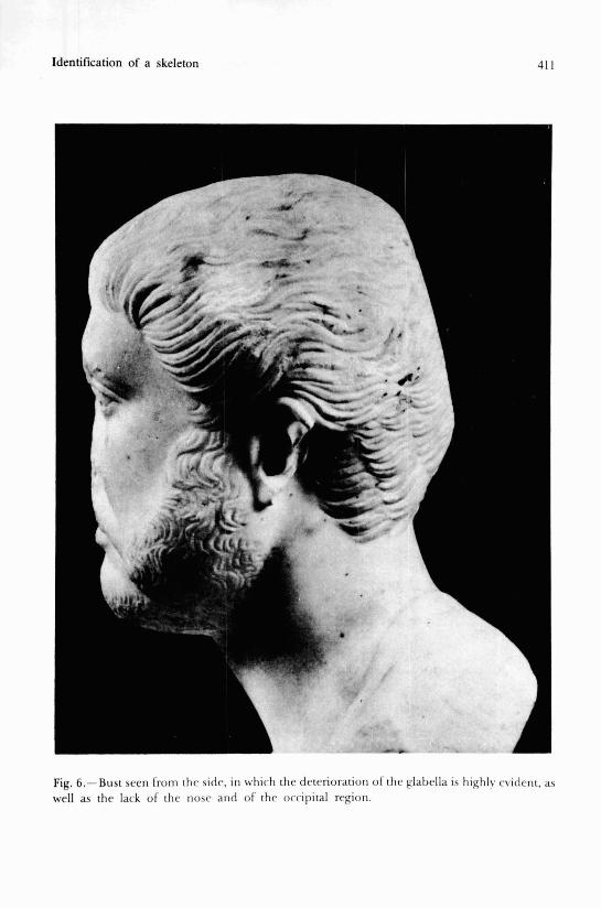
Closed to the interment a marble funeral bust was encountered, repre- senting a person with a beard, dated to the second half of the 1 century b.C. (fig. 5 and 6).
The data revealed lead us to attempt to establish the ihypothesis of a pos- sible connection between the three archeological settings just presented: Ins- cription compiled by Boy, bony remains and funeral objects contained within the sarcophagus, and lastly, the funeral bust.
Topographically no contradictions exist. Chonologi~cally a coincidence exists between the dates attributed to the funeral bust and to the inscription. With regard to the associated funeral objects the only component which could be out of place is the coin, dated without any doubt in the era of Tibe- rio. In our opinion, its presence in the interment implieis no problems as it was probably considered a coin of prestige from a collection no longer in circulation, which does not impede one giving the interrnent a later chrono- logy.
STUDY OF THE INDIVIDUAL IN TOMB Nr.2 AT ((ROBERT D'AGUILO)) STREET (TARRAGONA)
An almost complete, fairly well preserved skeleton was encountered with a conservation index of 59, in which almost al1 the boneis of the hands and
390 DOMINGO CAMPILLO y FRANCESC TARRATS
feet were missing. The ribs have several fractures incurred posthumously and some epiphyses are slightly damaged.
The whole skeleton is grey colured attributable to the earth in which it was deposited which left a thin layer of concretions.
Although the number of preserved bones is very important, the bony structure was weak due to taphonomic processes. In order to favour preser- vation we proceeded to submerge the bones in a 10% solution of ((Paraloid)) in toluene. The cranium, in the right parietotemporal region presented a posthumous fracture with loss of some fragments of bone, as well as other small areas which were dereriorated and required restoration with ((polies- ter» carried out by Mr. Jaume Mayas and assistants.
Anthropological study
(According to Martin's and Saller's technique)
CRANIUM
The complete cranium is in a fairly good state of preservation, with an inferior fragment of the right parietal bone missing, wich includes the pteri- goid region of the occipital and temporal bones with the mastoid proccess of the same side. The left condyle of the mandible is missing as well as al1 the teeth, although some were lost during the individual's lifetime.
ANTHROPOMETRY
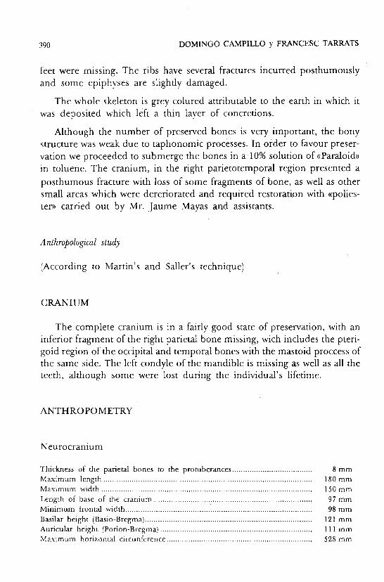
Neurocranium
Thickness of the parietal bones to the protuberances ..................................... Maximum length ............................................................................................... Maximum width .................................................................................................
......................................................................... Length of base of the cranium Minimum frontal width ....................................................................................
............................................................................. Basilar height (Basio-Bregma) ..................................................................... Auricular height (Porion-Bregma)
Maximum horizontal circunference ...................................................................
Identification of a skeleton 391
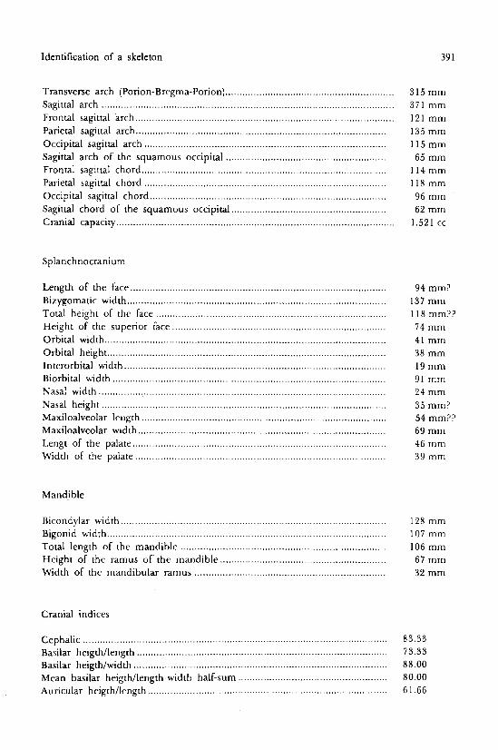
Transvefse arch (Porion-Bregma-Porion) ............................................................ Sagittal arch ........................................................................................................ Frontal sagittal arch ............................................................................................ Parietal sagittal arch ......................................................................................... Occipital sagittal arch ...................................................................................... Sagittal arch of the squamous occipital ......................................................... Frontal sagittal chord ...................................................................................... Parietal sagittal chord ...................................................................................... Occipital sagittal chord .................................................................................... Sagittal chord of the squamous occipital ....................................................... Cranial capacity ...................................................................................................
Splanchnocranium
Length of the face ........................................... ; ............................................... Bizygomatic width ............................................................................................ Total height of the face .................................................................................. Height of the superior face ............................................................................
.................................................................................................... Orbital width ................................................................................................... Orbital height
Interorbital width ............................................................................................. Biorbital width ................................................................................................. Nasal width ......................................................................................................
..................................................................................................... Nasal height Maxiloalveolar length ....................................................................................... Maxiloalveolar width ........................................................................................ Lengt of the palate .......................................................................................... Width of the palate .........................................................................................
315 mm 371 mm 121 mm 135 mm 115 rnm 65 mm
114 mm 118 mm 96 mm '
62 mm 1.521 cc
94 mm? 137 mm 1 18 mm?? 74 mm 41 mm 38 mm 19 mm 91 mm 24 mm 35 mm? 54 mm?? 69 mm 46 mm 39 mm
Mandible
Bicondylar width .............................................................................................. 128 mm Bigonid width ................................................................................................... 107 mm Total length of the mandible ......................................................................... 106 mm Height of the ramus of the mandible ........................................................... 67 mm Width of the mandibular ramus .................................................................... 32 rnm
Cranial indices
Cephalic ................... .. ..................................................................................... 83.33 Basilar heigthllength ......................................................................................... 73.33
.......................................................................................... Basilar heigthlwidth 8 8 .O0 ..................................................... Mean basilar heigthllength-widtb half-sum 80.00
Auricular heigthllength ..................................................................................... 6 1.66
392 DOMINGO CAMPILLO y FRANCESC TARRATS
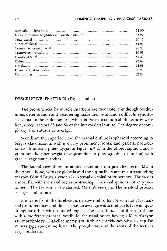
...................................................................................... Auricular heighdwidth Mean auricular heighdlength-width half-sum ................................................. Total facial ........................................................................................................
................................................................................................... Superior facial Transverse craneo-facial .................................................................................... Transverse frontal .............................................................................................
................................................................................................... Fronto-parietal Orbit al ............................................................................................................... Nasal .................................................................................................................. Flower's gnathic index ..................................................................................... Mandibular ........................................................................................................
DESCRIPTIVE FEATURES (Fig. 1 and 2)
The prominences for muscle insertions are moderate, eventhough posthu- mous deterioration and weathering make their evaluation difficult. Synosto- sis is total in the endocranium, whilst in the exocranium al1 the sutures were free, except sectors S3 and S4 of the interparietal suture. The degree of com- plexity the sutures is average.
Seen from the superior view, the cranial outline is spheroid according to Sergi's classification, with not very prominent frontal and parietal protube- rances. Moderate phenozygia (in Figure n.O 2, in the photographic craneo- grammes the phenozygia disappear due to photographic distorsion) with gracile zygomatic arches.
The lateral view shows arounded cranium from just after sector M2 of the frontal bone, with the glabella and the supraciliary arches corresponding to types IV and Broca's grade ((2)) external occipital protuberance. The face is almost flat with the nasal bones protruding. The nasal spine is not very pro- minent. The Pterion is «H» shaped, Martin's «e» type. The mastoid process is large and robust.
From the front, the forehead is narrow (index, 65.33) with not very mar- ked protuberances and the face has an average width (index 86.13) with qua- drangular orbits with rounded angles. The nasal fossa is piriform in shape with a moderate prenasal vestibule, the nasal bones having a Martin's type ((1 » morphology. Glabellar metopism. Robust cheekbones with a deep De Villiers type ((3)) canine fossa. The protuberance at the roots of the teeth is very moderate.
Identification of a skeleton 393
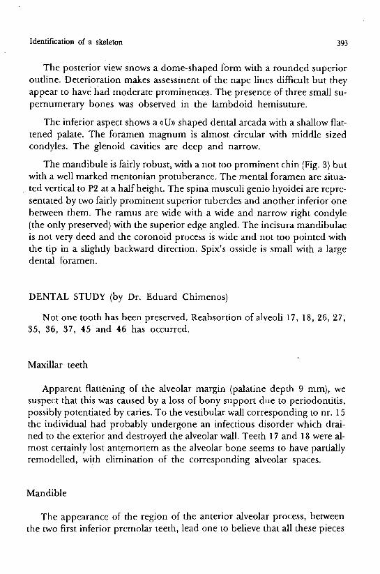
The posterior view snows a dome-shaped form with a rounded superior outline. Deterioration makes assessment of the nape lines difficult but they appear to have had moderate prominences. The presence of three small su- pernumerary bones was observed in the lambdoid hemisuture.
The inferior aspect shows a «U» shaped dental arcada with a shallow flat- tened palate. The foramen magnum is almost circular with middle sized condyles. The glenoid cavities are deep and narrow.
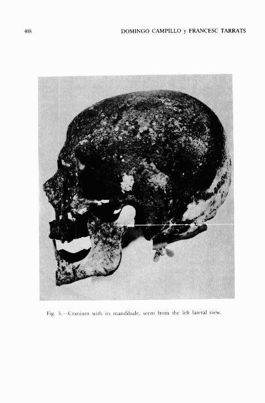
The mandibule is fairly robust, with a not tso prominent chin (Fig. 3) but with a well marked mentonian protuberance. The mental foramen are situa- ted vertical to P2 at a half height. The spina musculi genio hyoidei are repre- sentated by two fairly prominent superior tubercles and another inferior one between them. The ramus are wide with a wide and narrow right condyle (the only preserved) with the superior edge angled. The incisura mandibulae is not very deed and the coronoid process is wide and not too pointed with the tip in a slightly backward direction. Spix's ossicle is small with a large dental foramen.
DENTAL STUDY (by Dr. Eduard Chimenos)
Not one tooth has been preserved. Reabsortion of alveoli 17, 18, 26, 27, 35, 36, 37, 45 and 46 has occurred.
Maxillar teeth
Apparent flattening of the alveolar margin (palatine depth 9 mm), we suspect that this was caused by a loss of bony support due to periodontitis, possibly potentiated by caries. To the vestibular wall corresponding to nr. 15 the individual had probably undergone an infectious disorder which drai- ned to the exterior and destroyed the alveolar wall. Tee'th 17 and 18 were al- most certainly lost antemortem as the alveolar bone seems to have partially remodelled, with elimination of the corresponding alveolar spaces.
Mandible
The appearance of the region of the anterior alveolar process, between the two first inferior premolar teeth, lead one to believe that al1 these pieces
394 DOMINGO CAMPILLO y FRANCESC TARRATS
remained in situ until the individual's death, eventhough they must have been affected by a moderate periodontal disease (periodontitis) with a slight loss of bony support.
On the right side, teeth 45 and 46 were probably lost long before death, the alveolar bone having completely regenerated. On the left, teeth 35, 36 and 37 were lost antemortem, the alveolar bone also completely regenera- ting.
POSTCRANIAL SKELETON
No remarkable features of an anthropological nature can be described in the postcranial skeleton, as the alterations observed are anomalies and pat- hological alterations wich will be studied later. The study is completed by anthropological data.
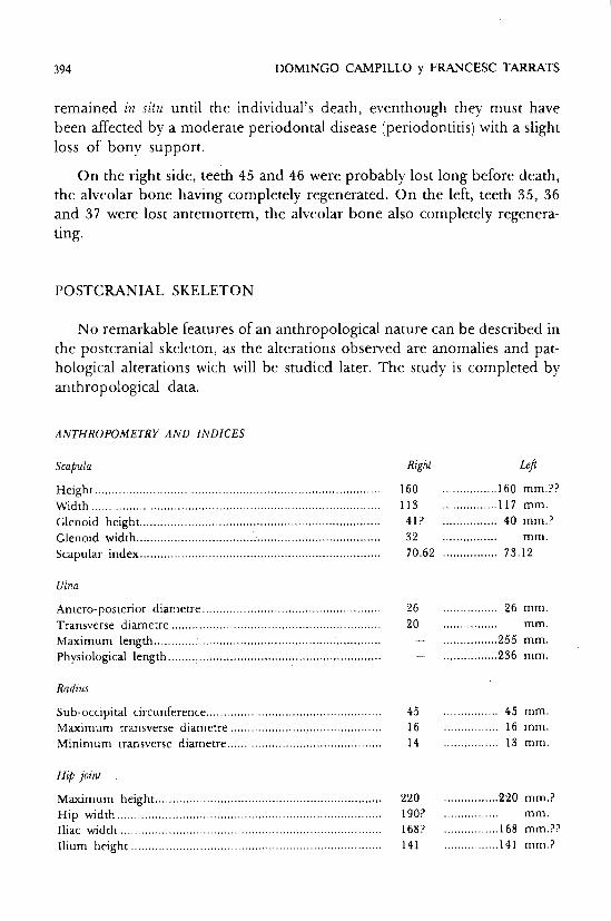
ANTHROPOMETRY AND INDICES
Scapula Righi L.ft ................ Height ................................................................................... 160 160 mm.??
Width .................................................................................... 113 ................ 1 1 7 m m . Glenoid height ...................................................................... 41? ................ 40 mm.?
................ Glenoidwidth ....................................................................... 32 mm. Scapular index ...................................................................... 70.62 ................ 75.12
Ulna
Antero-posterior dian~etre ............................................. 26 ................ 26 mm. ................ Transverse diametre . . . . . . . . . . . . . . . . . . . . . . . . . . . . . . . . . . . . . . . . . . . . . . . . . . . . . . . . . . . . . 20 mm. ................ Maximum length ..................... .. ......................................... - 255 mm.
Physiological length .............................................................. - ................ 236 mm.
Radius
Sub-occipital circunference .................................................. 45 ................ 45 mm. ..................... Maximum transverse diametre 16 ................ 16 mm.
Minimum transverse diametre ......................................... 14 ................ 13 mm.
Maximum height .................................................................. 220 ................ 220 mm.? ............................................................................. Hip width 190? ................ mm.
Iliac width ............................................................................ 168? ................ 168 mm.?? Ilium height ......................................................................... 141 ................ 141 mm.?
Identification of a skeleton 395
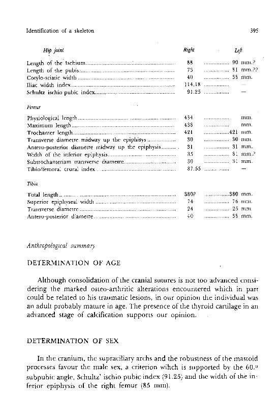
Hip joint Right b!
................ Length of the Ischium ......................................................... 88 90 mm.?
................ Length of the pubis ............................................................. 73 81 mm.??
................ Cotylo-sciatic width .............................................................. 40 38 mm. Iliac width index .................................................................. 1 14.18 ................ -
................ Schultz ischio-pubic index .............................................. 9 1.25 -
Physiological length .............................................................. Maximum length .................................................................. Trochanter length ................................................................. Transverse diametre midway up the epiphisys .................. Antero-posterior diametre midway up the epiphysis ......... Width of the inferior epiphysis ........................................... Subtrochanteriam transverse diametre .................................
.................................................. Tibiolfemoral crural index
4,34 ................ mm. ................ 438 mm. ................ 421 421 mm. ................ 30 30 mm. ................ 31 31 mm. ................ 135 81 mm.? ................ 30 31 mm.
13 7.55 ................ -
Tibia
................ Total length ......................................................................... 38O? 380 mm.
................ Superior epiph~seal width .................................................. 74 76 mm.
................ Transverse diametre ............................................................. 24 25 mm.
................ Antero-posterior diametre .................................................... 410 38 mm.
Anthropological summary
DETERMINATION OF AGE
Although consolidation of the cranial sutures is not too advanced consi- dering the marked osteo-arthritic alterations encountered which in part could be related to his traumatic lesions, in our opinion the individual was an adult probably mature in age. The presence of the thyroid cartilage in an advanced stage of calcification supports our opinion.
DETERMINATION OF §EX
In the cranium, the supraciliary archs and the robiistness of the mastoid processes favour the male sex, a criterion wihch is supported by the 60.O subpubic angle, Schultz' ischio pubic index (91.25) and the width of the in- ferior epiphysis of the right femur (85 m m ) .
396 DOMINGO CAMPILLO y FRANCESC TARRATS
DETERMINATION OF HEIGTH
In order to determinate this individual's height we have used Manouv- rier's and Trotter-Gleser's tables applied to the femurs and to the femur- tibia and have reached the conclusion that the height ranged from 165-169 cm.
TYPOLOGY
The morphology of the' spheroidal cranium, wide, globulous brachicra- nium (index 83.33) with patinocranium (index 88.00), the squamous occipi- tal bone curved with a rounded occiput, the glabella and the supraciliary archs are moderately prominent, the face is fairly wide with a wide mandible (index 82.81). The face is flet with prominent cheek-bones, the nasal bones are straight, the orbits quadrangular and the chin is small.
Likewise, the corporal height is average with wide shoulders and short limbs, features compatible with the alpine typology (bra~hice~halous curved occipital bones) according to Ferembach's classification.
Pathology study
As the anomalies and pathological alterations are numerous, an anato- mical order will be followed to describe them, ending the discussion with an interelation between same which allow us to reach aethiogenic conclusions.
CRANIUM
We only find dental pathology inclued within the anthropologicai study.
SPINE AND THORAX
Almost al1 the spine is preserved. The axis and vertebral badies of C-3, T-9, T-10 and L-1 are missing.
The signs of osteoarthritis are marked along the whole of the spine.
Identification of a skeleton 397
At cervical level an osteophytic reaction exists ori the articular surface of the anterior arch of the atlas at odontoid peg of the axis. Al1 the joints show exostosis and the bodies are deformed, with an osteophytic edge reaction (grade ((3)) according to Stewart's classification), mainly affecting the last four vertebrae of the sector.
In the thoracic spine the osteoarthitic signs ar~e moderate in the first three vertebrae (grade « 1 »). In the section involving T-4 to T-8 the alterations are intense, with large ((parrot-beaks)) (grade ((4))) plus synostosis of the bo- dies of T-5, T-6, and T-7, that of T-4 with T-5 and T-7 with T-8 are about to join. Unfortunately the bodies of T-9 and T-10 have been lost. In T-1 1 the osteoarthritic reaction is moderate (grade ((ID), but ithe upper edge of T-12 has grade ((3)) alterations with a Schmorl's hernia which reaches the vertebral foramen, the vertebral body presenting a cuneiform imorphology with a pos- terior base when it is observed laterally. The alterations of T-12 mentioned lead one to believe that a traumatic lesion of the vertebra ocurred due to hy- perflexion, probably with extrusion of the nucleus pulposus to the interior of the spinal canal. A herniated disc at this level is not very frequent, but it is not exceptional. Unfortunately, the body of h-1 has been lost and does not allow for a more precise evaluation.
The lumbar spine has only four vertebrae, as L-5 is sacralized. The body of L-1 as,we have already menationed, has been losit and L-2, L-3, and L-4 show moderate (grade ((2))) signs of osteoarthritis.
The sacrum is formed by six totally synostosed vertebrae. The signs of osteoarthritis at the basand at the small joints are very moderate. On the posterior face the foramina sacralis dorsalis are large and deep. The articular facets of the hip bones show a marked edge (the left one is almost destro-
yed).
There are no important lateral flattenings of the vertebral bodies to make one suspect a scoliosis.
Although there are many fractures Post mortem, the majority of the ribs are preserved most of which show signs of osteoarthitis on the articular facet at the transverse process and the head of the rib. Two fragments of rib which could be fourth and a seventh one, show consolidateid fractures with a bone callus.
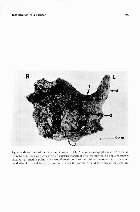
Of the sternum only the manubrium is preserved which is rather deterio- rated but which presents a typical sternocondral Srb ianomaly (1862) (Fig. 4)
398 DOMINGO CAMPILLO y FRANCESC TARRATS
and which according to Kohler and Zimmer (1 959) consists of a ((regression of the first or of the first two ribs due to partid fusion of both in small bony horn)). The articular facet on the internal epiphysis of the left clavicle shows marked signs of osteoarthritis (the external epiphysis is damaged).
A hyoid horn is also preserved and the thyroid cartilage partially calci- fied.
UPPER LIMBS
Of the clavicles the left is the only one preserved, with both epiphyses very much deteriorated, presenting in the middle portion a fairly large bone callus from a fracture which has consolidated well with no desviation and which implies prolonged survival after the trauma.
The scapulas present osteoarthritic edges at the glenoid cavities. The hu- merus and the ulna show signs of osteoarthritis at the epiphysis, somewhat more marked at the left ulna. The radii cannot be assessed due to posthu- mous deterioration and no alterations have been observed in the few remai- ning bones of the hands.
LOWER LIMBS
Hip bones
The subpubic angle has been calculated as being 60.0 at it is difficult to measure due to deterioration of the symphysis facets, which also makes age assessment difficult.
The morphology of the right hip (the left is more deteriorated and defor- med) is male and this is supported by the width indices of the iliac wing and of Schultz's ischiopubic index. As pathologic alterations, moderate signs of osteoarthritis can be observed at the cotyloid cell, at the descending ramus of the pubis and at the two rami of the ischium especially at the point of in- sertion of the gemelli pelvis at the tubercle of the ischium.
The left hip presents a severe traumatic lesion which, according to Judet and Letournel's classification (1974) would correspond to cthe anterior spine)), without a fracture of the middle ischiopubic ramus, associated with a fracture of the iliac wind.
Identification of a skeleton 399
DESCRIPTION OF THE FRACTURE OF THE LEFT HIP
Unfortunately, the iliac crest and the region of the articular facet are da- maged and an accurate assessment cannot be made.
On the external face, a well consolidated fracture juts out from the supe- rior angle of the obturator foramen, surronds the condyloid cavity and pe- netrates through the angle of the greater sciatic notch. In the depths of the acetabulum a diastasis of the margins of fracture exists with a cicatrical exos- tosic reaction. Outside the cotyloid cavity, to the exteri~al part and extending itself up to the angle of the said sciatic notch, the prcrsence of an exostosic pyramid which contacts the greater trochanter can be observed. Another fracture emerges from the external edge of the acetabiulum and rises until it reaches the middle of the two antero and postero-superior iliac spines. Cica- trization is defective, a diastasis has formed as newly formed bone bridges.
On the internal face, the fracture follows the innocninate crest and innu- merable newly formed vascular orifices or possibly the result of a cured su- puration can be seen.
As a result of these lesions, the cotyloid cavlty has cleepened and the iliac wing has undergone a downward deviation, adopting a more horizontal po- sition.
The deformity of the left half of the pelvis is evident and undoubtedly it bears a relationship to a fracture of the left femur which will be ,commented on below.
The osteoarthritic exostoses are more evident at the pubis and ischium on the right side.
FEMURS
in both femurs, the linea aspera is highly marked. In the right femur a moderare exostosis can be observed at the superior rnargin of the greater trochanter and to vertex of the trochanter minor and al crest at both condy- les.
The left femur lacks almost al1 the neck and the head of the femur, it not being at al1 easy to give an opinion on a probable post-traumatic necrosis of
400 DOMINGO CAMPILLO y FRANCESC TARRATS
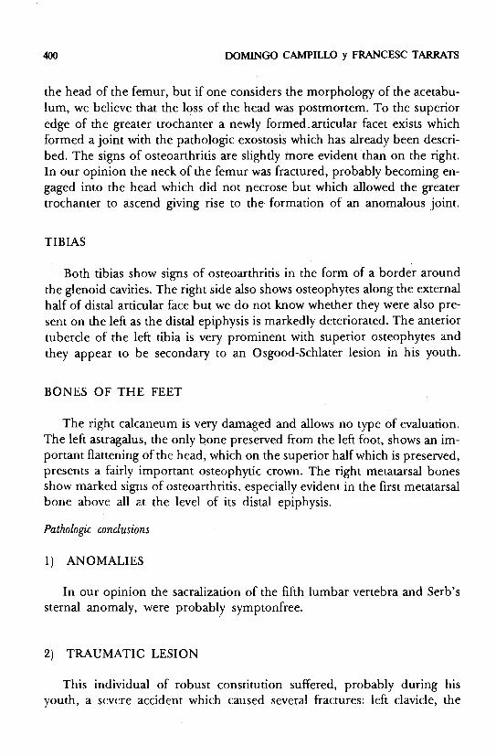
the head of the femur, but if one considers the morphology of the acetabu- lum, we believe that the loss of the head was postmortem. To the superior edge of the greater trochanter a newly formed-articular facet exists which formed a joint with the pathologic exostosis which has already been descri- bed. The signs of osteoarthritis are slightly more evident than on the right. In our opinion the neck of the femur was fractured, probably becoming en- gaged into the head which did not necrose but which allowed the greater trochanter to ascend giving rise to the formation of an anomalous joint.
TIBIAS
Both tibias show signs of osteoarthritis in the form of a border around the glenoid cavities. The right side also shows osteophytes along the external half of distal articular face but we do not know whether they were also pre- sent on the left as the distal epiphysis is markedly deteriorated. The anterior tubercle of the left tibia is very prominent with superior osteophytes and they appear to be secondary to an Osgood-Schlater lesion in his youth.
BONES OF THE FEET
The right calcaneum is very darnaged and allows no type of evaluation. The left astragalus, the only bone preserved from the left foot, shows an im- portant flattening of the head, which on the superior half which is preserved, presents a fairly important osteophytic crown. The right níetatarsal bones show marked signs of osteoarthritis, especially evident in the first metatarsal bone above al1 at the leve1 of its distal epiphysis.
Pathologic conclusions
1) ANOMALIES
In our opinion the sacralization of the fifth lumbar vertebra and Serb's sternal anomaly, were probably symptonfree.
2) TRAUMATIC LESION
This individual of robust constitution suffered, probably during his youth, a severe accident which caused severa1 fractures: left clavicle, the
Identification of a skeleton 40 1
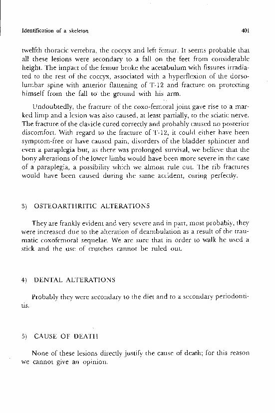
twelfth thoracic vertebra, the coccyx and left femur. It seems probable that al1 these lesions were secondary to a fa11 on the feet from considerable height. The impact of the femur broke the acetabulum with fissures irradia- ted to the rest of the coccyx, associated with a hyperflexion of the dorso- lumbar spine with anterior flattening of T-12 and fracture on protecting himself from the fdl to the ground with his arm.
Undoubtedly, the fracture of the coxo-femoral joint gave rise to a mar- ked limp and a lesion was dso caused, at least partialky, to the sciatic nerve. The fracture of the clavicle cured correctly and probablly caused no posterior discomfort. With regard to the fracture of T-12, it could either have been symptom-free or have caused pain, disorders of the bladder sphincter and even a paraplegia but, as there was prolonged survival, we believe that the bony alterations of the lower limbs would have been more severe in the case of a paraplegia, a possibility which we almost rule out. The rib fractures would have been caused during the same accident, curing perfectly.
3) OSTEOARTHRITIC ALTERATIONS
They are frankly evident and very severe and in part., most probably, they were increased due to the alteration of dearnbulation as a result of the trau- matic coxdfemorai sequelae. We are sure that in ordcr to walk he used a stick and the use of crutches cannot be ruled out.
4) DENTAL ALTERATIONS
Probably they were secondary to the diet and to a secondary periodonti- tis.
5 ) CAUSE OF DEATH
None of these lesions directly justifj the cause of death; for this reason we cannot give an opinion.
402 DOMINGO CAMPILLO y FRANCESC TARRATS
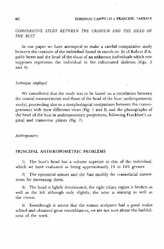
COMPARATIVE STUDY BETWEEN THE CRANIUM AND THE HEAD OF THE BUST
In our paper we have attempted to make a careful comparative study between the cranium of the individual found in «tomb nr. 2)) of Robert d'A- guiló Street and the head of the «bust of an unknown individual)) which one supposes represents the individual in the exhumated skeleton (Figs. 5 and 6).
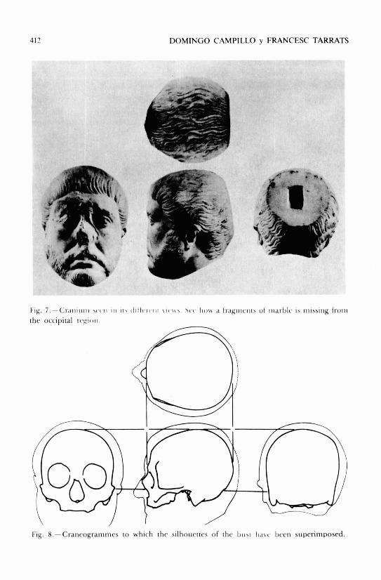
Technique employed
We considered that the study was to be based on a correlation between the cranial measurements and those of the head of the bust (anthropometric study), proceeding also to a morphological comparison between the craneo- grarnmes with their differents views (Fig. 1 and 2) and the photographs of the head of the bust in anthropometric projections, following Frankfurt's sa- gittal and transverse planes (Fig. 7).
PRINCIPAL ANTHROPOMETRIC PROBLEMS
1 ) The bust's head has a volume superior to that of the individual, which we have evaluated as being approximately 13 to 14% greater.
2) The epicranial tisSues and the hair modifi the craniofacial dimen- sions by increasing them.
3) The head is lightly deteriorated, the right ciliary region is broken as well as the left although only slightly, the nose is missing as well as the crown.
4) Eventhough it seems that the roman sculptors had a good realist school and obtained great resemblances, we are not sure about the faithful- ness of the work.
1 Identikation of a okeleton 403
CRITERION TO RESOLVE THE ANTHROPOMETRIC PROBLEMS
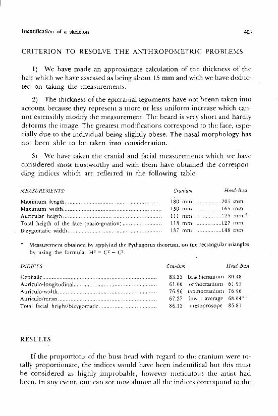
1) We have made an approximate calculation of the thickness of the hair which we have assessed as being about 15 mm and wich we have deduc- ted on taking the measurements.
2) The thickness of the epicranial teguments have not beenn taken into account because they represent a more or less uniform increase which can- not ostensibly modify the rneasurement. The beard is ,very short and hardly deforms the image. The greatest modifications corresp~ond to the face, espe- cially due to the individual being slightly obese. The nasal morphology has not been able to be taken into consideration.
3) We have taken the cranial and facial measurernents which we have considered most trustworthy and with them have obtained the correspon- ding indices which are reflected in the following table.
MEASUREMENTS: Cranium Head-Bust
Maximum length .............................................................. 180 mm. ................ 205 mm. Maximum width .......................................................... 150 mrn. ................ 165 mm. Auricular heigth ............................................................... 1 1 1 mrn. ................ 125 mm.' Total heigth of the face (nasio-gnation) ......................... 118 mm. ................ 127 mm.
............................................................ Bizygomatic width 137 mrn. ................ 148 mm.
* ~ e a s u r e m e n t obtained by applyind the Pythagorus theorum, on the rectangular triangles, by using the formula: H2 = C2 + C2.
INDICES: Cranium Head-Bust
Cephalic ....................................................................... 83.33 brachicranium 80.48 Aunculo-longitudinal ................................................... 6 1.66 orthocranium 6 1.95
............................................................... Auriculo-width 76.96 tapinocranium 76.96 Aunculo/mean .............................................................. 67.27 low i average 68.64*" Total facial height/bizygomatic .................................... 86.13 inesoprosope 85.8 1
RESULTS
If the proportions of the bust head with regard to the cranium were to- tally proportionate, the indices would have been indentifical but this must be considered as highly improbable, however meticulous the artist had been. In any event, one can see now almost al1 the indices correspond'to the
404 DOMINGO CAMPILLO y FRANCESC TARRATS
same typology, except the «aricular)mean width+length» index is ((average)) (68.64**), but is almost at the highest leve1 of the low results (68.00). In con- clusion, we consider that from the anthropometric point of view the cra- nium and the head are compatible.
PRINCIPAL PROBLEMS
1) The head of the bust is in an oblique position leaning towards the left and with a slight rotation towards the sarne side. This position means that Frankfurt's plane is not paralell to the ground and for this reason it be- comes difficult to centre the photographic camera.
2) determination of Frankfurt's plane is complex as the left infraorbi- tary point and both porions are difficult to determine. The porions do not coincide exactly with the superior part of the external auditive canal. The in-
congruente between Frankfurt's true plane and that which we have calcula- ted approximately, affects the anthropological c(views» specially the antero- posterior one.
3) Photographic distorsion rnakes the photographic craneagrammes not cornpletely superimposable with those drawn with the craneograph and with Martin's craneophor (Figs. 1 and 2). Although the photographs of the head of the bust have al1 been taken from the same distance, the focusing point is rather subjective, especially in the frontal view and small variations in the distance modify the distorsion.
TECHIQUE EMPLOYED
We have superimposed al1 the cranial views with those of the head of the bust, taking into account the approximate thickness of the teguments and of the hair and altogether we belive that they match correctly (Figs. 8 and 9) with the exception of the forehead, more affected by the non-correction of Frankfurt's plane and the photographic distorsion.
RESULTS
Even when taking into account the possible anomalies of any artistic re- production, we considerer that the craneogrammes of individual nr. 2 of
Identification of a skeleton 405
((Robert d'Aguiló Street)) are compatible with the morphologhy of the sculp- tured head which it is supposed belongs to it.
l
FINAL CONCLUSIONS
1) The morphology of the cranium is totally comipatible with the head of the sculpture.
2) The pyknik aspect is also compatible with its anthropological mor- phology.
3) The obesity could have been influenced by the difficulties for dearn- bulation, by the post-traumatic limp which would have forced him to a cer- tain degree of sedentariness.
4) The age is difficult to assess. The aspect of the head of the bust seems to be that of an individual slightly younger but, apart from the possible mo- difications made by the artist, the osteoarthritic alterations lead one to belie- ve he was a mature man, but they may also have been influenced indirectely by the severe fracture of the left lower limb. The possibility exists that the bust was made during a period well before death. Lastlly, we must emphasi- ze that we consider the skeleton to be betwen 45 and 55 years of age.
SIMILAR STUDIES
Exhaustive research of the literature has not been carried out but some papers have been found published by Genna (1958), Bartucz (1966) and Lengyel (1976), in which the skeletal remains of several known persons have been studied which are compared with their artistic representations, pain- tings and sculptures.
FINAL COMMENTS
On terminating this paper we consider it necessary to insist on the fact that it is not possible to reach more than a simple hypolthesis, starting from an unquestionable fact. Tiberius Claudius Apollinaris is, up to the present, the first known doctor from Tarragona.
406 DOMINGO CAMPILLO y FRANCESC TARRATS
ACKNOWLEDGEMENTS
This paper would not have been possible without the meticulous colla- boration of the Photographers Mr. Oriol Clavel1 and Julia Martínez. 1 would also like to thank Mrs. Antonia Grau for help with the drawings and also the restoration of the cranium carried out by Mr. Jaume Mayas and his associa- tes. For other concepts 1 would like to express my gratitude to Mr. Joseph Barberá.
Identification of a skeleton
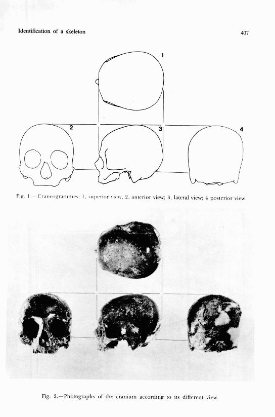
F . 1 . - ~r; l i l<~O~~; l l l1111l~\ : 1 . \iil)carior viih\v. 2. aritrrior view; 3, lateral view; 4 posterior vicw.
Fig. 2.-Photographs of the cranium according to its diffcrent view.
DOMINGO CAMPILLO y FRANCESC TARRATS
Fig. 3.-Critiiitiiii witli i t 5 iiiaridibulr, scciii from thc lrfi latcral vicw.
Identification of a skeleton 409
Fig. 4.-Manubriuni ot ilic srcriiurii. R, riglii; L, Iclt. A, aiioiiinloii?. apopliyhib wiili Sib'a iiial- formation. 1 , line along which thc left cxtcrnal margin of thc stcrntim would bc approximatelv situated; 2, junction point which would correspond to the midlino bctween the first and sc- cond nbs; S, ossificd fasciclc of union betwern thc second rib and thc bodv of the sternum.
410 DOMINGO CAMPILLO y FRANCESC TARRATS
Fig. 5.-Bust secn froiii thc fiont ir1 which onc can apprcciaic thc dctcrioration of thc right su- praciliary arch, of thr head of the lcft evebrow and thc nose.
Identification of a skeleton 41 1
Fig. 6.-Bust seen from thc sidc, in which thc deterioration of thc p;labclla is highly rvident, as well as the lack of thc nose and of thc occipital rcgion.
417 DOMINGO CAMPILLO y FRANCESC TARRATS
Fig. 7.-Craiiiiiiii \ ( . ( , I I I I I 1 1 , ~ 1 1 1 1 i ~ t ~ ~ i 1 1 \ I W \ . \ ( . c . Iio\\. a Iragiiiciii!, o1 iiiai.b1i i b iiiissiiig I r o i i i
the occipital rc>pioii.
Fig. 8.-Cranrogrammes to which the silhouettes of the I>iirc Ii,i\c. been superirnposed.
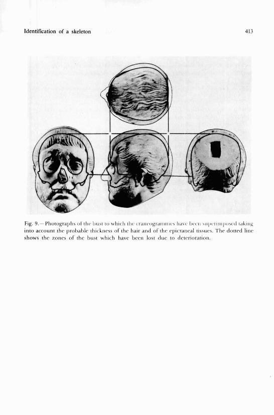
Identification of a skeleton 413
Fig. 9.-Photograplis ot rlic hrisi to wtiicli ilic ci.aiicogi.aiiitiic,\ lidte I)ccii \ L I I K ~ I ~ I I I I ~ ) ~ ~ ~ ~ ~ r.il\iii# into account the probablr thickncss of the hair and of thr epicrancal tissucs. The dotted linc shows the zones of the bust which have been lost due to deterioration.
414 DOMINGO CAMPILLO y FRANCESC TARRATS
RARTUCZ, L. ( 1 966). Palaropatho10,qia I I I . Budapest, Ed. Franklin-nvomda, 61 2 pp. BROCA, P. ( 1 785). Instructions craniologiques ct craniornétriqucs. MPm. Sor. d'Anthrop. dr Paris,
63, 1- 103. GENNA, G. (1958). Lorenzo il Magnifico e il suo fratclo Giuliano. «Esirato dai Rendironti drll'Ac-
cndrmia Narionale dri X L ( X 0 . O dalla fondazione»). Seri IV(VIII ) : 1-95 + XLV T. JUDET, R. ct LETOURNEL, E. (1974): LPS jact11rrs d~ cotvle. Paris. Ed, Masson, 178 pp. KOHLER, A. y ZIMMER, E. A. (1959): Rorr11,qenoloqia. Barcelona, Ed. Labor, 708 pp. LENGYEL, 1. (1976). Laboraro- cxarnination oí' skclctal remains froni J. C. Lavatrr's grave.
G P S ~ P T P I L S , ?9:381- 384. L ~ P E Z - P I Ñ E R O , J. M. (Dir.) (1988): Historia dr la Mrdiria Valrnciaria, I. Valencia, Ed. Viccnt
Garcia, 162 pp. L ~ P E Z TERRADA, M. L. (1988): La medicina en la Valcncia romana. En Hisl. Mrd. Valrririarra,
dir. por J . M. Lópcz- Piñero. 1: 59-61. MANDUVRIER, L. (1893): La détcrmination de la taillr d'aprbs les grands rnernbres. Mcm. Soc.
d'Anthrop. (ir Paris, 4:347-402. MARTIN, R. urid SALLER, K. (1957): Lrhrblich drr Anlliropolo,qir, I I I . Stutgart, Ed. Custav Fis-
chcr, 634 pp. OLIVIER, C. (1960): Pralrqiir Arilhropoloffque. Paris, Ed. Vigot, 299 pp. PIULACHS, P. (1958): L~rcrori~r dr patología qz~ini;fica, II (1.O port.). Barcelona, Ed. Torav,
935 111'.
SCHMORL, G. (1932): Uber Vcrlagerung von Randschcilbcngcwcbr iiñnd ihrc Folgcn. Arrh. Klin. Chir., 1 72:34-72
SCHULTZ, A. H. (1949): Scx diffrcnccs in thc pelvis of ~ r ima tcs . A7n. J. ofPhvs. Anthrop., Z:401- 423.
SRB, (1962): M P ~ . Jh. K.K. GPS. d. A~rz t$-V?fna. STEWARD, T. D. (1958): Thcratc of dcvclopmcnt of vertebral ostco-arthritis in Amcrica Whi-
tes and its signiticaricc in skclctal agc idcntification. Johanncsburgh, Thr Le~ch, 2X:144- 151.
7 I R R A C 0 , Ohjrclr i I,nal,q~. Tarragona, Ed. Cencralitat de Catalunva i Diputació de Tarragona, 144 pp.
TROTTER, M. and GLESER, G: C. (1958): A re-cvaluation of cstimation of staturc bascd on mrasurcrncnts of staturc takcn during lifc arid of long boncs after dcath. Am. J. of Phvs. Anlhrop., 16:79-124.
VILLIERS, H . (1968): T ~ P Skt~l l o/ lhr Soltlh Afiican Nr,qo. Johanncsburgh, Ed. Witwatcrsrand Univ. Prcss.. 159.