Organic Marketing - An Evolving Landscape (Michael Fleischner)

Fleischner Society
Society for Thoracic Imaging and Diagnosis
June 14 -17, 2017
Beverly, Massachusetts
Annual Meeting 48th
2017 2017
Fleischner Society June 14-17, 2017 48th AnnualMeeting Beverly, Massachusetts
Table of Contents
Page
Welcome Letter from the President Alexander Bankier, MD 1
Welcome Letter from the Academic Development Committee Chair 3 Ji n Mo Goo, MD, & Scientific Program Committee Chair, Jeffrey D. Galvin, MD
Members in Attendance 5
Fleischner Society Presidents 1970 – 2017 7
Corporate Sponsors 9
Schedule at a Glance 11
Full Program 13
THURSDAY, JUNE 15, 2017 Session 1: New Insights in Interstitial Lung Disease 21 Session 2: Diffuse Lung Parenchymal Disease 23Session 3: Interstitial Lung Disease and Fibrosis 25 Session 4: The Fleischner Lecture 30 Session 5: Lung Microstructures 32 Session 6: Chronic Obstructive Lung Disease 37 Session 7: Physiology and Pathophysiology of Lung Disease 40 Session 8: Pulmonary Embolism and Pulmonary Hypertension 45 Session 9: Infections 51
FRIDAY, JUNE 16, 2017 Session 10: Big Data and Deep Learning 57 Session 11: New Approaches in Lung Cancer 61 Session 12: Lung Nodule Management 65 Session 13: Assessment of Lung Cancer and Lung Nodule 70 Session 14: Various Approaches 75 Session 15: Mediastinum and Pluera 78 Session 16: Biopsy and Localization of Lung Nodules 82 Session 17: Lung Adenocarinoma and Genomics 85

Fleischner Society June 14-17, 2017 48th AnnualMeeting Beverly, Massachusetts
Dear Fleischner Society Members:
I am very pleased to welcome you to the Wylie Inn and Conference Center, Beverly, for the 48th annual meeting of our Society. The meeting this year is again remarkable for the many excellent abstracts submitted for scientific presentation. This year’s program will feature high level scientific presentations by Active members and Members‐Elect, as well as updates on our recent white papers. A highlight of the meeting will be the annual Felix Fleischner Lecture delivered by Dr. Massimo Pistolesi.
We extend an especially warm welcome to our industry representatives, who will attend our many presentations and participate in discussions and networking opportunities. For the second time, we are introducing roundtable sessions for the Platinum and Gold sponsors. We hope these in depth scientific discussions on topics of mutual interest can lead to improvements in diagnostic methods and therapies in thoracic disease, and stimulate new research in our field.
On Thursday evening, our Presidential Dinner will be held at the Willowdale Estate and we welcome our colleagues from industry to join us for this special event. On Saturday, a post Meeting tour will take place for Fleischner members only. This tour will bring us to the newly re‐designed Harvard Art Museums and to the Harvard Faculty Club, where our lunch will be enhanced with a presentation given by Dr. Herbert Y. Kressel, Editor‐in‐Chief of the journal RADIOLOGY.
I would like to extend my thanks to the members of the Executive Committee: David Lynch, Charles Powell, Bill Travis and Giichi Inoue, for their advice and guidance during this year. I would also like to thank the members of the Scientific Meeting Committee, especially the chair Jin Mo Goo, who, together with Ann NC Leung, Talissa Altes, and Jeffrey Kline, has done an incredible amount of work to put together the best possible program. In addition, my thanks go to the Membership subcommittee with its chair, Cornelia Schaefer‐Prokop, who was supported by Connie Hsia, Yoshiharu Ohno, and Charles White. I am also deeply indebted to Luca Richeldi for his valuable contributions as Chair of the ad‐hoc Development Committee. Finally, my thanks go to Larry Goodman, who has successfully instilled new spirits into our initiative to more actively involve our Senior Members into ongoing activities of the Society.
Special thanks go to our new Executive Director Barbara Hickman from the ACR who has done an enormous amount of work. Barbara has also worked hard on the many applications for funding and on planning our annual meeting.
With the ongoing implementation of our Strategic Plan, this has been a very productive year for the Fleischner Society. It is also a very exciting time, as we consider and shape together the future direction of our field.
It has been my great pleasure to work with this wonderful Fleischner team and to serve as the President of this distinguished society over the past year.
Sincerely,
Alexander A Bankier, MD, PhD
1

2

Fleischner Society June 14-17, 2017 48th AnnualMeeting Beverly, Massachusetts
Dear Members,
It is our great pleasure to welcome you to the 2017 Annual Meeting of the Fleischner Society. This year’s program consists of 43 presentations including the Fleischner Lecture, which is the highest number in recent years. Most presentations are 10 minutes long but a generous amount of time is allotted for discussion as usual for the exchange of multifaceted scientific expertise of members. These presentations will deal with most important issues in thoracic imaging including recently emerging topics of big data and deep learning. We are sure that the membership will benefit from sharing our knowledge and exciting multidisciplinary discussion.
Sincerely,
Charles A. Powell, MD Jin Mo Goo, MD, PhD Chair, Academic Development Committee Chair, Scientific Program subcommittee
Scientific Program Subcommittee Jin Mo Goo, MD, PhD, Chair Ann N. C. Leung, MD Talissa A. Altes, MD Jeffrey Kline, MD
3

4

Fleischner Society June 14-17, 2017 48th AnnualMeeting Beverly, Massachusetts
Members in Attendance
Prasad S. Adusumilli, MD Talissa Altes, MD Alexander A. Bankier, MD, PhD Graham Barr, MD, DrPH Phillip M. Boiselle, MD Alan S. Brody, MD Andrew Bush, MD, FRCP, FRCPCH, FERS Yeun-Chung Ray Chang, MD, PhD David C. Christiani, MD, MPH, MS Thomas V. Colby, MD Joel D. Cooper, MD Harvey O. Coxson, PhD, BSc James D. Crapo, MD Sujal R. Desai, MD, FRCP, FRCR Tomas Franquet, MD Jeffrey R Galvin, MD Jin Mo Goo, MD, PhD Lawrence R. Goodman, MD Philippe A. Grenier, MD Linda B. Haramati, MD, MS Hiroto Hatabu, MD, PhD, FACR Christian J. Herold, MD Eric A. Hoffman, PhD Jim C. Hogg, MD, PhD Connie C. Hsia, MD Jung-Gi Im, MD, PhD Yoshikazu Inoue, MD, PhD Murray L. Janower, MD Takeshi Johkoh, MD, PhD Larry R. Kaiser, MD, FACS Jeffrey S. Klein, MD Kyung Soo Lee, MD, PhD Ann N. C. Leung, MD, FRCP
Heber MacMahon, MB, BCh Klaus Markstaller, MD John R. Mayo, MD Theresa C. McLoud, MD Atul C. Mehta, MD, FACP, FCCP, MB David P. Naidich, MD Matthias Ochs, MD Yoshiharu Ohno, MD, PhD Edward (Ned) F. Patz, MD Massimo Pistolesi, MD Charles A. Powell, MD Mathias Prokop, MD, PhD Suhail Raoof, MD, FCCP, MACP, FCCM Martine Remy-Jardin, MD, PhD Geoffrey D. Rubin, MD, MBA, FACR, FNASCI, FSCBTMR Raul San Jose Estepar, PhD Cornelia Schaefer-Prokop, MD, PhD Mark L. Schiebler, MD Neil Schluger, MD Joon Beom Seo, MD, PhD Edwin K. Silverman, MD, PhD Nicola Sverzellati, MD, PhD Noriyuki Tomiyama, MD, PhD William D. Travis, MD Edwin J. Van Beek, MD, PhD, Med, FRCR, FRCPE Bram Van Ginneken, PhD Paul E. Van Schil, MD, PhD Johny Verschakelen, MD, PhD Charles White, MD Per Wollmer, MD, PhD Yasushi Yatabe, MD, PhD Jae-Joon Yim, MD
David Lynch, MB, BCh
5

6
Fleischner Society June 14-17, 2017 48th AnnualMeeting Beverly, Massachusetts
Presidents of the Fleischner Society
1970 Robert G. Fraser, MD 1971 Robert G. Fraser, MD 1972 Leo G. Rigler, MD 1973 Robert E. Steiner, OBE, MD 1974 Richard H. Greenspan, MD 1975 Benjamin Felson, MD 1976 Morris Simon, MD 1977 Lynne M. Reid, MD 1978 E. Robert Heitzman, MD1979 Björn Nordenström, MD 1980 Eric N.C. Milne, MD, 1981 Kent Ellis, MD 1982 John F. Murray, MD 1983 David H. Trapnell, MD, MRCP 1984 Norman Blank, MD 1985 John B. West, MD, PhD, DSc 1986 Melvin M. Figley, MD 1987 Reginald E. Greene, MD 1988 Ewald R. Weibel, MD 1989 Alexander Gottschalk, MD 1990 William H. Northway, MD 1991 Edward A. Gaensler, MD 1992 John H. M. Austin, MD 1993 E. James Potchen, MD
1994 Paul J. Friedman, MD 1995 Christopher Flower, MD 1996 Robert B. Mellins, MD 1997 Carl E. Ravin, MD 1998 Charles (Chick) Kuhn III, MD 1999 Gordon Gamsu, MD 2000 Philip O. Alderson, MD 2001 Theresa C. McLoud, MD 2002 Philippe Grenier, MD 2003 Lawrence R. Goodman, MD 2004 James D. Crapo, MD 2005 Christian J. Herold, MD 2006 Stephen J. Swensen, MD 2007 Hugh M. O'Brodovich, MD 2008 Heber MacMahon, MB, BCh 2009 David P. Naidich, MD 2010 Massimo Pistolesi, MD 2011 Geoffrey D. Rubin, MD 2012 David M. Hansell, MD 2013 Johny A. Verschakelen, MD, PhD 2014 Hans– Ulrich Kauczor, MD 2015 William Travis, MD 2016 Alexander A. Bankier,MD,PhD
7

8

Fleischner Society June 14-17, 2017
48th AnnualMeeting Beverly, Massachusetts
GOLD SPONSORS:
The Fleischner Society expresses its sincere gratitude to the
following sponsors for their generous support of the 2017 meeting:
Corporate Sponsors
Boehringer-Ingelheim Pharmaceuticals, Inc. Tia Hubert, MS, Associate Director, Medical Science Liaison
Veracyte Inc. Pauline A. Bianchi, Senior Director, Medical Affairs
Siemens Healthineers Matthew K. Fuld, PhD, CT Clinical Excellence Segment Product Manager Razvan Ionasec, PhD, Head of Product Management Clinical Applications CT
Fuld Ionasec
Promedior, Inc. Brian J. Bartholmai, MD, Radiologist, Mayo Clinic Bernt van den Blink, MD, PhD, Medical Director
Bartholmai Blink
Genentech, Inc. Will Chou, MD, JD, Senior Medical Director Karin E. Rosen, MD, Group Medical Director
Chou Rosen
9

Fleischner Society June 14-17, 2017
48th AnnualMeeting Beverly, Massachusetts
EDUCATIONAL GRANT Olea Medical Solutions Inc.
Asahi Kasei Pharma Corporation Koji Bessho, Researcher
SILVER SPONSOR:
GE Healthcare
Imbio LLC Steve Kohlmyer, Chief Technology Officer
No Picture
Available
Integrated Diagnostics Steven C. Springmeyer, MD, FCCP, Chief Medical Officer
VIDA Diagnostics Jered P. Sieren, Director of Imaging Services
COPPER SPONSORS:
Corporate Sponsors (cont’d).
10

Fleischner Society June 14-17, 2017 48th AnnualMeeting Beverly, Massachusetts
Schedule at a Glance (times are subject to change)
FLEISCHNER SOCIETY ANNUAL MEETING June 14-17, 2017
Wylie Inn & Conference Center Beverly, Massachusetts
Wednesday, June 14 3:30 PM Executive Committee meeting 6:30 PM Welcome Reception for members/guests (Tupper Manor Conservatory)
Thursday, June 15 06:30 AM Registration (Outside Beverly Auditorium) 06:30 AM Breakfast (Tupper Manor Conservatory) 07:00 AM Industry Meetings/Roundtables (Tupper Manor Garden Room & Library) 08:00 AM Presidential welcome and scientific meeting (Beverly Auditorium) 09:20 AM Break (25 minutes) 11:50 AM Lunch (Tupper Manor Conservatory) 1:30 PM Scientific meeting continues (Beverly Auditorium) 3:35 PM Break (30 minutes) 6:00 PM Scientific meeting adjourns 7:30 PM Group dinner at Willowdale Estate (bus transportation provided)
Friday, June 16
06:30 AM Breakfast (Tupper Manor Conservatory) 07:00 AM Industry Meetings/Roundtables (Tupper Manor Garden Room & Library) 08:00 AM Scientific meeting (Beverly Auditorium) 10:15 AM Break (25 minutes) 11:30 AM Members’ business lunch (Tupper Manor Conservatory) 1:15 PM Group Photo 2:10 PM Scientific meeting continues (Beverly Auditorium) 3:25 PM Break (30 minutes) 5:30 PM Scientific meeting adjourns
Evening on your own: no group event planned
Saturday, June 17 07:00 AM Breakfast (Tupper Manor Conservatory) 07:30 AM White Paper working groups (Tupper Manor Garden Room, Library, Oval Room) 09:00 AM Depart for Boston/Cambridge and post-meeting social program 10:30 AM Tour of Harvard Fogg Art Museum and lunch at Harvard Faculty Club 3:00 PM Bus departs for downtown Boston
11

12

Fleischner Society June 14-17, 2017 48th AnnualMeeting Beverly, Massachusetts
Thursday, June 15, 2017
Session 1 New Insights in Interstitial Lung Disease
Moderators Hiroto Hatabu, MD, PhD and Lawrence Goodman, MD
8:00 AM-8:10 AM Presidential Welcome Alexander A Bankier, MD, PhD
8:10 AM-8:20 AM 18F-Fluciclatide positron emission tomography for identification of integrin receptor activation in IPF and systemic sclerosis Edwin Van Beek, MD, PhD, Med, FRCR, FRCPE
8:20 AM-8:30 AM Restrictive allograft syndrome after lung transplantation: New radiological insights Johny Verschakelen, MD, PhD
8:30 AM-8:45 AM Discussion
Session 2 Diffuse Lung Parenchymal Disease
Moderators David Lynch, MB, BCh and Jonathan Goldin, MB ChB, PhD
8:45 AM-8:55 AM Dyspneic current and former cigarette smokers referred for open lung biopsy: unexpected findings related to marijuana exposure Jeffrey Galvin, MD
8:55 AM-9:05 AM Lymphoid interstitial pneumonia based on recent diagnostic criteria; CT-pathologic correlation in 10 patients Takeshi Johkoh, MD, PhD
9:05 AM-9:20 AM Discussion
9:20 AM-9:50 AM Coffee break
Session 3 Interstitial Lung Disease and Fibrosis
Moderators Thomas Colby, MD and Nicola Sverzellati, MD, PhD
9:50 AM-10:00 AM Mortality and undiagnosed interstitial lung abnormalities Hiroto Hatabu, MD, PhD, FACR
10:00 AM-10:10 AM Diagnosis of idiopathic interstitial pneumonias: the roles of serial multidisciplinary discussions Yoshikazu Inoue, MD, PhD
13

Fleischner Society June 14-17, 2017 48th AnnualMeeting Beverly, Massachusetts
10:10 AM-10:25 AM Diagnostic criteria for Idiopathic Pulmonary Fibrosis: a Fleischner Society White Paper David Lynch, MB, BCh
10:25 AM-10:50 AM Discussion
Session 4 The Fleischner Lecture
Moderators Alexander Bankier, MD, PhD and Philippe Grenier, MD
10:50 AM-11:30 AM Structure and function in COPD: a pulmonologist look beyond FEV1 Massimo Pistolesi, MD
11:30 AM-11:50 AM Discussion
11:50 AM-1:20 PM Lunch
Session 5 Lung Microstructures
Moderators Johny Verschalkelen, MD, PhD, and Matthias Ochs, MD
1:20 PM-1:30 PM Quantitation of lung microstructure via micro CT: 3 mouse strains, age dependence and acinar location Eric Hoffman, PhD
1:30 PM-1:40 PM Small airways disease in Idiopathic Pulmonary Fibrosis James Hogg, MD, PhD
1:40 PM-1:50 PM Airspace dimension assessment (AiDA) – further developments Per Wollmer, MD, PhD
1:50 PM-2:10 PM Discussion
Session 6 Chronic Obstructive Lung Disease
Moderators James Crapo, MD and James Hogg, MD, PhD
2:10 PM-2:20 PM Cardiopulmonary function in COPD: Insights from 4-D flow MRI. Preliminary results from the MESA COPD Study II Graham Barr, MD, DrPH
2:20 PM-2:30 PM CT total airway count explains airflow limitation in COPD patients without emphysema Harvey Coxson, PhD, BSc
14

Fleischner Society June 14-17, 2017 48th AnnualMeeting Beverly, Massachusetts
2:30 PM-2:45 PM Discussion
Session 7 Physiology and Pathophysiology of Lung Disease
Moderators Andrew Bush, MD and Klaus Markstaller, MD
2:45 PM-2:55 PM Hyperpolarized gas MRI of non-sedated infants and young children Talissa Altes, MD
2:55 PM-3:05 PM Effects of pulmonary perfusion disturbance on regional lung mechanics and post-pneumonectomy compensatory lung growth assessed by high resolution computed tomography (HRCT) Connie Hsia, MD
3:05 PM-3:15 PM Mapping regional functional lung characteristics by computed tomography-enhanced electrical impedance tomography – Proof of concept study Christian J. Herold, MD
3:15 PM-3:35 PM Discussion
3:35 PM-4:05 PM Coffee break
Session 8 Pulmonary Embolism and Pulmonary Hypertension
Moderators Theresa McLoud, MD and Ann Leung, MD
4:05 PM-4:20 PM New Member Presentation: Whirlwind tour of pulmonary embolism imaging: A Bronx perspective Linda Haramati, MD, MS
4:20 PM-4:30 PM PET-MR of a new fibrin binding agent in a porcine model of acute pulmonary embolism Mark Schiebler, MD
4:30 PM-4:40 PM Chronic pulmonary silicone embolism from breast augmentation is not a common finding in explanted lungs Atul C. Mehta, MD, FACP, FCCP, MB
4:40 PM-4:50 PM Dual-energy CT (DECT) lung perfusion in chronic thromboembolic pulmonary hypertension (CTEPH): diagnostic accuracy and concordance with radionuclide scintigraphy Martine Remy-Jardin, MD, PhD
4:50 PM-5:10 PM Discussion
15

Fleischner Society June 14-17, 2017 48th AnnualMeeting Beverly, Massachusetts
Session 9 Infections
Moderators Tomas Franquet, MD and Neil Schluger, MD
5:10 PM-5:20 PM Revisit to tree-in-bud lesions, cluster of micronodules, and route of spread in pulmonary tuberculosis: radiology-pathology correlation Jung-Gi Im, MD, PhD
5:20 PM-5:30 PM Substitution of ethambutol with linezolid during the intensive phase of treatment of pulmonary tuberculosis: Preliminary results for a prospective, multicenter, randomized, open-label, phase II trial Jae-Joon Yim, MD
5:30 PM-5:45 PM Discussion
Friday, June 16, 2017
Session 10 Big Data and Deep Learning
Moderators Mathias Prokop, MD, PhD and Edwin Silverman, MD, PhD
8:00 AM-8:15 AM New Member Presentation: Automated detection of tuberculosis on chest radiographs: ready for implementation Bram Van Ginneken, PhD
8:15 AM-8:25 AM Direct patient outcome prediction from chest CT images using deep learning Raul San Jose Estepar, PhD
8:25 AM-8:35 AM Application of deep learning in chest imaging: from quantification of DILD to reading chest radiographs Joon Beom Seo, MD, PhD
8:35 AM-9:00 AM Discussion
Session 11 New Approaches in Lung Cancer
Moderators Kyung Soo Lee, MD, PhD, and Charles Powell, MD
9:00 AM-9:10 AM Immunomodulation by micrometastatic lung cancer cells Prasad Adusumilli, MD
9:10 AM-9:20 AM CEST imaging: Challenging for lung cancer by high-field MRI Yoshiharu Ohno, MD, PhD
9:20 AM-9:35 AM Discussion
16

Fleischner Society June 14-17, 2017 48th AnnualMeeting Beverly, Massachusetts
Session 12 Lung Nodule Management
Moderators Cornelia Schaefer-Prokop, MD, PhD and David Naidich, MD
9:35 AM-9:45 AM Variability in characterisation and management of incidental CT-detected lung nodules: An international collaborative study Sujal Desai, MD, FRCP, FRCR
9:45 AM-9:55 AM Low-dose CT lung cancer screening in Korea Jin Mo Goo, MD, PhD
9:55 AM-10:05 AM Accuracy of the Vancouver Risk Calculator compared to ACR LungRads applied to the National Lung Screening Trial Charles White, MD
9:55 AM-10:15 AM Discussion
10:15 AM-10:40 AM Coffee break
Session 13 Assessment of Lung Cancer and Lung Nodule
Moderators Heber MacMahon, MB, BCh and Charles White, MD
10:40 AM-10:50 AM Development of a prognostic marker for lung cancer using analysis of tumor evolution Edward Patz, MD
10:50 AM-11:00 AM How radiologic-pathologic correlation can help in lung cancer assessment William Travis, MD
11:00 AM-11:10 AM Peripheral vision at the moment of recognition for lung nodules in CT scans: Inter-reader variations and associations with detection performance Geoffrey Rubin, MD, MBA, FACR, FNASCI, FSCBTMR
11:10 AM-11:30 AM Discussion
11:30 AM-2:00 PM Lunch and Business Meeting
Session 14 Various Approaches
Moderators Phillip Boiselle, MD and David Christiani, MD
2:00 PM-2:10 PM Using thoracic imaging outcomes in research Alan Brody, MD
17

Fleischner Society June 14-17, 2017 48th AnnualMeeting Beverly, Massachusetts
2:10 PM-2:20 PM An algorithmic approach to cavitary lung disease Suhail Raoof, MD, FCCP, MACP, FCCM
2:20 PM-2:30 PM Spectrum of non-communicable lung disease in Ethiopia Neil Schluger, MD
2:30 PM-2:50 PM Discussion
Session 15 Mediastinum and Pluera
Moderators Jeffrey Galvin, MD and Joel Cooper, MD
2:50 PM-3:00 PM Volumetric analysis of the thymic epithelial tumors: Correlation with WHO classification and Masaoka-Koga staging Noriyuki Tomiyama, MD, PhD
3:00 PM-3:10 PM Malignant pleural mesothelioma: what's new in staging and surgery? Paul Van Schil, MD, PhD
3:10 PM-3:25 PM Discussion
3:25 PM-3:55 PM Coffee break
Session 16 Biopsy and Localization of Lung Nodules
Moderators John Mayo, MD and Larry Kaiser, MD
3:55 PM-4:05 PM Preoperative and intraoperative dye localization for lung nodule resection using video-assisted thoracoscopic surgery (VATS) Yeun-Chung Chang, MD, PhD
4:05 PM-4:15 PM Risk factors associated with CT-guided transthoracic needle lung biopsy-induced pneumothorax Jeffrey Klein, MD
4:15 PM-4:30 PM Discussion
Session 17 Lung Adenocarinoma and Genomics
Moderators Christian Herold, MD, and Yasushi Yatabe, MD, PhD
4:30 PM-4:40 PM Solitary nodular invasive mucinous adenocarcinoma (IMA) of the lung: specific diagnosis with the so-called ‘morphologic-metabolic dissociation’ sign Kyung Soo Lee, MD, PhD
18

Fleischner Society June 14-17, 2017 48th AnnualMeeting Beverly, Massachusetts
4:40 PM-4:50 PM Radiogenomics map of non-small cell lung cancer Ann Leung, MD, FRCP
4:50 PM-5:00 PM Functional genomics of the progression of lung adenocarcinoma Charles Powell, MD
5:00 PM-5:20 PM Discussion
19

20

Fleischner Society June 14-17, 2017 48th AnnualMeeting Beverly, Massachusetts
Title:
18F¬Fluciclatide positron emission tomography for identification of integrin receptor activation in IPF and systemic sclerosis
Authors:
Saeed Mirsadraee, Aleksander Marin, William Jenkins, Martin Connell, Adriana Tavares, Alison Fletcher, Nikhil Hirani, Edwin JR van Beek
Affiliations:
Edinburgh Imaging, University of Edinburgh, U.K.
Purpose:
Integrin pathways (especially αvβ3 and αvβ5) are activated in lung fibrosis. 18F-fluciclatide is a highly selective ligand for the αvβ3 and αvβ5 integrin receptors. The aim of this study was to evaluate 18F- fluciclatide uptake in lung fibrosis as a potential biomarker of integrin activation in these patients.
Materials and Methods:
Healthy volunteers and patients with systemic sclerosisinterstitial lung disease (SSc- ILD) or idiopathic pulmonary fibrosis (IPF) were recruited. SUV (standardised uptake value) images for 50 to 60 minutes after tracer injection were reconstructed using an optimised iterative time-of-flight reconstruction algorithm. To estimate total lung 18Ffluciclatide uptake, whole lung segmentation masks were generated from CT images, the masks were then morphologically eroded to reduce uptake from the liver, and the resulting volumes of interest (VOIs) were transferred to the PET images to determine mean lung SUVs. Qualitative assessment of uptake was performed on fibrotic and normal looking lung regions, based on low dose CT images.
Results:
8 healthy volunteers and 16 patients with SScILD (n=6) or IPF (n=10) were recruited. The mean whole lungs SUV for fibrotic and control lungs were 1.4 g/mL (+ 0.5) and 0.7 g/mL (+ 0.3) (Figure 1; Mann-Whitney Test, p=0.002). Qualitative review of images demonstrated higher uptake in fibrotic areas compared to normal looking lung regions.
Conclusions:
18Ffluciclatide PET imaging is a promising biomarker for abnormal αvβ3 and αvβ5 expression in patients with pulmonary fibrosis. The technique may be used to monitor response to treatments that target integrin receptors in these patients.
21

Fleischner Society June 14-17, 2017 48th AnnualMeeting Beverly, Massachusetts
Title:
Restrictive allograft syndrome after lung transplantation: new radiological insights
Authors:
Johny A Verschakelen, Adriana Dubbeldam, Johan Coolen, Walter De Wever
Affiliations:
Department of Radiology, UZ Leuven, Leuven Belgium
Purpose:
To describe the CT changes in patients with restrictive allograft syndrome (RAS) after lung transplantation before and after diagnosis.
Materials and Methods:
Twenty-two patients with the clinical diagnosis of RAS ( FEV1 and TLC decline of respectively >20% and >10%) were included. All available CT scans were analyzed for the appearance and evolution of lungabnormalities.
Results:
In 14 patients non-regressing nodules and reticulations predominantly affecting the upper lobes developed an average of 13.9 months prior to the diagnosis of RAS. Median graft survival after onset of these abnormalities was 13.9 months with most patients still in follow-up (9/14).
In 8 patients, a sudden appearance of diffuse consolidations mainly affecting both upper and lower lobes was seen an average of 2.8 months prior to the diagnosis of RAS. Median graft survival was 6.4 months with graft loss in most patients (6/8).
Conclusions:
Patients developing RAS are not a homogeneous group. Our study shows that some patients show slow progression while others have a rapid progression which is reflected in a different onset and progression of CT changes.
22

Fleischner Society June 14-17, 2017 48th AnnualMeeting Beverly, Massachusetts
Title:
Dyspneic current and former cigarette smokers referred for open lung biopsy: unexpected findings related to marijuana exposure
Authors:
Jeffrey Galvin and Teri J. Franks
Affiliations:
American Institute for Radiologic Pathology and The Joint Pathology Center, Silver Spring Maryland
Purpose:
To describe the radiologic and histologic findings in patients with unexpected marijuana exposure
Materials and Methods:
Over a period of 24 months the biopsy material and CT scans of 10 dyspneic cigarette smokers were referred to the Joint Pathology center for secondary review. These patients were selected based on the presence of black particulates in a lymphatic distribution. Histologic material and CT scans were reviewed separately with subsequent combined assessment.
Results:
All of the patients were male with a mean age of 56 years (range 48-74 years). The history of marijuana exposure was not included at the time of assessment and was only obtained after the question of marijuana exposure was raised by histologic findings. 6/10 patients demonstrated large, thin-walled cysts in the mid and lower lung zones on chest imaging. All patients demonstrated a constellation of other findings associated with cigarette smoke exposure including: emphysema, macrophages both around airways and extending into alveolar spaces. Small airway injury including, fibrosis, distortion and bronchiolar metaplasia. Airway centered organizing pneumonia was common and there was variable alveolar wall fibrosis
Conclusions:
There are increasing number of dyspneic patients referred for open lung biopsy who have been exposed to marijuana smoke. They are predominantly older males and the suspicion of exposure should be raised by the presence of black particles in a “lymphatic distribution” on histology and lower lobe cysts on imaging. The relationship between marijuana smoke exposure and findings usually associated with tobacco smoke such as respiratory bronchiolitis, emphysema, pulmonary langerhans’ cell histiocytosis and diffuse pulmonary fibrosis is uncertain.
23

Fleischner Society June 14-17, 2017 48th AnnualMeeting Beverly, Massachusetts
Title:
Lymphoid Interstitial Pneumonias (LIP) based on recent diagnostic criteria; CT-pathologic correlation in 10 patients.
Authors:
Takeshi Johkoh, Noriyuki Tomiyama
Affiliations:
Department of Radiology, Kinki Central Hospital of Mutual Add Association of Public School Tearchesr
Purpose:
Although LIP was originally rare diseases, it has been a much rarer entity after revision of diagnostic criteria with ATS-ERS 02 and 13 consensus classification of idiopathic interstitial pneumonias (IIPs). The objective of the present study was to precisely correlate CT findings of LIP based on recent diagnostic criteria with the pathologic ones
Materials and Methods:
Based on the definition of both ATS/ERS02 and 13 consensus classifications of IIPs, 10 cases diagnosed as LIP (4 female, 6 male, mean age; 52 years old) with surgical lung biopsy were enrolled in the study、1 to 3 specimens in each case were obtained and total 23 specimens were collected. CT findings corresponding to the 23 specimens were precisely correlated with pathologic ones
Results:
CT showed lower lobe predominance, ground-glass opacities (GGO), and cyst in all cases, thickening of interlobular septa in 5, cenrtilobular nodules and pleural thickening in 4, respectively, and thickening of bronchovascuar bundels in 2.
Regarding corresponding areas to 23 surgical biopsy sites, CT demonstrated GGO in all 23 sites, centrilobular nodules and thickening of interlobular septa in 3, respectively, and pleural thickening and cysts in2, respectively,
Pathologically, GGO correlated with the infiltration of lymphoid cells to alveolar septa and centrilobular nodules corresponded to either the infiltration of lymphoid cells to alveolar septa along bronchioles and lymphoid follicles in bronchial walls. Thickening of interlobular septa and pleura on CT corresponded to the lymphoid infiltration and lymphoid follicles in interlobular septa and pleural. Cysts pathologically correlated with destruction and elastolysis of alveolar septa surrounding aggregated lymphoid cells.
Conclusions:
The common CT finding of LIP is ground-glass opacity due to pathologically the infiltration of lymphoid cells to alveolar septa. Cyst formation is related to elastolysis of alveolar septa.
24

Fleischner Society June 14-17, 2017 48th AnnualMeeting Beverly, Massachusetts
Title:
Mortality and Undiagnosed Interstitial Lung Abnormalities Authors:
Rachel K. Putman MD, Hiroto Hatabu MD PhD, Tetsuro Araki MD PhD, Gunnar Gudmundsson MD PhD, Mizuki Nishino MD, Edwin K. Silverman MD PhD, George T. O’Connor MD, George R. Washko MD, Ivan O. Rosas MD, and Gary M. Hunninghake MD MPH et al.
Affiliations:
Brigham and Women’s Hospital/ Harvard Medical School
Purpose:
Interstitial Lung Abnormalities (ILA) have been associated with physiologic abnormalities, including decreased six minute walk distance, decreased total lung capacity and diffusion capacity for carbon monoxide, suggestive of an early or milder stage of pulmonary fibrosis; however the association between ILA and mortality has not been previously investigated.
Materials and Methods:
Chest computed tomography (CT) scans and information about mortality were available on 2633, 5320, 2045, and 1670 participants from the Framingham Heart Study (FHS), Age Gene/Environment Susceptibility (AGES)-Reykjavik, COPDGene, and Evaluation of COPD Longitudinally to Identify Predictive Surrogate End-points (ECLIPSE) cohorts, respectively. To evaluate the association between ILA and mortality we used logistic regression (for absolute mortality) and Cox proportional hazards models (for models evaluating time-to-mortality).
Results:
ILA were present in 177 (7%), 378 (7%), 156 (8%), 157 (9%) of the participants from FHS, AGES-Reykjavik, COPDGene, and ECLIPSE cohorts, respectively. Over median follow-up times of ~3-9 years there were more deaths among those with ILA than in those without ILA in each of these cohorts. After adjustment for covariates [including age, race, gender, body mass index, pack-years of smoking, current smoking status and Global Initiative for chronic Lung Disease stage (where available)] , ILA was associated with an increase in the risk of death in the FHS (HR=2.7, 95% CI, 1.1-65, P=0.03), AGES-Reykjavik (Hazard Ratio [HR] 1.3, 95% Confidence Interval [CI] 1.2-1.4, P<0.001), COPDGene (HR=2.2, 95% CI, 1.1-4.3, P=0.03), and ECLIPSE (HR=1.4, 95% CI, 1.1-2, P=0.02) cohorts. There was no evidence that these associations could be explained by smoking, reports of cancer, or measures of chronic obstructive pulmonary disease (COPD) or coronary artery disease (CAD). In the AGES-Reykjavik cohort the increased rate of mortality was explained by an increased rate of death due to respiratory disease in general, and specifically pulmonary fibrosis. After adjusting for important covariates (age, gender, race, body mass index, pack-years of smoking, current smoking status), those with ILA had a 140% increase in their odds to have died from a respiratory cause (Odds Ratio 2.4, 95% CI 1.7-3.4, P<0.001) compared to those without ILA.
25

Fleischner Society June 14-17, 2017 48th AnnualMeeting Beverly, Massachusetts
Conclusions:
Our findings demonstrate that ILA, present in approximately 1 in 11 to 1 in 14 research participants, is an important risk factor for mortality among older persons.
(JAMA. 2016;315(7): 672-681)
26
Fleischner Society June 14-17, 2017 48th AnnualMeeting Beverly, Massachusetts
Title:
Diagnosis of idiopathic interstitial pneumonias: The roles of serial multidisciplinary discussions
Authors:
Inoue Y (1), Kitaichi M (2), Akira M (1), Johkoh T (3), Hebisawa A (4), Yamadori I (5), Arai T (1), Matsumuro A (1), Kasai T (1), Wallace W (6), Hansell D (7), Travis W (8), NHO Respiratory Research Network, Osaka Respiratory Symposium.
Affiliations:
(1) NHO Kinki-Chuo Chest Medical Center, Clinical Research Center, Osaka, Japan.
(2) NHO Minami Wakayama Medical Center, Pathology, Wakayama, Japan.
(3) Kinki Central Hospital, Radiology, Hyogo, Japan.
(4) NHO Tokyo National Hospital, Pathology, Tokyo, Japan.
(5) Fukuyama Medical Association, Clinical Pathology Center, Hiroshima, Japan.
(6) David Geffen School of Medicine at UCLA, pathology, Los Angeles, USA.
(7) Royal Brompton Hospital, radiology, London, United Kingdom.
(8) Memorial Sloan Kettering Cancer Center, Pathology, New York, USA.
Purpose:
The ATS/ERS Updated International Multidisciplinary Consensus Classification of Idiopathic Interstitial Pneumonias (IIPs) (AJRCCM, 2013) and the ATS/ERS/JRS/ALAT international guideline of Idiopathic Pulmonary Fibrosis (IPF) (AJRCCM, 2011) emphasized the role of clinical radiological and pathological (CRP) diagnosis and multidisciplinary discussion (MDD). The purpose pf this study is to clarify the roles of serial MDDs.
Materials and Methods:
181 biopsy proven cases with IIPs were retrospectively registered in Japan in 2011, and followed until 2015 (prospective). We compared the diagnosis by the local hospital (LocalDx), and the 1st MDD (2012-2013, 1stMDD) and the 2nd MDD (2914-2015, 2ndMDD) according to the diagnostic criteria in 2013. This is a retrospective and prospective multi-center cohort study of IIPs sponsored by National Hospital Organization (NHO) Respiratory Network (Japan), and The Osaka Respiratory Symposium Group.
27

Fleischner Society June 14-17, 2017 48th AnnualMeeting Beverly, Massachusetts
Results:
181 cases with surgical lung biopsy proven IIPs (localDx) were registered from the 33 NHO hospitals. The numbers at LocalDx of IPF, iNSIP, RB-ILD, DIP, COP, AIP, iLIP, iPPFE, unclassifiable IIPs (UIIPs), other type of IIPs, and not IIPs (excluded) were 70, 68, 1, 2, 3, 0, 1, 2, 7, 27, and 0. The numbers at 1stMDD were 77, 48, 0, 3, 3, 0, 0, 5, 0, 40, and 5. The numbers at 2ndMDD were 78, 48, 0, 3, 3, 0, 0, 7, 0, 24, and 18. 25% of IPF, 43% of iNSIP, 44% of UIIPs (UIIPs) were changed at 1stMDD. 4% of IPF, 0% of iNSIP, 45%of UIIPs were changed at 2ndMDD. 14 patients were excluded from IIPs at MDDs. Disease behabiourduring was important in MDDs. Kappa statistics were 0.49 and 0.85 between LocalDx vs 1stMDD, and1stMDD vs 2ndMDD. Kaplan–Meier analysis of survival showed well discriminated IPF after MDDs.
Conclusions:
Serial MDD seems to be necessary for the diagnosis of IIPs and better management. Because most of local hospitals may have difficulty to perform routine MDD, simple and useful strategy for the standardized diagnosis is required.
28

Fleischner Society June 14-17, 2017 48th AnnualMeeting Beverly, Massachusetts
Title:
Diagnostic criteria for Idiopathic Pulmonary Fibrosis: a Fleischner Society White Paper
Authors:
Fleischner Society IPF White Paper Writing Group
Affiliations:
National Jewish Health
Purpose:
Recent data indicate the need to revisit previous diagnostic criteria for idiopathic pulmonary fibrosis (IPF). The purpose of this Fleischner Society White Paper was to review the literature and provide updated guidelines for the diagnosis of IPF.
Materials and Methods:
We performed a systematic search of the literature based on key questions, and combined this with expert opinion to develop an updated approach to the diagnosis of IPF.
Results:
A systematic clinical checklist is provided for evaluating the patient with suspected IPF and excluding alternative diagnoses. Four categories are used for both CT and histopathologic evaluation: Typical usual interstitial pneumonia (UIP)/IPF, Probable UIP/IPF, Indeterminate for UIP/IPF, and features most consistent with an alternative diagnosis. The multidisciplinary team conference is emphasized as a critical tool for establishing the diagnosis, determining indications for biopsy, reviewing histology in conjunction with clinical and imaging data, and re-reviewing cases with atypical behavior.
Conclusions:
We anticipate that use of these criteria should facilitate a multidisciplinary diagnosis of IPF.
29

Fleischner Society June 14-17, 2017 48th AnnualMeeting Beverly, Massachusetts
Title:
Structure and function in COPD: a pulmonologist look beyond FEV1
Authors:
Massimo Pistolesi, Mariaelena Occhipinti, Matteo Paoletti, Francesca Bigazzi, Gianna Camiciottoli
Affiliations:
Department of Experimental and Clinical Medicine, University of Florence, Thoracic Department of the Careggi University Hospital, Florence, Italy
Purpose/Results/Conclusions:
Clinical and pharmacological trials by which evidence-based medicine recommendations on COPD are based (e.g. GOLD recommendations) enroll patients using only one or few variables. COPD patients have many clinical and functional attributes. The use of a single functional measurement, such as FEV1, or of clinical variables such as symptoms and exacerbations underrepresent the degree of complexity of COPD. The so called evidence-based knowledge does not take into account patients heterogeneity, but considers instead just an average patient derived from group analysis of randomized clinical trials. To move towards personalized therapy with specific treatments targeting the different pathophysiologic mechanisms of airflow limitation in COPD it is necessary to define in each patient whether airway-obstructive or emphysematous lung changes predominate. The recent statement of the Fleischner Society contributed to the qualitative and quantitative classification of the CT subtypes of COPD (Lynch D et al. Radiology 2015).
Software developed for the CT quantitative analysis of COPD made it possible to classify in vivo severity and phenotype of COPD with an accuracy comparable to that of autopsy studies. In recently published studies we have shown that the pathologic changes assessed by quantitative CT scan can be predicted by clinical and functional variables combined in statistical models. The predominant CT phenotype of COPD can be predicted by combining diffusing capacity (DLCO), sputum purulence, and total lung capacity (TLC). On the other hand, CT severity of COPD can be predicted by combining degree of airflow obstruction (FEV1/VC), sputum purulence, and functional residual capacity (FRC) (Camiciottoli G. et al. Eur Respir J 2013). Considering that CT density analysis is based on Hounsfields Units (HU) thresholds, we also studied the possibility to predict by lung function data the percentage of lung parenchyma with CT attenuation values below predetermined inspiratory and expiratory CT density thresholds.
We found that the correlation between lung function and CT attenuation data is not linear and that an accurate estimation of the inspiratory CT density at -950 HU (r=0.87) and the expiratory CT density at -910 HU (r=0.91) can only be made by nonlinear multiple models based on FEV1/VC, DLCO and FRC (Paoletti M. et al. Radiology 2015). COPD has a high prevalence in the general population (about 10% in subjects older than 65) and, consequently, CT scan cannot be used in all patients with COPD.
30
Fleischner Society June 14-17, 2017 48th AnnualMeeting Beverly, Massachusetts
The predictive models already identified and those presently under development to separate the emphysematous (CT persistent low density area, pLDA) from the non-emphysematous (CT functional low density area, fLDA) lung structural changes deriveded by co-registration of inspiratory and expiratory CT scans (pPRM) (Occhipinti M. et al. submitted) may be further steps to identify the COPD underlying pathophysiologic mechanism (endotype) and to address specific therapy toward the different clinical presentation (phenotype) of each patient.
31

Fleischner Society June 14-17, 2017 48th AnnualMeeting Beverly, Massachusetts
Title:
Quantitation of Lung Microstructure Via Micro CT: 3 Mouse Strains, Age Dependence and Acinar Location*
Authors:
1A.S. KizhakkePullyakote, 2D.M Vasilescu, 3E.A. Hoffman (presenter)
Affiliations:
1University of California-San Diego, 2University of British Columbia, 3University of Iowa
Purpose:
The characterization of the normal pulmonary acinus is a necessary first step in understanding the nature of normal structure-function relationships in the lung and in assessing the etiology of pulmonary pathology. Murine models play a vital role in the advancement of current understanding of the dynamics of gas exchange, particle deposition and the manifestations of diseases such as Chronic Obstructive Pulmonary Disease, Cystic Fibrosis and Asthma. With the advent of interior tomography, high-resolution micro computed tomography (µCT) systems provide the ability to nondestructively assess the pulmonary acinus at micron and sub-micron resolutions. Through the development of new center-line-based image analysis methods coupled with the development of imaging protocols incorporating Systematic Uniform Random Sampling (SURS) principles applied to intact ex-vivo lungs fixed in-situ, we seek to utilize interior tomography to characterize the structure of pulmonary acini in mice and study the variations across the variables of age, location within the lung and strain phenotypes.
Materials and Methods:
Lungs from mice of three common research strains were perfusion fixed in-situ, and imaged using a multi-resolution µCT system (Micro XCT 400, Zeiss Inc.). Using lower resolution whole lung images, SURS methods were used for identification of region-specific acini for high-resolution imaging. Acinar morphometric metrics included diameters, lengths and branching angles for each alveolar duct and total path lengths from entrance of the acinus to the terminal alveolar sacs. In addition, other metrics such as acinar volume, alveolar surface area and surface area/volume ratios were assessed.
Results:
A generation-based analysis demonstrated significant differences in acinar morphometry across young and old age groups and across the three strains. In addition we have demonstrated distinct structural differences between central and surface acini. The method was successfully adapted to large animals and the data from one porcine specimen has been presented. The registration framework provides a direct technique to assess acinar deformations and provides critical physiological information about the state of alveolar ducts and individual alveoli at different phases of respiration.
32

Fleischner Society June 14-17, 2017 48th AnnualMeeting Beverly, Massachusetts
Conclusions:
The techniques presented here allow us to perform direct assessment of the three-dimensional structure of the pulmonary acinus in previously unavailable detail and present a unique technique for comprehensive quantitative analysis. The acinar morphometric parameters will help develop improved mathematical and near-anatomical models that can accurately represent the geometric structure of acini, leading to improved assessment of flow dynamics in the normal lung.
*This work derives from the Biomedical Engineering PhD thesis of A.S. KizhakkePullyakote, Univ of Iowa
33
Fleischner Society June 14-17, 2017 48th AnnualMeeting Beverly, Massachusetts
Title:
Small airways disease in Idiopathic Pulmonary Fibrosis
Authors:
Stijn E. Verleden1*, Naoya Tanabe2*, Wim A. Wuyts1, Davide Piloni1,3, Stijn Willems1, John E. McDonough1, Dragos M. Vasilescu2, Cindy Mai4, Jeroen Hostens5, Joel Cooper6, Naftali Kaminski7 David Ruttens1, Elly Vandermeulen1, Erik K. Verbeken1, Johny Verschakelen4, Craig J Galban8, Brian D Ross8, Dirk E. Van Raemdonck1, Marc Decramer1, Geert M. Verleden1, Bart M. Vanaudenaerde1*, James C. Hogg2*
Affiliations:
1 KU Leuven - University of Leuven, Department of Clinical and Experimental Medicine, Division of Respiratory diseases, B-3000 Leuven, Belgium, B-3000 Leuven, Belgium,² University of British Columbia, Center for Heart and lung innovation, St. Paul's Hospital, Vancouver, BC, Canada.3 Respiratory Disease Unit, Fondazione IRCCS Policlinico San Matteo, University of Pavia, Pavia, Italy.4 KU Leuven - University of Leuven, University Hospitals Leuven, Department of radiology, B-3000 Leuven, Belgium5 Bruker microCT, Kontich, Belgium6 Department of Thoracic Surgery University of Pennsylvania, Philadelphia, PA, USA.7Department of Medicine.. Yale University, New Haven Connecticut.8Department of Radiology, University of Michigan, Ann Arbor, Michigan
Purpose:
To reexamine the hypothesis that that disease in the small conducting airways is an important feature of the pathology of idiopathic pulmonary fibrosis (IPF)
Materials and Methods:
Explanted lungs donated by patients (n=9) treated for severe IPF by lung transplantation were compared to unused donor lungs (n=8) that served as controls. These lungs (n=17) were inflated with air, frozen solid in liquid nitrogen vapor and kept frozen. While a multi-dimensional computed tomography (MDCT) specimen scan was obtained and the lung was cut into contiguous transverse 2cm thick slices. In the first set of experiments random samples of IPF (n=72) and control lung (n=78) were examined using a combination of microCT and histology. In addition a second set of experiments was conducted to compare lung samples selected from minimal (n=27) and established (n=27) regions of fibrosis identified by (JV) on the pre-operative Thoracic MDCT scans.
Results:
The visibility of airways ~2mm in diameter increased (p=0.018) on MDCT specimen scans in IPF, and in addition the micro-CT scans showed a sharp reduction in total numbers of terminal bronchioles / ml of lung tissue in IPF compared to control lung tissue (p=0.0006). Further microCT studies show that terminal bronchiolar destruction is present in both minimal and established regions of fibrosis identified on the preoperative MDCT scans. Further the histology shows that the tissue undergoing remodeling and destruction is infiltrated by CD4,CD8 and B cell lymphocytes that form tertiary lymphoid organs.
34

Fleischner Society June 14-17, 2017 48th AnnualMeeting Beverly, Massachusetts
Conclusions:
Destruction of the terminal bronchioles is an early and important feature of the pathology of IPF that can be identified when thoracic CT scans are combined with microCT and histology.
35

Fleischner Society June 14-17, 2017 48th AnnualMeeting Beverly, Massachusetts
Title:
Airspace dimension assessment (AiDA) – further developments
Authors:
Per Wollmer, Laura Aaltonen, Jonas Jakobsson, Jakob Löndahl
Affiliations:
Depts. Of Translational Medicine and Ergonomics and Aerosol Science, Lund University, Sweden
Purpose:
Inhaled nanoparticles deposit in the peripheral air spaces by diffusion only. The probability of deposition of a nanoparticle of a given size therefore depends on its residence time in the lung and the distance to an airway wall. We have previously shown that the recovery of nanoparticles in an alveolar sample of exhaled gas after a single-breath inhalation of a nanoaerosol followed by a standardized breath-hold is higher in subjects with COPD than in normal subjects. The recovery of nanoparticles also correlates with indices of emphysema obtained by CT. The purpose of this study was to characterize further the deposition of nanoparticles.
Materials and Methods:
We performed modelling of the deposition of nanoparticles by diffusion in a lung model of randomly oriented, cylindrical tubes. We also performed measurements of the recovery of nanoparticles in normal subjects after varying breath-hold time and with sampling at varying volumetric lung depth.
Results:
Recovery of nanoparticles declines exponentially with increasing breath-hold time. By fitting a monoexponential equation to experimental data using three breath-hold times, we obtained a rate constant for the decline in recovery. The rate constant is proportional to the air space radius squared. Measurements in normal subjects gave an air space radius of approximately 300 nm at a volumetric lung depth exceeding 1 litre.
Conclusions:
It is possible to derive quantitative information about the dimensions of peripheral air spaces from measurements of the recovery of exhaled nanoparticles.
36

Fleischner Society June 14-17, 2017 48th AnnualMeeting Beverly, Massachusetts
Title:
Cardiopulmonary function in COPD: insights from 4-D flow MRI. Preliminary Results from the MESA COPD Study II
Authors:
Ozair Rahman1, Pallavi Balte2, Carmen Blanken1, Kenichiro Suwa1, Martin Prince2, James Carr1, Dave Bluemke3, Oliver Wieben3, Joao Lima4, Stephen Dashnaw2, R Graham Barr2, Michael Markl1
Affiliations:
1Northwestern University, 2Columbia University,3University of Wisconsin, 4Johns Hopkins
Purpose:
Chronic obstructive pulmonary disease (COPD) and pulmonary emphysema are associated with large decrements in pulmonary microvascular blood flow, left heart filling and cardiac output. While the classic cardiac complication of COPD is cor pulmonale, recent MRI studies find a small right ventricle in emphysema and COPD, suggesting upstream resistance. We used 4D flow MRI to evaluate venous return to and blood flow in the right heart in COPD.
Materials and Methods:
The MESA COPD Study II evaluated 220 participants with COPD and smoking controls. Participants underwent 4D flow MRI at 1.5T. Data analysis included flow quantification in the caval veins, right atrium and pulmonary arteries (figure A) and 3D flow visualization to grade vortical flow in the right atrium (figure B,C) using 3 criteria: 1.) Vortex present/absent, 2.) Vortex visualized throughout the cardiac cycle/non persistent vortex, 3.) Rotational direction (figure B,C). In 20 subjects, intra/inter-observer variability was assessed.
Results:
Of the 53 subjects (49% male, age: 72±7 years) in this preliminary analysis, 30% had COPD, 8% had emphysema on CT, and 62% were controls. The flow rates were consistent in the main pulmonary artery (70± 21mL/cycle) vs. left and right pulmonary arteries (sum: 68±24mL/Cycle), across the Tricuspid valve (71 ± 26mL/cycle) vs. superior and inferior vena cava (sum: 74±30 mL/cycle) vs. pulmonary veins (sum: 83± 38mL/cycle) illustrating the conservation of flow and accuracy of 4D flow. RA vortex flow was seen in 55% of cases with COPD compared to 73% of healthy smokers, with excellent overseer-variability (ICC:0.85). Retrograde fraction showed significant differences when comparing caval and pulmonary venous circulation between patients with COPD and controls.
Conclusions:
Venous 4D flow MRI was reproducible and flow rates were conserved, suggesting accuracy. Preliminary result suggest different right atrial vortex flow in COPD, potentially suggesting a disruption of blood flow in the right atrium in COPD.
37

Fleischner Society June 14-17, 2017 48th AnnualMeeting Beverly, Massachusetts
38

Fleischner Society June 14-17, 2017 48th AnnualMeeting Beverly, Massachusetts
Title:
CT Total Airway Count Explains Airflow Limitation in COPD Patients without Emphysema
Authors:
Miranda Kirby PhD1,2, Naoya Tanabe MD1, Wan C Tan MD1, Guohai Zhou1, Ma’en Obeidat PhD1, Cameron J. Hague MD2, Jonathon Leipsic MD2, Jean Bourbeau MD3,4, Don D. Sin MD1, James C Hogg MD1 andHarvey O Coxson PhD1,2 for the CanCOLD Collaborative Research Group and the Canadian RespiratoryResearch Network
Affiliations:
1UBC Centre for Heart Lung Innovation, St. Paul's Hospital; 2Department of Radiology, University of British Columbia; 3The Montreal Chest Institute of the Royal Victoria Hospital, McGill University Health Centre; 4Respiratory Epidemiology and Clinical Research Unit, McGill University, Canada.
Purpose:
We hypothesized that the total number of airways quantified in vivo using computed tomography (CT) reflects early airway-related disease changes and is associated with lung-function decline independent of emphysema in COPD.
Materials and Methods:
Participants in the multi-centre, population-based, longitudinal CanCOLD study underwent inspiratory/expiratory CT at visit-1; spirometry was performed at four visits over 6-years. Emphysema and gas trapping were quantified as the CT inspiratory-low-attenuation-areas-below -950HU (LAA950) and expiratory-low-attenuation-areas-below -856HU (LAA856), respectively. CT total-airway-count (TAC) was measured as well as airway inner diameter and wall thickness (Pi10) using anatomically equivalent airways.
Results:
Participants included never-smokers (n=286), smokers with normal spirometry at risk for COPD (n=298), GOLD-I (n=361) and GOLD-II COPD (n=239). TAC was significantly reduced by 19% in GOLD-I and GOLD-II compared with never-smokers (p<0.0001) and by 17% in GOLD-I and GOLD-II compared to at risk participants (p<0.0001) after adjusting for LAA950. Further analysis revealed parent airways with missing daughter branches had reduced airway inner diameters (p<0.0001) and thinner walls (p<0.0001) compared to those without missing daughter branches. Among all CT measures, TAC had the greatest influence on FEV1 (p<0.0001), FEV1/FVC (p<0.0001) and bronchodilator responsiveness (p<0.0001). TAC was independently associated with lung-function decline (FEV1:p=0.02; FEV1/FVC:p=0.01).
Conclusions:
Reduced TAC reflects airway-related changes that occur in early/mild COPD and predicts subsequent lung-function decline irrespective of emphysema, indicating that CT TAC measurements may be a biomarker to predict accelerated disease progression in COPD.
39

Fleischner Society June 14-17, 2017 48th AnnualMeeting Beverly, Massachusetts
Title:
Hyperpolarized gas MRI of non-sedated infants and young children
Authors:
Talissa Altes (1), Craig Meyer (2), Alix Paget-Brown (2), Sean Fain (3), John P Mugler (2)
Affiliations:
1) University of Missouri2) University of Virginia3) University of Wisconsin
Purpose:
To develop and evaluate a protocol for hyperpolarized helium-3 ventilation magnetic resonance imaging (MRI) of the lungs of non-sedated infants and children.
Materials and Methods:
Hyperpolarized helium-3 ventilation MRI was performed on seven children ≤ 4 years old. Contiguous 2D-spiral helium-3 images were acquired sequentially with a scan time of ≤0.2 seconds/slice.
Results:
Motion-artifact–free, high signal-to-noise ratio (SNR) images of lung ventilation were obtained. Gas was homogeneously distributed in healthy individuals; focal ventilation defects were found in some patients with respiratory diseases, Figure 1.
Figure 1: Hyperpolarized helium-3 coronal MR images of a 14-month-old non-sedated, unrestrained infant with CF. A small focal ventilation defect is seen in the right lung (arrow).
Conclusions:
Hyperpolarized helium-3 ventilation MRI can aid assessment of pediatric lung disease even at a young age.
40

Fleischner Society June 14-17, 2017 48th AnnualMeeting Beverly, Massachusetts
Title:
Effects of pulmonary perfusion disturbance on regional lung mechanics and post-pneumonectomy compensatory lung growth assessed by high resolution computed tomography (HRCT)
Authors:
D. Merrill Dane1, Nick Tustison3, Cuneyt Yilmaz1, Aaron S. Estrera2, James C. Gee4, and Connie C.W. Hsia1
Affiliations:
Departments of 1Internal Medicine and 2Cardiothoracic Surgery, University of Texas Southwestern Medical Center, Dallas, TX. 3Department of Radiology, University of Virginia School of Medicine. 4Department of Radiology, Perelman School of Medicine, University of Pennsylvania, Philadelphia, PA.
Purpose:
Following surgical removal of the right lung by pneumonectomy (PNX) in adult canines, the remaining lobes undergo compensatory growth and remodeling. Increased alveolar tissue-capillary mechanical stress/strain resulting from expansion and perfusion perturbations are major putative stimuli for the adaptive response. We previously showed that selective redistribution of lobar perfusion by surgical pulmonary artery banding (PAB) of one lobe impairs post-PNX volume expansion and alveolar tissue growth while enhancing capillary formation in all remaining lobes (J Appl Physiol 121:312-23, 2016). Here we examined whether these perfusion and growth perturbations are associated with alterations in regional parenchymal mechanics assessed by HRCT.
Materials and Methods:
Adult male dogs (20.3±2.5 kg body weight) underwent PAB of the left caudal lobe, restricting its circumference to the relaxed state (1.8 cm). Control animals underwent sham-PAB (n=4 each). After recovery (4wk), right PNX was performed. Before and 3mo post-PNX, HRCT was performed at two lung volumes (transpulmonary pressures = 10 and 30 cm H2O) in supine and prone positions. Using non-rigid registration (Advanced Normalization Tools), linear strain magnitude along 3D axes, principal strains and shear distortion in xy, xz, and yz planes were estimated in each remaining lobe.
Results:
In both PAB and Sham-PAB groups pre- and post-PNX, parenchymal strains were significantly higher in the left caudal lobe than other lobes in supine but not prone position. Pre-PNX, the highest shear is supine in the right infra-cardiac lobe in yz plane, and shear is lower in prone than supine posture in all lobes. Post-PNX specific compliance is reduced and shear is increased in all lobes in the xz plane. PAB further increased shear distortion in the xy and yz planes. Compared to Sham-PAB, post-PNX shear in PAB group was higher in all lobes in xy plane and in the caudal lobe in yz plane; shear in the yz plane also failed to decline from supine to prone.
41

Fleischner Society June 14-17, 2017 48th AnnualMeeting Beverly, Massachusetts
Conclusions:
Results demonstrate significant mechanical heterogeneity in both normal and post-PNX lungs especially in the caudal lobe in supine posture while PAB accentuates shear distortion in both banded and unbanded lobes. Mechanical derangement of the parenchyma especially shear stress may play a role in modulating the magnitude and course of post-PNX compensatory alveolar-capillary growth and remodeling in the remaining lobes.
42

Fleischner Society June 14-17, 2017 48th AnnualMeeting Beverly, Massachusetts
Title:
Mapping regional functional lung characteristics by Computed Tomography-enhanced Electrical Impedance Tomography – Proof of Concept Study
Authors:
C. Herold 1), S. Böhme2), J. Hofmanninger1) , E. Kaniusas3), G. Langs1), H. Prosch1), K. Markstaller2)
Affiliations:
1) Department of Biomedical Imaging and Image-guided Therapy, Medical University of Vienna2) Department of Anesthesia, General Intensive Care Medicine and Pain Medicine, Medical
University of Vienna3) University of Technology, Vienna
Purpose:
Mechanical ventilation of the acutely injured lung can itself lead to secondary lung damage (i.e. of VILI), and setting the ventilator to the patient’s individual needs remains a major challenge. Although sophisticated radiological imaging technologies exist that can provide functional and structural information about lung tissue at high spatial resolution, there is still a major gap in translating the monitoring of lung mechanics dynamically and without radiation exposure to the patient’s bedside.
To address this goal, our “Vienna EIT Cluster” (consisting of radiologists, anesthesiologists and bioengineers) introduced a novel bedside pulmonary imaging approach: “Computed Tomography (CT) enhanced Electrical Impedance Tomography (EIT)”. Here, highly dynamic and non-invasive EIT images are enhanced by single static but highly spatially resolved CT-images – offering novel and meaningful EIT image analyses.
Our aim is to precisely map the regional structural behavior of lung tissue during mechanical ventilation by distinguishing - within each EIT pixel - between lung tissue that is either hyperinflated, normally aerated, poorly aerated, or non-aerated. To show the feasibility of this approach, we propose to use complex curve progression analysis to attribute the regional relative impedance changes (RICs) by “CT-enhanced EIT” to dynamic CT (dCT) measures.
Materials and Methods:
With ethics committee approval, 8 anaesthetized and mechanically ventilated piglets (25-35 kg) were studied in healthy conditions and after saline lavage injury. An initial MCT FLUOROSCOPYscan was performed to establish the CT-enhanced EIT method (by integrating anatomical a priori information of thorax, lung and heart contours derived from the MCT FLUOROSCOPYinto EIT reconstruction algorithms). Thereafter, time synchronized measurements of CT-enhanced EIT (PioneerSet, Swisstom, Switzerland) and CT fluoroscopy (Emotion 16, Siemens AG, Germany) at the level of the EIT sensor belt were recorded during a defined pressure ramp maneuver: 0 to 50 mbar (5 mbar/sec) followed by a 5 sec plateau phase at 50 mbar, then deflation by 5 mbar/sec until 0 mbar (Elisa 800, Salvia Medical, Germany). CT-enhanced EIT data was captured at 50 Hz temporal resolution, while CT fluoroscopy scans were acquired at 1 frame per second.
43

Fleischner Society June 14-17, 2017 48th AnnualMeeting Beverly, Massachusetts
To map the regional structural behavior of lung tissue by both methods during the ventilation maneuver, the following post-processing steps were applied:
From the CT-enhanced EIT images, the RICs over time were post-processed pixel wise for each EIT pixel within the lung borders. For the analysis of CT fluoroscopy the aim was to track corresponding positions in the lung over time. Therefore, we compensated lung movement and the movement of the heart by means of a geodesic registration approach in a first step. Then, in a second step, we assessed the behavior of HU over time by fitting a curve model at each position (regionally) in the lungs. As a third step, we created a common reference space by means of group wise registration, which facilitated the comparison of the gained parameters on corresponding positions among different subjects. Further, for a direct comparison to CT-enhanced EIT, the CT analyses were performed in sublevel “region of interests” according to EITs spatial resolution (32x32 pixels). Finally, after normalization, the RICs over time plots and the HU over time plots could be analyzed by complex curve progression in regard to the defined HU borders of hyperinflated, normally aerated, poorly aerated, or non-aerated lung tissue.
Results:
Overall, both methods (CT-enhanced EIT and CT fluoroscopy) could follow the dynamic changes induced by the respiratory maneuver - reflecting the physiologic behavior of lung tissue during the respiratory maneuver. When looking at the CT fluoroscopy derived curve shapes of the local HU changes over time, we found quantifiable differences in regard to the functional behavior of the lung. In correspondence to CT fluoroscopy, these differences occurred in-line in the local RICs time plots assessed by CT-enhanced EIT.
Moreover, the performed CT fluoroscopy analysis by local curve models allowed interpreting and analyzing the local behavior of lung tissue by model parameters such as local tidal intensity differences or time and slope of the inspiration and expiration curves. Tidal intensity distributions, timing and slope showed particularly clear and smooth variations along the ventral-dorsal dimension. Further, we could illustrate distinct local differences of the model parameters, in both, healthy and diseased lung state that could be attributed to functional lung compartments. In a next step, we are working on the identification of curve shape thresholds for the categorization of the CT-enhanced EIT lung pixels to either hyperinflated, normally aerated, poorly aerated, or non-aerated.
Conclusions:
Our results provide first evidence that mapping lung compartmental behavior by CT-enhanced EIT based on regional lung inflation and deflation curve progression analysis seems feasible. This physiologically motivated approach has a high potential to obtain absolute parameters of lung function - although the EIT method per se provides only relative measures.
Although still a far way to go, providing CT-enhanced EIT image-maps categorized into hyperinflated, normally aerated, poorly aerated, and non-aerated lung compartments would be of outstanding clinical interest and could serve as a ideal bedside monitoring tool to individually tailor mechanical ventilation in the future.
This project is funded by the Vienna Science and Technology Fund (WWTF, LS14-069)
44

Fleischner Society June 14-17, 2017 48th AnnualMeeting Beverly, Massachusetts
Title:
Whirlwind Tour of Pulmonary Embolism Imaging: A Bronx Perspective
Authors:
Linda B. Haramati and many collaborators
Affiliations:
Departments of Radiology and Medicine, Montefiore Medical Center/Albert Einstein College of Medicine, Bronx, NY
Purpose:
Pulmonary embolism (PE) is a fascinating disease process with varied clinical presentations, multi-faceted diagnostic strategies and a central role for judicious imaging.
Materials and Methods:
Both CT pulmonary angiography and V/Q scintigraphy are alive and well in the Bronx. This unusual practice environment, along with excellent collaborations, has been a source of rich inspiration in my research. I will give a 15-year overview of our PE research observations.
Results:
In 2001 CTPA overtook V/Q scanning as the most frequently performed PE imaging modality. This shift was accompanied by an increase in imaging utilization. Understanding has grown since then that CTPA resulted in the diagnosis of a different spectrum of PE disease, notably milder than the clinical scenario described in the 1960 landmark Lancet article that validated anticoagulant therapy for PE. Recognition that over diagnosis of PE exists has become accepted. Our PE work, enhanced by broad clinical collaborations includes population based analysis, outcomes research, cohort studies, imaging descriptions and results reporting. This has helped us develop a deeper understanding of pulmonary thromboembolic disease and the role of imaging in guiding patient care.
Conclusions:
Insights from pulmonary embolism imaging, enhanced by clinical collaborations and ongoing inquiry continue to shed light on pulmonary thromboembolic disease, a complex entity requiring a nuanced approach to optimize clinical practice.
45

Fleischner Society June 14-17, 2017 48th AnnualMeeting Beverly, Massachusetts
Title:
PET-MR of a new fibrin binding agent in a porcine model of acute pulmonary embolism
Authors:
ML Schiebler 1, T Schubert1, W Cai1, P Caravan 2, D Consigny1, S Cho1
Affiliations:
1 University of Wisconsin-Madison, 2. Martinos Center for Biomedical Imaging, MGH
Purpose:
Create a porcine model for acute pulmonary embolism and test the binding of a new PET agent to fibrin ( 64Cu-FBP8).
Materials and Methods:
After animal use approval by IACUC, five 50 kg domestic swine were studied using a new method for the introduction of acute clot. We catheterized the LLL pulmonary artery and introduced silk suture material until a subsegmental pulmonary artery was occluded. After two hours of waiting for in-situ fibrin formation within the occluded vessel the animal was brought down to the PET/MR instrument for imaging. A total of 4 hours of continuous PET imaging was performed along with multiple MRA exams.
Results:
One animal was used to test the formation of clot without the PET agent. Three animals were successfully imaged with the PET/MR machine. We found binding of the agent (64Cu-FBP8) to the area of in-situ thrombus formation. This was shown by increased PET activity within the occluded vessel and contemporaneous MR angiographic confirmation of this occlusion. One of the five animals was not able to tolerate the procedure.
Conclusions:
64Cu-FBP8 is a fibrin specific binding agent coupled to an isotope of Cu that emits a positron. We were able to show that this agent works to bind the fibrin associated with a silk suture generated acute pulmonary embolism in a large animal model.
46
Fleischner Society June 14-17, 2017 48th AnnualMeeting Beverly, Massachusetts
Title:
Chronic Pulmonary Silicone Embolism from Breast Augmentation is Not a Common Finding in Explanted Lungs
Authors:
ATUL C MEHTA, MD, FACP, FCCP
Affiliations:
CLEVELAND CLINIC, CLEVELAND, OHIO, USA
Purpose:
Acute pulmonary silicone embolism (APSE) from cosmetic silicone injections is well known to cause lung disease. Recently, a few cases of pathologically confirmed chronic pulmonary silicone embolism (CPSE) from silicone as well as saline breast implants have been reported. The clinical course and morbidity associated with CSPE is not well characterized. Therefore, this study was done 1) To determine the prevalence of CPSE in female lung transplant recipients with a history of breast implantation; 2) To determine whether breast augmentation plays a role in chronic lung disease requiring lung transplantation.
Materials and Methods:
The study population consisted of female lung transplant recipients who had undergone breast implantation prior to the lung transplant surgery. A retrospective chart review of the clinical history as well as imaging was performed to identify the women with breast implants. Data was gathered for the following variables - demographics, current status of patient (alive or deceased), smoking history, type of breast implant (saline or silicone), duration of breast augmentation prior to transplantation, history of implant rupture, unilateral versus bilateral lung transplantation, primary diagnosis leading to lung transplantation, radiologic and pathologic findings, and survival of candidate after transplantation. Detailed histological examination of explanted lungs was performed by a fellowship-trained pulmonary pathologist with expertise in lung transplantation and pulmonary silicone embolism. Standard protocol which included analyzing 3 sections (peripheral, mid and central) of alveolated lung per lobe was used.
Results:
Of 1518 lung transplant recipients, 578 were females. 10 females (1.73%) had history of breast augmentation, 3 had silicone and 1 had saline implant, and in 6, the type of implant could not be determined. The mean duration of breast implantation prior to lung transplantation was 29.25 years (4/10). 2 females had evidence of implant rupture on gross examination at the time of surgery. Lung transplantation was unilateral in 7 and bilateral in 3, for COPD in 7and cystic fibrosis, ciliary dyskinesia, A1ATD one patient each. 4 patients were alive at the time of this study, with a mean age 66.25 years. The mean age at death was 52.2 years. A total of 84 H & E stained slides from the explanted lungs of the 10 patients (range 3-23, mean 8.4) were examined for evidence of silicone emboli within and around the pulmonary vasculature. None of the slides showed pathologic evidence of silicone embolism
47
Fleischner Society June 14-17, 2017 48th AnnualMeeting Beverly, Massachusetts
Conclusions:
Breast augmentation is not a significant contributor to pulmonary disease causing lung transplantation. Further studies are required to determine the prevalence of CPSE in the general breast implant population and to define the relationship between breast augmentation, CPSE and pulmonary disease
48
Fleischner Society June 14-17, 2017 48th AnnualMeeting Beverly, Massachusetts
Title:
Dual-energy CT (DECT) lung perfusion in chronic thromboembolic pulmonary hypertension (CTEPH): diagnostic accuracy and concordance with radionuclide scintigraphy
Authors:
Matthieu Masy1, Jessica Giordano1, Claude Hossein-Foucher2, Pascal DeGroote3, Marie Fertin 3, Nicolas Lamblin3, Jean-François Bervar4, Alain Duhamel5, Jacques Remy1, Martine Remy-Jardin1
Affiliations:
1Department of Thoracic Imaging, Hospital Calmette, University Centre of Lille – France
2Department of Nuclear Medicine, Hospital Salengro, University Centre of Lille – France
3Department of Cardiology, Cardiology Hospital, University Centre of Lille – France
4Department of Pulmonology, Hospital Calmette, University Centre of Lille – France
5 Department of Biomedical Statistics, University Centre of Lille – France
Purpose:
To evaluate (1) the concordance rate of DECT lung perfusion and ventilation/perfusion (V/Q) scintigraphy in diagnosing chronic thromboembolic pulmonary hypertension (CTEPH) amongst patients with proven pulmonary hypertension (PH); and (2) the diagnostic accuracy of DECT when combining the reading of lung perfusion and CT angiographic (CTA) images.
Materials and Methods:
80 consecutive patients with proven PH underwent V/Q scintigraphy and DECT lung perfusion on a 2nd- and 3rd-generation dual-source CT system. Diagnosis of CTEPH was established when at least one segmental triangular perfusion defect was detected on DECT perfusion images while requiring a characteristic V/Q mismatch on scintigraphic examinations. In the subgroup of CTEPH patients, the reading of CTA images was undertaken three weeks later. Results:
The study population comprised 36 patients with CTEPH (“CTEPH group”) and 44 patients with other aetiologies of PH (i.e. “non CTEPH group”). The diagnosis of CTEPH was established as follows: (a) on DECT perfusion images alone, there were 35 true positive, 6 false positive and 1 false negative (sensitivity: 0.97 ; specificity : 0.86 ; positive predictive value : 0.85 ; negative predictive value : 0.97) ; (b) on V/Q scans, there were 36 true positive et 1 false positive (sensitivity: 1 ; specificity : 0.97 ; positive predictive value : 0.97 ; negative predictive value: 1). There was good agreement between DECT perfusion and V/Q scintigraphy (kappa: 0.80) in diagnosing CTEPH. Combined information from DECT lung perfusion and CT angiographic images enabled reclassification of (a) the 6 false-positive cases of DECT lung perfusion into the “non CTEPH group”; in the absence of CT features of chronic PE, CT features of small airway disease could explain the presence of triangular perfusion defects;
49

Fleischner Society June 14-17, 2017 48th AnnualMeeting Beverly, Massachusetts
(b) the false negative case into the “CTEPH group”, based on the finding of mild central and extensiveperipheral chronic PE in the lower lobes.
Conclusions:
There is good agreement between DECT perfusion and V/Q scintigraphy. The diagnostic accuracy of DECT is reinforced by the morpho-functional analysis of datasets.
50

Fleischner Society June 14-17, 2017 48th AnnualMeeting Beverly, Massachusetts
Title:
Revisit to tree-in-bud lesions, cluster of micronodules, and route of spread in pulmonary tuberculosis: radiology-pathology correlation
Authors:
Jung-Gi Im, MD., Harumi Itoh, MD.
Affiliations:
Seoul National University Hospital, Fukui University Medical College
Purpose:
To clarify the pathologic basis of tree-in-bud lesions, cluster of micronodules(galaxy appearance), and route of spread in adult pulmonary tuberculosis
Materials and Methods:
High-resolution CT (HRCT) images, contact radiographs of the resected lung specimens who died with active pulmonary tuberculosis were reviewed and correlated with special reference to tree-in-bud lesions, cluster of micronodules, and route of spread of tuberculosis.
Results:
Terminal clubbing of tree-in-bud lesions was due to filling of caseous material into branching alveolar ducts in narrow angle. Cluster of micronodules seen on HRCT corresponded with compact aggregation of tree-in-bud lesions on pathology. Tuberculous lesions on postmortem radiographs showed evidence of bronchogenic spread without any evidence of lymphatic spread.
51
Fleischner Society June 14-17, 2017 48th AnnualMeeting Beverly, Massachusetts
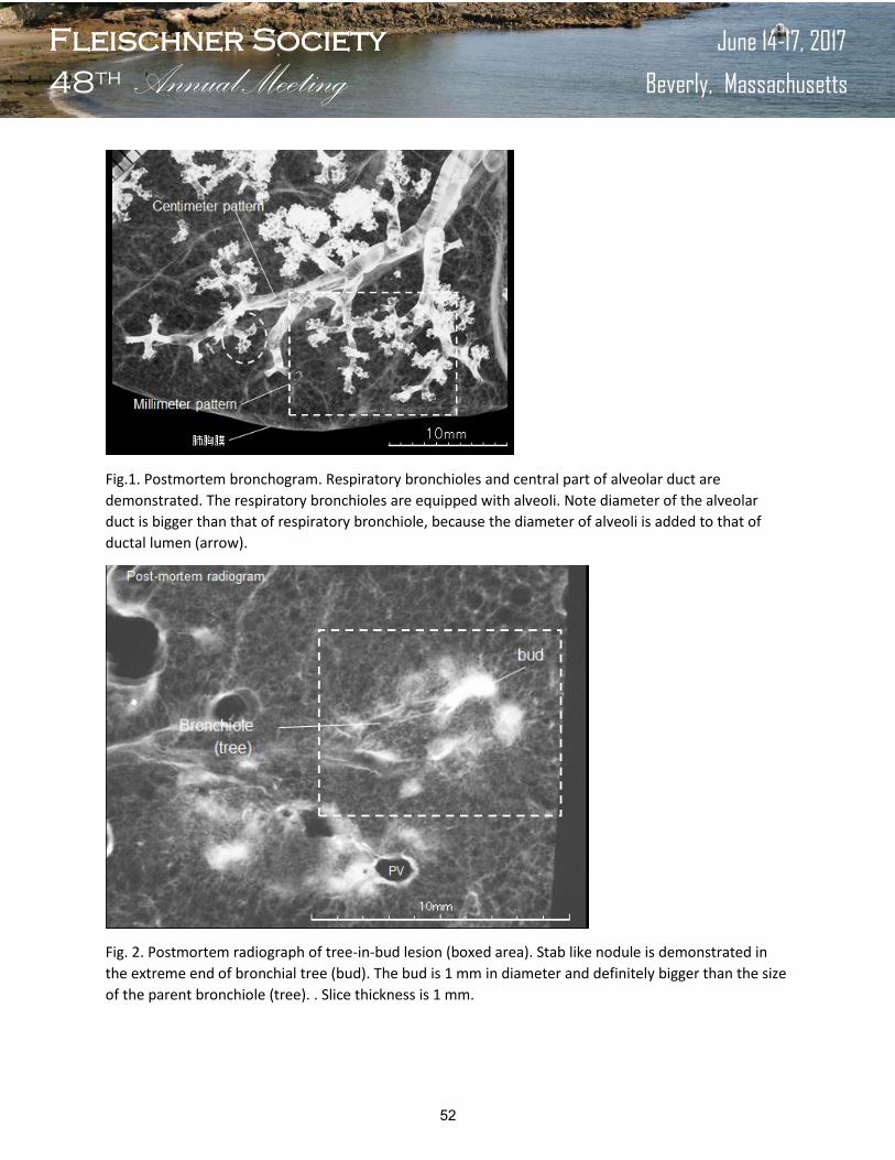
Fig.1. Postmortem bronchogram. Respiratory bronchioles and central part of alveolar duct are demonstrated. The respiratory bronchioles are equipped with alveoli. Note diameter of the alveolar duct is bigger than that of respiratory bronchiole, because the diameter of alveoli is added to that of ductal lumen (arrow).
Fig. 2. Postmortem radiograph of tree-in-bud lesion (boxed area). Stab like nodule is demonstrated in the extreme end of bronchial tree (bud). The bud is 1 mm in diameter and definitely bigger than the size of the parent bronchiole (tree). . Slice thickness is 1 mm.
52

Fleischner Society June 14-17, 2017 48th AnnualMeeting Beverly, Massachusetts
Fig 3. Postmortem radiograph prior to slicing (slice thickness is 7mm) showing cluster of micronodules (galaxy appearance on CT). Note that margins of the collected tree- in –bud nodules are straight, suggesting perilobular distribution of the nodules.
Fig. 4. Serial postmortem radiographs show tree-in bud lesions tend to gather close to pulmonary veins (white asterisks) , sparing relatively around central bronchioles(white circles). Slice thickness is 1 mm.
Conclusions:
Tree-in-bud lesions and cluster of micronodules in patients with active tuberculosis are the findings of bronchogenic spread.
53

Fleischner Society June 14-17, 2017 48th AnnualMeeting Beverly, Massachusetts
Title:
Substitution of ethambutol with linezolid during the intensive phase of treatment of pulmonary tuberculosis: Preliminary results for a prospective, multicenter, randomized, open-label, phase II trial
Authors:
Lee JK2 , Lee JY1, Kim DK2, Yoon HI3, Jeong I1, Heo E2, Park YS4, Jo YS4, Lee JH3, Park SS2, Park JS3, Kim JH1, Lee SM4, Joh JS1, Lee CH4, Lee J4, Choi SM4, Park JH2, Lee SH3, Cho YJ3, Lee YJ3, Kim SJ3, Hwang YR2, Kim H4, Ki J1, Choi H3, Lee M5, Song T5, Kim HS6, Han J6, Ahn H6, Hahn S6, Yim JJ4
Affiliations:
1Division of Pulmonary and Critical Care Medicine, Department of Internal Medicine, National Medical Center, Seoul, Republic of Korea, Division of Pulmonary and Critical Care Medicine, Department of Internal Medicine, 2Seoul National University Boramae Medical Center, Seoul, Republic of Korea, 3Division of Pulmonary and Critical Care Medicine, Department of Internal Medicine, Seoul National University Bundang Hospital, Seongnam, Republic of Korea, Division of Pulmonary and Critical Care Medicine, 4Department of Internal Medicine, 5International TB Research Center, 6Medical Research Collaborating Center, Seoul National University Hospital, Seoul, Republic of Korea
Purpose:
Linezolid, an oxazolidinone, substantially improves treatment outcomes of multidrug-resistant tuberculosis and extensively drug-resistant tuberculosis. We set to test whether the use of linezolid instead of ethambutol could increase the rate of sputum culture conversion as of 8 weeks of treatment in patients with drug-susceptible tuberculosis.
Materials and Methods:
We conducted a phase II, multicenter, randomized study with three arms for patients with pulmonary tuberculosis without rifampicin resistance screened by the Xpert MTB/RIF® assay. One group of patients received isoniazid (6 months), rifampicin (6 months), pyrazinamide (2 months), and ethambutol (2 months) (control arm). In second group, we replaced ethambutol with linezolid (600 mg/day) for 2 weeks (LZD 2 weeks arm). In third group, we replaced ethambutol with linezolid (600 mg/day) for 4 weeks (LZD 4 weeks arm). The primary outcome was the sputum culture conversion rate on liquid media after 2 months of treatment.
Results:
Preliminary analysis was performed. Of 429 participants enrolled, 420 were eligible for the primary efficacy analysis. In the modified intention-to-treat analysis, negative cultures at 2 months of treatment were observed in 87/141 (61.7%) participants in control arm, 105/142 (73.9%, P=0.03) participants in LZD 2 weeks arm, 98 /137 (71.5%, P=0.08) participants in LZD 4 weeks arm. Time to culture conversion was 33 days in control arm, 28 days in LZD 2 weeks arm (P=0.05), and 21 days in LZD 4 weeks arm (P=0.03).
54

Fleischner Society June 14-17, 2017 48th AnnualMeeting Beverly, Massachusetts
Conclusions:
Substitution of ethambutol with linezolid during the intensive phase of treatment resulted in higher rates of culture conversion at 2 months and shorter time to culture conversion.
* Acknowledgement: Linezolid (Zyvox®) was donated by Pfizer Inc.
55

56

Fleischner Society June 14-17, 2017 48th AnnualMeeting Beverly, Massachusetts
Title:
Automated detection of tuberculosis on chest radiographs: ready for implementation
Authors:
Bram Van Ginneken
Affiliations:
Radboudumc
Purpose:
Chest radiography is an important tool for diagnosis of tuberculosis (TB), particularly as a triage test when screening large groups where those with X-ray abnormalities undergo a confirmatory bacteriological test. The main limitation for this approach is the lack of trained human readers in countries with a high burden of TB.
Materials and Methods:
We have developed an automated algorithm for reading chest radiographs for signs of TB. This project started in 1996, and was restarted in 2008 and led to a first release in 2011 and is currently in active use as a CE certified software package (CAD4TB) in over 15 countries in Africa, Asia and Europe. The software has been validated in over 10 studies.
Results:
Automated reading of chest radiographs has similar performance to reading by clinical officers in countries with a high burden of TB. A cost-effectiveness modeling study suggests that use of the software can reduce the cost per notified TB case. The WHO has investigated what further studies are needed before the technology can be incorporated in TB guidelines.
Conclusions:
Automated chest radiography interpretation may improve the effectiveness of the diagnostic process for tuberculosis.
57

Fleischner Society June 14-17, 2017 48th AnnualMeeting Beverly, Massachusetts
Title:
Direct Patient Outcome Prediction from Chest CT Images using Deep Learning
Authors:
Raul San Jose Estepar, German Gonzalez, Samuel Y Ash, George R Washko
Affiliations:
Applied Chest Imaging Laboratory, Radiology, Brigham and Women’s Hospital
Purpose:
To determine if a deep learning analysis, specifically convolutional neural network (CNN), based approach could detect and stage individuals with chronic obstructive pulmonary disease (COPD) and predict acute respiratory disease events (ARD) and mortality in smokers
Materials and Methods:
CNN-based models were created and tested in the CT scans of subjects enrolled in the COPDGene Study. These models were then tested in a separate cohort of unrelated smokers from the ECLIPSE study. CNN models were trained to predict mortality and acute episodes of respiratory disease (ARD) and to estimate GOLD Stage and forced expiratory volume in one second (FEV1). The C-index, net-reclassification index (NRI), area under the curve (AUC) and correlation coefficient (r) were used to quantify the models’ performance.
Results:
The CNN model correctly determined the diagnosis of COPD in 773 of the 1,000 COPDGene testing subjects (c-statistic 0.856) and accurately staged them 51.1% of the time and was correct or off by 1 stage 74.95% of the time. The same model applied to the ECLIPSE cohort correctly predicted the GOLD stage in 29.4% of the cases and was correct or off by 1 stage in 74.6%. In the COPDGene testing cohort, the CNN model of ARD events had a c-statistic of 0.64 and was well calibrated with regard to risk prediction with Hosmer-Lemeshow p=0.502, suggesting no evidence of poor calibration. In the ECLIPSE cohort the CNN model of ARD events had a c-statistic of 0.55 and the model was well calibrated with regard to risk prediction with Hosmer-Lemeshow p=0.380, suggesting no evidence of poor calibration. In both the COPDGene testing cohort and ECLIPSE, the CNN model for mortality showed fair discrimination based on c-indices of 0.72 (CI: 0.50, 0.90) and 0.60 (CI: 0.49, 0.71) respectively, and no evidence of poor calibration as indicated by non-significant Greenwood-Nam-D’Agnostino p-values of 0.307 and 0.331 respectively.
58

Fleischner Society June 14-17, 2017 48th AnnualMeeting Beverly, Massachusetts
Conclusions:
A deep learning approach based on convolutional neural networks (CNN) that uses only CT imaging data can identify those smokers who have COPD and predict which are most likely to have ARD events and who have the highest mortality. Further work is needed that utilizes both imaging and other clinical data, but at a population level CNN analysis may be a powerful tool for risk assessment.
59

Fleischner Society June 14-17, 2017 48th AnnualMeeting Beverly, Massachusetts
Title:
Application of Deep Learning in Chest Imaging: From Quantification of DILD to Reading Chest Radiographs
Authors:
Joon Beom Seo, Namkug Kim, Beomhee Park, Yongwon Cho
Affiliations:
University of Ulsan College of Medicine, Asan Medical Center
Purpose:
To apply new convolutional neural network (CNN) machine learning algorithm in chest imaging such as automated classification of regional disease pattern on CT images of diffuse lung disease and automated detection and classification of disease pattern on chest radiographs.
Materials and Methods:
Simple CNN architecture was applied to develop automated classification of 6 different regional patterns (normal, ground-glass opacity, reticulation, honeycombing, consolidation, and emphysema) using 767 cubic 3D typical VOIs from CTs of diffuse lung diseases. The accuracy of classification was tested with different data set.
For automated detection and classification of chest radiographs, we extended the YOLO model for detecting and classifying multiple abnormalities simultaneously, which include nodule, consolidation, interstitial opacity, cardiomegaly, pleural effusion, and pneumothorax. For training and testing, 4070 data with various abnormalities were used. The abnormal lesions were labelled by chest radiologists.
Results:
The accuracy of 84% for automated classification of regional disease pattern was achieved using new CNN approach. The algorithm was successfully applied to whole volumetric assessment.
The mean accuracy in detecting and localizing disease pattern on chest radiographs was about 74%.
Conclusions:
CNN is new and promising method for automatic detection and classification of various abnormalities on chest CT and radiographs.
60
Fleischner Society June 14-17, 2017 48th AnnualMeeting Beverly, Massachusetts
Title:
Immunomodulation by micrometastatic lung cancer cells
Authors:
Prasad S. Adusumilli, Sarina Bains, Takashi Eguchi, Navin K. Chintala, William D. Travis
Affiliations:
Memorial Sloan Kettering Cancer Center, New York, NY, USA
Purpose:
Even after surgical resection and adjuvant chemotherapy for early-stage non-small cell lung cancer (NSCLC), there are micrometastatic cancer cells that often go undetected and untreated that result in recurrence. Among patients with stage IA NSCLC (T1, ≤3 cm) following curative-intent resection, 15–30% will experience recurrence within 5 years. During our investigation of the biological reasons underlying these recurrences we have identified aerogenous invasion of isolated tumor cells away from the main tumor. This is a new form of invasion that we have termed “spread through alveoloar spaces” (STAS), and it is a potential cause associated with higher rates of recurrence and poorer survival. STAS is now a recognized form of invasion in the new 2015 World Health Organization classification of lung adenocarcinoma (ADC). The purpose of this investigation is to elicit co-localization of STAS cells with immune cells as our hypothesis is that STAS cells modulate the immune microenvironment, thus facilitating tumor growth.
Materials and Methods:
We performed immunohistochemistry (IHC) for CK7+ cancer cells with surrounding macrophages, and immunofluorescence (IF) for tumor-infiltrating immune cells—CD3+ T cells, CD4+ helper T cells, CD8+ cytotoxic T cells, FoxP3+ regulatory cells, and CD163+ M-2 macrophages.
Results:
We observed that STAS cells within normal lung and lymph nodes are associated with immunosuppressive macrophages, T regulatory cells, and neutrophils. In a preliminary analysis (n = 40, small lung ADCs; cohort now expanded) of tumor, stroma, and peritumoral normal lung parenchyma, STAS+ tumor cells were associated with: (a) a higher number of FoxP3 cells, a lower number of CD3+ cells, and a high FoxP3/CD3 ratio in the normal lung parenchyma; and (b) a higher number of CD163+ macrophages in the tumor and a high FoxP3/CD3 ratio in the tumoral stroma. Fig 7
61

Fleischner Society June 14-17, 2017 48th AnnualMeeting Beverly, Massachusetts
Conclusions:
The above observations, coupled with the observed differences between smokers and non-smokers, are intriguing and provide rationale to investigate the relationship between immune cells in the tumor, stroma, and normal lung parenchyma that harbor STAS cells.
62
Fleischner Society June 14-17, 2017 48th AnnualMeeting Beverly, Massachusetts
Title:
CEST Imaging: Challenging for Lung Cancer by High-Field MRI
Authors:
Yoshiharu Ohno, MD, PhD
Affiliations:
Division of Functional and Diagnostic Imaging Research, Department of Radiology, Kobe University Graduate School of Medicine
Purpose:
To directly and prospectively compare the capability for differentiation of malignant from benign pulmonary nodules and/ or masses among chemical exchange saturation transfer (CEST) imaging targeted to amide (-NH)groups, diffusion-weighted MR imaging (DWI) and FDG-PET/CT.
Materials and Methods:
Eighty-eight consecutive patients (54 men and 34 women; mean age 70 years) with pulmonary nodules and/ or masses underwent CEST imaging and DWI at a 3T MR system, FDG-PET/CT and pathological and/or follow-up examinations. According to final diagnoses, all lesions were divided into malignant (n=49) and benign (n=39) groups. To obtain CEST imaging data in each subject, respiratory-synchronized fast advanced spin-echo images were conducted following a series of magnetization transfer (MT) pulses. Then, magnetization transfer ratio asymmetry (MTRasym) was calculated from z-spectra at 3.5ppm in each pixel, and MTRasym map was computationally generated. To evaluate the capability for differentiation between two groups at each lesion, MTRasym, apparent diffusion coefficient (ADC) and SUVmax were assessed by ROI measurements. To compare each index between two groups, Student’s t-test was performed. Then, ROC analysis was performed to determine each feasible threshold value for differentiation of two groups. Finally, sensitivity, specificity and accuracy were compared each other by McNemar’s test.
Results:
Mean MTRasym (1.97±6.38%), ADC (1.17±0.25×10⁻³mm2/sec) and SUVmax (3.19±1.60) of malignant group had significant difference with those of benign group (MTRasym: -2.9±4.9%, p=0.0002; ADC: 1.33±0.18×10⁻³mm2/sec, p=0.0024, SUVmax: 2.27±0.48, p=0.0008). Results of ROC analysis showed that there were no significant differences of area under the curve (Az) among all indexes (MTRasym: Az=0.72, ADC: Az=0.67, SUVmax: Az=0.72) (p>0.05). When applied each feasible threshold value, there were no significant differences of diagnostic performance among all indexes (p>0.05).
63

Fleischner Society June 14-17, 2017 48th AnnualMeeting Beverly, Massachusetts
Conclusions:
CEST imaging is considered at least as valuable as DWI and FDG-PET/CT for differentiation of malignant from benign pulmonary lesions
64

Fleischner Society June 14-17, 2017 48th AnnualMeeting Beverly, Massachusetts
Title:
Variability in Characterisation and Management of Incidental CT-Detected Lung Nodules: An International Collaborative Study
Authors:
Nair A, Bartlett E, Walsh SLF, Bhalla S, Devaraj A, Goo JM, Klein JS, MacMahon H, Seo JB, Sverzellati N, Schaefer-Prokop C, Desai SR
Affiliations:
King’s College, London
Purpose:
To evaluate interobserver variation among an international cohort of thoracic radiologists for the characterization and initial management of incidentally-detected pulmonary nodules on computed tomography (CT).
Materials and Methods:
Targeted CT images stacks and clinical data, for 70 nodules in consecutive patients, were reviewed by 107 radiologists (attending/consultant, n=91 versus trainee, n=16; median experience=9 years, range=1-50) from 25 countries. Nodule morphology (solid versus part-solid versus pure ground-glass) togetherwith a level of certainty (to the nearest 5%) for each choice was scored. The maximum diameter of eachnodule (including, where relevant, the solid component) was measured and the presence/absence ofspecific morphological features recorded. Preferences for i) initial management (i.e. follow-up versusdischarge) and, if considering follow-up, ii) invasive management or CT follow-up ± PET-CT were stated.Factors influencing the management option were also recorded. Unweighted multirater Fleiss k wasused to compare agreement for first-choice nodule categorization, morphological features and for initialmanagement choice. Associations between nodule categorization, nodule size and level of certaintywere tested with Spearman’s rank correlation or one-way analysis of variance (ANOVA) as appropriate.
Results:
Agreement for overall first-choice nodule categorization (k=0.65) was good but better for solid (k=0.74) than part-solid nodules (k=0.53). Most observers (median=103 [96%], range=87-107) assigned a 70% or greater certainty for the first-choice category; certainty levels did not correlate with median nodule diameter but were significantly different between nodule categories (one-way ANOVA, p<0.001). Interobserver agreement for morphological features was only fair to moderate (k=0.24-0.53; including spiculation [k=0.35]). Agreement for initial management option was only fair (k=0.33) and similar between experienced (>10 years; n=45) and less experienced (≤10 years; n=62) observers (k=0.31 versus k=0.34, respectively). For 16/70 (23%) nodules, all 107 observers agreed on the need for follow-up but
65

Fleischner Society June 14-17, 2017 48th AnnualMeeting Beverly, Massachusetts
agreement on the method of follow-up (CT follow-up versus invasive management) (k=0.36) or for performing PET-CT (k=0.32) was only fair. The dominant factor in determining management was nodule morphology in 65/70 (93%) cases but there was wide variation in the proportion of observers rating nodule morphology as the most influential (median 62% of observers, range 37% to 86%). Nodule dimension was the dominant factor in 4/70 (6%).
Conclusions:
The level of certainty for first-choice nodule categorization was high among international thoracic radiologists. Overall interobserver agreement for first-choice categorization was good but is lowest for part-solid nodules. Agreement among thoracic radiologists on the need for, and nature of, initial follow-up for incidental pulmonary nodules is only fair.
66

Fleischner Society June 14-17, 2017 48th AnnualMeeting Beverly, Massachusetts
Title:
Low-dose CT lung cancer screening in Korea
Authors:
Jin Mo Goo, MD, PhD
Affiliations:
Department of Radiology, Seoul National University Hospital
Purpose:
To describe the current status of lung cancer screening with low-dose CT (LDCT) in Korea.
Materials and Methods:
In Korea the National Cancer Screening Program is running for five types of cancers including stomach, liver, colorectal, breast, and uterine cervix. Although lung cancer is the number one cause of cancer deaths, lung cancer screening has been performed in a form of individualized health assessment. In 2015, the Korean guidelines for lung cancer screening was published and annual LDCT screening is recommended to current smokers and ex-smokers who had quit less than 15 years earlier, aged 55 to 74 years with a smoking history of 30 pack-years or more. Based on this, the Korean LUng CAncer Screening (LUCAS) project is planned.
Results:
The Korean LUCAS project will evaluate the feasibility in establishing a nationwide lung cancer screening program. It starts in 2017 targeting 8000 participants at 11 centers including National Cancer Center, 9 regional cancer centers, and Seoul National University Hospital. LDCT will be obtained with CT scanners with at least 16 detector rows and thin-section images < 1.5 mm will be reconstructed. A network-based CAD center will play an important role in collecting data from each center, storing data in cloud system, providing computer applications including detection aid, segmentation tools, and categorization tools, and on-line quality assessment for uploaded CT data.
Conclusions:
Korean LUCAS project may provide results for the feasibility in implementing systems recommended by many guidelines.
67
Fleischner Society June 14-17, 2017 48th AnnualMeeting Beverly, Massachusetts
Title:
Accuracy of the Vancouver Risk Calculator compared to ACR LungRads applied to the National Lung Screening Trial
Authors:
CS White1, S Dalal2, E Dharaiya2
Affiliations:
University of Maryland1, Philips Medical Systems2
Purpose:
To compare the accuracy of the Vancouver Risk Calculator (VRC) with ACR LungRADs in determining malignancy among solid nodules identified in the National Lung Screening Trial (NLST).
Materials and Methods:
The study received approval from the IRB and the NCI CDAS system to use NLST data. All nodules detected in the CT arm of the NLST were considered for inclusion. After exclusions due to inability to determine malignant vs benign nodule status (no gold standard), incomplete data sheets preventing the use of the risk calculator, and subsolid nodule type, 4078 nodules were available for evaluation. The 4078 solid nodules were scored using the VRC consisting of 9 nodule and patient parameters (output = % likelihood of malignancy, malignancy threshold tested at >5 likelihood) and LungRADS (output = Category 2-4B, malignancy defined as 4A or 4B, malignancy defined as >5% likelihood by LungRADS table). Comparison was performed between LungRADS and the VRC to determine their respective sensitivity, specificity and accuracy to distinguish between benign and malignant nodules.
Results:
For ACR LungRADS category 2 (non-malignant), there were 2,141 nodules of which 3 were malignant. The VRC designated 0 malignant with 0 False Positives (FP) for the 5% threshold. For LungRADS category 3 (non-malignant) there were 1,136 nodules of which 7 were malignant. The VRC designated 3 malignant, with 31 false positives. For LungRADS category 4A (malignant), there were 630 nodules of which 26 were malignant. The VRC designated 25 malignant with 244 FP. For LungRADS category 4B (malignant), there were 171 nodules of which 64 were malignant. VRC designated 64 malignant with 107 FP. Overall sensitivity, specificity and accuracy for LungRADS was 90.0%, 82.1% and 82.3%, respectively and for VRC 5% it was 92.0% 90.4% and 90.4%, respectively.
68

Fleischner Society June 14-17, 2017 48th AnnualMeeting Beverly, Massachusetts
Conclusions:
In comparison with ACR LungRads, the Vancouver risk model at a 5% threshold demonstrated higher sensitivity , specificity and accuracy in determining malignancy among solid nodules in the NLST.
69

Fleischner Society June 14-17, 2017 48th AnnualMeeting Beverly, Massachusetts
Title:
Development of a Prognostic Marker for Lung Cancer Using Analysis of Tumor Evolution
Authors:
Edward F. Patz, Jr., Michael Campa, Rick Durrett, Allen Rodrigo
Affiliations:
Duke University
Purpose:
To apply evolutionary analytical methods developed to study expansion and migration of populations to tumor biology in order to produce a prognostic marker in cancer.
Materials and Methods:
Whole exome sequencing (WES) is performed on individual tumor cells isolated from the tumors of non-small cell lung cancer (NSCLC) patients (half early stage, half late stage). We then reconstruct each tumor’s phylogenetic tree, and compare the tree patterns from early stage NSCLC (indolent tumors without metastasis) to those from late stage disease (tumors with metastasis). We use a combination of tree features to generate a prognostic score that separates tumors with very different phenotypes (indolent vs. aggressive).
Results:
To date we have developed the methods to isolate single tumor cells and performed WES. We have just begun to analyze patient specimens in the effort to develop a classifier.
Conclusions:
Evolutionary analysis of single tumor cells is a potential novel approach to phenotype tumors.
70

Fleischner Society June 14-17, 2017 48th AnnualMeeting Beverly, Massachusetts
Title:
How Radiologic-Pathologic Correlation Can Help in Lung Cancer Assessment
Authors:
William D. Travis, M.D. and Prasad Adusumilli, M.D.
Affiliations:
Departments of Pathology and Thoracic Surgery, Memorial Sloan Kettering Cancer Center, New York, New York
Purpose:
The purpose of this presentation is to summarize how radiologic pathologic correlation can improve pathologic assessment of lung cancer specimens. This approach is currently not standard of care.
Materials and Methods:
During the past 7 years of my clinical practice I have routinely accessed our PACSWEB system during my daily sign outs and exported selected images from cases that illustrate the value of radiologic pathologic correlation in improving pathologic diagnosis of lung cancer. Selected key examples will be presented, particularly focusing on relevance for implementing the recent proposed 8th Edition TNM Classification, recommending the use of invasive rather than total size in nonmucinous lung adenocarcinomas for the size T factor.
Results:
There are three major areas where radiologic pathologic correlation assists in achieving a more accurate pathologic diagnosis: 1) differential diagnosis in small biopsies, 2) assessment of tumor size for TNM staging and 3) classification of single vs multiple tumors.
In small biopsies that show a lepidic pattern of nonmucinous adenocarcinoma, evaluation of the CT scan can suggest whether the diagnosis is more likely to be AIS, MIA, lepidic predominant or invasive predominant adenocarcinoma.
Pathologic assessment of total vs invasive tumor size in resected nonmucinous lung adenocarcinomas with a lepidic component can be improved by reviewing CT scans because the lepidic component is often poorly appreciated on gross exam. In addition, tumor size can be more accurately assessed after radiologic pathologic correlation in the following settings: 1) Sausage or bilobed shaped tumors where the maximum single diameter may be better assessed using all three views (axial, coronal and saggital) rather than just axial alone, 2) Tumors removed in multiple parts, 3) Intraoperative defects in tumors, 4) Marked non-neoplastic reactions, 5) Mistaken pathologic assessment.
71

Fleischner Society June 14-17, 2017 48th AnnualMeeting Beverly, Massachusetts
Tumor size estimation in the setting of neoadjuvant therapy can result in multiple foci of viable tumor within a tumor bed. Radiologic correlation can clarify whether there is a single tumor or intrapulmonary metastases.
Assessment of pathology specimens is sometimes ambiguous whether one or more tumors are present and whether the nodules represent intrapulmonary metastases or separate primaries. In such cases, correlation with CT scans along with comprehensive histologic assessment can be help to resolve these questions.
Conclusions:
Lung cancer specimen assessment is greatly enhanced by radiologic pathologic correlation in specific settings, particularly nonmucinous adenocarcinomas with lepidic components, tumors with non-spherical shapes, tumors with intraoperative defects, marked non-neoplastic reactions, tumor size in the neoadjuvant setting and multiple tumors.
72

Fleischner Society June 14-17, 2017 48th AnnualMeeting Beverly, Massachusetts
Title:
Peripheral Vision at the Moment of Recognition for Lung Nodules in CT Scans: Inter-reader Variations and Associations with Detection Performance
Authors:
Geoffrey D. Rubin, Brian Harrawood, Kingshuk RoyChoudhury, Justus E. Roos, Lukas Ebner. Martin Tall. Sandy Napel
Affiliations:
Duke and Stanford Universities
Purpose:
To detect and characterize the moment when radiologists recognize that a lung nodule is present within a chest CT scan and thus accommodate for peripheral vision when assessing radiologists’ search strategies.
Materials and Methods:
Under an institutional review board approved and HIPAA compliant protocol, time-varying three-dimensional gaze traces acquired from 13 radiologists using stacked section paging and eye tracking during the interpretation of 40 chest CT scans enriched with 157 simulated 5-mm solid lung nodules were subdivided into periods of nodule visibility (exposures). “Gaze distances” were measured between gaze points and both true positive (TP) and false negative (FN) lung nodules during nodule exposures to quantify relationships between gaze distance, duration, and lung nodule detection. An automated algorithm was developed to detect the moment of recognition (MoR) within true positive (TP) exposures at a time point immediately preceding the saccade that converged upon the to be detected nodule. MoR distances from TP nodules were measured and characterized as central (foveal) versus peripheral vision based upon a 5° gaze angle threshold.
Results:
Of 9,751 nodule exposures, 997 concluded with TP detections. On average, exposure to FN nodules was 3.5 times longer than TP nodules across gaze angles of 2.5–25.5° (p<0.00001). Across all TP detections, median MoR was 32.1 mm (5.5° gaze angle) and 53.5% (534/997) of TP detections were initially recognized with peripheral vision. MoR distance was inversely correlated with detection difficulty (p<0.0001). Readers tendency to detect nodules with central versus peripheral vision varied substantially. The fraction of lung volume exposed to the fovea was inversely correlated with per reader median MoR (r=-0.64, p=0.02), but neither total search duration nor reader sensitivity correlated with median MoR distance.
73

Fleischner Society June 14-17, 2017 48th AnnualMeeting Beverly, Massachusetts
Conclusions:
Across readers, peripheral vision was variably responsible for lung nodule detection in CT scans, however greater reliance on central versus peripheral vision was not associated with diagnostic performance.
74

Fleischner Society June 14-17, 2017 48th AnnualMeeting Beverly, Massachusetts
Title:
Using thoracic Imaging Outcomes in Research
Authors:
Alan Brody
Affiliations:
Cincinnati Children’s Hospital
Purpose:
To discuss a possible Fleischner Society white paper
Materials and Methods:
Imaging outcomes are commonly used in clinical and laboratory research. There is little guidance available for researchers and thoracic imagers on selecting and performing imaging studies for use as outcome measures. A document presenting current uses of thoracic imaging outcomes and guidance for their use in future research could be useful.
Results:
If the members of the Society conclude that this would be a useful effort, a writing team would be formed and a detailed outline presented at a future meeting.
Conclusions:
Will depend on the response of the members.
75

Fleischner Society June 14-17, 2017 48th AnnualMeeting Beverly, Massachusetts
Title:
An algorithmic approach to cavitary lung disease
Authors:
Kevin Brown, MD, David Naidich, MD, Jay Ryu, MD, Khalid Ghafoor, MD, Shalin Patel, MD, Suhail Raoof, et al
Affiliations:
Lenox Hill Hospital, NY; National Jewish Hospital , Denver; New York University, NY; Mayo Medical Clinic, Rochester
Purpose:
To develop an algorithmic approach to diagnosing cavitary lung disease
Materials and Methods:
Literature review using Pubmed was carried out. Key words entered for search included cavitary lung disease, lung cavitation, lung cavities, diffuse lung disease and cysts. Abstracts in the English language were reviewed to assess for relevance to our article. Radiologists, pulmonologists and pathologists developed an algorithm that was predicated on clinical features and high resolution CT scan of the chest. The clinical and radiographic information was amalgamated to develop a stepwise logical approach that clinicians can follow to develop a short and accurate differential diagnosis
Results: The 4 main categories of conditions associated with diffuse cavitary lung disease include: 1. Infections (acute and chronic)2. Malignancy (Primary and metastatic)3. Inflammatory/immunological4. MiscellaneousThe HRCT findings of each entity are described along with the salient clinical features.
Conclusions:
We describe a simple algorithmic approach to diagnosis of cavitary lung disease by amalgamating both clinical and radiographic features
76

Fleischner Society June 14-17, 2017 48th AnnualMeeting Beverly, Massachusetts
Title:
Spectrum of Non-communicable Lung Disease in Ethiopia
Authors:
Neil Schluger, Teddy Haile, Dawit Kebede, Charles Sherman
Affiliations:
Columbia University, Addis Ababa University, Brown University
Purpose:
Most of the focus on lung disease in Ethiopia, a low-income country with a rapidly developing economy, has been on tuberculosis and acute respiratory infections. Little is known about the extent and cause of chronic lung disease in Ethiopia, the second largest country in Africa, with a population of roughly 100 million people.
Materials and Methods:
We examined the extent and burden of non-communicable lung disease, particularly asthma, COPD and lung cancer in patients seen in the pulmonary clinics of Tikur Anbessa (Black Lion) Specialized Hospital (TASH), the largest public hospital in Addis Ababa, and the main teaching affiliate of the Addis Ababa School of Medicine. TASH is also the site of the East Africa Training Initiative in Pulmonary Medicine, a two-year pulmonary fellowship program that is training the first cadre of lung specialists in East Africa.
Results:
Asthma, COPD and lung cancer are common illnesses among patients presenting to the Chest Clinic at TASH. Patients with lung cancer especially are younger than generally observed in high-income countries. Unlike patients in high-income countries with chronic lung diseases such as COPD and lung cancer, patients in Ethiopia have a very low prevalence of tobacco use: fewer than 7% of men and 1% of women smoke cigarettes. The most significant risk for chronic lung disease in Ethiopia appears to be household air pollution related to biomass fuel use for cooking.
Conclusions:
The burden of chronic lung disease in Ethiopia is considerably higher than previously realized. Indoor air pollution from biomass fuel burning is likely an important cause of COPD and lung cancer there. Use of cleaner fuels might ameliorate this problem to a degree. There is a need for greater access to diagnostic and therapeutic services for non-communicable lung diseases in East Africa.
77

Fleischner Society June 14-17, 2017 48th AnnualMeeting Beverly, Massachusetts
Title:
Volumetric Analysis of the Thymic Epithelial Tumors: Correlation with WHO classification and Masaoka-Koga Staging
Authors:
Noriyuki Tomiyama, Yukinaga Sato, Masahiro Yanagawa
Affiliations:
Osaka University Graduate School of Medicine
Purpose:
The purpose of our study was to investigate the correlation between tumor volume and each subtype of thymic epithelial tumors based on the World Health Organization (WHO) classification and Masaoka-Koga staging.
Materials and Methods:
The study was approved by the Institutional Review Board. Sixty-one consecutive patients (45 thymomas and 16 thymic carcinomas) were included. All 61 thymic epithelial tumors were classified according to Masaoka-Koga staging: 31 non-invasive thymic epithelial tumors (Stage I) and 30 invasive tumors (8 Stage II, 11 Stage III, 3 Stage IVa, and 8 Stage IVb). Tumor volumes on thin-section CT were automatically calculated using our software. The correlation of tumor volume with each WHO subtype and Masaoka-Koga staging was analyzed using Mann-Whitney U test and Scheffe’s F test.
Results:
The tumor volumes (mean ± SD) of each WHO histologic type were 127.5 ± 105.6cm3 in type A; 42.0±37.9 cm3 in type AB; 61.8±121.5 cm3 in type B1; 48.6±62.6 cm3 in type B2; and 44.2±48.2 cm3 in type B3 thymomas, and 117.5±143.6 cm3 in thymic carcinoma. Thymic carcinomas were significantly larger than thymomas (p = .0016). The tumor at Stage IVb (190.8± 156.8 cm3) was significantly larger than stage I (33.1± 42.6 cm3) (p<.05). Invasive thymic epithelial tumors were significantly larger than non-invasive tumors (p= .0016).
Conclusions:
Our software could measure the volume of thymic epithelial tumor. The volume of invasive thymic epithelial tumors may be larger than non-invasive tumors at the time of initial presentation.
78

Fleischner Society June 14-17, 2017 48th AnnualMeeting Beverly, Massachusetts
Title:
Malignant pleural mesothelioma: what's new in staging and surgery?
Authors:
Paul E. Van Schil, MD, PhD; Jeroen M. Hendriks, MD, PhD; Patrick Lauwers, MD
Affiliations:
Department of Thoracic and Vascular Surgery, Antwerp University Hospital and Antwerp University, Belgium
Purpose:
To describe the most important changes in the 8th TNM edition for malignant mesothelioma and provide an overview of current surgical management.
Results:
Incidence of malignant pleural mesothelioma will increase until 2020. As this tumor has a specific longitudinal growth pattern along the pleural surfaces, precise staging remains difficult and has not yet been standardised. In the 8th edition of the TNM classification substantial changes were introduced; most important ones are the grouping of T1a and T1b into a single T1 category, and the classification of mediastinal nodal involvement as N1 disease [1].
Prognosis of patients with malignant pleural mesothelioma remains poor and it is clear that single modality therapy will not improve long-term survival. Combined modality therapies are further evaluated but due to lack of large, controlled randomised trials the relative contribution of each treatment modality, chemotherapy, radiotherapy and surgery, has not been fully established [2].
Equally, the most efficient surgical procedure remains a hotly debated topic. To better compare results from different institutions, uniform definitions for surgical interventions including extrapleural pneumonectomy and the different forms of pleurectomy have been described by a task force of the International Association for the Study of Lung Cancer (IASLC) [3]. The confusing term “radical pleurectomy” is not used anymore. Currently, there is a shift towards extended pleurectomy/decortication when a macroscopic complete resection of all tumor can be obtained by this procedure instead of the classical extrapleural pneumonectomy. In recent trials induction chemotherapy has been administered to improve surgical resection rates but pathologically complete responses are infrequently observed.
The European Society of Medical Oncology (ESMO) recommendations state that “surgery may be indicated to perform a macroscopic complete resection by means of pleurectomy/decortication or extrapleural pneumonectomy” [4].
79
Fleischner Society June 14-17, 2017 48th AnnualMeeting Beverly, Massachusetts
The latter procedure is still used when there is extensive ingrowth inside the fissures or pulmonary parenchyma. The role of postoperative radiotherapy seems to be limited after completion of the recent SAKK 17/04 trial [5].
Best survival results have been reported after multimodality therapy including surgery. However, although some of these studies are prospective, there were no control groups and selection of suitable patients may introduce an important bias [6-9]. On the other hand, the final results of these trials represent a major benchmark to which other treatment modalities should be compared. The role of induction versus adjuvant chemotherapy has to be further elucidated. The incorporation of targeted therapies is a potentially valid therapeutic option. Other treatment options that are currently explored include hyperthermic intrapleural chemotherapy, immunotherapy, gene therapy and photodynamic therapy. However, no randomised comparisons are available yet. In the meantime therapeutic nihilism should not prevail and every patient with suspected or proven mesothelioma should be discussed within a multidisciplinary tumour board to provide the most adapted, personalized treatment.
Further clinical research is certainly required and hopefully, the prospective database of the IASLC and ongoing clinical trials will provide new, relevant information and guidelines for this dreadful disease.
Conclusions:
Prognosis of patients with mesothelioma remains poor. In the 8th edition of the TNM classification of mesothelioma substantial changes were introduced in the T and N descriptors. Every patient with mesothelioma should be discussed within a multidisciplinary tumor board. In patients with a good performance status multimodality therapy is indicated. Regarding surgical management there is currently a shift towards extended pleurectomy/ decortication to obtain a macroscopic complete resection. Newer treatment modalities are presently evaluated to obtain better local and distant control.
80

Fleischner Society June 14-17, 2017 48th AnnualMeeting Beverly, Massachusetts
References
1. Rusch VW et al. The IASLC Mesothelioma Staging Project: Proposals for the M Descriptors and forRevision of the TNM Stage Groupings in the Forthcoming (Eighth) Edition of the TNM Classificationfor Mesothelioma. J Thorac Oncol. 2016; 11:2112-9.
2. Van Schil P et al. Multimodal management of malignant pleural mesothelioma: where are wetoday? Eur Respir J. 2014; 44:754-64.
3. Rice D et al. Recommendations for uniform definitions of surgical techniques for malignant pleuralmesothelioma: a consensus report of the international association for the study of lung cancerinternational staging committee and the international mesothelioma interest group. J Thorac Oncol2011; 6:1304-12.
4. Baas P et al. Malignant pleural mesothelioma: ESMO clinical practice guidelines for diagnosis,treatment and follow-up. Ann Oncol 2015; 26 suppl 5: v31-9.
5. Stahel R et al. Neoadjuvant chemotherapy and extrapleural pneumonectomy of malignant pleuralmesothelioma with or without hemithoracic radiotherapy (SAKK 17/04): a randomised,international, multicentre phase 2 trial. Lancet Oncol 2015; 16:1651-8.
6. Van Schil PE et al. Trimodality therapy for malignant pleural mesothelioma: results from an EORTCphase II multicentre trial. Eur Resp J 2010; 36:1362-9.
7. Treasure T et al. Extra-pleural pneumonectomy versus no extra-pleural pneumonectomy forpatients with malignant pleural mesothelioma: clinical outcomes of the Mesothelioma and RadicalSurgery (MARS) randomised feasibility study. The lancet oncology. 2011; 12:763-72.
8. Cao C et al. A systematic review and meta-analysis of surgical treatments for malignant pleuralmesothelioma. Lung Cancer 2014; 83:240-5.
9. Taioli E et al. Meta-analysis of survival after pleurectomy/decortication versus extrapleuralpneumonectomy in mesothelioma. Ann Thorac Surg 2015; 99:472-81.
81
Fleischner Society June 14-17, 2017 48th AnnualMeeting Beverly, Massachusetts
Title:
Preoperative and intraoperative dye localization for lung nodule(s) resection using video-assisted thoracoscopic surgery (VATS)
Authors:
Yeun-Chung Chang, Wei-Chun Ko, Yao-Hui Tseng, Mong-Wei Lin, Yee-Fan Lee, Ning Chien , Hsao-Hsun Hsu, Jo-Yu Chen, Jang-Ming Lee, Pei-Ming Huang, Jin-Shing Chen Affiliations:
Departments of Medical Imaging and Surgery, National Taiwan University Hospital; Departments of Radiology and Surgery, National Taiwan University College of Medicine, Taipei, Taiwan
Purpose:
Due to the limitation of small incision, an ideal preoperative localization technique is essential for surgical resection of small pulmonary nodules by video-assisted thoracoscopic surgery (VATS). We would like to demonstrate clinical feasibility of CT-guided dye localization of preoperative small pulmonary nodules before VATS and using robotic C-arm CT in a hybrid operation room (OR) for VATS.
Materials and Methods:
Patent Blue Vital (PBV) dye localization was performed in all subjects (177 patients with 196 nodules for uniportal VATS, 217 nodules in 100 patients for resection of multiple nodules during one operative sesson, 25 patients for intraoperative robotic C-arm CT before VATS in a hybrid OR) using a 21 G needle under image guidance. Results:
All nodules were successfully resected using uniportal VATS without any conversion to thoracotomy. The
postoperative course was smooth, with a short mean hospital stay (3.3 ± 1.2 days) and a low morbidity
rate (0.6%, 1/177). For multiple nodule localization, the overall success rate was 99% by nodule. Failed localization of two nodules in two patients was due to poor dye visualization (n=1) and significant pneumothorax (n=1). 100% (25/25) success rate for the intraoperative localization in a hybrid OR.
Conclusions:
CT-guided PBV dye localization for multiple small pulmonary nodules before VATS is a safe, feasible, and accurate method with high success rate. This approach makes it easy to perform uniportal VATS, multiple nodule resections during one VATS operation, or integrating with VATS procedure in a hybrid OR.
82

Fleischner Society June 14-17, 2017 48th AnnualMeeting Beverly, Massachusetts
Title:
Risk factors associated with CT-guided transthoracic needle lung biopsy-induced pneumothorax
Authors:
Sergey Leshchinskiy, Ryan Walsh, Anthony Kopecky, Richard Watts, Jeffrey Klein
Affiliations:
University of Vermont College of Medicine, Department of Radiology, Burlington, VT, USA
Purpose:
The most common complication of CT-guided transthoracic needle biopsy (CT-TNB) is pneumothorax. We analyzed our experience with coaxial CT-TNB to determine if hyperlucency or localized emphysema as seen on CT within lung traversed by a biopsy needle was a better predictor of pneumothorax development and pleural drainage compared to pre-procedure forced expiratory volume in 1 second (FEV1).
Materials and Methods:
We retrospectively reviewed the medical records and pre-procedural/biopsy CT studies of 497 adult patients who underwent CT-TNB of a lung lesion over a recent five-year period. Only lesions accessed via aerated lung were analyzed. 234 patients had a recent FEV1 value available in the medical record. Pertinent demographic and procedural data were recorded. The presence of hyperlucency or emphysema along the biopsy needle tract was determined from review of pre-procedural CT scans. Complications recorded included post-procedural bleeding, pneumothorax, and need for pleural drainage.
Results:
The incidence of CT-TNB-induced pneumothorax was 35.2%(175/497) with pleural drainage required in 5.6%(28) of cases. Multivariate logistic regression analysis revealed that factors significantly associated with post-procedural pneumothorax included lesion lobe, lesion size, lesion depth, and needle dwell time; hyperlucency or emphysema along the biopsy needle tract (Odds ratio [O.R.] = 2.03; 95% confidence interval [C.I.] = 1.29-3.2) and lower lobe location (O.R.= 2.03; C.I. = 1.36-3.06) were most significant. Need for pleural drainage was strongly associated with hyperlucency or emphysema along the needle tract (O.R.= 4.01; C.I. = 1.13-14.26). Subanalysis of patients with pre-procedural FEV1 values showed no significant association between FEV1 and CT-TNB-induced pneumothorax or pleural drainage.
83

Fleischner Society June 14-17, 2017 48th AnnualMeeting Beverly, Massachusetts
Conclusions:
The presence of hyperlucency or emphysema peripheral to a lung lesion undergoing CT-TNB is associated with a significantly higher rate of pneumothorax and pleural drainage, while pre-procedural FEV1 shows no such association. This may help identify patients of highest risk for CT-TNB-induced pneumothorax.
84

Fleischner Society June 14-17, 2017 48th AnnualMeeting Beverly, Massachusetts
Title:
Solitary Nodular Invasive Mucinous Adenocarcinoma (IMA) of the Lung: Specific Diagnosis with the so-called 'Morphologic-Metabolic Dissociation Sign’
Authors:
Cha MJ, Lee KS
Affiliations:
Samsung Medical Center, Sungkyunkwan University School of Medicine (SKKU-SOM)
Purpose:
To evaluate the diagnostic performance of morphologic-metabolic (M-M) dissociation sign in making a diagnosis of solitary pulmonary nodular invasive mucinous adenocarcinoma (IMA)
Materials and Methods:
Thirty-five patients with nodular (< 3 cm in diameter) IMAs and 329 patients with nodular invasive nonmucinous adenocarcinomas (ADC) who underwent both CT and PET/CT preoperatively were included. Morphologic malignancy was determined when the tumor shadow disappearance rate was less than 0.5 for a nodule showing lobulated or spiculated margin on CT. Dissociated metabolic finding was SUVmax of < 3.5 on PET/CT. Diagnostic performance of M-M dissociation sign was evaluated.
Results:
Among 35 nodular IMAs (size; 21 ± 7 mm, SUVmax; 1.8 ± 2.0) and 329 invasive nonmucinous ADCs (size; 21 ± 6 mm, SUVmax; 4.8 ± 4.2), 19 IMAs (54%) and 34 nonmucinous ADCs (10%) showed positive M-M dissociation sign, which differed significantly between the two groups (p < .001). The diagnostic performance of the M-M dissociation sign in discriminating IMA from nonmucinous ADCs was as follows: sensitivity (19 of 35, 54.3%), specificity (295 of 329, 89.7%), accuracy (314 of 364, 86.3%), positive predictive value (19 of 53, 35.8%), and negative predictive value (295 of 311, 94.9%). Irrespective of histologic subtypes (mucinous vs. nonmucious), patients with M-M dissociation sign-positive tumors showed relatively longer disease free survival and overall survival compared with those with M-M dissociation sign negative tumors (p = 0.037 and p=0.059, respectively).
Conclusions:
Identification of the absence of M-M dissociation sign is an accurate indication to exclude IMA among lung ADCs. Additionally, regardless of histologic subtypes, patients with lung ADCs showing the sign have better survival than those with tumors showing negative dissociation sign.
85

Fleischner Society June 14-17, 2017 48th AnnualMeeting Beverly, Massachusetts
Title:
Radiogenomics map of non-small cell lung cancer
Authors:
Zhou M, Leung AN, Echegaray S, Gentles A, Shrager JB, Jensen KC, Berry GJ, Plevritis SK, Rubin DL, Napel S, Gevaert O.
Affiliations:
Stanford University
Purpose:
To create a radiogenomics map linking CT image features and gene expression profiles of non-small cell lung cancer (NSCLC).
Materials and Methods:
We enrolled 113 patients with NSCLC who had preoperative thin-section (≤2.5 mm) CT scans and available tumor tissue. For each tumor, a thoracic radiologist recorded 87 semantic features including characteristics of nodule shape, margin, and attenuation as well as those of the background lung parenchyma. Total RNA was extracted from the tissue specimens and analyzed with RNA sequencing technology. A radiogenomics map linking semantic image features to highly co-expressed gene clusters (metagenes) was build using the Pearson correlation metric with multiple testing correction.
Results:
A radiogenomics map was created with 32 statistically significant correlations between semantic features and metagenes. Nodule margination showed a significant correlation (P-value <0.05) to metagene 65, genes associated with the epithelial to mesenchymal transition: over- and underexpression were associated with poorly defined and smooth nodule margins, respectively. Metagene 19, which reflects the activity of the EGF pathway, was found to be significantly correlated (P-value <0.05) with 6 semantic features describing nodule margination and attenuation. High expression of metagene 19 is correlated with a high proportion of ground glass opacity and irregular or poorly defined margins.
Conclusions:
Radiogenomic analysis in NSCLC revealed multiple associations between semantic features and metagenes representing canonical molecular pathways.
86

Fleischner Society June 14-17, 2017 48th AnnualMeeting Beverly, Massachusetts
Title:
Functional Genomics of the Progression of Lung Adenocarcinoma
Authors:
Charles. A. Powell, Seungyeul Yoo, Seeojeong Kang, Pierre P. Massion, Jun Zhu, Alain C. Borczuk
Affiliations:
Icahn School of Medicine at Mount Sinai, Weill Cornell Medicine
Purpose:
Lung Adenocarcinoma is a heterogeneous tumor in terms of pathology, radiology and clinical course. This heterogeneity is captured in the revised WHO Lung Classification that creates new categories of pre-invasive tumors, Adenocarcinoma in situ, Microinvasive, and Lepidic Predominant adenocarcinomas distinct from invasive subtypes. Our aim is to identify the mechanisms and genomic drivers of progression that distinguish pre-invasive from invasive lung adenocarcinoma for the purpose of developing molecular diagnostic and therapeutics targeted to this transition.
Materials and Methods:
We profiled the transcriptome of 53 lung adenocarcinoma tumors using RNAseq and genomic copy number alterations using array platforms.
Results:
Transcriptomic analysis identified 1214 genes that reproducibly distinguished pre-invasive from invasive carcinomas. The signature was validated in in two other independent dataset; TCGA LUAD1 and Shedden et al., and was correlated with survival. Genomic DNA copy number analysis identified novel regions of DNA amplification that were associated with invasive status. Gene targets were validated by immunohistochemistry on lung tissue microarrays and tested in cell systems.
Conclusions:
Transcriptomic and Genomic analysis of lung adenocarcinoma have identified unique signatures and copy number targets that distinguish pre-invasive from invasive lung adenocarcinoma. These signatures provide insights into the mechanisms of cancer progression and leads for new approaches to diagnose and treat lung adenocarcinoma .
87

Fleischner Society June 14-17, 2017 48th AnnualMeeting Beverly, Massachusetts
88
Notes

Fleischner Society June 14-17, 2017 48th AnnualMeeting Beverly, Massachusetts
Notes
89

Fleischner Society June 14-17, 2017 48th AnnualMeeting Beverly, Massachusetts
Notes
90

Fleischner Society June 14-17, 2017 48th AnnualMeeting Beverly, Massachusetts
Notes
91

Fleischner Society June 14-17, 2017 48th AnnualMeeting Beverly, Massachusetts
Notes
92

Fleischner Society June 14-17, 2017 48th AnnualMeeting Beverly, Massachusetts
The Fleischner Society
47th Annual Meeting
June 10 - 12, 2016
NEW YORK

Future Meeting Dates and Locations
2018 Fleischner Society Annual Meeting June 8-9, 2018
Royal College of Physicians Ireland Dublin, Ireland
The Fleischner Society 1891 Preston White Drive
Reston, VA 20191 USA
www.fleischnersociety.org