Announcements - Boston University
15
Announcements • Quiz on Chapter 5a today, Chapter 5b and 5c quiz next week • Rest of semester schedule: Discussion Dates Lab Dates Lab Due Dates Chapter 5a Mon. 11/5 Chapter 5b 11/5 – 11/7 11/7 – 11/12 Chapter 5c 11/12 – 11/14 11/14 – 11/19 11/28 – 12/3 in lab Chapter 6a 11/26 – 11/28 11/28 – 12/3 Chapter 6b 12/3 – 12/5 12/5 – 12/10 12/12, All Sections! Lab Exam 12/10 – 12/12
Transcript of Announcements - Boston University

Announcements • Quiz on Chapter 5a today, Chapter 5b and 5c quiz
next week
• Rest of semester schedule:
Discussion Dates Lab Dates Lab Due Dates
Chapter 5a Mon. 11/5
Chapter 5b 11/5 – 11/7 11/7 – 11/12
Chapter 5c 11/12 – 11/14 11/14 – 11/19 11/28 – 12/3 in lab
Chapter 6a 11/26 – 11/28 11/28 – 12/3
Chapter 6b 12/3 – 12/5 12/5 – 12/10 12/12, All Sections!
Lab Exam 12/10 – 12/12

Chapter 5: Structural Characterization of LDH
Purpose:
Learn how to:
1) Pour an SDS Polyacrylamide gel
2) Prepare samples for SDS-PAGE
3) Determine purity and subunit MW of your LDH

Chapter 5: Overview ● Week 1: Gel Filtration Chromatography
● Separate LDH from standard proteins by native size
● Determine Native MW
● Week 2: Denaturing Polyacrylamide Gel Electrophoresis (SDS-PAGE)
● Separate LDH from other proteins by subunit size
● Determine Subunit MW
● Week 3: Native Electrophoresis (Zymograms)
● Confirm the quaternary structure of LDH from Weeks 1 & 2

Separating Proteins by Electrophoresis
● Applied electric field separates proteins
● Many factors effect rate:
● Isoelectric points
● Titration curves
● Molecular weight
● Hydrodynamic properties
● Native Electrophoresis
● Separation of full protein by size and charge
● Maintains tertiary and quaternary structure
● Denaturing Electrophoresis
● Denature of protein into subunits
● Separation of those denatured subunits by size
● 2D Electrophoresis
● Separation of protein by pI in first direction
● Separation by size in second direction

Polyacrylamide Gels
● Matrix for separation
● Acts as homogeneous support to prevent diffusion of proteins out of gel
● Acts as molecular sieve slowing the migration of proteins in proportion to their charge-to-mass ratio
● Concentration can be varied to separate wide range of proteins
● Speed of separation is related to electrophoretic mobility

Polyacrylamide Gel Formation
Need to remove any un-polymerized monomer – it can react with protein functional groups!

Denaturing Polyacrylamide Gel Electrophoresis – SDS-PAGE
● Method to separate proteins by molecular weight of denatured subunits
● Treat protein with strong denaturant (SDS) and sulfhydryl compound (βME or DTT)
● Unfolds polypeptide chains into random coils
● Coats protein surface to give uniform negative charge
● Prevents disulfide bonds from forming between subunits
● Denatured subunits can be permanently separated with SDS containing buffers
● Large excess of SDS
● SDS binds to any protein at 1.4 g SDS/g of protein
● Gives all proteins same charge/mass ratio
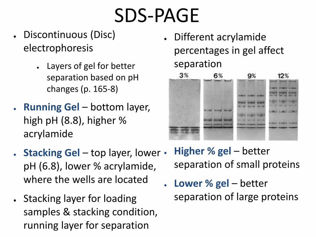
SDS-PAGE ● Discontinuous (Disc)
electrophoresis
● Layers of gel for better separation based on pH changes (p. 165-8)
● Running Gel – bottom layer, high pH (8.8), higher % acrylamide
● Stacking Gel – top layer, lower pH (6.8), lower % acrylamide, where the wells are located
● Stacking layer for loading samples & stacking condition, running layer for separation
● Different acrylamide percentages in gel affect separation
● Higher % gel – better separation of small proteins
● Lower % gel – better separation of large proteins
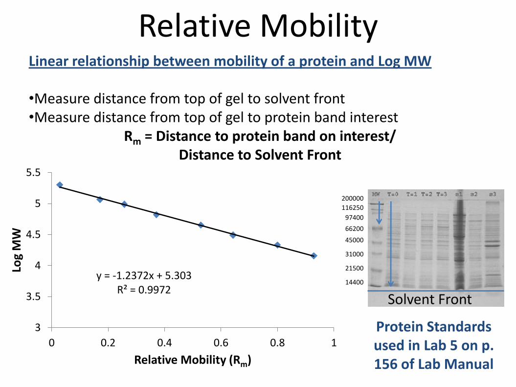
Relative Mobility Linear relationship between mobility of a protein and Log MW •Measure distance from top of gel to solvent front •Measure distance from top of gel to protein band interest
Rm = Distance to protein band on interest/ Distance to Solvent Front
Solvent Front
Protein Standards used in Lab 5 on p. 156 of Lab Manual
y = -1.2372x + 5.303 R² = 0.9972
3
3.5
4
4.5
5
5.5
0 0.2 0.4 0.6 0.8 1
Log
MW
Relative Mobility (Rm)
200000
116250
97400
66200
45000
31000
21500
14400
Procedure: Chapter 5-Week 2
● Prepare Samples
● Assemble Gel Apparatus
● Load Samples
● Run Gel
● Pour Gels (for the next lab section)
● Staining and Visualization

● Prepare Samples
● Crude Extract – 30-50 μg
● 3P-Dialyzed – 15-30 μg
● Purified LDH – 2-5 μg
● SDS-PAGE Standards – 5 μl aliquot, ready to load, 1 per gel
● Total Sample Volume = 20 μl
● 2 μl 10X Sample Buffer (thick blue liquid)
● Up to 18 μl of sample to put at the appropriate concentration of protein
● Water to bring total volume to 20 μl
● Denature samples 1-2 min at ~ 90°C
Procedure: Chapter 5-Week 2 Use concentration from Dye-
Binding from Chapter 3!
Calculate volumes before you come to the lab!
Standards: Bio-Rad Broad Range
See Table p. 156 Marked with ¶ Symbol

Procedure: Chapter 5-Week 2 ● Assemble Gel Apparatus:
● 2 groups/gel, 2 gels/gel box
● Pictures p. 135-136 and demo!
● Load Samples:
● Use gel loading tips
● Yellow loading guides can help line up wells
● Write down your loading scheme!
● Run Gel:
● 50 V until through the stacking gel, 200V after that
● Run until blue tracking dye reaches bottom of gel
● Total time ~ 1 hr

Procedure: Chapter 5-Week 2 ● Pour Gels:
● Put together short plate and spacer plate (1.5 mm)
● Lock together with casting clamp and stand (pictures p. 133)
● Check plates for leaks with water
● Prepare Running Gel – Recipe p. 134 (100 µl APS)
– Pour or pipet into plate, save extra and see when it polymerizes
– Overlay with isobutanol
● Prepare Stacking Gel – Recipe p. 134 (40 µl APS)
– Pour or pipet into plate, save extra and see when it polymerizes
– Insert comb to form wells
● Put poured gel in buffer at 4°C for the next section

Procedure: Chapter 5-Week 2 ● Staining and Visualization:
● Remove gel from plates
● Nick a corner so you know the orientation
● Stain with Coomassie Brilliant Blue R-250
● Destain with 1:5:5 Acetic Acid/Methanol/Water solution
● Image on gel doc
● Calculate Rm values for standards and unknown protein bands
● Make graph of Log MW vs Rm

Linear relationship between mobility of a protein and Log MW •Measure distance from top of gel to solvent front •Measure distance from top of gel to protein band interest
Rm = Distance to protein band on interest/ Distance to Solvent Front
Solvent Front
Protein Standards used in Lab 5 on p. 156 of Lab Manual
y = -1.2372x + 5.303 R² = 0.9972
3
3.5
4
4.5
5
5.5
0 0.2 0.4 0.6 0.8 1
Log
MW
Relative Mobility (Rm)
200000
116250
97400
66200
45000
31000
21500
14400
Calculate Rm Values Make Plot of Log MW vs. Rm