Ankylosis - ASO · ankylosis this is likely to result in tipping of adjacent teeth, space loss and...
4
Creating Brighter Futures University of Sydney Ankylosis
Transcript of Ankylosis - ASO · ankylosis this is likely to result in tipping of adjacent teeth, space loss and...
Creating Brighter Futures
University of Sydney
Ankylosis
However detection is difficult because fusion of bone and cementum may be minute or concealed.10
4. Percussion. A change in tone on percussion is audible when at least 20% of the root surface is ankylosed.8,11
5. Orthodontic traction. Failure of an unobstructed tooth to move following application of orthodontic forces probably indicates ankylosis.4
Deciduous Tooth AnkylosisPrevalence rates of ankylosed deciduous teeth vary from 1.3% to 14.3% of the population, depending on the criteria used, with a significantly higher incidence between siblings.5.8 Deciduous mandibular molars are affected with more than ten times the frequency of deciduous maxillary molars. Kurol and Magnusson have found that in young children the ankylotic area is often in the apical part of the root, while in older children, it is located more coronally.7 Where a permanent successor is present the ankylosed deciduous molar usually resorbs normally and the effects on occlusal development are temporary. Normal conditions will usually resume following exfoliation and subsequent eruption of the permanent successor.6 Ankylosed second deciduous molars can often be associated with agenesis of second premolars and therefore do not usually exfoliate at the normal time.8
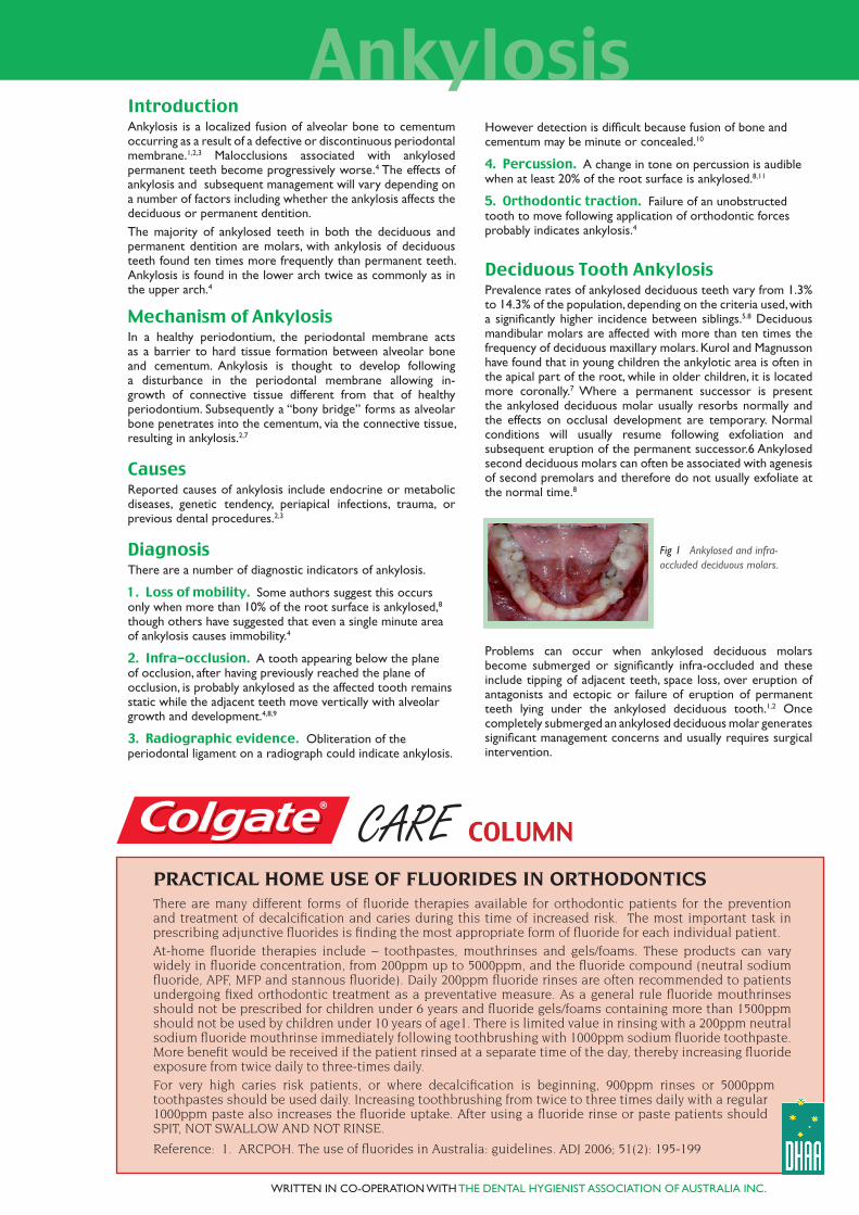
Fig 1 Ankylosed and infra-occluded deciduous molars.
Problems can occur when ankylosed deciduous molars become submerged or significantly infra-occluded and these include tipping of adjacent teeth, space loss, over eruption of antagonists and ectopic or failure of eruption of permanent teeth lying under the ankylosed deciduous tooth.1,2 Once completely submerged an ankylosed deciduous molar generates significant management concerns and usually requires surgical intervention.
WRITTEN IN CO-OPERATION WITH THE DENTAL HYGIENIST ASSOCIATION OF AUSTRALIA INC.
PRACTICAL HOME USE OF FLUORIDES IN ORTHODONTICSThere are many different forms of fluoride therapies available for orthodontic patients for the prevention and treatment of decalcification and caries during this time of increased risk. The most important task in prescribing adjunctive fluorides is finding the most appropriate form of fluoride for each individual patient.
At-home fluoride therapies include – toothpastes, mouthrinses and gels/foams. These products can vary widely in fluoride concentration, from 200ppm up to 5000ppm, and the fluoride compound (neutral sodium fluoride, APF, MFP and stannous fluoride). Daily 200ppm fluoride rinses are often recommended to patients undergoing fixed orthodontic treatment as a preventative measure. As a general rule fluoride mouthrinses should not be prescribed for children under 6 years and fluoride gels/foams containing more than 1500ppm should not be used by children under 10 years of age1. There is limited value in rinsing with a 200ppm neutral sodium fluoride mouthrinse immediately following toothbrushing with 1000ppm sodium fluoride toothpaste. More benefit would be received if the patient rinsed at a separate time of the day, thereby increasing fluoride exposure from twice daily to three-times daily.
For very high caries risk patients, or where decalcification is beginning, 900ppm rinses or 5000ppm toothpastes should be used daily. Increasing toothbrushing from twice to three times daily with a regular 1000ppm paste also increases the fluoride uptake. After using a fluoride rinse or paste patients should SPIT, NOT SWALLOW AND NOT RINSE.
Reference: 1. ARCPOH. The use of fluorides in Australia: guidelines. ADJ 2006; 51(2): 195-199
IntroductionAnkylosis is a localized fusion of alveolar bone to cementum occurring as a result of a defective or discontinuous periodontal membrane.1,2,3 Malocclusions associated with ankylosed permanent teeth become progressively worse.4 The effects of ankylosis and subsequent management will vary depending on a number of factors including whether the ankylosis affects the deciduous or permanent dentition. The majority of ankylosed teeth in both the deciduous and permanent dentition are molars, with ankylosis of deciduous teeth found ten times more frequently than permanent teeth. Ankylosis is found in the lower arch twice as commonly as in the upper arch.4
Mechanism of AnkylosisIn a healthy periodontium, the periodontal membrane acts as a barrier to hard tissue formation between alveolar bone and cementum. Ankylosis is thought to develop following a disturbance in the periodontal membrane allowing in-growth of connective tissue different from that of healthy periodontium. Subsequently a “bony bridge” forms as alveolar bone penetrates into the cementum, via the connective tissue, resulting in ankylosis.2,7
CausesReported causes of ankylosis include endocrine or metabolic diseases, genetic tendency, periapical infections, trauma, or previous dental procedures.2,3
DiagnosisThere are a number of diagnostic indicators of ankylosis.
1. Loss of mobility. Some authors suggest this occurs only when more than 10% of the root surface is ankylosed,8 though others have suggested that even a single minute area of ankylosis causes immobility.4
2. Infra-occlusion. A tooth appearing below the plane of occlusion, after having previously reached the plane of occlusion, is probably ankylosed as the affected tooth remains static while the adjacent teeth move vertically with alveolar growth and development.4,8,9
3. Radiographic evidence. Obliteration of the periodontal ligament on a radiograph could indicate ankylosis.
CARE COLUMN
Ankylosis
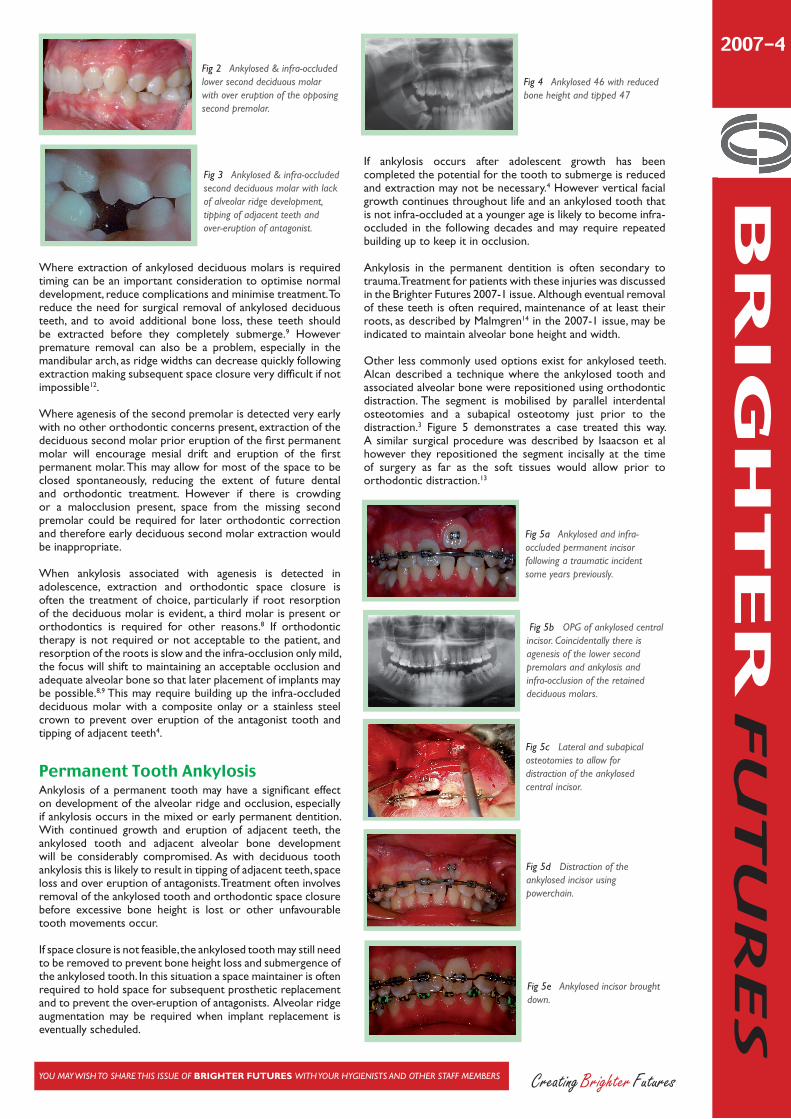
Fig 2 Ankylosed & infra-occluded lower second deciduous molar with over eruption of the opposing second premolar.
Fig 3 Ankylosed & infra-occluded second deciduous molar with lack of alveolar ridge development, tipping of adjacent teeth and over-eruption of antagonist.
Where extraction of ankylosed deciduous molars is required timing can be an important consideration to optimise normal development, reduce complications and minimise treatment. To reduce the need for surgical removal of ankylosed deciduous teeth, and to avoid additional bone loss, these teeth should be extracted before they completely submerge.9 However premature removal can also be a problem, especially in the mandibular arch, as ridge widths can decrease quickly following extraction making subsequent space closure very difficult if not impossible12.
Where agenesis of the second premolar is detected very early with no other orthodontic concerns present, extraction of the deciduous second molar prior eruption of the first permanent molar will encourage mesial drift and eruption of the first permanent molar. This may allow for most of the space to be closed spontaneously, reducing the extent of future dental and orthodontic treatment. However if there is crowding or a malocclusion present, space from the missing second premolar could be required for later orthodontic correction and therefore early deciduous second molar extraction would be inappropriate.
When ankylosis associated with agenesis is detected in adolescence, extraction and orthodontic space closure is often the treatment of choice, particularly if root resorption of the deciduous molar is evident, a third molar is present or orthodontics is required for other reasons.8 If orthodontic therapy is not required or not acceptable to the patient, and resorption of the roots is slow and the infra-occlusion only mild, the focus will shift to maintaining an acceptable occlusion and adequate alveolar bone so that later placement of implants may be possible.8,9 This may require building up the infra-occluded deciduous molar with a composite onlay or a stainless steel crown to prevent over eruption of the antagonist tooth and tipping of adjacent teeth4.
Permanent Tooth AnkylosisAnkylosis of a permanent tooth may have a significant effect on development of the alveolar ridge and occlusion, especially if ankylosis occurs in the mixed or early permanent dentition. With continued growth and eruption of adjacent teeth, the ankylosed tooth and adjacent alveolar bone development will be considerably compromised. As with deciduous tooth ankylosis this is likely to result in tipping of adjacent teeth, space loss and over eruption of antagonists. Treatment often involves removal of the ankylosed tooth and orthodontic space closure before excessive bone height is lost or other unfavourable tooth movements occur.
If space closure is not feasible, the ankylosed tooth may still need to be removed to prevent bone height loss and submergence of the ankylosed tooth. In this situation a space maintainer is often required to hold space for subsequent prosthetic replacement and to prevent the over-eruption of antagonists. Alveolar ridge augmentation may be required when implant replacement is eventually scheduled.
Fig 4 Ankylosed 46 with reduced bone height and tipped 47
If ankylosis occurs after adolescent growth has been completed the potential for the tooth to submerge is reduced and extraction may not be necessary.4 However vertical facial growth continues throughout life and an ankylosed tooth that is not infra-occluded at a younger age is likely to become infra-occluded in the following decades and may require repeated building up to keep it in occlusion.
Ankylosis in the permanent dentition is often secondary to trauma. Treatment for patients with these injuries was discussed in the Brighter Futures 2007-1 issue. Although eventual removal of these teeth is often required, maintenance of at least their roots, as described by Malmgren14 in the 2007-1 issue, may be indicated to maintain alveolar bone height and width. Other less commonly used options exist for ankylosed teeth. Alcan described a technique where the ankylosed tooth and associated alveolar bone were repositioned using orthodontic distraction. The segment is mobilised by parallel interdental osteotomies and a subapical osteotomy just prior to the distraction.3 Figure 5 demonstrates a case treated this way. A similar surgical procedure was described by Isaacson et al however they repositioned the segment incisally at the time of surgery as far as the soft tissues would allow prior to orthodontic distraction.13
Fig 5a Ankylosed and infra-occluded permanent incisor following a traumatic incident some years previously.
Fig 5b OPG of ankylosed central incisor. Coincidentally there is agenesis of the lower second premolars and ankylosis and infra-occlusion of the retained deciduous molars.
Fig 5c Lateral and subapical osteotomies to allow for distraction of the ankylosed central incisor.
Fig 5d Distraction of the ankylosed incisor using powerchain.
Fig 5e Ankylosed incisor brought down.
2007-4B
RIG
HT
ER
FU
TU
RE
S
Creating Brighter FuturesYOU MAY WISH TO SHARE THIS ISSUE OF BRIGHTER FUTURES WITH YOUR HYGIENISTS AND OTHER STAFF MEMBERS
BRIGHTER FUTURES
Alternatively it is possible to surgically luxate a tooth in its alveolus and then promptly apply vertical traction to attempt to move the tooth into occlusion.10 With this procedure it is important to preserve the existing cortical bone and sever the bony bridge of ankylosis without injury to the periodontium and nutrient vessels at the apices.4 However this procedure bears the significant risks of devitalisation, root fracture, root resorption and recurrent ankylosis.
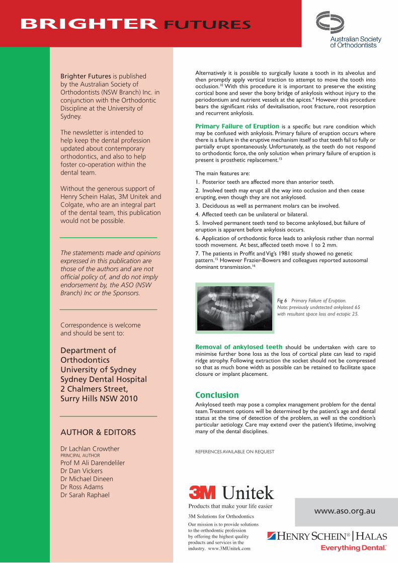
Primary Failure of Eruption is a specific but rare condition which may be confused with ankylosis. Primary failure of eruption occurs where there is a failure in the eruptive mechanism itself so that teeth fail to fully or partially erupt spontaneously. Unfortunately, as the teeth do not respond to orthodontic force, the only solution when primary failure of eruption is present is prosthetic replacement.15
The main features are: 1. Posterior teeth are affected more than anterior teeth.2. Involved teeth may erupt all the way into occlusion and then cease erupting, even though they are not ankylosed.3. Deciduous as well as permanent molars can be involved.4. Affected teeth can be unilateral or bilateral.5. Involved permanent teeth tend to become ankylosed, but failure of eruption is apparent before ankylosis occurs.6. Application of orthodontic force leads to ankylosis rather than normal tooth movement. At best, affected teeth move 1 to 2 mm.7. The patients in Proffit and Vig’s 1981 study showed no genetic pattern.15 However Frazier-Bowers and colleagues reported autosomal dominant transmission.16
Fig 6 Primary Failure of Eruption. Note: previously undetected ankylosed 65 with resultant space loss and ectopic 25.
Removal of ankylosed teeth should be undertaken with care to minimise further bone loss as the loss of cortical plate can lead to rapid ridge atrophy. Following extraction the socket should not be compressed so that as much bone width as possible can be retained to facilitate space closure or implant placement.
ConclusionAnkylosed teeth may pose a complex management problem for the dental team. Treatment options will be determined by the patient’s age and dental status at the time of detection of the problem, as well as the condition’s particular aetiology. Care may extend over the patient’s lifetime, involving many of the dental disciplines.
REFERENCES AVAILABLE ON REQUEST
Brighter Futures is published by the Australian Society of Orthodontists (NSW Branch) Inc. in conjunction with the Orthodontic Discipline at the University of Sydney.
The newsletter is intended to help keep the dental profession updated about contemporary orthodontics, and also to help foster co-operation within the dental team.
Without the generous support of Henry Schein Halas, 3M Unitek and Colgate, who are an integral part of the dental team, this publication would not be possible.
The statements made and opinions expressed in this publication are those of the authors and are not official policy of, and do not imply endorsement by, the ASO (NSW Branch) Inc or the Sponsors.
Correspondence is welcome and should be sent to:
Department of OrthodonticsUniversity of SydneySydney Dental Hospital2 Chalmers Street, Surry Hills NSW 2010
AUTHOR & EDITORS
Dr Lachlan CrowtherPRINCIPAL AUTHOR
Prof M Ali DarendelilerDr Dan VickersDr Michael DineenDr Ross AdamsDr Sarah Raphael
Products that make your life easier www.aso.org.au
3M Solutions for Orthodontics Our mission is to provide solutions to the orthodontic profession by offering the highest quality products and services in the industry. www.3MUnitek.com
b