Anisotropic dehydration of hydrogel surfacesoro.open.ac.uk/51753/1/51753.pdfORIGINAL RESEARCH...
8
ORIGINAL RESEARCH Anisotropic dehydration of hydrogel surfaces Georgia Kaklamani 1 • David Cheneler 2 • Liam M. Grover 3 • Michael J. Adams 3 • Spiros H. Anastasiadis 1,4 • James Bowen 5 Received: 20 June 2017 / Accepted: 14 October 2017 Ó The Author(s) 2017. This article is an open access publication Abstract Efforts to develop tissue-engineered skin for regenerative medicine have explored natural, synthetic, and hybrid hydrogels. The creation of a bilayer material, with the stratification exhibited by native skin, is a complex problem. The mechanically robust, waterproof epidermis presents the stratum corneum at the tissue/air interface, which confers many of these protective properties. In this work, we explore the effect of high temperatures on algi- nate hydrogels, which are widely employed for tissue engineering due to their excellent mechanical properties and cellular compatibility. In particular, we investigate the rapid dehydration of the hydrogel surface which occurs following local exposure to heated surfaces with tempera- tures in the range 100–200 °C. We report the creation of a mechanically strengthened hydrogel surface, with improved puncture resistance and increased coefficient of friction, compared to an unheated surface. The use of a mechanical restraint during heating promoted differences in the rate of mass loss; the rate of temperature increase within the hydrogel, in the presence and absence of restraint, is simulated and discussed. It is hoped that the results will be of use in the development of processes suitable for preparing skin-like analogues; application areas could include wound healing and skin restoration. Keywords Alginate Á Dehydration Á Hydrogel Á Polysaccharide Á Skin Á Stratification Introduction Biological tissues are highly organized and stratified structures. Complex tissue architectures are composed of different cell types with specific functions and locations. The interaction between them is conducted via their extracellular matrix. The interfaces between tissues are also complex and not easily distinguishable (Dormer et al. 2010). Tissue engineering aims to develop and mimic such architectures in vitro; various methods having been developed to simulate tissue complexity and allow inter- action between different cell types, proteins, implanted materials, and scaffolds (Lee et al. 2009). Methods include 3D printing, electrospinning, and cell-sheeting engineering, often requiring complicated manipulations or lengthy constructions (Grossin et al. 2009). Skin, the largest organ of the body, exhibits a stratified and organized structure, providing a protective layer with multiple functions (Horch et al. 2005). Nerve fibres and sensory receptors permit the detection of touch, pain, and temperature (Adams et al. 2007; Adams et al. 2013). There is significant interest in developing skin analogues suit- able for replacing real skin, accelerating wound healing, restoring burns, and functioning like native skin (MacNeil 2007). Skin tissue is composed of two layers: epidermis, a waterproof barrier that excludes microbes and retains body & James Bowen [email protected] 1 Institute of Electronic Structure and Laser, Foundation for Research and Technology Hellas, Heraklion, Crete, Greece 2 Engineering Department, Lancaster University, Bailrigg, Lancaster LA1 4YR, UK 3 School of Chemical Engineering, The University of Birmingham, Edgbaston B15 2TT, UK 4 Department of Chemistry, University of Crete, Heraklion, Crete, Greece 5 Department of Engineering and Innovation, The Open University, Milton Keynes MK7 6AA, UK 123 Prog Biomater DOI 10.1007/s40204-017-0075-9
Transcript of Anisotropic dehydration of hydrogel surfacesoro.open.ac.uk/51753/1/51753.pdfORIGINAL RESEARCH...

ORIGINAL RESEARCH
Anisotropic dehydration of hydrogel surfaces
Georgia Kaklamani1 • David Cheneler2 • Liam M. Grover3 • Michael J. Adams3 •
Spiros H. Anastasiadis1,4 • James Bowen5
Received: 20 June 2017 / Accepted: 14 October 2017
� The Author(s) 2017. This article is an open access publication
Abstract Efforts to develop tissue-engineered skin for
regenerative medicine have explored natural, synthetic, and
hybrid hydrogels. The creation of a bilayer material, with
the stratification exhibited by native skin, is a complex
problem. The mechanically robust, waterproof epidermis
presents the stratum corneum at the tissue/air interface,
which confers many of these protective properties. In this
work, we explore the effect of high temperatures on algi-
nate hydrogels, which are widely employed for tissue
engineering due to their excellent mechanical properties
and cellular compatibility. In particular, we investigate the
rapid dehydration of the hydrogel surface which occurs
following local exposure to heated surfaces with tempera-
tures in the range 100–200 �C. We report the creation of a
mechanically strengthened hydrogel surface, with
improved puncture resistance and increased coefficient of
friction, compared to an unheated surface. The use of a
mechanical restraint during heating promoted differences
in the rate of mass loss; the rate of temperature increase
within the hydrogel, in the presence and absence of
restraint, is simulated and discussed. It is hoped that the
results will be of use in the development of processes
suitable for preparing skin-like analogues; application
areas could include wound healing and skin restoration.
Keywords Alginate � Dehydration � Hydrogel �Polysaccharide � Skin � Stratification
Introduction
Biological tissues are highly organized and stratified
structures. Complex tissue architectures are composed of
different cell types with specific functions and locations.
The interaction between them is conducted via their
extracellular matrix. The interfaces between tissues are
also complex and not easily distinguishable (Dormer et al.
2010). Tissue engineering aims to develop and mimic such
architectures in vitro; various methods having been
developed to simulate tissue complexity and allow inter-
action between different cell types, proteins, implanted
materials, and scaffolds (Lee et al. 2009). Methods include
3D printing, electrospinning, and cell-sheeting engineering,
often requiring complicated manipulations or lengthy
constructions (Grossin et al. 2009).
Skin, the largest organ of the body, exhibits a stratified
and organized structure, providing a protective layer with
multiple functions (Horch et al. 2005). Nerve fibres and
sensory receptors permit the detection of touch, pain, and
temperature (Adams et al. 2007; Adams et al. 2013). There
is significant interest in developing skin analogues suit-
able for replacing real skin, accelerating wound healing,
restoring burns, and functioning like native skin (MacNeil
2007).
Skin tissue is composed of two layers: epidermis, a
waterproof barrier that excludes microbes and retains body
& James Bowen
1 Institute of Electronic Structure and Laser, Foundation for
Research and Technology Hellas, Heraklion, Crete, Greece
2 Engineering Department, Lancaster University, Bailrigg,
Lancaster LA1 4YR, UK
3 School of Chemical Engineering, The University of
Birmingham, Edgbaston B15 2TT, UK
4 Department of Chemistry, University of Crete, Heraklion,
Crete, Greece
5 Department of Engineering and Innovation, The Open
University, Milton Keynes MK7 6AA, UK
123
Prog Biomater
DOI 10.1007/s40204-017-0075-9

fluids; and dermis, beneath the epidermis, which is a col-
lagen-rich connective tissue (Hunt et al. 2009). Melano-
cytes impart skin colour and are found at the lower level of
the epidermis, and fibroblasts are found at the dermal layer
and are responsible for the strength of the skin (MacNeil
2007; Bannasch et al. 2003). Epidermis, the outer skin
layer, has a surface called stratum corneum, which is a less
hydrated surface layer presented at the air/skin interface.
Stratum corneum is composed of dead cells formed from
keratin and with thickness that varies from 10 to 15 lm for
humans (Johnson et al. 1993).
Significant progress has been made in the development
of tissue-engineered skin (Shevchenko et al. 2010), the
skin analogue produced using cells, extracellular matrix,
and combinations thereof, although the use of autografts
and allografts is associated with several limitations (Priya
et al. 2008; Bello et al. 2001). Various methods have been
utilized to achieve skin regeneration (Yang et al. 2000;
Lechler and Fuchs 2005; Fuchs and Horsley 2008), or to
manufacture skin products for wound healing. Most of the
skin products that have been reported use a natural,
synthetic, or hybrid hydrogel as a scaffold (Priya et al.
2008; Currie et al. 2001; Bakakrishnan et al. 2005;
Boucard et al. 2007; Powell and Boyce 2009), for
example the use of collagen matrix encapsulated with
fibroblasts and seeded with keratinocytes (Yang et al.
2000). Due to the poor mechanical properties and diffi-
culties of handling collagen hydrogel, hybrid collagen/
alginate scaffolds have also been produced (Kim et al.
2011). Other hybrid scaffolds for skin regeneration
include chitosan–gelatin bilayers (Mao et al. 2003).
Stratified materials have also been produced using poly-
electrolyte multilayers alternated with cell-containing gel
layers (Grossin et al. 2009).
Alginate has been used in tissue engineering for the
regeneration of skin, bone, and cartilage due to its
mechanical properties and cellular compatibility, having
been used to support the growth of fibroblasts and ker-
atinocytes (Hunt et al. 2009; Kim et al. 2011; Alsberg et al.
2001; Stevens et al. 2004). Alginate is a naturally occur-
ring, non-toxic polysaccharide derived from brown algae
(Augst et al. 2006). It is biodegradable and can be used to
form hydrogels under cytocompatible conditions. Alginate
hydrogels are formed through ionotropic gelation of dis-
solved alginate in the presence of multivalent cations such
as Mg2?, Ca2?, Sr2?, and Ba2? (Morch et al. 2006; Topuz
et al. 2012), and can be gelled using the internal (Chan
et al. 2002a, b) or the external gelation method (Hunt et al.
2010; Kaklamani et al. 2014). Alginate has also been used
to produce hybrid hydrogels for tissue engineering appli-
cations (Choi et al. 1999), but no solution proposed yet
fulfils all the requirements needed to accomplish functional
stratified structures.
The aim of this work was to investigate processes by
which hydrogels can be modified to provide a stratum
corneum-like dehydrated surface layer. Using a high-tem-
perature heat source directly in contact with the hydrogel,
the effect of rapid dehydration by evaporative loss of water
from the hydrogel surface was studied. The influence of
temperature and contact duration was considered. The
external gelation method was employed to produce
hydrogel samples; alginate concentration was also varied
and investigated. The purpose of this research is to develop
processes which might be suitable for the preparation of
skin-like analogues.
Experimental
Materials
All chemicals were sourced from Sigma Aldrich (UK)
unless otherwise stated. Purities were[ 99% in all cases.
HPLC-grade H2O was employed throughout.
Hydrogel preparation
Hydrogels (HGs) were prepared using a previously repor-
ted external gelation method (Kaklamani et al. 2014).
Briefly, sodium alginate (NaAlg) solution was poured into
a poly(styrene) mould (141.4 mm inside diameter, 9.0 mm
inside height, Sterilin, UK) to a liquid height of 6 mm and
allowed to gel in the presence of an aqueous solution of
CaCl2 held at the upper and lower boundaries by porous
microcellulose sheets. Prior to the addition of the aqueous
NaAlg solution, stainless steel cylindrical spacers (21 mm
diameter, 6 mm height, Longshore Systems Engineering,
UK) were placed at 60 o intervals around the inner edge of
the mould to support the upper sheet. The volume of
aqueous NaAlg solution required was 81.75 mL. The upper
sheet was held in place from above using a poly(styrene)
support, filled with water to maintain close contact between
the upper sheet and the NaAlg solution as it gelled, since
some shrinkage was observed at the sample edges.
NaAlg powder was dissolved in H2O under agitated
conditions at a temperature of 70 �C and stirred for a
minimum of 2 h using a Stuart US-152 hot plate stirrer
(Appleton Woods, UK). The concentration of the NaAlg
solution was 2.5% (w/v) and 5.0% (w/v). An aqueous
CaCl2 solution of 2 M concentration was created by dis-
solving CaCl2 powder in water, whilst stirring, at room
temperature; the solution was allowed to cool to room
temperature of 18 �C prior to use. Microcellulose sheets
(QL100, Fisherbrand, UK) were trimmed to match the
shape of the poly(styrene) mould and immersed in aqueous
CaCl2 solution for 5 min immediately prior to use. Samples
Prog Biomater
123

were allowed to gel at 18 �C for 60 min. The gelation
geometry and schematic are shown in Fig. 1, reproduced
from (Kaklamani et al. 2014). The solid gels which formed
are hereafter referred to by their Young’s modulus, which
have also been previously reported (Kaklamani et al.
2014), and are 189 ± 25 kPa (2.5% w/v) and
433 ± 17 kPa (5.0% w/v).
High-temperature dehydration
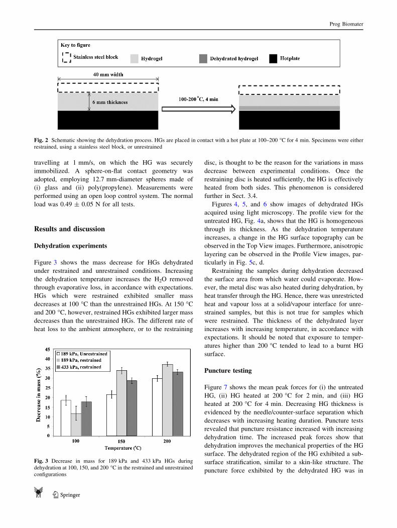
Following HG preparation, rectangular specimens
(40 mm 9 40 mm) were extracted using a cutting tool. HG
specimens were heated for 4 min using a hot plate (Fisher
Scientific, UK) at temperatures of 100, 150 and 200 �C,i.e., above the boiling point of water. Two different con-
ditions were investigated—restrained and unrestrained. In
the restrained condition, a 40 mm diameter stainless steel
block (mass = 80 g) was used to ensure intimate contact
between the specimen and the hot plate. In the unrestrained
condition, samples were prone to migrate across the surface
of the hot plate, a thin layer of water vapour lubricating the
contact. A schematic is shown in Fig. 2. The face of the
specimen adjacent to the hot plate is hereafter referred to as
the Dehydrated Surface, while the opposite face is referred
to as the Upper Surface. The mass of each specimen was
measured before and after dehydration.
Puncture testing
Puncture testing was performed at 18 �C and 40% relative
humidity using a Z030 mechanical tester (Zwick/Roell,
UK). A stainless steel needle (gauge 19G, bevel point,
Fisher Scientific, UK) was attached to a 5 N load cell. The
puncture procedure involved approaching the needle
towards the HG at a velocity of 5 mm/s. The specimen was
oriented with the Dehydrated Surface presented upwards,
facing the needle. Load–displacement data were recorded
at 100 Hz.
Friction measurements
Tribological measurements were performed on the Dehy-
drated Surface using a custom-built tribometer (Longshore
Systems Engineering, UK). Tests employed a rotating stage
Fig. 1 Gelation geometry (a) and gelation schematic (b) showing
(i) ungelled alginate solution, (ii)–(iii) progression of the gelation
through the alginate solution as the cations diffuse into the alginate
solution, generating cross-links, and (iv) the final, gelled alginate.
Reproduced from (Kaklamani et al. 2014)
Prog Biomater
123
travelling at 1 mm/s, on which the HG was securely
immobilized. A sphere-on-flat contact geometry was
adopted, employing 12.7 mm-diameter spheres made of
(i) glass and (ii) poly(propylene). Measurements were
performed using an open loop control system. The normal
load was 0.49 ± 0.05 N for all tests.
Results and discussion
Dehydration experiments
Figure 3 shows the mass decrease for HGs dehydrated
under restrained and unrestrained conditions. Increasing
the dehydration temperature increases the H2O removed
through evaporative loss, in accordance with expectations.
HGs which were restrained exhibited smaller mass
decreases at 100 �C than the unrestrained HGs. At 150 �Cand 200 �C, however, restrained HGs exhibited larger mass
decreases than the unrestrained HGs. The different rate of
heat loss to the ambient atmosphere, or to the restraining
disc, is thought to be the reason for the variations in mass
decrease between experimental conditions. Once the
restraining disc is heated sufficiently, the HG is effectively
heated from both sides. This phenomenon is considered
further in Sect. 3.4.
Figures 4, 5, and 6 show images of dehydrated HGs
acquired using light microscopy. The profile view for the
untreated HG, Fig. 4a, shows that the HG is homogeneous
through its thickness. As the dehydration temperature
increases, a change in the HG surface topography can be
observed in the Top View images. Furthermore, anisotropic
layering can be observed in the Profile View images, par-
ticularly in Fig. 5c, d.
Restraining the samples during dehydration decreased
the surface area from which water could evaporate. How-
ever, the metal disc was also heated during dehydration, by
heat transfer through the HG. Hence, there was unrestricted
heat and vapour loss at a solid/vapour interface for unre-
strained samples, but this is not true for samples which
were restrained. The thickness of the dehydrated layer
increases with increasing temperature, in accordance with
expectations. It should be noted that exposure to temper-
atures higher than 200 �C tended to lead to a burnt HG
surface.
Puncture testing
Figure 7 shows the mean peak forces for (i) the untreated
HG, (ii) HG heated at 200 �C for 2 min, and (iii) HG
heated at 200 �C for 4 min. Decreasing HG thickness is
evidenced by the needle/counter-surface separation which
decreases with increasing heating duration. Puncture tests
revealed that puncture resistance increased with increasing
dehydration time. The increased peak forces show that
dehydration improves the mechanical properties of the HG
surface. The dehydrated region of the HG exhibited a sub-
surface stratification, similar to a skin-like structure. The
puncture force exhibited by the dehydrated HG was in
Fig. 2 Schematic showing the dehydration process. HGs are placed in contact with a hot plate at 100–200 �C for 4 min. Specimens were either
restrained, using a stainless steel block, or unrestrained
Fig. 3 Decrease in mass for 189 kPa and 433 kPa HGs during
dehydration at 100, 150, and 200 �C in the restrained and unrestrained
configurations
Prog Biomater
123

accordance with the puncture forces reported for needle
insertion into skin (vanGerwen et al. 2012). The puncture
resistance of HGs can be tailored by changing the cauter-
ization time, an area for further investigation.
Tribological testing versus glass and polypropylene
The results of tribological testing of untreated and dehy-
drated (200 �C, 4 min) HGs versus glass and poly(propy-
lene) are presented in Table 1. The friction coefficient of
the HG surface against both materials more than doubled
due to dehydration. The glass surface is hydrophilic
(hH2O = 0�), whereas poly(propylene is hydrophobic
(hH2O = 112�). Hence, the HG/glass adhesion is greater
than the HG/poly(propylene) adhesion, and therefore the
friction coefficient is correspondingly larger for the
HG/glass than for HG/poly(propylene). Further, the dehy-
drated surface did not visibly undergo syneresis when
under contact pressure from the spherical probe, which
meant that lubricating effects were negligible compared to
the untreated surface. The friction coefficients of the
dehydrated HG surface compare favourably with those of
human skin: lglass = 0.26 and lPP = 0.35 (Adams et al.
2007).
Effect of restraint on heating rate
A heat transfer simulation of heating the restrained and
unrestrained HG samples was performed using Energy2D
(v2.9.5) (Xie 2012). The material properties used are
shown in Table 2. The initial hot plate temperatures of 100,
Fig. 4 Top view and profile view for 189 kPa HGs during dehydration at 100, 150, and 200 �C in the unrestrained configuration
Fig. 5 Top view and profile view for 189 kPa HGs during dehydration at 100, 150, and 200 �C in the restrained configuration
Prog Biomater
123

150, and 200 �C were specified; changes in sample thick-
ness due to evaporative losses were not incorporated.
Figure 8 shows the effect of incorporating the restrain-
ing disc on the temperature of the upper surface of the
433 kPa HG. The presence of the restraining disc decreases
the rate of temperature increase within the HG. The disc
also increases in temperature, and is typically 15–20 �Clower than the HG. In the restrained condition, energy from
the hot plate accumulates in the aluminium disc and the
HG. In the unrestrained condition, energy is lost more
easily, because the HG is a good thermal conductor. This
disattenuates the energy loss at the upper surface, by
preventing heat transfer from the HG to the ambient
atmosphere. The disc also means that the HG is heated
from above and below when restrained.
Considering the E = 189 kPa HGs in Fig. 3, at
T = 100 �C the restrained HG loses less mass than the
unrestrained HG. This is because the temperature of the
restrained HG increases more slowly than the unrestrained
HG. The HG temperature does not reach 100 �C, the
boiling point of water, for either the restrained or unre-
strained condition. At T = 150 and 200 �C, the restrained
HGs exhibit greater mass loss than the unrestrained HGs;
why might this be? Here, the HG temperature rises to an
Fig. 6 Top view and profile view for 433 kPa HGs during dehydration at 100, 150, and 200 �C in the restrained configuration
Fig. 7 a Puncture schematic, showing dehydrated hydrogel as the upper surface. b Puncture test data for 433 kPa HGs: untreated (green);
dehydrated at 200 �C for 2 min (blue); dehydrated at 200 �C for 4 min (red)
Table 1 Friction coefficient of
cauterized alginate against glass
and poly(propylene)
Hydrogel substrate Friction coefficient vs glass
lglass (-)
Friction coefficient vs poly(propylene)
lPP (-)
Untreated 0.31 ± 0.01 0.12 ± 0.01
Dehydrated, 200 �C, 4 min 0.63 ± 0.02 0.28 ± 0.01
Prog Biomater
123

excess of 100 �C for both the restrained and unrestrained
conditions. It is suggested that energy retained within the
restrained HG/aluminium disc increases the rate of water
evaporation, which otherwise would have caused an
increase in temperature. In comparison, this energy is
conducted through the unrestrained HG and is lost at the
HG/air interface. The difference in behaviour occurs when
the hot plate temperature is increased above the boiling
point of water.
Conclusions
We report the effect of local exposure to high temperatures
on the surface properties of alginate hydrogels. Rapid
dehydration of the surface using temperatures in the range
100–200 �C leads to the creation of a mechanically
strengthened hydrogel surface layer, exhibiting improved
puncture resistance and increased coefficient of friction
compared to the unheated surface. Direct contact with a
surface at 200 �C for 4 min produced the most mechani-
cally robust layer. Mechanical constraint of the hydrogel
sample was important during processing, preventing cur-
vature of the sample during cauterization, particularly at
the edges.
Further investigations will study a greater variety of HG
compositions, as well as explore in more detail the
importance of dehydration temperature, duration, the
thermal conductivity of the HG, and the nature of the
restraint material. A modified apparatus for dehydration
within a humidified atmosphere, for reducing evaporative
loss, is currently being designed. The possible benefit of
performing the heating whilst the HG is immersed in an
aqueous solution, perhaps a physiologically relevant buffer,
is also under consideration. Finally, an improved under-
standing of the material structure remaining following the
dehydration process would be useful.
Acknowledgements This work was supported by the European
Union under the FP7 programme (NANOBIOTOUCH Project: FP7-
NMP-228844). The Zwick/Roell Z030 mechanical tester and Long-
shore Systems Engineering tribometer used in this research were
obtained through Birmingham Science City: Innovative Uses for
Advanced Materials in the Modern World (West Midlands Centre for
Advanced Materials Project 2), with support from Advantage West
Midlands (AWM) and partly funded by the European Regional
Development Fund (ERDF). The authors thank the assistance of
Elaine Mitchell throughout the course of this research.
Compliance with ethical standards
Conflict of interest All authors declare that they have no conflict of
interest.
Ethical approval This article does not contain any studies with
human participants or animals performed by any of the authors.
Table 2 Parameters used for
heat transfer simulationsMaterial Density
q (kg m-3)
Heat capacity
Cp (kJ kg-1 K-1)
Thermal conductivity
k (W m-1 K-1)
Air 1.204 1.012 0.025
Aluminium hot platea 2700 0.921 205
Hydrogel 1070 4.184 0.591
Steel disc 8000 0.502 50
aConstant temperature
Fig. 8 Temperature of the HG sample upper surface when heated from below using a hot plate, a unrestrained, and b restrained
Prog Biomater
123

Open Access This article is distributed under the terms of the
Creative Commons Attribution 4.0 International License (http://crea
tivecommons.org/licenses/by/4.0/), which permits unrestricted use,
distribution, and reproduction in any medium, provided you give
appropriate credit to the original author(s) and the source, provide a
link to the Creative Commons license, and indicate if changes were
made.
References
Adams MJ, Briscoe BJ, Johnson SA (2007) Friction and lubrication of
human skin. Tribol Lett 26:239–253. doi:10.1007/s11249-007-
9206-0
Adams MJ, Johnson SA, Lefevre P, Levesque V, Hayard V (2013)
Finger pad friction and its role in grip and touch. J R Soc
Interface 10:1–19
Alsberg E, Anderson KW, Albeiruti A, Franceschi RT, Mooney DJ
(2001) Cell-interactive alginate hydrogels for bone tissue
engineering. J Dental Res 80:2025–2029
Augst AD, Kong JK, Mooney DJ (2006) Alginate hydrogels as
biomaterials. Macromol Biosci 6:623–633
Bakakrishnan B, Mohanty M, Umashankar PR, Jayakrishnan A
(2005) Evaluation of an in situ forming hydrogel wound dressing
based on oxidized alginate and gelatin. Biomaterials
26:6335–6342
Bannasch H, Fohn M, Unterberg T, Bach AD, Weyand B (2003) Skin
tissue engineering. Clin Plast Surg 30:573–579
Bello YM, Falabella AF, Eaglstein WH (2001) Tissue engineered
skin. Am J Clin Dermatol 2:305–313
Boucard N, Viton C, Agay D, Mari E, Roger T (2007) The use of
physical hydrogels of chitosan for skin regeneration following
third-degree burns. Biomaterials 28:3478–3488
Chan LW, Jin Y, Heng PWS (2002a) Cross-linking mechanisms of
calcium and zinc in production of alginate microspheres. Int J
Pharm 242:255–258
Chan LW, Lee H, Heng PWS (2002b) Production of alginate
microspheres by internal gelation using an emulsification
method. Int J Pharm 242:259–262
Choi YS, Hong SR, Lee YM, Song KW, Moon YP (1999) Study on
gelatin-containing artificial skin: I. Preparation and characteris-
tics of novel gelatin-alginate sponge. Biomaterials 20:409–417
Currie LJ, Sharpe JR, Martin R (2001) The use of fibrin glue in skin
grafts and tissue-engineered skin replacements: a review. Plast
Reconstr Surg 108:1713–1726
Dormer NH, Berkland CJ, Detamore MS (2010) Emerging techniques
in stratified designs and continues gradients for tissue engineer-
ing of interfaces. Ann Biomed Eng 38:2121–2141
Fuchs E, Horsley V (2008) More than one way to skin. Genes
Develop 22:976–985
Grossin L, Cortial D, Saulnier B, Felix O, Chassepot A, Decher G
(2009) Step-by-step built-up of biologically active cell-contain-
ing stratified films aimed at tissue engineering. Adv Mater
21:650–655
Horch RE, Kopp J, Kneser U, Beier J, Bach AD (2005) Tissue
engineering of cultured skin substrates. J Cell Mol Med
9:592–608
Hunt NC, Shelton RM, Grover LM (2009) An alginate hydrogels
matrix for the localized delivery of a fibroblast/keratinocyte co-
culture. Biotechnol J 4:730–737
Hunt NC, Smith AM, Gbureck U, Shelton RM, Grover LM (2010)
Encapsulation of fibroblasts causes accelerated alginate hydrogel
degradation. Acta Biomater 6:3649–3656
Johnson SA, Gorman DM, Adams MJ, Briscoe BJ (1993) The friction
and lubrication of human stratum corneum. Thin Films Tribol
25:663–672
Kaklamani G, Cheneler D, Grover LM, Adams MJ, Bowen J (2014)
Mechanical properties of alginate hydrogels using external
gelation. J Mech Behav Biomed Mater 36:135–142
Kim GH, Ahn S, Kim YY, Cho Y, Chun W (2011) Coaxial structured
collagen-alginate scaffolds: fabrication, physical properties, and
biomedical application for skin tissue regeneration. J Mater
Chem 21:6165–6172
Lechler T, Fuchs E (2005) Asymmetric cell divisions promote
stratification and differentiation of mammalian skin. Nature
437:275–280
Lee W, Debasitis JC, Lee VK, Lee JH, Fischer K (2009) Multi-
layered culture of human skin fibroblasts and keratinocytes
through three-dimensional freeform fabrication. Biomaterials
30:1587–1595
MacNeil S (2007) Progress and opportunities for tissue-engineered
skin. Nature 445:874–880
Mao J, Zhao L, Yao K, Shang Q, Yang G, Cao Y (2003) Study of
novel chitosan-gelatin artificial skin in vitro. J Biomed Mater
Res A 64:301–308
Morch YA, Donati I, Strand BL, Skjak-Bræk G (2006) Effect of
Ca2?, Ba2?, and Sr2? on alginate microbeads. Biomacromol
7:1471–1480
Powell HM, Boyce ST (2009) Engineered human skin fabricated
using electrospun collagen-PCL blends: morphogenesis and
mechanical properties. Tissue Eng A 15:2177–2187
Priya SG, Jungvid H, Kumar A (2008) Skin tissue engineering for
tissue repair and regeneration. Tissue Eng B 14:105–118
Shevchenko RV, James LS, James SE (2010) A review of tissue-
engineered skin bioconstructs available for skin reconstruction.
J R Soc Interface 7:229–258
Stevens MM, Qanadilo HF, Langer R, Shastri VP (2004) A rapid-
curing alginate gel system: utility in periosteum-derived carti-
lage tissue engineering. Biomaterials 25:887–894
Topuz F, Henke A, Richtering W, Groll J (2012) Magnesium ions and
alginate do form hydrogels: a rheological study. Soft Matter
8:4877–4881
VanGerwen DJ, Dankelman J, van den Dobbelstein JJ (2012) Needle–
tissue interaction forces—a survey of experimental data. Med
Eng Phys 34:665–680
Xie C (2012) Interactive heat transfer simulations for everyone. Phys
Teach 50:237–240
Yang EK, Seo YK, Youn HH, Lee DH, Park SN, Park JK (2000)
Tissue engineered artificial skin composed of dermis and
epidermis. Artif Organs 24:7–17
Publisher’s Note
Springer Nature remains neutral with regard to jurisdictional claims in
published maps and institutional affiliations.
Prog Biomater
123