Anionic Order and Band Gap Engineering in Vacancy Ordered ...
19
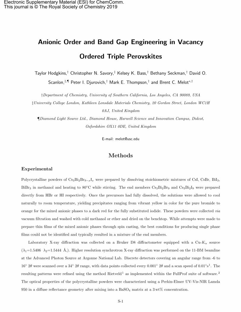
Anionic Order and Band Gap Engineering in Vacancy Ordered Triple Perovskites Taylor Hodgkins, † Christopher N. Savory, ‡ Kelsey K. Bass, † Bethany Seckman, † David O. Scanlon, ‡,¶ Peter I. Djurovich, † Mark E. Thompson, † and Brent C. Melot *,† †Department of Chemistry, University of Southern California, Los Angeles, CA 90089, USA ‡University College London, Kathleen Lonsdale Materials Chemistry, 20 Gordon Street, London WC1H 0AJ, United Kingdom ¶Diamond Light Source Ltd., Diamond House, Harwell Science and Innovation Campus, Didcot, Oxfordshire OX11 0DE, United Kingdom E-mail: [email protected] Methods Experimental Polycrystalline powders of Cs 3 Bi 2 Br 9-x I x were prepared by dissolving stoichiometric mixtures of CsI, CsBr, BiI 3 , BiBr 3 in methanol and heating to 80 ◦ C while stirring. The end members Cs 3 Bi 2 Br 9 and Cs 3 Bi 2 I 9 were prepared directly from HBr or HI respectively. Once the precursors had fully dissolved, the solutions were allowed to cool naturally to room temperature, yielding precipitates ranging from vibrant yellow in color for the pure bromide to orange for the mixed anionic phases to a dark red for the fully substituted iodide. These powders were collected via vacuum filtration and washed with cold methanol or ether and dried on the benchtop. While attempts were made to prepare thin films of the mixed anionic phases through spin casting, the best conditions for producing single phase films could not be identified and typically resulted in a mixture of the end members. Laboratory X-ray diffraction was collected on a Bruker D8 diffractometer equipped with a Cu-K α source (λ 1 =1.5406 λ 2 =1.5444 ˚ A,). Higher resolution synchrotron X-ray diffraction was performed on the 11-BM beamline at the Advanced Photon Source at Argonne National Lab. Discrete detectors covering an angular range from -6 to 16 ◦ 2θ were scanned over a 34 ◦ 2θ range, with data points collected every 0.001 ◦ 2θ and a scan speed of 0.01 ◦ s 1 . The resulting patterns were refined using the method Rietveld 1 as implemented within the FullProf suite of software. 2 The optical properties of the polycrystalline powders were characterized using a Perkin-Elmer UV-Vis-NIR Lamda 950 in a diffuse reflectance geometry after mixing into a BaSO 4 matrix at a 3 wt% concentration. S-1 Electronic Supplementary Material (ESI) for ChemComm. This journal is © The Royal Society of Chemistry 2019
Transcript of Anionic Order and Band Gap Engineering in Vacancy Ordered ...
Anionic Order and Band Gap Engineering in Vacancy
Ordered Triple Perovskites
Taylor Hodgkins,† Christopher N. Savory,‡ Kelsey K. Bass,† Bethany Seckman,† David O.
Scanlon,‡,¶ Peter I. Djurovich,† Mark E. Thompson,† and Brent C. Melot∗,†
†Department of Chemistry, University of Southern California, Los Angeles, CA 90089, USA
‡University College London, Kathleen Lonsdale Materials Chemistry, 20 Gordon Street, London WC1H
0AJ, United Kingdom
¶Diamond Light Source Ltd., Diamond House, Harwell Science and Innovation Campus, Didcot,
Oxfordshire OX11 0DE, United Kingdom
E-mail: [email protected]
Methods
Experimental
Polycrystalline powders of Cs3Bi2Br9−xIx were prepared by dissolving stoichiometric mixtures of CsI, CsBr, BiI3,
BiBr3 in methanol and heating to 80◦C while stirring. The end members Cs3Bi2Br9 and Cs3Bi2I9 were prepared
directly from HBr or HI respectively. Once the precursors had fully dissolved, the solutions were allowed to cool
naturally to room temperature, yielding precipitates ranging from vibrant yellow in color for the pure bromide to
orange for the mixed anionic phases to a dark red for the fully substituted iodide. These powders were collected via
vacuum filtration and washed with cold methanol or ether and dried on the benchtop. While attempts were made to
prepare thin films of the mixed anionic phases through spin casting, the best conditions for producing single phase
films could not be identified and typically resulted in a mixture of the end members.
Laboratory X-ray diffraction was collected on a Bruker D8 diffractometer equipped with a Cu-Kα source
(λ1=1.5406 λ2=1.5444 A,). Higher resolution synchrotron X-ray diffraction was performed on the 11-BM beamline
at the Advanced Photon Source at Argonne National Lab. Discrete detectors covering an angular range from -6 to
16◦ 2θ were scanned over a 34◦ 2θ range, with data points collected every 0.001◦ 2θ and a scan speed of 0.01◦s1. The
resulting patterns were refined using the method Rietveld1 as implemented within the FullProf suite of software.2
The optical properties of the polycrystalline powders were characterized using a Perkin-Elmer UV-Vis-NIR Lamda
950 in a diffuse reflectance geometry after mixing into a BaSO4 matrix at a 3 wt% concentration.
S-1
Electronic Supplementary Material (ESI) for ChemComm.This journal is © The Royal Society of Chemistry 2019

Computational
Density Functional Theory (DFT) calculations were performed within periodic boundary conditions using the Vienna
Abinitio Simulation Package (VASP) to simulate the effect of iodine substitution on the electronic structure within
the Cs3Bi2Br9 structure.3–6 The projector-augmented wave method is used to describe the interaction between
valence and core electrons in all calculations, and scalar-relativistic pseudopotentials were used, with Bi 5d electrons
explicitly treated as valence.7 All possible symmetry-inequivalent iodine-substituted structures within the unit cell of
Cs3Bi2Br9 were exhaustively generated through the SOD program.8 Geometry relaxation and energetic ordering of
these structures was then performed using the PBEsol functional,9 which has been used to successfully describe the
structural properties of Cs3Bi2Br9 and other bismuth halides in previous reports.10–12 Each structure was optimized
until the force on each atom did not exceed 0.01 eV A−1, and the lowest energy polymorph for each substitutional
configuration was then used as the basis for electronic structure calculations. The electronic properties, density
of states and band structures were calculated using the HSE06 functional,13 which with the addition of spin-orbit
coupling, has previously been used to accurately predict the electronic structures, including band gaps, of bismuth
halide semiconductors.11,14,15 A cutoff energy of 400 eV and a k-mesh of 3×3×3 was used in all calculations.
The equilibrium substituted structures reported are provided in an online repository: https://github.com/SMTG-
UCL/cesium-bismuth-halides.
S-2

References
(1) Rietveld, H. M. A profile refinement method for nuclear and magnetic structures. Journal of Applied Crystal-
lography 1969, 2, 65.
(2) Rodrıguez-Carvajal, J. Recent advances in magnetic structure determination by neutron powder diffraction.
Physica B 1993, 192, 55–69.
(3) Kresse, G.; Hafner, J. Ab initio molecular dynamics for liquid metals. Physical Review B 1993, 47, 558–561.
(4) Kresse, G.; Hafner, J. Ab initio molecular-dynamics simulation of the liquid-metal amorphous-semiconductor
transition in germanium. Physical Review B 1994, 49, 14251–14269.
(5) Kresse, G.; Furthmuller, J. Efficiency of ab initio total energy calculations for metals and semiconductors using
a plane wave basis set. Computational Materials Science 1996, 6, 15.
(6) Kresse, G.; Furthmuller, J. Efficiency of ab initio total energy calculations for metals and semiconductors using
a plane wave basis set. Computational Materials Science 1996, 6, 15–50.
(7) Blochl, P. E.; Jepsen, O.; Andersen, O. K. Improved tetrahedron method for Brillouin-zone integrations. Phys.
Rev.B 1994, 49, 16223.
(8) Grau-Crespo, R.; Hamad, S.; Catlow, C. R. A.; de Leeuw, N. H. Symmetry-adapted configurational modelling
of fractional site occupancy in solids. Journal of Physics: Condensed Matter 2007, 19, 256201.
(9) Perdew, J. P.; Ruzsinszky, A.; Csonka, G. I.; Vydrov, O. a.; Scuseria, G. E.; Constantin, L. A.; Zhou, X.;
Burke, K. Restoring the Density-Gradient Expansion for Exchange in Solids and Surfaces. Physical Review
Letters 2008, 100, 136406.
(10) Savory, C. N.; Walsh, A.; Scanlon, D. O. Can Pb-Free Halide Double Perovskites Support High-Efficiency Solar
Cells? ACS Energy Letters 2016, 1, 949–955.
(11) Bass, K. K.; Estergreen, L.; Savory, C. N.; Buckeridge, J.; Scanlon, D. O.; Djurovich, P. I.; Bradforth, S. E.;
Thompson, M. E.; Melot, B. C. Vibronic Structure in Room Temperature Photoluminescence of the Halide
Perovskite Cs3Bi2Br9. Inorganic Chemistry 2017, 56, 42–45.
(12) Ganose, A. M.; Matsumoto, S.; Buckeridge, J.; Scanlon, D. O. Defect Engineering of Earth-Abundant Solar
Absorbers BiSI and BiSeI. Chemistry of Materials 2018, 30, 3827–3835.
(13) Krukau, A. V.; Vydrov, O. A.; Izmaylov, A. F.; Scuseria, G. E. Influence of the exchange screening parameter
on the performance of screened hybrid functionals. The Journal of Chemical Physics 2006, 125, 224106.
S-3

(14) Lehner, A. J.; Fabini, D. H.; Evans, H. A.; Hbert, C.-A.; Smock, S. R.; Hu, J.; Wang, H.; Zwanziger, J. W.;
Chabinyc, M. L.; Seshadri, R. Crystal and Electronic Structures of Complex Bismuth Iodides A3Bi2I9 (A = K,
Rb, Cs) Related to Perovskite: Aiding the Rational Design of Photovoltaics. Chemistry of Materials 2015, 27,
7137–7148.
(15) Lehner, A. J.; Wang, H.; Fabini, D. H.; Liman, C. D.; Hbert, C.-A.; Perry, E. E.; Wang, M.; Bazan, G. C.;
Chabinyc, M. L.; Seshadri, R. Electronic structure and photovoltaic application of BiI3. Applied Physics Letters
2015, 107 .
S-4

(a) (b)
Cs3Bi2Br9 Cs3Bi2I9Figure S 1: Comparison of the close-packing sequence in the (a) trigonal bromide and (b) hexagonal iodide structures.Note the bromide results in layers of corner-sharing octahedra whereas the iodide produces dimerized units of face-sharing octahedra as described in the main text.
S-5

2 4 6 8 10 12 14 16 18 202θ (deg) [λ = 0.414613Å]
Inte
nsity
(ar
b. u
nits
)
Figure S 2: Results of the Rietveld refinement of the structure for Cs3Bi2Br9 against synchrotron X-ray diffractiondata collected on the 11-BM beamline (λ=0.414613A). Details of the fitted parameters can be found in the followingtable.
S-6

2 4 6 8 10 12 14 16 18 202θ (deg) [λ = 0.412602Å]
Inte
nsity
(ar
b. u
nits
)
Figure S 3: Results of the Rietveld refinement of the structure for Cs3Bi2Br8I1 against synchrotron X-ray diffractiondata collected on the 11-BM beamline (λ=0.412602A). Details of the fitted parameters can be found in the followingtable.
S-7
2 4 6 8 10 12 14 16 18 202θ (deg) [λ = 0.414613Å]
Inte
nsity
(ar
b. u
nits
)
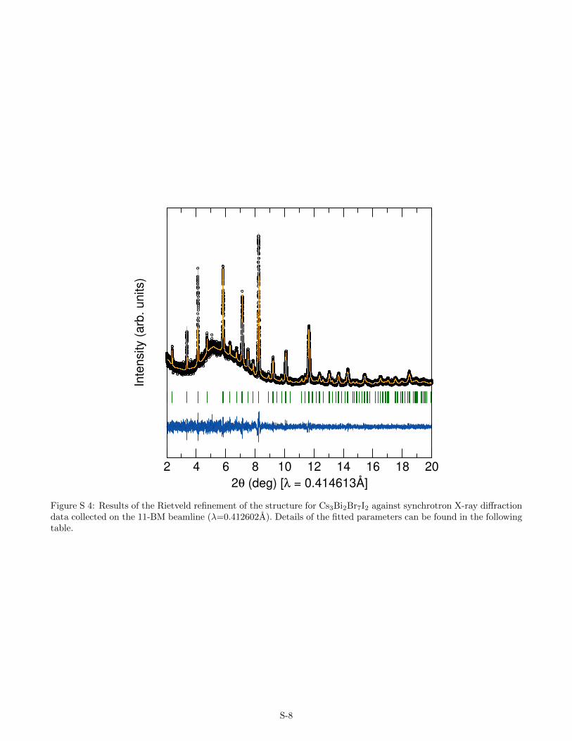
Figure S 4: Results of the Rietveld refinement of the structure for Cs3Bi2Br7I2 against synchrotron X-ray diffractiondata collected on the 11-BM beamline (λ=0.412602A). Details of the fitted parameters can be found in the followingtable.
S-8

2 4 6 8 10 12 14 16 18 202θ (deg) [λ = 0.414613Å]
Inte
nsity
(ar
b. u
nits
)
Figure S 5: Results of the Rietveld refinement of the structure for Cs3Bi2Br6I3 against synchrotron X-ray diffractiondata collected on the 11-BM beamline (λ=0.414613A). Details of the fitted parameters can be found in the followingtable.
S-9

2 4 6 8 10 12 14 16 18 202θ (deg) [λ = 0.414633Å]
Inte
nsity
(ar
b. u
nits
)
Figure S 6: Results of the Rietveld refinement of the structure for Cs3Bi2Br5I4 against synchrotron X-ray diffractiondata collected on the 11-BM beamline (λ=0.414633A). Details of the fitted parameters can be found in the followingtable.
S-10

2 4 6 8 10 12 14 16 18 202θ (deg) [λ = 0.414613Å]
Inte
nsity
(ar
b. u
nits
)
Figure S 7: Results of the Rietveld refinement of the structure for Cs3Bi2Br4I5 against synchrotron X-ray diffractiondata collected on the 11-BM beamline (λ=0.414613A). Details of the fitted parameters can be found in the followingtable.
S-11

2 4 6 8 10 12 14 16 18 202θ (deg) [λ = 0.414613Å]
Inte
nsity
(ar
b. u
nits
)
Figure S 8: Results of the Rietveld refinement of the structure for Cs3Bi2Br3I6 against synchrotron X-ray diffractiondata collected on the 11-BM beamline (λ=0.414613A). Details of the fitted parameters can be found in the followingtable.
S-12

Table S 1: Results of the Rietveld refinement of the structure for Cs3Bi2Br9 against synchrotron X-ray diffractiondata collected on the 11-BM beamline (λ=0.414613A). a = 7.96150(2), c = 9.84784(3). RBragg =3.42%
atom x y z OccupancyBi1 0.6667 0.3333 0.1919 0.3333Cs1 0.0000 0.0000 0.0000 0.1667Cs2 0.6667 0.3333 0.6662 0.3333Br1 0.5000 0.5000 0.0000 0.5000Br2 0.3353 0.1676 0.3399 1.0000
atom β11 β22 β33 β12 β13 β23Bi1 0.0080 0.0080 0.0027 0.0040 0.0000 0.0000Cs1 0.0224 0.0224 0.0176 0.0112 0.0000 0.0000Cs2 0.0217 0.0217 0.0097 0.0108 0.0000 0.0000Br1 0.0347 0.0347 0.0127 0.0263 -0.0060 0.0060Br2 0.0126 0.0230 0.0114 0.0063 0.0018 0.0009
S-13
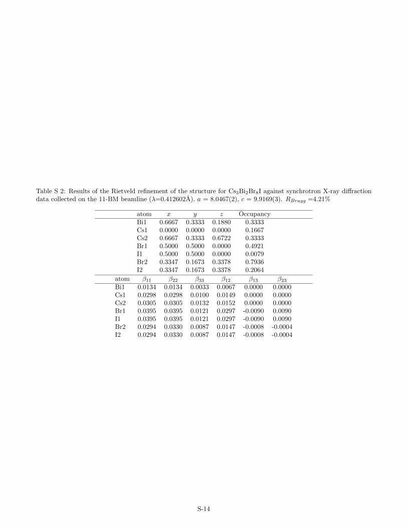
Table S 2: Results of the Rietveld refinement of the structure for Cs3Bi2Br8I against synchrotron X-ray diffractiondata collected on the 11-BM beamline (λ=0.412602A). a = 8.0467(2), c = 9.9169(3). RBragg =4.21%
atom x y z OccupancyBi1 0.6667 0.3333 0.1880 0.3333Cs1 0.0000 0.0000 0.0000 0.1667Cs2 0.6667 0.3333 0.6722 0.3333Br1 0.5000 0.5000 0.0000 0.4921I1 0.5000 0.5000 0.0000 0.0079Br2 0.3347 0.1673 0.3378 0.7936I2 0.3347 0.1673 0.3378 0.2064
atom β11 β22 β33 β12 β13 β23Bi1 0.0134 0.0134 0.0033 0.0067 0.0000 0.0000Cs1 0.0298 0.0298 0.0100 0.0149 0.0000 0.0000Cs2 0.0305 0.0305 0.0132 0.0152 0.0000 0.0000Br1 0.0395 0.0395 0.0121 0.0297 -0.0090 0.0090I1 0.0395 0.0395 0.0121 0.0297 -0.0090 0.0090Br2 0.0294 0.0330 0.0087 0.0147 -0.0008 -0.0004I2 0.0294 0.0330 0.0087 0.0147 -0.0008 -0.0004
S-14

Table S 3: Results of the Rietveld refinement of the structure for Cs3Bi2Br7I2 against synchrotron X-ray diffractiondata collected on the 11-BM beamline (λ=0.412602A). a = 8.1073(2), c = 9.9778(3). RBragg =2.80%
atom x y z OccupancyBi1 0.6667 0.3333 0.1869 0.3333Cs1 0.0000 0.0000 0.0000 0.1667Cs2 0.6667 0.3333 0.6746 0.3333Br1 0.5000 0.5000 0.0000 0.4699I1 0.5000 0.5000 0.0000 0.0301Br2 0.3306 0.1653 0.3390 0.6158I2 0.3306 0.1653 0.3390 0.3842
atom β11 β22 β33 β12 β13 β23Bi1 0.0097 0.0097 0.0012 0.0048 0.0000 0.0000Cs1 0.0366 0.0366 0.0052 0.0183 0.0000 0.0000Cs2 0.0244 0.0244 0.0109 0.0122 0.0000 0.0000Br1 0.0469 0.0469 0.0176 0.0383 0.0003 -0.0003I1 0.0469 0.0469 0.0176 0.0383 0.0003 -0.0003Br2 0.0244 0.0347 0.0027 0.0122 0.0120 0.0060I2 0.0244 0.0347 0.0027 0.0122 0.0120 0.0060
S-15

Table S 4: Results of the Rietveld refinement of the structure for Cs3Bi2Br6I3 against synchrotron X-ray diffractiondata collected on the 11-BM beamline (λ=0.414613A). a = 8.1626(1), c = 10.0316(1). RBragg =3.73%
atom x y z OccupancyBi1 0.6667 0.3333 0.1843 0.3333Cs1 0.0000 0.0000 0.0000 0.1667Cs2 0.6667 0.3333 0.6761 0.3333Br1 0.5000 0.5000 0.0000 0.4412I1 0.5000 0.5000 0.0000 0.0588Br2 0.3313 0.1656 0.3383 0.4509I2 0.3313 0.1656 0.3383 0.5491
atom β11 β22 β33 β12 β13 β23Bi1 0.0096 0.0096 0.0027 0.0048 0.0000 0.0000Cs1 0.0261 0.0261 0.0121 0.0131 0.0000 0.0000Cs2 0.0253 0.0253 0.0109 0.0127 0.0000 0.0000Br1 0.0423 0.0423 0.0174 0.0301 -0.0042 0.0042I1 0.0423 0.0423 0.0174 0.0301 -0.0042 0.0042Br2 0.0238 0.0279 0.0109 0.0119 0.0098 0.0049I2 0.0238 0.0279 0.0109 0.0119 0.0098 0.0049
S-16
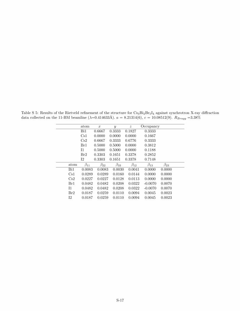
Table S 5: Results of the Rietveld refinement of the structure for Cs3Bi2Br5I4 against synchrotron X-ray diffractiondata collected on the 11-BM beamline (λ=0.414633A). a = 8.21314(6), c = 10.08512(9). RBragg =3.38%
atom x y z OccupancyBi1 0.6667 0.3333 0.1827 0.3333Cs1 0.0000 0.0000 0.0000 0.1667Cs2 0.6667 0.3333 0.6776 0.3333Br1 0.5000 0.5000 0.0000 0.3812I1 0.5000 0.5000 0.0000 0.1188Br2 0.3303 0.1651 0.3378 0.2852I2 0.3303 0.1651 0.3378 0.7148
atom β11 β22 β33 β12 β13 β23Bi1 0.0083 0.0083 0.0030 0.0041 0.0000 0.0000Cs1 0.0289 0.0289 0.0160 0.0144 0.0000 0.0000Cs2 0.0227 0.0227 0.0128 0.0113 0.0000 0.0000Br1 0.0482 0.0482 0.0208 0.0322 -0.0070 0.0070I1 0.0482 0.0482 0.0208 0.0322 -0.0070 0.0070Br2 0.0187 0.0259 0.0110 0.0094 0.0045 0.0023I2 0.0187 0.0259 0.0110 0.0094 0.0045 0.0023
S-17

Table S 6: Results of the Rietveld refinement of the structure for Cs3Bi2Br4I5 against synchrotron X-ray diffractiondata collected on the 11-BM beamline (λ=0.414613A). a = 8.2881(1), c = 10.1679(2). RBragg =3.71%
atom x y z OccupancyBi1 0.6667 0.3333 0.1816 0.3333Cs1 0.0000 0.0000 0.0000 0.1667Cs2 0.6667 0.3333 0.6799 0.3333Br1 0.5000 0.5000 0.0000 0.3895I1 0.5000 0.5000 0.0000 0.1105Br2 0.3292 0.1646 0.3365 0.1098I2 0.3292 0.1646 0.3365 0.8902
atom β11 β22 β33 β12 β13 β23Bi1 0.0093 0.0093 0.0036 0.0046 0.0000 0.0000Cs1 0.0281 0.0281 0.0155 0.0140 0.0000 0.0000Cs2 0.0212 0.0212 0.0152 0.0106 0.0000 0.0000Br1 0.0370 0.0370 0.0211 0.0174 -0.0113 0.0113I1 0.0370 0.0370 0.0211 0.0174 -0.0113 0.0113Br2 0.0274 0.0222 0.0131 0.0137 0.0086 0.0043I2 0.0274 0.0222 0.0131 0.0137 0.0086 0.0043
S-18
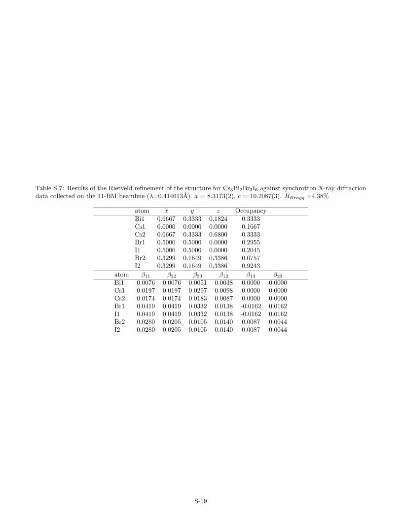
Table S 7: Results of the Rietveld refinement of the structure for Cs3Bi2Br3I6 against synchrotron X-ray diffractiondata collected on the 11-BM beamline (λ=0.414613A). a = 8.3173(2), c = 10.2087(3). RBragg =4.38%
atom x y z OccupancyBi1 0.6667 0.3333 0.1824 0.3333Cs1 0.0000 0.0000 0.0000 0.1667Cs2 0.6667 0.3333 0.6800 0.3333Br1 0.5000 0.5000 0.0000 0.2955I1 0.5000 0.5000 0.0000 0.2045Br2 0.3299 0.1649 0.3386 0.0757I2 0.3299 0.1649 0.3386 0.9243
atom β11 β22 β33 β12 β13 β23Bi1 0.0076 0.0076 0.0051 0.0038 0.0000 0.0000Cs1 0.0197 0.0197 0.0297 0.0098 0.0000 0.0000Cs2 0.0174 0.0174 0.0183 0.0087 0.0000 0.0000Br1 0.0419 0.0419 0.0332 0.0138 -0.0162 0.0162I1 0.0419 0.0419 0.0332 0.0138 -0.0162 0.0162Br2 0.0280 0.0205 0.0105 0.0140 0.0087 0.0044I2 0.0280 0.0205 0.0105 0.0140 0.0087 0.0044
S-19