AnimalTransport Unit One Cells, Exchange and Transport AS Biology OCR Specification.
102
AnimalTransport Unit One Cells, Exchange and Transport AS Biology OCR Specification
-
Upload
lucas-allison -
Category
Documents
-
view
214 -
download
0
Transcript of AnimalTransport Unit One Cells, Exchange and Transport AS Biology OCR Specification.

AnimalTransport
Unit OneCells, Exchange and Transport
AS BiologyOCR Specification
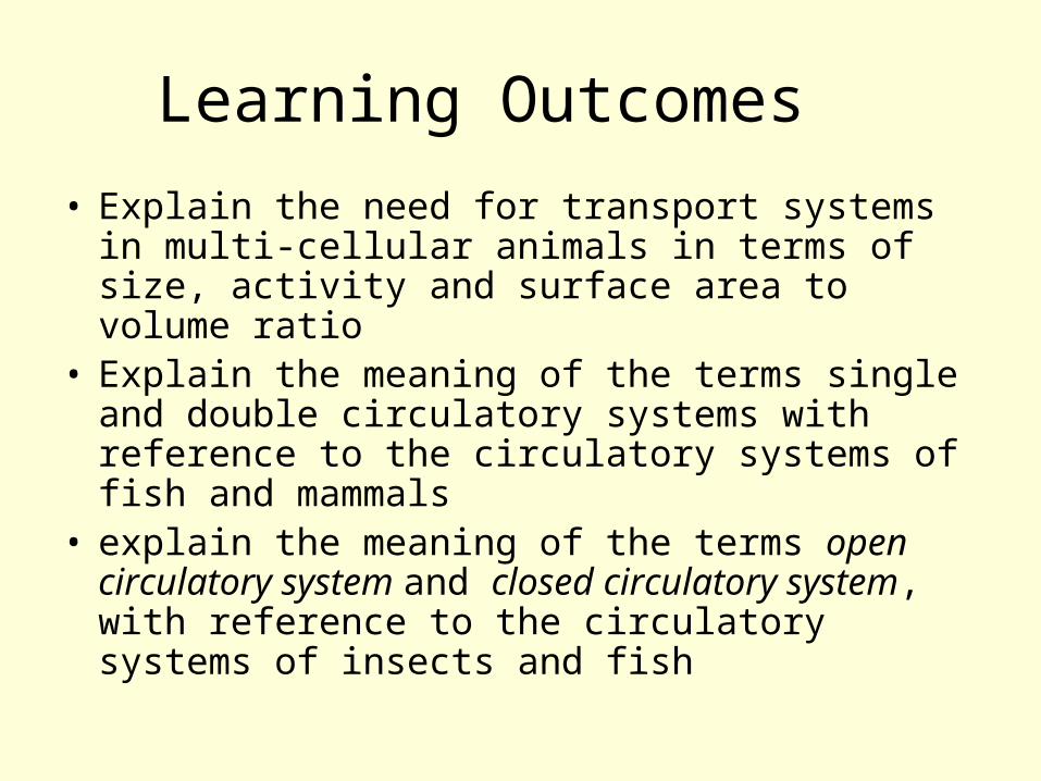
Learning Outcomes
• Explain the need for transport systems in multi-cellular animals in terms of size, activity and surface area to volume ratio
• Explain the meaning of the terms single and double circulatory systems with reference to the circulatory systems of fish and mammals
• explain the meaning of the terms open circulatory system and closed circulatory system, with reference to the circulatory systems of insects and fish
The Mammalian Transport System
Why do multi-cellular animals require a transport System?
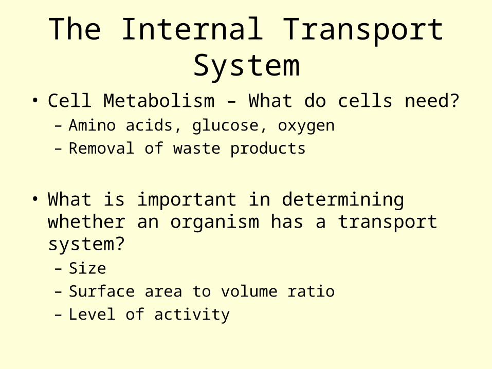
The Internal Transport System
• Cell Metabolism – What do cells need?– Amino acids, glucose, oxygen– Removal of waste products
• What is important in determining whether an organism has a transport system?– Size– Surface area to volume ratio– Level of activity
Revision Activity
• Using the table on the next slide, determine the importance of the three factors and give information to support your answers?
• Size• Surface area to volume ratio• Level of activity
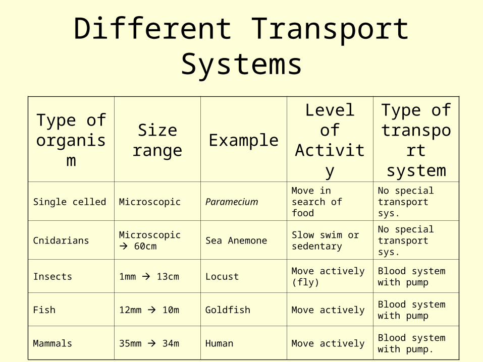
Different Transport Systems
Type of organis
m
Size range
ExampleLevel of Activity
Type of transport system
Single celled Microscopic ParameciumMove in search of food
No special transport sys.
CnidariansMicroscopic 60cm
Sea AnemoneSlow swim or sedentary
No special transport sys.
Insects 1mm 13cm Locust Move actively (fly)
Blood system with pump
Fish 12mm 10m Goldfish Move activelyBlood system with pump
Mammals 35mm 34m Human Move activelyBlood system with pump.
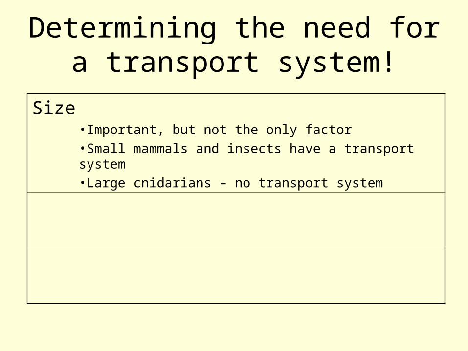
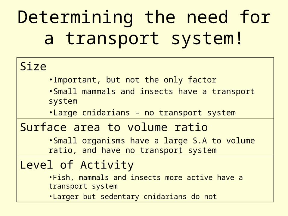
Determining the need for a transport system!
Size•Important, but not the only factor•Small mammals and insects have a transport system•Large cnidarians – no transport system
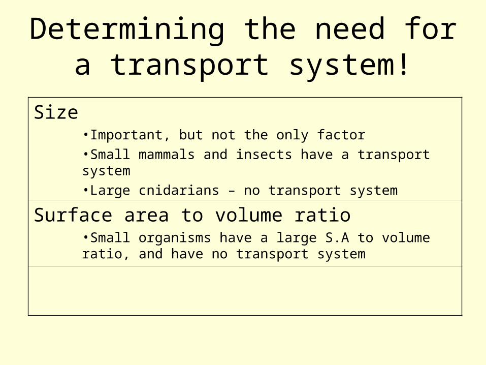
Determining the need for a transport system!
Size•Important, but not the only factor•Small mammals and insects have a transport system•Large cnidarians – no transport system
Surface area to volume ratio•Small organisms have a large S.A to volume ratio, and have no transport system
Determining the need for a transport system!
Size•Important, but not the only factor•Small mammals and insects have a transport system•Large cnidarians – no transport system
Surface area to volume ratio•Small organisms have a large S.A to volume ratio, and have no transport system
Level of Activity•Fish, mammals and insects more active have a transport system•Larger but sedentary cnidarians do not
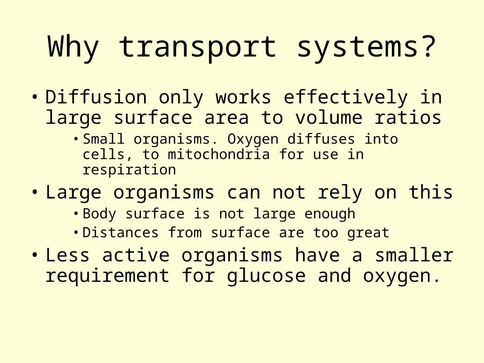
Why transport systems?
• Diffusion only works effectively in large surface area to volume ratios
• Small organisms. Oxygen diffuses into cells, to mitochondria for use in respiration
• Large organisms can not rely on this• Body surface is not large enough• Distances from surface are too great
• Less active organisms have a smaller requirement for glucose and oxygen.
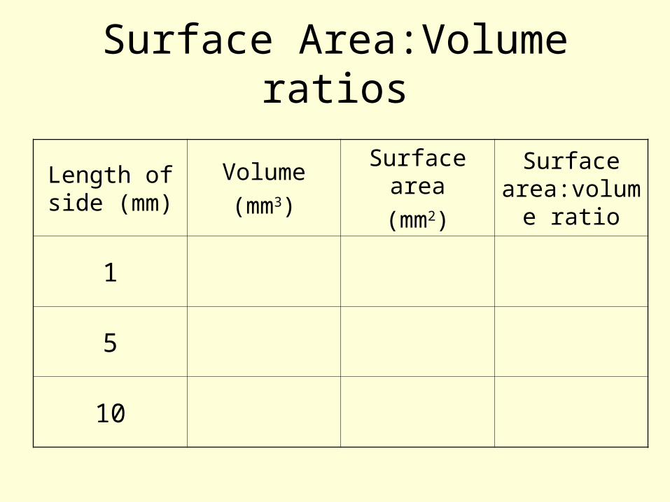
Surface Area:Volume ratios
Length of side (mm)
Volume(mm3)
Surface area
(mm2)
Surface area:volum
e ratio
1
5
10
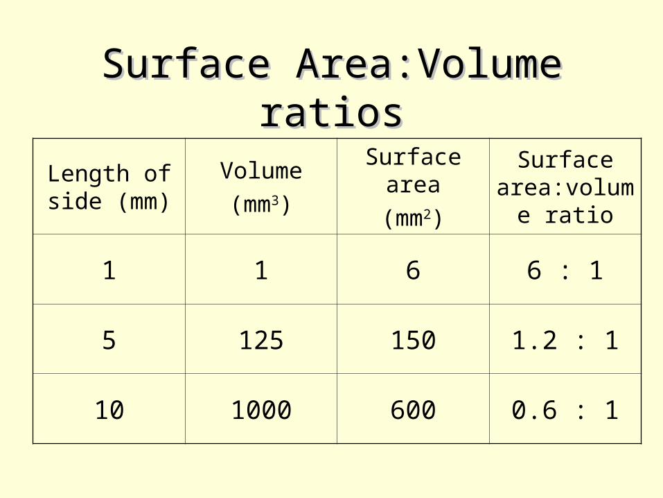
Surface Area:Volume Surface Area:Volume ratiosratios
Length of side (mm)
Volume(mm3)
Surface area
(mm2)
Surface area:volum
e ratio
1 1 6 6 : 1
5 125 150 1.2 : 1
10 1000 600 0.6 : 1
Surface area: volume ratio
• With a cube shape– As it gets bigger the volume increases
faster than the surface area– Larger multi-cellular animals need a
transport system and special gas exchange surfaces
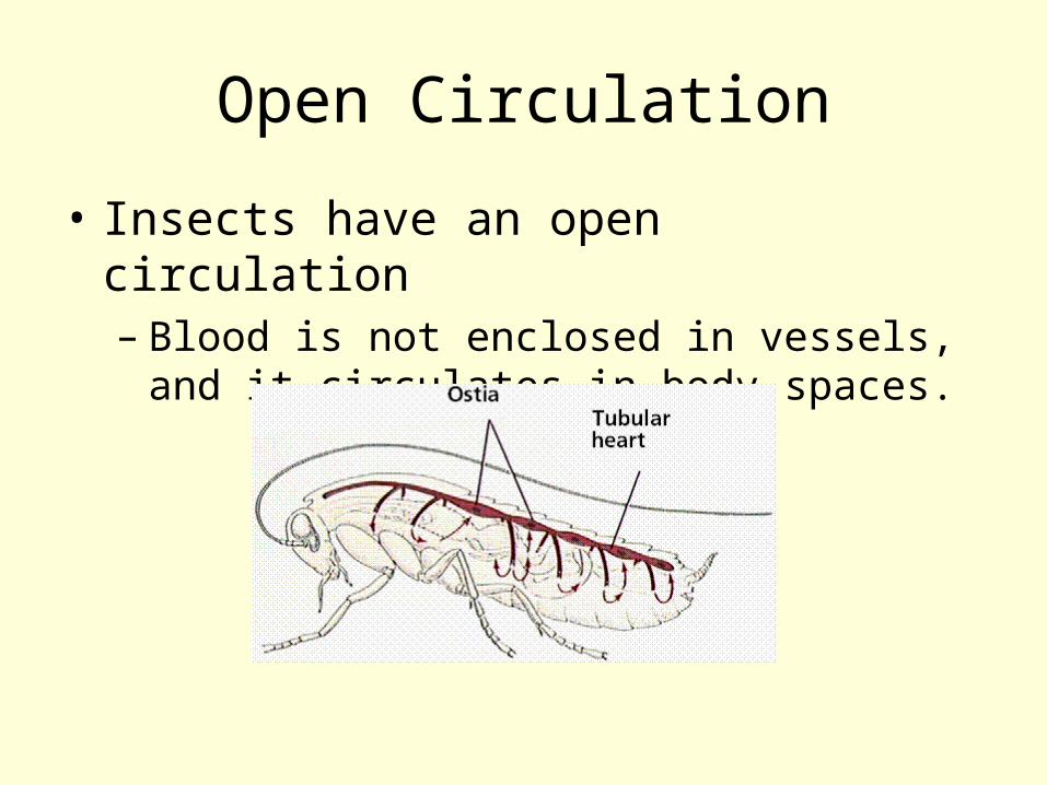
Open Circulation
• Insects have an open circulation– Blood is not enclosed in vessels, and it
circulates in body spaces.
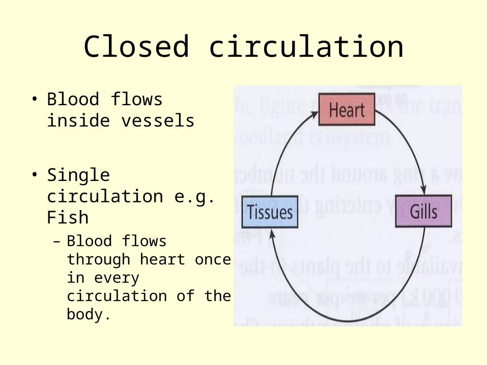
Closed circulation
• Blood flows inside vessels
• Single circulation e.g. Fish– Blood flows through
heart once in every circulation of the body.
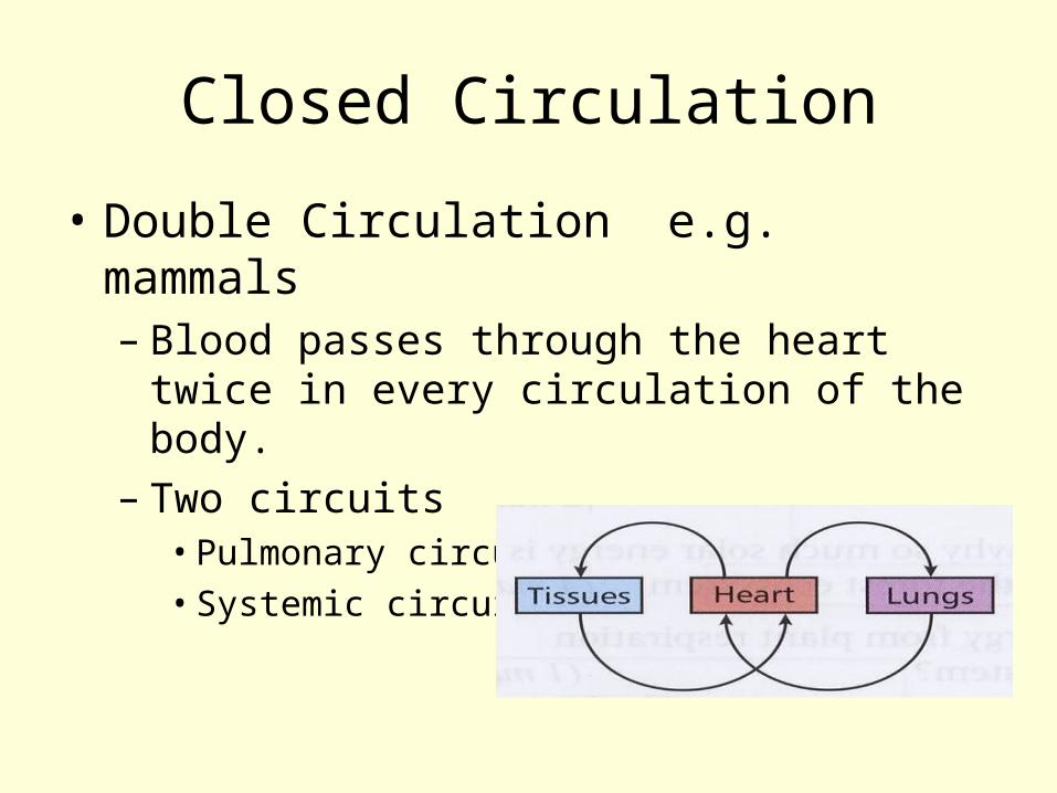
Closed Circulation
• Double Circulation e.g. mammals– Blood passes through the heart twice in
every circulation of the body.– Two circuits
• Pulmonary circuit• Systemic circuit
Advantages of a double circulation
• Simultaneous high pressure delivery of oxygenated blood to all regions of the body
• Oxygenated blood reaches respiring cells undiluted by deoxygenated blood.
The Mammalian Heart
Structure of the HeartDissection
Learning Outcomes
• describe, with the aid of diagrams and photographs, the external and internal structure of the mammalian heart;
• explain, with the aid of diagrams, the differences in the thickness of the walls of the different chambers of the heart in terms of their functions;
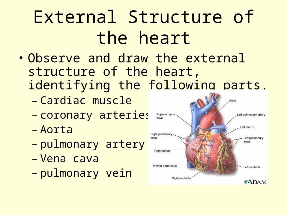
External Structure of the heart
• Observe and draw the external structure of the heart, identifying the following parts.– Cardiac muscle– coronary arteries– Aorta– pulmonary artery– Vena cava– pulmonary vein
Internal structure of the heart
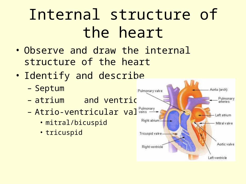
• Observe and draw the internal structure of the heart
• Identify and describe – Septum– atrium and ventricle– Atrio-ventricular valves
• mitral/bicuspid• tricuspid
Revision of structure of heart
• Label the diagram of the heart– Right atria / left atria– Right ventricle / left ventricle– Aorta / pulmonary artery– Vena cava / pulmonary vein
• Colour in deoxygenated blood blue / oxygenated blood red
The Mammalian Heart
The Cardiac Cycle
Learning outcomes
• describe the cardiac cycle, with reference to the action of the valves in the heart;
Cardiac Cycle
• The sequence of events of a heart beat
• Alternate contractions (systole) and relaxations (diastole)
• Between 70 and 75 bpm
Cardiac Cycle
• Blood flows through the heart– Muscles contract– Volume chamber decreases– Pressure increases– Blood forced to a region of lower
pressure– Valves prevent backflow
Cardiac Cycle
• There are 3 main stages to the cardiac cycle– Atrial systole– Ventricular systole– Diastole
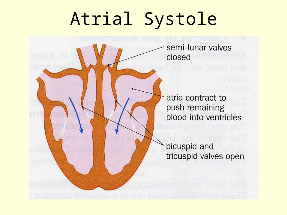
Atrial Systole
• Heart is full of blood and ventricles relaxed
• Both atria contract• Blood passes into ventricles• A-V valves open due to pressure• 70% blood flows passively atria -
ventricle
Atrial Systole
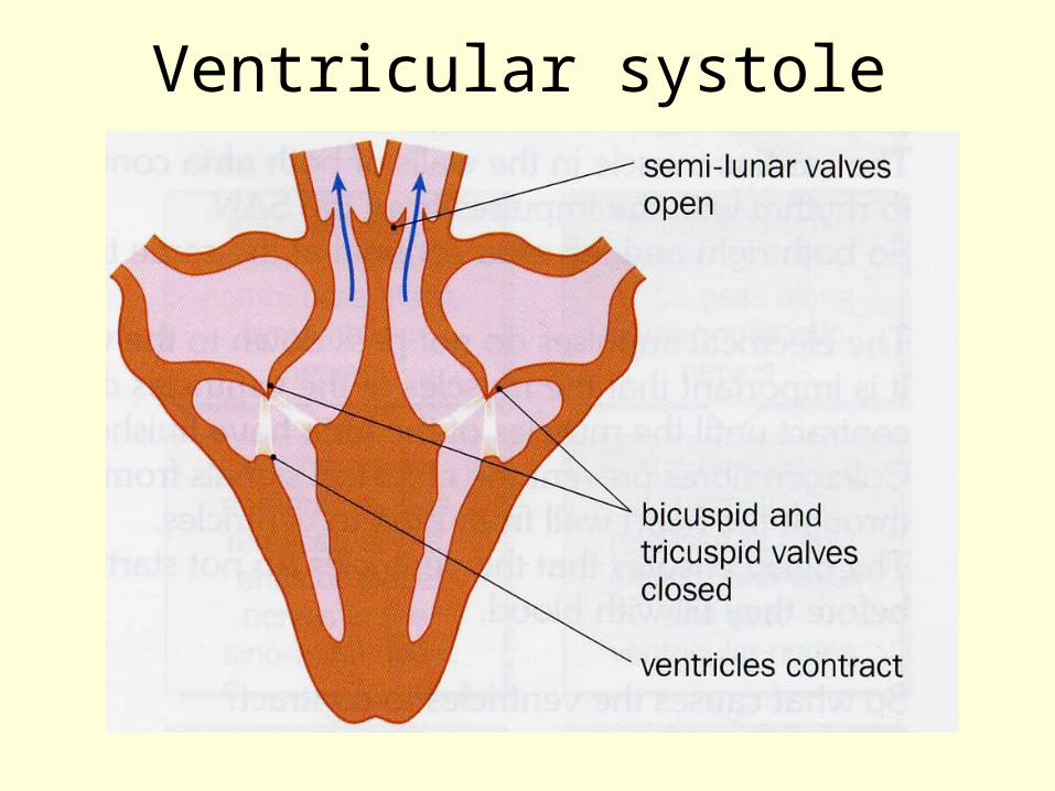
Ventricular Systole
• Atria relax• Ventricles contract• Forces blood into pulmonary artery
and aorta• A-V valves close (lub)• S-L valves open• Pulse is generated
Ventricular systole
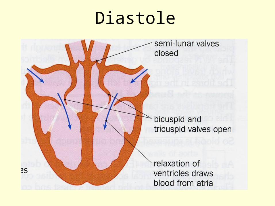
Diastole
• Ventricles relax• Pressure in ventricle < pressure in
arteries• High pressure blood in arteries cause
S-L valves to shut (dub)• All muscles relax• Blood from vena cava and pulmonary
vein enter atria
Diastole
Structure and function of heart muscle
• Ventricle walls are thicker– Need greater force when contract
• R. Ventricle –force relatively small, pumps to lungs
• L. Ventricle – sufficient to push blood around body
• Thickness left > right
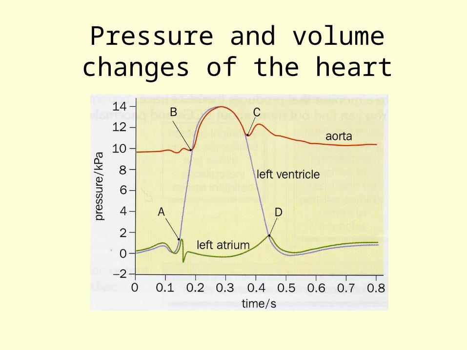
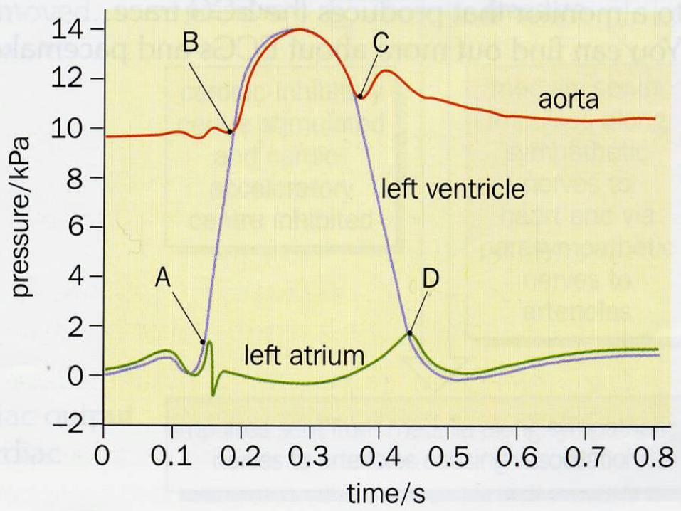
Pressure and volume changes of the heart
Learning outcomes
• Describe how heart action is coordinated with reference to the sinoatrial node (SAN), the atrioventricular node (AVN) and the Purkyne tissue.
• Interpret and explain electrocardiogram (ECG) traces, with reference to normal and abnormal heart activity.
Control of Heart Beat
• Myogenic – heart muscle contracts and relaxes without having to receive impulses from the nervous system– Sino-atrial node– Atrio-ventricular node
Sino-atrial Node
• Special cardiac muscle tissue in right atrium
• a.k.a. SAN or Pacemaker• Sets the rhythm at which all other
cardiac muscle cells beat• Sends excitation wave
(depolarisation) over atrial walls
What happens next?
• Collagen fibres prevent the wave of excitation from passing from the atria to the ventricle walls
• Allows the ventricle to fill before they contract
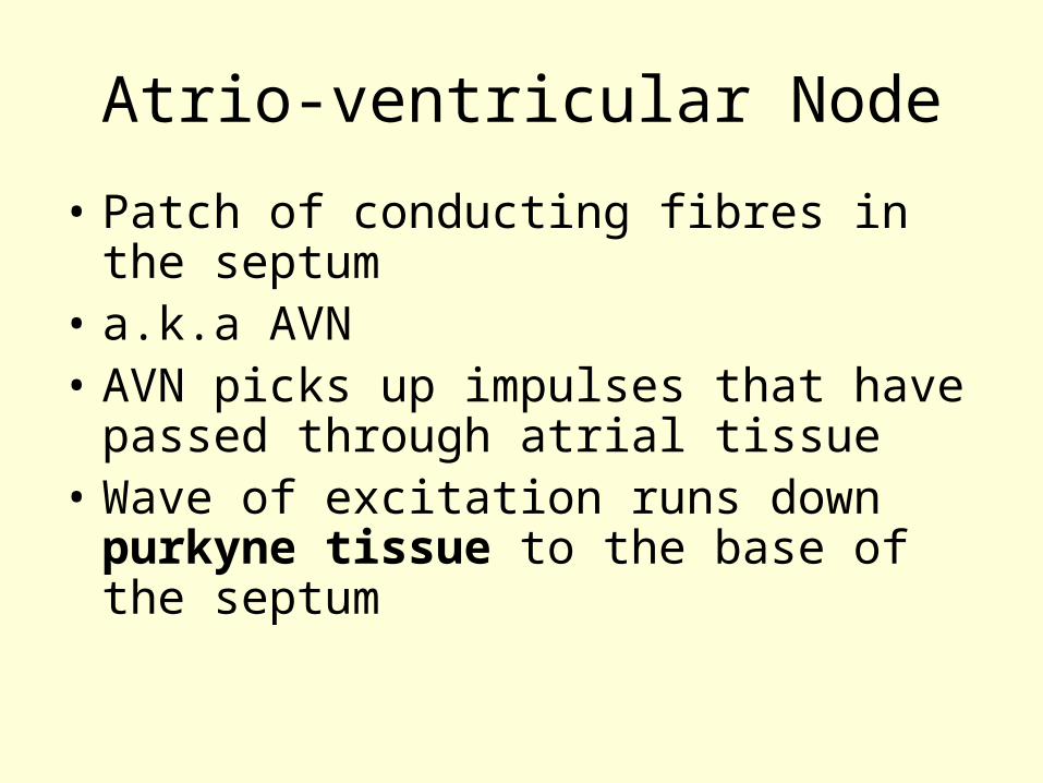
Atrio-ventricular Node
• Patch of conducting fibres in the septum
• a.k.a AVN• AVN picks up impulses that have
passed through atrial tissue• Wave of excitation runs down
purkyne tissue to the base of the septum
Atrio-ventricular Node
• Wave spreads upwards and outwards through the ventricular walls
• Blood is squeezed up and out through arteries.
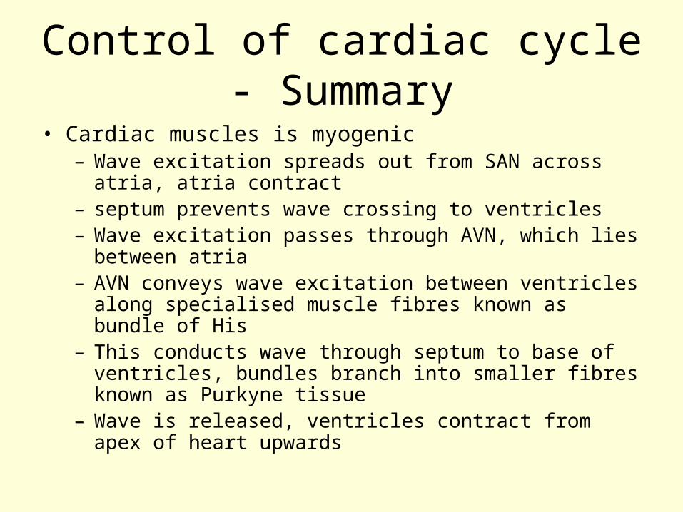
Control of cardiac cycle - Summary
• Cardiac muscles is myogenic– Wave excitation spreads out from SAN across atria,
atria contract– septum prevents wave crossing to ventricles– Wave excitation passes through AVN, which lies
between atria– AVN conveys wave excitation between ventricles
along specialised muscle fibres known as bundle of His
– This conducts wave through septum to base of ventricles, bundles branch into smaller fibres known as Purkyne tissue
– Wave is released, ventricles contract from apex of heart upwards
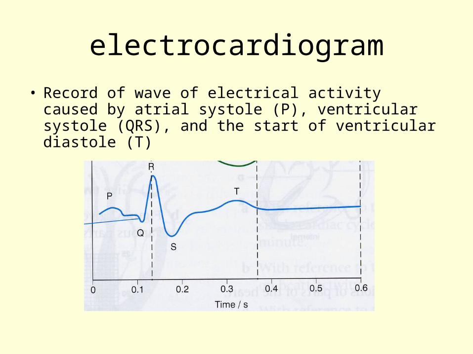
electrocardiogram
• Record of wave of electrical activity caused by atrial systole (P), ventricular systole (QRS), and the start of ventricular diastole (T)
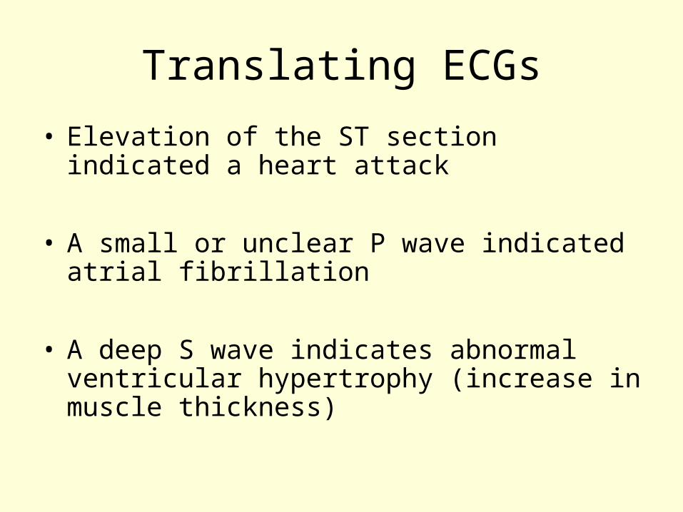
Translating ECGs
• Elevation of the ST section indicated a heart attack
• A small or unclear P wave indicated atrial fibrillation
• A deep S wave indicates abnormal ventricular hypertrophy (increase in muscle thickness)
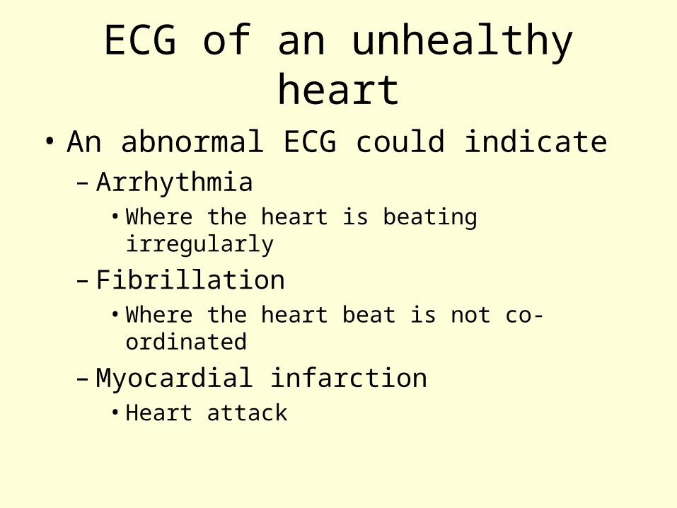
ECG of an unhealthy heart
• An abnormal ECG could indicate– Arrhythmia
• Where the heart is beating irregularly
– Fibrillation• Where the heart beat is not co-ordinated
– Myocardial infarction• Heart attack
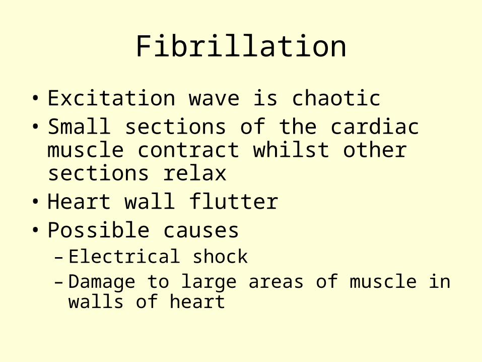
Fibrillation
• Excitation wave is chaotic• Small sections of the cardiac muscle
contract whilst other sections relax• Heart wall flutter• Possible causes
– Electrical shock– Damage to large areas of muscle in
walls of heart
Exam Question
• Answer the practice exam questions on Moodle on all objectives done so far on the Heart and the need for a transport system
The Mammalian Transport System
Structure and function of Arteries, Veins and Capillaries
Learning Outcomes
• describe, with the aid of diagrams and photographs, the structures and functions of arteries, veins and capillaries;
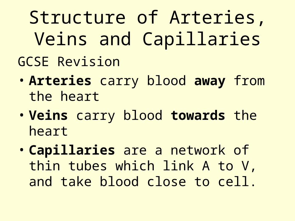
Structure of Arteries, Veins and Capillaries
GCSE Revision• Arteries carry blood away from the
heart• Veins carry blood towards the
heart• Capillaries are a network of thin
tubes which link A to V, and take blood close to cell.
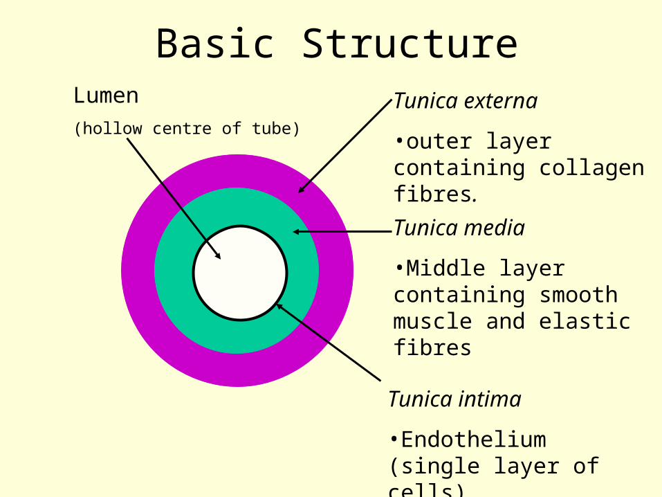
Basic Structure
Tunica intima
•Endothelium (single layer of cells)
Tunica media
•Middle layer containing smooth muscle and elastic fibres
Tunica externa
•outer layer containing collagen fibres.
Lumen (hollow centre of tube)
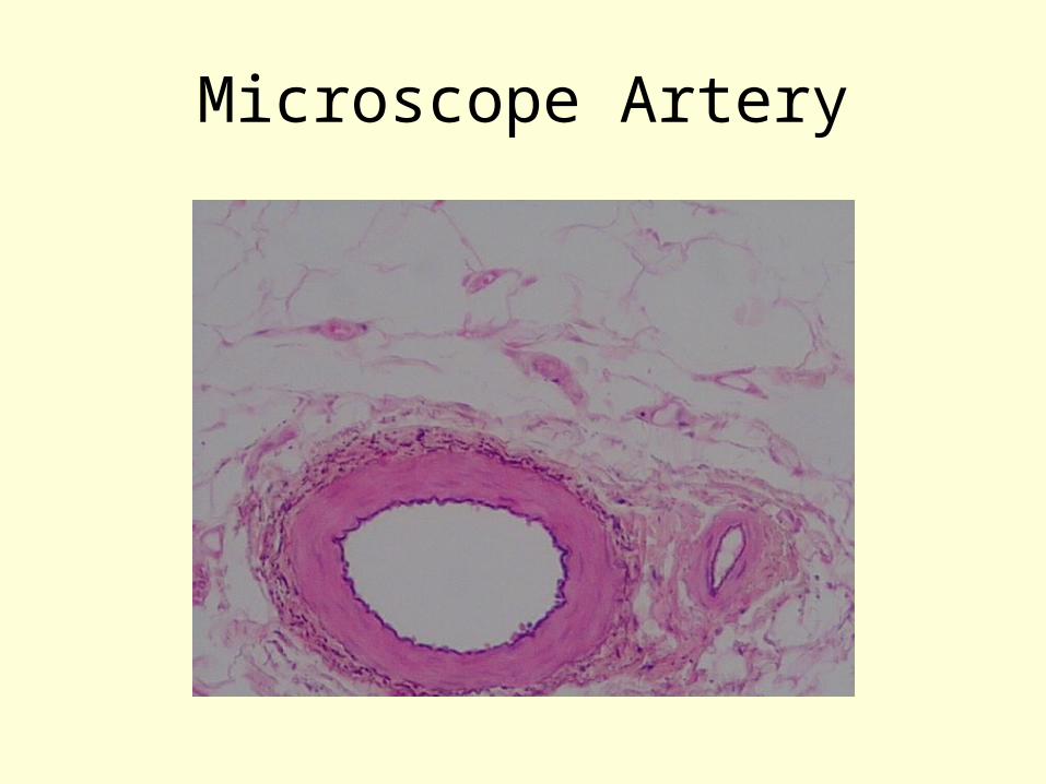
Microscope Artery
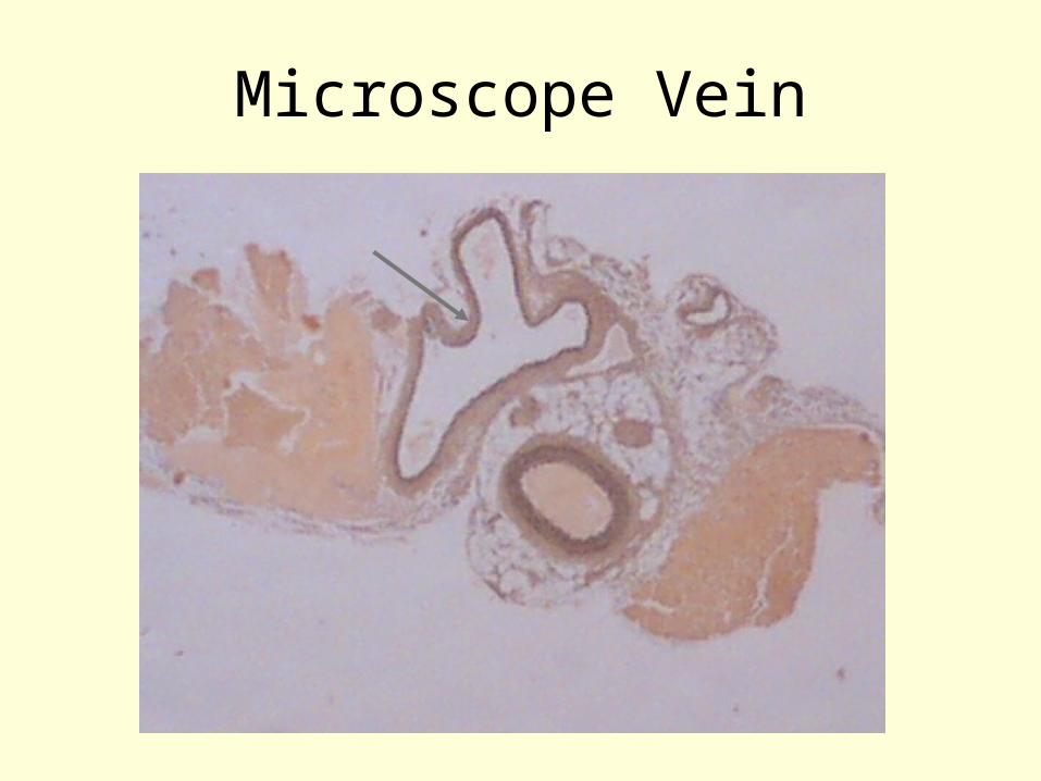
Microscope Vein
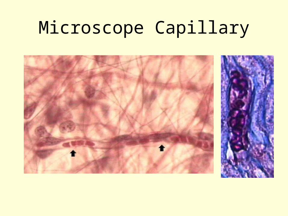
Microscope Capillary
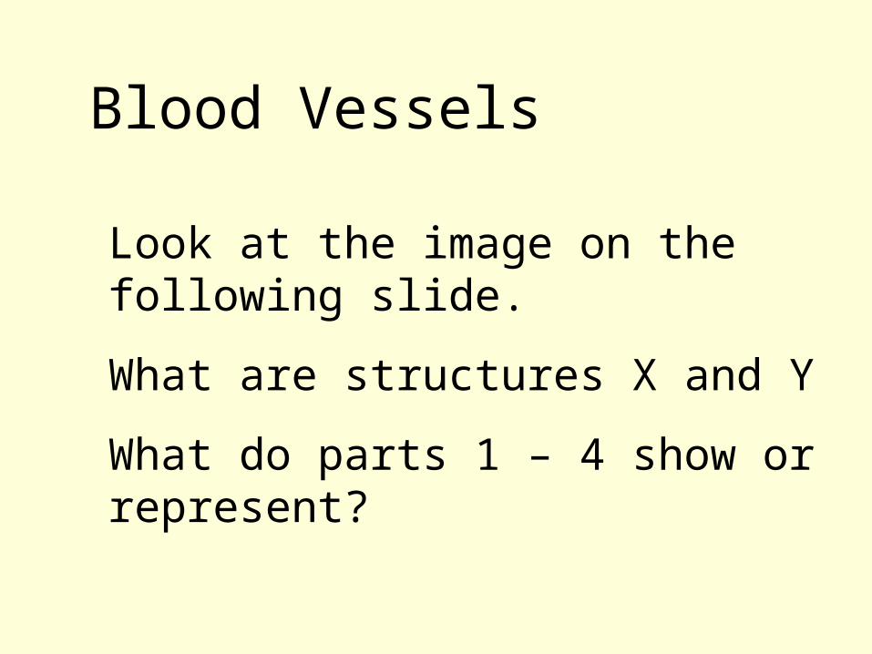
Look at the image on the following slide.
What are structures X and Y
What do parts 1 – 4 show or represent?
Blood Vessels
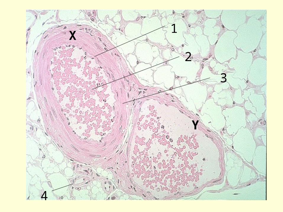
X
Y
1
3
2
4
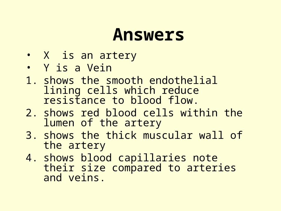
Answers• X is an artery • Y is a Vein1. shows the smooth endothelial lining
cells which reduce resistance to blood flow.
2. shows red blood cells within the lumen of the artery
3. shows the thick muscular wall of the artery
4. shows blood capillaries note their size compared to arteries and veins.
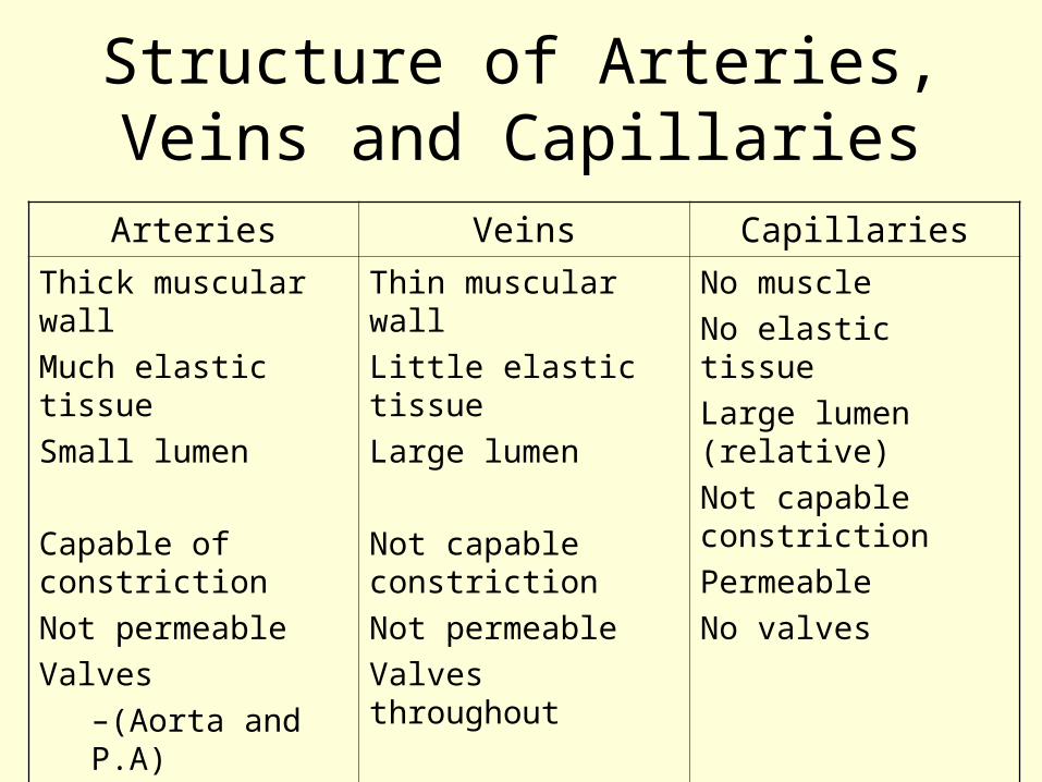
Structure of Arteries, Veins and Capillaries
Arteries Veins Capillaries
Thick muscular wallMuch elastic tissueSmall lumen
Capable of constrictionNot permeableValves
–(Aorta and P.A)
Thin muscular wallLittle elastic tissueLarge lumen
Not capable constrictionNot permeableValves throughout
No muscleNo elastic tissueLarge lumen (relative)Not capable constrictionPermeableNo valves
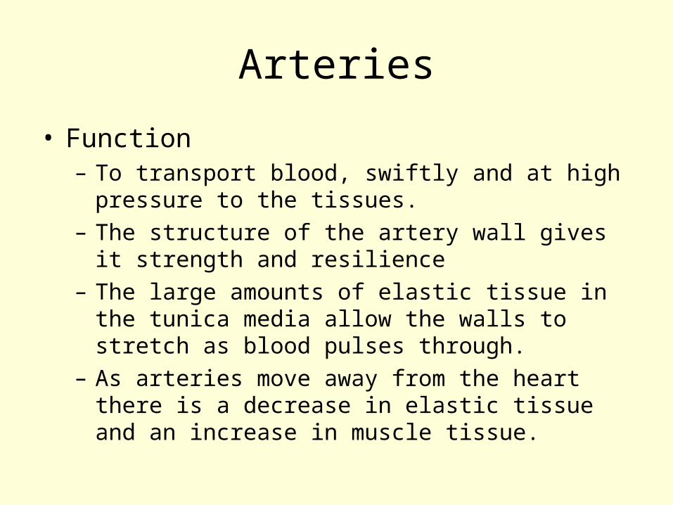
Arteries
• Function– To transport blood, swiftly and at high pressure
to the tissues.– The structure of the artery wall gives it
strength and resilience– The large amounts of elastic tissue in the
tunica media allow the walls to stretch as blood pulses through.
– As arteries move away from the heart there is a decrease in elastic tissue and an increase in muscle tissue.
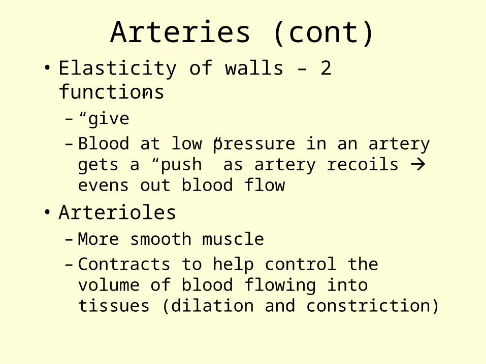
Arteries (cont)• Elasticity of walls – 2 functions
– “give”– Blood at low pressure in an artery
gets a “push” as artery recoils evens out blood flow
• Arterioles– More smooth muscle– Contracts to help control the volume
of blood flowing into tissues (dilation and constriction)
Capillaries• Function
– To take blood as close as possible to all cells, allowing rapid transfer of substances between cells and blood
• Network of capillaries capillary bed
Veins• Venules/veins
– Return blood to the heart
• Low venous pressure
• Semi-lunar valves– Form from endothelium– Allow blood to travel to the heart– Prevents the back flow of blood
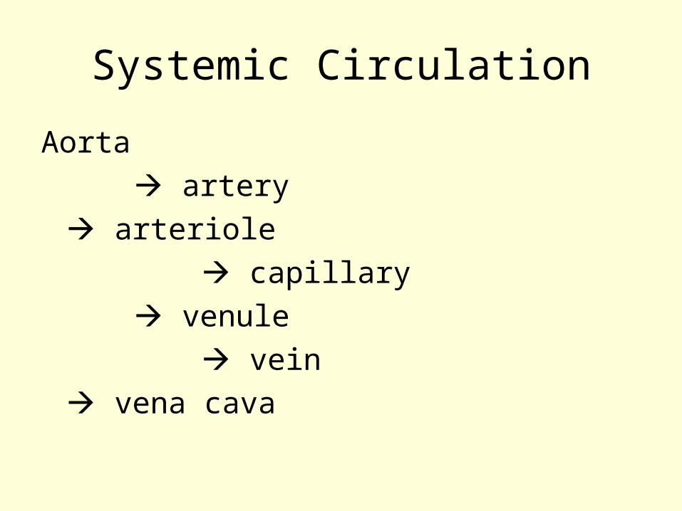
Systemic Circulation
Aorta artery
arteriole capillary
venule vein
vena cava
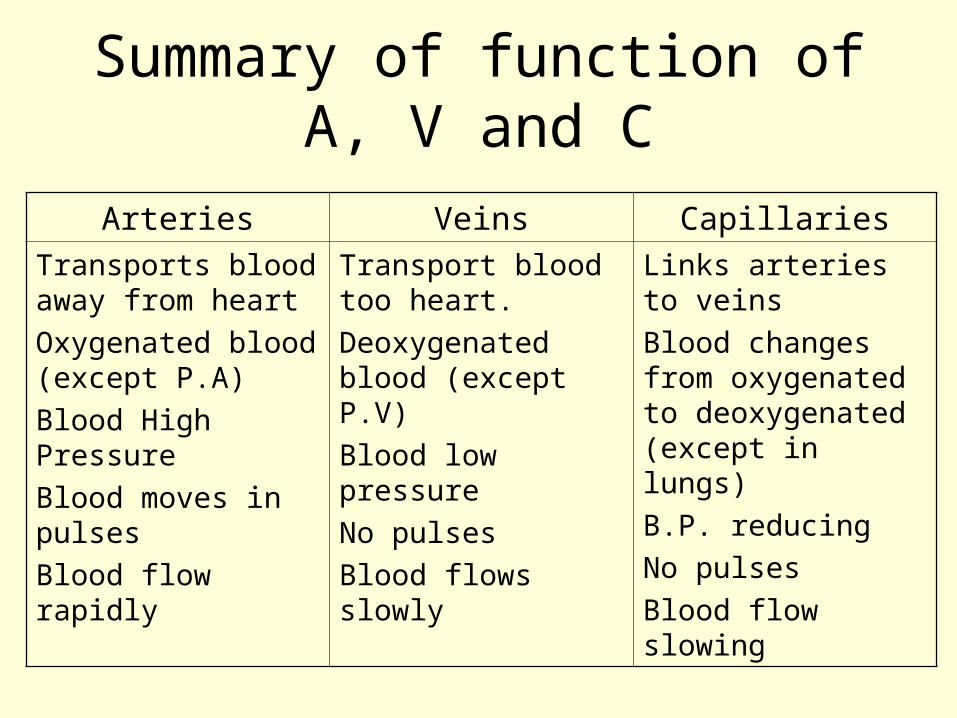
Summary of function of A, V and C
Arteries Veins CapillariesTransports blood away from heartOxygenated blood (except P.A)Blood High PressureBlood moves in pulsesBlood flow rapidly
Transport blood too heart.Deoxygenated blood (except P.V)Blood low pressureNo pulsesBlood flows slowly
Links arteries to veinsBlood changes from oxygenated to deoxygenated (except in lungs)B.P. reducingNo pulsesBlood flow slowing
Revision Questions (1)
– Suggest why arteries close to the heart have more elastic fibres in walls than arteries further away from the heart.
– Suggest why there are no blood capillaries in the cornea of the eye. How might the cornea be supplied with its requirements?
Revision Questions (2)
• Suggest reasons for the following:1. Normal venous pressure in the feet is
about 25mm Hg. When a soldier stands at attention the blood pressure in their feet rises very quickly to about 90mm Hg.
2. When you breathe in (volume thorax increases), blood moves through the veins towards the heart.
Blood, Tissue fluid and Lymph
Blood – the transport medium• Plasma
– Straw coloured, alkaline liquid– Consists mainly of water
• Functions of blood– Defends body against disease– Maintains diffusion gradients– Acts as a buffer– Provides pressure– Distributes heat around body
Blood plasma
• Water with dissolved substances– Nutrients e.g. glucose– Waste products e.g. urea– Plasma proteins
• Buffers• Solute potential
Red Blood CellsErythrocytes
• Origin– Bone marrow
• Mature RBC transport respiratory gases
• Life span 120 days• No nucleus/ cell organelles• Cytoplasm full of haemoglobin
• Biconcave disc• Large SA: volume ratio
White Blood CellsLeucocytes
• Protect body as part of the immune system
• Originate in bone marrow thymus and lymph for growth and development
• Lymphocytes– Production of antibodies
• neutrophils, monocytes– phagocytosis
Platelets(cell fragments)
• Tiny packages cytoplasm containing vesicles with thromboplastins– Clotting factors
• Made in bone marrow• Last 6 – 7 days
Pupil Activity
• Which of these functions could, or could not, be carried out by a RBC.
• Protein synthesis• Cell division• Lipid synthesis• Active transport
Answers SAQ
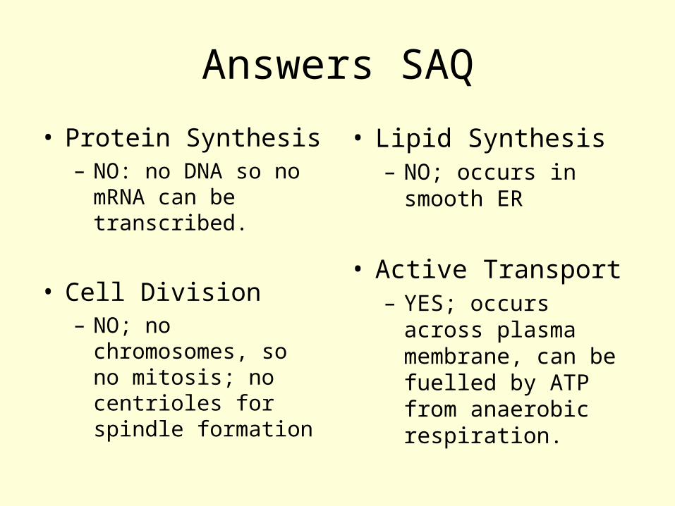
• Protein Synthesis– NO: no DNA so no
mRNA can be transcribed.
• Cell Division– NO; no
chromosomes, so no mitosis; no centrioles for spindle formation
• Lipid Synthesis– NO; occurs in
smooth ER
• Active Transport– YES; occurs across
plasma membrane, can be fuelled by ATP from anaerobic respiration.
Tissue Fluid
• Immediate environment of each individual body cell.
• Homeostasis maintains composition of tissue fluid at a constant level to provide the optimum environment in which cells can work.
• Contains less proteins than Blood plasma
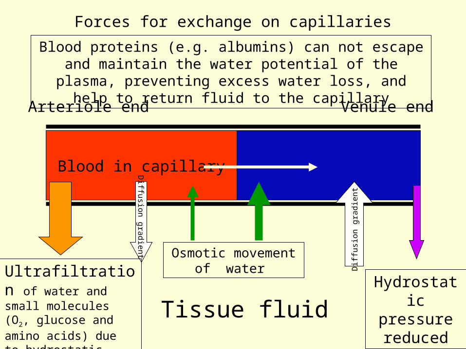
Forces for exchange on capillaries
Blood in capillary
Arteriole end Venule end
Blood proteins (e.g. albumins) can not escape and maintain the water potential of the plasma,
preventing excess water loss, and help to return fluid to the capillary
Diff
usio
n g
radie
nt
Osmotic movement of water Ultrafiltration of
water and small molecules (O2, glucose and amino acids) due to hydrostatic pressure
Diff
usi
on g
radie
nt
Hydrostatic pressure reducedTissue fluid
Lymph• Similar composition to plasma with
less proteins• Lipids absorbed in lacteals, give
lymph milky appearance• Tiny blind ending vessels• Tiny valves in walls allow large
molecules to pass in.• Drains back into blood plasma in
subclavian vein.
oedema
• If lymph does not take away proteins in tissue fluid between cells, YOU could die in 24 hours.
• Get a build up in tissue fluid, called oedema.
Movement in lymph capillaries
• Contraction of muscles around vessels
• Valves• Slow movement
– Diagram: the relationship between blood, tissue fluid and lymph at a capillary network
» Diagram: the lymph system
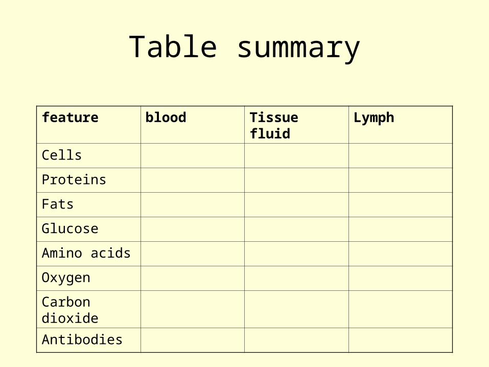
Table summary
feature blood Tissue fluid Lymph
Cells
Proteins
Fats
Glucose
Amino acids
Oxygen
Carbon dioxide
Antibodies
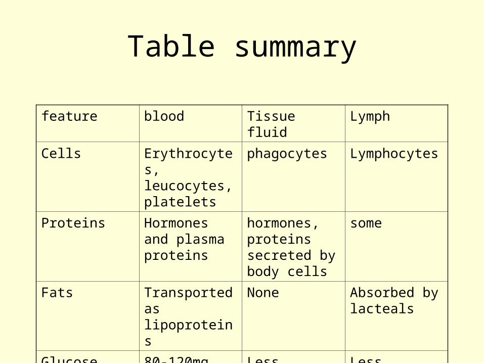
Table summary
feature blood Tissue fluid Lymph
Cells Erythrocytes, leucocytes, platelets
phagocytes Lymphocytes
Proteins Hormones and plasma proteins
hormones, proteins secreted by body cells
some
Fats Transported as lipoproteins
None Absorbed by lacteals
Glucose 80-120mg per 100cm3
Less Less
Amino acids more less less
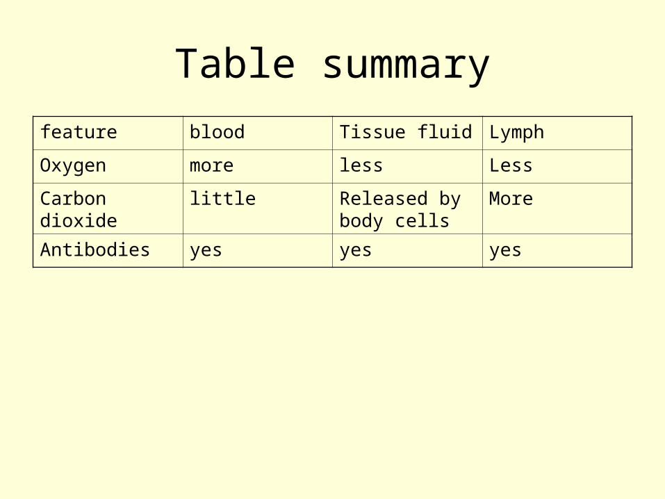
Table summary
feature blood Tissue fluid Lymph
Oxygen more less Less
Carbon dioxide
little Released by body cells
More
Antibodies yes yes yes
The Mammalian Transport System
Transport of Oxygen and Carbon Dioxide
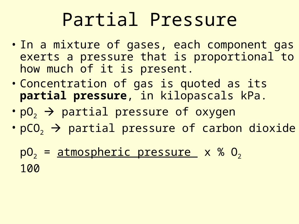
Partial Pressure• In a mixture of gases, each component
gas exerts a pressure that is proportional to how much of it is present.
• Concentration of gas is quoted as its partial pressure, in kilopascals kPa.
• pO2 partial pressure of oxygen• pCO2 partial pressure of carbon dioxide
pO2 = atmospheric pressure x % O2
100
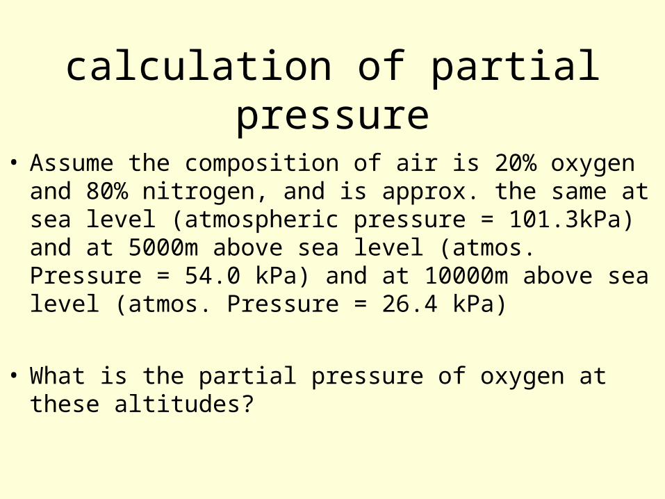
calculation of partial pressure
• Assume the composition of air is 20% oxygen and 80% nitrogen, and is approx. the same at sea level (atmospheric pressure = 101.3kPa) and at 5000m above sea level (atmos. Pressure = 54.0 kPa) and at 10000m above sea level (atmos. Pressure = 26.4 kPa)
• What is the partial pressure of oxygen at these altitudes?
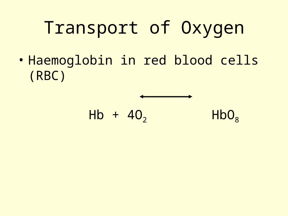
Transport of Oxygen
• Haemoglobin in red blood cells (RBC)
Hb + 4O2 HbO8
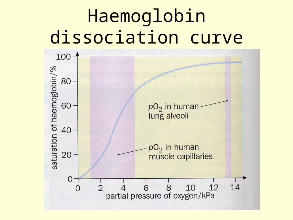
Haemoglobin dissociation curve
• A graph showing the amount of oxygen combining with haemoglobin at different partial pressures.
• High pO2 – haemoglobin saturated with oxygen
• Low pO2 – oxyhaemoglobin gives up its oxygen to respiring cells (dissociates)
Haemoglobin dissociation curve
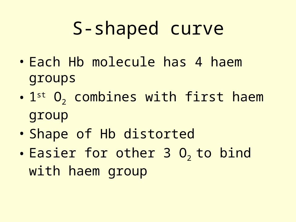
S-shaped curve
• Each Hb molecule has 4 haem groups
• 1st O2 combines with first haem group
• Shape of Hb distorted
• Easier for other 3 O2 to bind with haem group
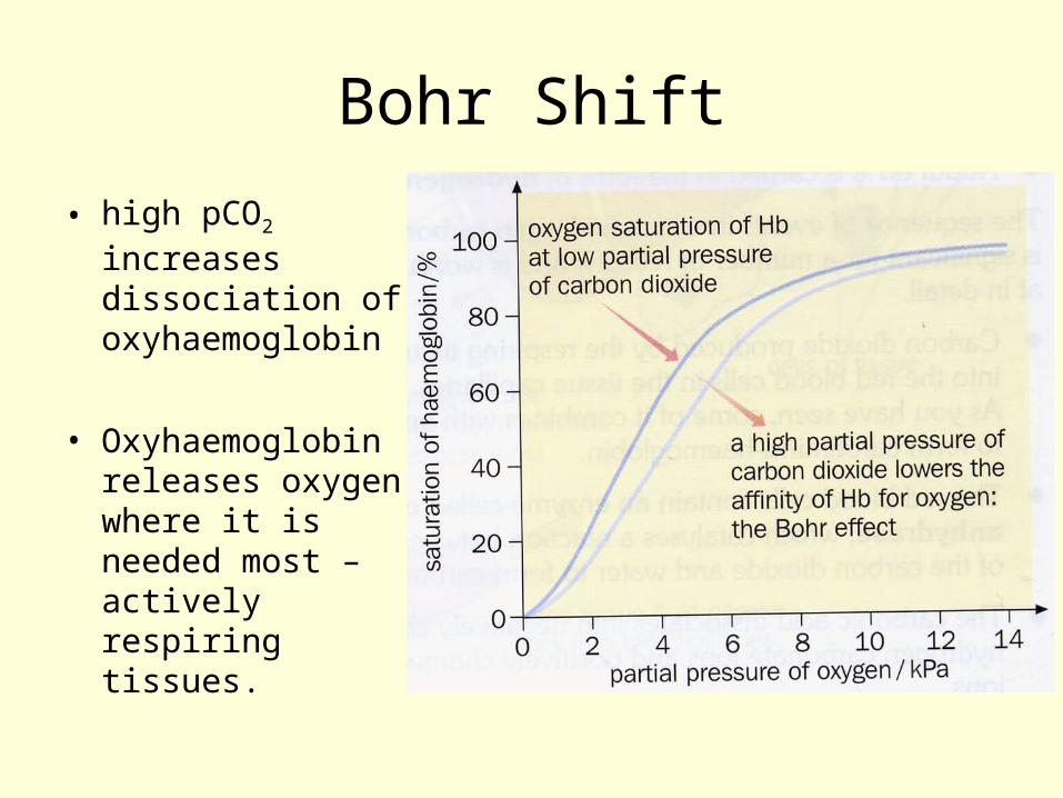
Bohr Shift
• high pCO2 increases dissociation of oxyhaemoglobin
• Oxyhaemoglobin releases oxygen where it is needed most – actively respiring tissues.
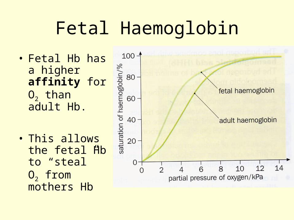
Fetal Haemoglobin
• Fetal Hb has a higher affinity for O2 than adult Hb.
• This allows the fetal Hb to “steal” O2 from mothers Hb
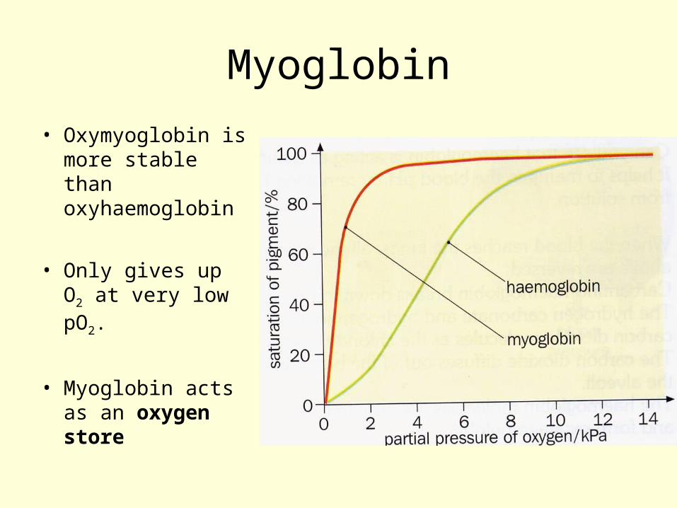
Myoglobin
• Oxymyoglobin is more stable than oxyhaemoglobin
• Only gives up O2 at very low pO2.
• Myoglobin acts as an oxygen store
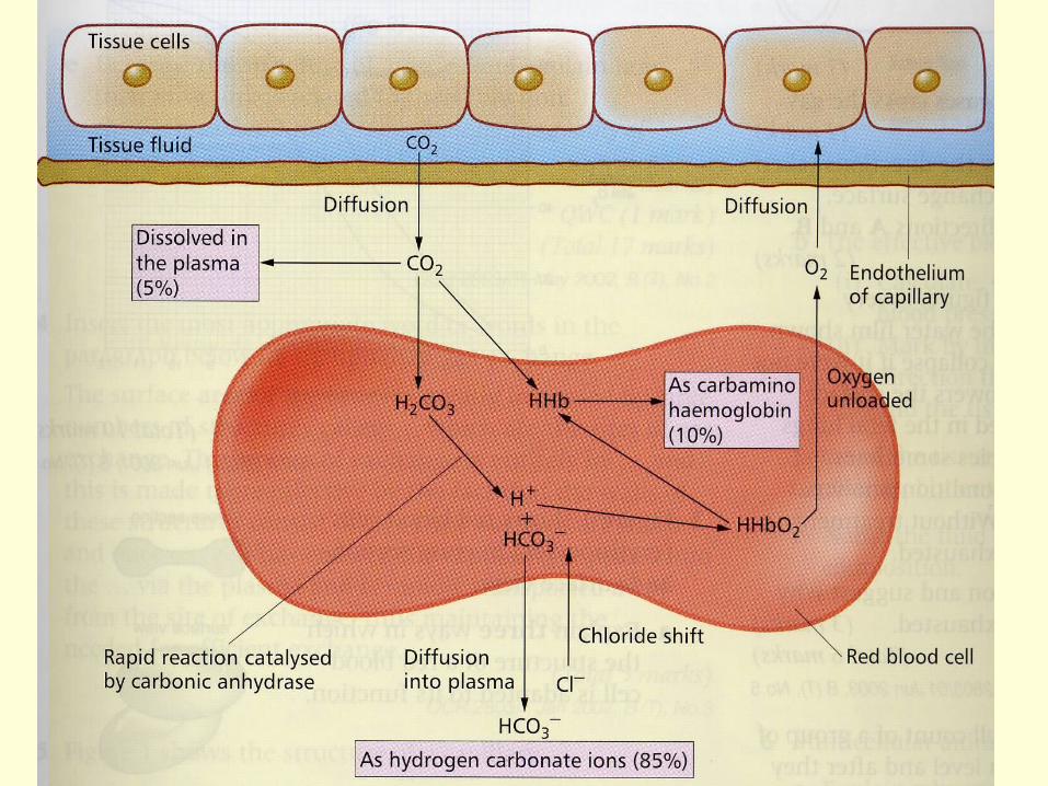
Carbon Dioxide Transport
• CO2 carried in three ways
– 5% in solution in plasma as CO2
– 10% combines with amino groups in Hb molecule (carbamino haemoglobin)
– 85% hydrogen carbonate ions
Carbon dioxide transport
• Transported in blood as hydrogen carbonate ions
• Carbonic anhydrase catalyses the reaction
CO2 + H2O H2CO3
Carbon Dioxide Transport
• Carbonic acid dissociates
H2CO3 H+ + HCO3-
• H+ ions associate with haemoglobin (buffer)
• Haemoglobinic acid (HHb)
• Contributes to Bohr effect
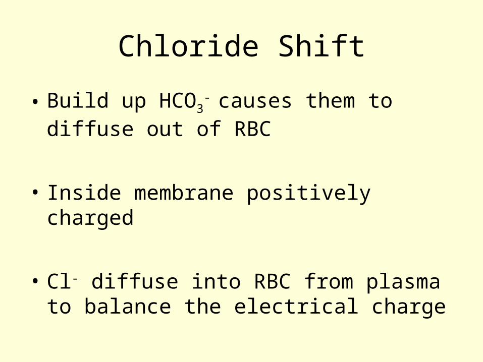
Chloride Shift
• Build up HCO3- causes them to diffuse
out of RBC
• Inside membrane positively charged
• Cl- diffuse into RBC from plasma to balance the electrical charge
Problems with Oxygen Transport
Carbon Monoxide• Haemoglobin combines readily with
carbon monoxide to form carboxyhaemoglobin (stable compound)
• Carbon monoxide has a higher affinity with haemoglobin than oxygen does
• 0.1% CO in air can cause death by asphyxiation.
High Altitude
– Question• Atheletes often prepare themselves for
important competitions by spending several months training at high altitude. Explain how this could improve their performance.
Training at high altitude
• Spending a length of time at high altitude stimulates the body to produce more red blood cells
• When an athlete returns to sea level, these “extra” RBC remain in the body for sometime, and can supply extra oxygen to muscles enabling them to work harder and for longer than they would otherwise.