ANIMAL MODELS OF HYPERTENSION AND EFFECT OF DRUGS
14
Indian Journal of Pharmacology 2003; 35: 349-362 EDUCATIONAL FORUM ANIMAL MODELS OF HYPERTENSION AND EFFECT OF DRUGS D.K. BADYAL, H. LATA*, A.P. DADHICH Department of Pharmacology, Christian Medical College, Ludhiana-141 008. *Department of Physiology, Dayanand Medical College, Ludhiana-141 001. Manuscript Received: 9.12.2002 Revised: 13.6.2003 Accepted: 19.8.2003 Hypertension, the most common cardiovascular disease is the primary cause of stroke, coronary artery disease and sudden cardiac death. Many factors are believed to be involved in the pathogenesis of hypertension and its complications. Experimental models of human disease are used to study pathophysiological factors involved in hypertension and assess antihypertensive agents. Today different strains of rats with genetic hypertension are available and in most laboratories, therapeutic studies on hypertension are carried out on these models. As new insights in to the pathogenesis of hypertension are revealed, new models are being developed to produce hypertension in animals. This article reviews experimental models of hypertension. Both conventional and genetic models of hypertension are discussed. Experimental hypertension blood pressure cardiovascular disease ABSTRACT KEY WORDS Introduction Hypertension is the most common cardiovascular disease and is a major public health issue in developed as well as developing countries. Although it is common and readily detectable, it can often lead to lethal complications if left untreated. Because of its high incidence and morbidity, various classes of drugs and regimens have been advocated for the control of hypertension. Despite the large armamentaria of drugs being available for the treatment of hypertension, the last two decades have witnessed the introduction of a number of new antihypertensive drugs. Recent research during this period has also added considerably to our knowledge of the mechanisms involved in the pathogenesis of hypertension. Human essential hypertension is a complex, multifactorial, quantitative trait under polygenic control. In order to understand the pathogenesis and to study the treatment and prevention of a disease, it is useful to develop animal models. Various models of experimental hypertension have been primarily developed to obtain information on the etiopatho- genesis of hypertension. These models are also used in the pharmacological screening of potential antihypertensive agents. In the past, hypertensive animal models have been used infrequently for testing antihypertensive potential of drugs. As new molecules are being synthesized in a large number, the use of animal models is increasing for testing these molecules. New animal models of hypertension are being developed as new insights in to the pathogenesis of hypertension are revealed. The animal models of hypertension share many features which are common to human hypertension. Many of these models have been developed by utilizing the etiological factors that are presumed to be responsible for human hypertension such as excessive salt intake, hyperactivity of renin- angiotensin-aldosterone system (RAAS) and genetic factors. Since regulation of blood pressure (BP) is multifactorial, the effectiveness of an antihypertensive agent in one model does not necessarily mean that the mechanism of action of a given agent in a given model is related to the pathogenesis of elevated blood pressure. Correspondence: D.K. Badyal E-mail: [email protected]
Transcript of ANIMAL MODELS OF HYPERTENSION AND EFFECT OF DRUGS

D.K. BADYAL et al.
Indian Journal of Pharmacology 2003; 35: 349-362 EDUCATIONAL FORUM
ANIMAL MODELS OF HYPERTENSION AND EFFECT OF DRUGS
D.K. BADYAL, H. LATA*, A.P. DADHICH
Department of Pharmacology, Christian Medical College, Ludhiana-141 008. *Department of Physiology,Dayanand Medical College, Ludhiana-141 001.
Manuscript Received: 9.12.2002 Revised: 13.6.2003 Accepted: 19.8.2003
Hypertension, the most common cardiovascular disease is the primary cause of stroke, coronary arterydisease and sudden cardiac death. Many factors are believed to be involved in the pathogenesis ofhypertension and its complications. Experimental models of human disease are used to studypathophysiological factors involved in hypertension and assess antihypertensive agents. Today differentstrains of rats with genetic hypertension are available and in most laboratories, therapeutic studies onhypertension are carried out on these models. As new insights in to the pathogenesis of hypertension arerevealed, new models are being developed to produce hypertension in animals. This article reviewsexperimental models of hypertension. Both conventional and genetic models of hypertension are discussed.
Experimental hypertension blood pressure cardiovascular disease
ABSTRACT
KEY WORDS
Introduction
Hypertension is the most common cardiovasculardisease and is a major public health issue in developedas well as developing countries. Although it is commonand readily detectable, it can often lead to lethalcomplications if left untreated. Because of its highincidence and morbidity, various classes of drugs andregimens have been advocated for the control ofhypertension. Despite the large armamentaria of drugsbeing available for the treatment of hypertension, thelast two decades have witnessed the introduction of anumber of new antihypertensive drugs. Recentresearch during this period has also addedconsiderably to our knowledge of the mechanismsinvolved in the pathogenesis of hypertension.
Human essential hypertension is a complex,multifactorial, quantitative trait under polygeniccontrol. In order to understand the pathogenesis andto study the treatment and prevention of a disease, itis useful to develop animal models. Various modelsof experimental hypertension have been primarilydeveloped to obtain information on the etiopatho-
genesis of hypertension. These models are also usedin the pharmacological screening of potentialantihypertensive agents. In the past, hypertensiveanimal models have been used infrequently for testingantihypertensive potential of drugs. As new moleculesare being synthesized in a large number, the use ofanimal models is increasing for testing thesemolecules. New animal models of hypertension arebeing developed as new insights in to thepathogenesis of hypertension are revealed.
The animal models of hypertension share manyfeatures which are common to human hypertension.Many of these models have been developed byutilizing the etiological factors that are presumed tobe responsible for human hypertension such asexcessive salt intake, hyperactivity of renin-angiotensin-aldosterone system (RAAS) and geneticfactors. Since regulation of blood pressure (BP) ismultifactorial, the effectiveness of an antihypertensiveagent in one model does not necessarily mean thatthe mechanism of action of a given agent in a givenmodel is related to the pathogenesis of elevated bloodpressure.
Correspondence: D.K. BadyalE-mail: [email protected]

ANIMAL MODELS OF HYPERTENSION
An ideal animal model of hypertension should fulfillthe following criteria:
• It should be feasible in small animals.
• It should be simple to perform and uniformlyreproducible.
• It should be able to predict the potentialantihypertensive properties of an agent.
• It should consume minimal quantities ofcompounds.
• It should be comparable to some form of humanhypertension.
Animals used: Whereas in the past, most studieson experimental hypertension were carried out ondogs, currently rat is the preferred animal species.Spontaneous hypertensive rat (SHR), the geneticstrain of hypertensive rat, is the animal of choice forscreening antihypertensive agents. SHR is thecornerstone of medical research in experimentalhypertension1. Rabbits, monkeys, pigs and mice arealso used to produce experimental hypertension2.
The various types of animal models of hypertensionbeing used are:
1. Renovascular hypertension
2. Dietary hypertension
3. Endocrine hypertension
4. Neurogenic hypertension
5. Psychogenic hypertension
6. Genetic hypertension
7. Other models
1. Renovascular hypertension:
This is a very commonly used model of hypertension.In renovascular hypertension, the RAAS plays animportant role3,4. Experimentally, renal hypertensionis produced by renal artery constriction, whichactivates peripheral RAAS and sympathetic nervoussystem5. A number of factors like decreased bloodvolume may lead to sympathetic stimulation in thismodel. Renin is secreted by kidneys whensympathetic activity is increased. Renin convertsangiotensinogen to angiotensin-I. Angiotensin-I isconverted to angiotensin-II by angiotensin convertingenzyme (ACE). Angiotensin-II is a potent vaso-constrictor and increases BP. Angiotensin-II also
causes release of aldosterone leading to salt andwater retention resulting in increased blood volumeand hypertension (Figure 1)3,4,6.
The various methods of inducing renovascularhypertension are as follows:
I. Goldblatt method: Goldblatt et al (1934)reported that a partial constriction of renalarteries in dogs produced hypertension7. Thistype of hypertension has also been induced inrabbits, rats and monkeys8. In rabbits and rats,a U-shaped silver ribbon clip is used toconstrict the renal arteries9. Rats weighing from120 to 200 g are anaesthetized with hexo-barbitone sodium (40 mg/kg body weight) anda silver clip of 0.2 mm internal diameter isplaced on the left renal artery close to theaorta10. The renal artery in rats can also beligated with 4-0 silk suture11. Constriction ofrenal artery should be more than 50%. Theanimal is considered hypertensive if systolicBP is more than 160 mm Hg for twoconsecutive days after 4 weeks of ligation5.
In dogs and rabbits, retroperitoneal approachcan be used and the operation is carried outthrough an oblique lateral abdominal incision,just below and parallel to costal margin.Muscles are split and peritoneum is strippedaway from the kidney. The kidney and the renalartery are easily identifiable. The first part ofrenal artery is cleaned for 1 cm or so from itsorigin from the aorta9. In dogs, renal arteriescan also be constricted with a small adjustablesilver clamp or with silk sutures12.
In rabbits, bilateral constriction of renal arteriesis achieved in two stages with an interval of1-4 weeks between operations. Persistenthypertension, which is moderate to severe, willdevelop over a period of 12 months9. The BPhas been reported to increase from normalmean level of 106 to 160-190 mm Hg from 4weeks to 9 months in 65% of rabbits8. The mostsatisfactory method in rabbits is to perform aright nephrectomy and two or more weeks laterto apply a clip to the left renal artery13.
Three types of hypertension are produced byGoldblatt method:
350

D.K. BADYAL et al.
Figure 1. Renin angiotensin system and Goldblatt hypertension.
a. Two kidney one clip (2K1C) hypertension:The renal artery is constricted on only oneside with the other artery (or kidney) leftuntouched. This results in a sustainedincrease in BP due to increased plasma reninactivity (PRA), which in turn increasescirculating angiotensin-II, a potent vaso-constrictor. However, there is no salt andwater retention because of the other normalkidney being intact. Thus, the resultanthypertension at this stage is renin-angiotensindependent. After about 6 weeks, theincreased angiotensin-II releases aldo-sterone from adrenal cortex leading to agradual retention of salt and water. Retentionof salt and water leads to decreased renin
351
(-)
production (Figure 1). From this stageonwards, hypertension is volume depen-dent4,6,9. Hence, salt and water balance iscritically involved in the pathogenesis ofrenovascular hypertension. Increased BP andincreased renin activity returns to normal byunclipping or removal of the affected kidney6.
b. One kidney one clip (1K1C) hypertension:Constriction of renal artery is done on one sideand the contralateral kidney is removed. Thereis an increase in BP within a few hours. Sincethere is no other kidney, there is no pressurediuresis and natriuresis, so there is rapid saltand water retention (Figure 1). Plasma reninactivity is usually normal. Hypertension soonbecomes volume dependent4,6,9.

ANIMAL MODELS OF HYPERTENSION
c. Two kidney two clip (2K2C) hypertension:Constriction of aorta or both renal arteries isdone. There is a patchy ischaemic kidneytissue, which secretes renin leading toincreased BP. The remaining kidney tissueretains salt and water. Indeed, one of the mostcommon causes of renal hypertension inhuman beings is such a patchy ischaemickidney disease4.
II. Hypertension induced by externalcompression of renal parenchyma: Thistype of hypertension is produced in dogs,rabbits and rats. The following methods areused to produce this type of hypertension:
a. Page hypertension: A sheet of cellophane isplaced around the kidney and held in place bysilk sutures tied loosely around the renal hilus.Both kidneys are wrapped or one kidney iswrapped and other is removed14. Afibrocollagenous shell is formed around thekidney in 3-5 days because of reaction of thetissue to the foreign material. This shellcompresses renal parenchyma leading todecreased renal vascular pressure. Thisexpands the extracellular volume leading toincreased peripheral resistance and henceincreased BP. In rabbits, chronic hypertensionwith normal or decreased renin activity isproduced over a period of approximately 6weeks 6,15. However, a high proportion ofanimals develops severe hypertension anddies within 2 months8.
b. Grollman hypertension: In this method,kidney tissue is compressed by securing a'figure of 8' ligature around the kidney16. Theligature around the kidney forms a figureresembling number 8. This type of hyper-tension can be produced in dogs, rabbits andrats. It is of two types:
1. Two kidney one ligature (2K1L): Ligature isapplied to one kidney and contralateral kidneyis left untouched.
2. One kidney one ligature (1K1L): Ligature isapplied to one kidney and contralateral kidneyis removed.
III. Coarctation of aorta: Renal blood flow can
be decreased by compressing the aorta.Coarctation can be done just above the renalarteries, between renal arteries and superiormesenteric arteries or between two renalarteries with the right artery above and theleft artery below the site of coarctation17. Anincrease in BP similar to 2K1C model can beproduced by applying a rubber band toabdominal aorta along with constriction of rightrenal artery for 8 weeks18. Coarctation can befollowed by unilateral nephrectomy to producethis type of hypertension6,19.
IV. Reduced renal mass: Reducing renal tissueto five-sixth (5/6th) by renal mass ablationproduces hypertension. In this method, theright kidney is removed and 2 or 3 branchesof left renal artery are ligated to produceinfarction of approximately 2/3rd of the leftkidney20.
V. Glomerular sclerosis secondary to micro-sphere embolisation is also used to producerenal hypertension21.
The mean arterial pressure in renal hyper-tensive rats produced by above methods(231.64±3.1 mm Hg) is significantly higher thanthe mean arterial pressure in normotensivecontrol rats (160±2.84 mm Hg)22.
2. Dietary hypertension:
I. Increased salt intake: Physiologically, normalkidney has the ability to excrete easily the dailysalt load without allowing a marked rise inextracellular volume. However, generalepidemiological data have shown that higherthe average sodium intake in a givenpopulation, the greater will be the prevalenceof hypertension. Chronic ingestion of excesssalt produces hypertension in rats, whichmimics human hypertension morphologically23.High salt intake hypertension has beenproduced in rats, rabbits and chicks byreplacing drinking water with 1-2% sodiumchloride for 9-12 months8,24.
High salt intake along with unilateralnephrectomy produces accelerated hyper-tension within 3-4 weeks in dogs25. Acceleratedrenal hypertension in rats is produced by
352

D.K. BADYAL et al.
applying a 'figure of eight' ligature to one kidneyand removing the other in a single stageoperation performed under strict asepticconditions using pentobarbitone sodium (20mg/kg, i.p. ) and ether anaesthesia. Thedrinking water is replaced with physiologicalsaline for three weeks and the animals areused 4 to 5 weeks after the operation22.
3. Endocrine hypertension:
I. Mineralocorticoid induced hypertension:Mineralocorticoids cause retention of sodiumand water in the body until escape diuresisoccurs due to increased pressure on thekidneys. No further retention of sodium andwater occurs, but general level of body sodiumand water is slightly raised.
Selye et al was the first to demonstrate thatdeoxycorticosterone acetate (DOCA)produces hypertension in rats26. There isincreased DOCA-induced reabsorption of saltand water leading to increased blood volumeand hence increased BP. There is alsoincreased secretion of vasopressin leading towater retention and vasoconstriction. Inaddition, altered activity of RAAS leads toincreased sympathetic activity 23,27. Rats,especially female and young, are prone toDOCA-salt induced hypertension23,24. This typeof hypertension can also be produced in dogsand pigs28. Other mineralocorticoids (e.g. ,aldosterone) and glucocorticoids can alsoproduce this type of hypertension26,29.
DOCA induced hypertension is salt dependentsince neither administration of DOCA norpartial removal of renal mass is effective inincreasing BP when applied without saltadministration23. To produce hypertension, ratsweighing about 100 g are kept on a diet highin sodium chloride and drinking water isreplaced by 2% sodium chloride solutionad libitum. After they attain a weight of about250 g, they are given DOCA dissolved insesame seed oil at a dose of 10 mg/kg, twiceweekly for 43 days24,30.
In another method, unilateral nephrectomy isperformed followed by DOCA administration31.
All operated rats receive an injection ofampicillin (10 mg/kg, i.m.) daily for 5 days andlocal application of neosporin-H to preventinfection. A week later, DOCA (25 mg/kg/wk,s.c. for 5 wk) dissolved in cotton seed oil, isinjected into nephrectomised rats. Alternatively,nephrecto-mised rats could receive DOCAfrom silicon rubber implants (200 mg/rat)implanted subcutaneously 32. NaCl (1.0%)solution is substituted for drinking water andgiven ad libitum 24.
II. Adrenal regeneration hypertension:Hypertension is produced in rats by unilateralnephrectomy followed by removal of rightadrenal gland and enucleation of left adrenalgland. Enucleation is carried out by making asmall incision in the capsule of adrenal glandthrough which the bulk of glandular tissue isextruded by gentle application of pressure withcurved forceps. Drinking water is replaced with1% saline. Hypertension develops duringregeneration of adrenal glands in about 2weeks8. Once hypertension has beenestablished, neither removal of the re-generated adrenal nor substitution of salinewith drinking water brings about a return of BPto normal33.
This type of hypertension develops morereadily in female and young rats. This modelis used mainly to study the role of variousfactors e.g., steroids, in the pathophysiologyof hypertension33,34.
4. Neurogenic hypertension:
One of the most important negative feedback in thecontrol of BP originates from baroreceptors locatedin the carotid sinus and aortic arch. Afferents ofbaroreceptors travel along 9th and 10th cranialnerves. The first synapse is present in the nucleustractus solitarius (NTS) of the medulla oblongata. NTSis not merely a relay center, it also contains complexsynaptic connections and innervation from many brainareas and receives information from periphery.Stimulation of baroreceptors causes inhibition ofvasomotor center leading to vasodilatation,bradycardia and decrease in BP8. Sectioning of thebaroreceptor nerves leads to persistent rise in BP indogs, cats and rabbits35. Neurogenic hypertensioncan be produced by the following methods:
353
ANIMAL MODELS OF HYPERTENSION
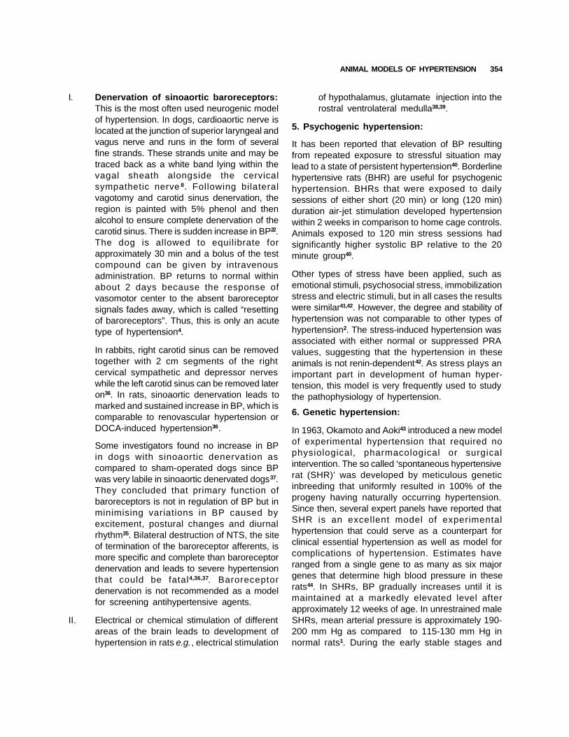
I. Denervation of sinoaortic baroreceptors:This is the most often used neurogenic modelof hypertension. In dogs, cardioaortic nerve islocated at the junction of superior laryngeal andvagus nerve and runs in the form of severalfine strands. These strands unite and may betraced back as a white band lying within thevagal sheath alongside the cervicalsympathetic nerve 8. Following bilateralvagotomy and carotid sinus denervation, theregion is painted with 5% phenol and thenalcohol to ensure complete denervation of thecarotid sinus. There is sudden increase in BP22.The dog is allowed to equilibrate forapproximately 30 min and a bolus of the testcompound can be given by intravenousadministration. BP returns to normal withinabout 2 days because the response ofvasomotor center to the absent baroreceptorsignals fades away, which is called “resettingof baroreceptors”. Thus, this is only an acutetype of hypertension4.
In rabbits, right carotid sinus can be removedtogether with 2 cm segments of the rightcervical sympathetic and depressor nerveswhile the left carotid sinus can be removed lateron36. In rats, sinoaortic denervation leads tomarked and sustained increase in BP, which iscomparable to renovascular hypertension orDOCA-induced hypertension36.
Some investigators found no increase in BPin dogs with sinoaortic denervation ascompared to sham-operated dogs since BPwas very labile in sinoaortic denervated dogs37.They concluded that primary function ofbaroreceptors is not in regulation of BP but inminimising variations in BP caused byexcitement, postural changes and diurnalrhythm35. Bilateral destruction of NTS, the siteof termination of the baroreceptor afferents, ismore specific and complete than baroreceptordenervation and leads to severe hypertensionthat could be fatal4,36,37. Baroreceptordenervation is not recommended as a modelfor screening antihypertensive agents.
II. Electrical or chemical stimulation of differentareas of the brain leads to development ofhypertension in rats e.g., electrical stimulation
of hypothalamus, glutamate injection into therostral ventrolateral medulla38,39.
5. Psychogenic hypertension:
It has been reported that elevation of BP resultingfrom repeated exposure to stressful situation maylead to a state of persistent hypertension40. Borderlinehypertensive rats (BHR) are useful for psychogenichypertension. BHRs that were exposed to dailysessions of either short (20 min) or long (120 min)duration air-jet stimulation developed hypertensionwithin 2 weeks in comparison to home cage controls.Animals exposed to 120 min stress sessions hadsignificantly higher systolic BP relative to the 20minute group40.
Other types of stress have been applied, such asemotional stimuli, psychosocial stress, immobilizationstress and electric stimuli, but in all cases the resultswere similar41,42. However, the degree and stability ofhypertension was not comparable to other types ofhypertension2. The stress-induced hypertension wasassociated with either normal or suppressed PRAvalues, suggesting that the hypertension in theseanimals is not renin-dependent42. As stress plays animportant part in development of human hyper-tension, this model is very frequently used to studythe pathophysiology of hypertension.
6. Genetic hypertension:
In 1963, Okamoto and Aoki43 introduced a new modelof experimental hypertension that required nophysiological, pharmacological or surgicalintervention. The so called ‘spontaneous hypertensiverat (SHR)’ was developed by meticulous geneticinbreeding that uniformly resulted in 100% of theprogeny having naturally occurring hypertension.Since then, several expert panels have reported thatSHR is an excellent model of experimentalhypertension that could serve as a counterpart forclinical essential hypertension as well as model forcomplications of hypertension. Estimates haveranged from a single gene to as many as six majorgenes that determine high blood pressure in theserats44. In SHRs, BP gradually increases until it ismaintained at a markedly elevated level afterapproximately 12 weeks of age. In unrestrained maleSHRs, mean arterial pressure is approximately 190-200 mm Hg as compared to 115-130 mm Hg innormal rats1. During the early stable stages and
354

D.K. BADYAL et al.
developmental phase of hypertension, elevated BPis maintained in large part by enhanced centralsympathetic outflow45. In the later stages increasedtotal peripheral resistance with a normal cardiacoutput and decreased permeability of the glomerularmembranes form the basis for the long termmaintenance of the hypertension. Comparisonbetween spontaneous and essential hypertensionreveal that both are caused by multifactorialinheritance and have in common an increasedvascular resistance which is caused by neuralvasomotor tone and non-neural structural alterations.Guidelines have been published for the maintenanceand use of SHRs46.
Spontaneous hypertension has been observed in anumber of different strains of common laboratoryanimals, at least 6 different strains of rats, and atleast one strain each of dogs and rabbits. The NewZealand strain of Smirk seems to be most similar tothe Japanese SHR, though this has not been studiedas widely47. In contrast, the Milan strain developedby Bianchi et al, seems to be different involvingprimarily alterations in renal sodium and watermetabolism; therefore it may not be analogous toessential hypertension48. A fourth strain developedby Dahl shows a high sensitivity (“S” strain) to sodiumintake in comparison to its normotensive control (“R”strain), which is sodium resistant49. Furthermore, afifth strain of hypertensive rats ‘the Sabra strain’ havebeen bred recently in Israel, and a sixth strain inFrance, the Lyon strain50,51.
Among these strains, SHRs develop not onlymoderate to severe hypertension but also typicalcomplications of hypertension. Stroke prone SHRs(SHRSP), which are selectively bred from amongSHRs, are extreme examples that developcerebrovascular lesions spontaneously in over 80%of rats52. Because of high mortality rate of stroke inman, the similarity of stroke in SHRSP is importantfor further application of this model to studies ofstroke in man. Other variants derived from SHRsare obese SHRs and arteriolipidosis-proneSHRs53,54. Extreme environmental conditions suchas stress and excess salt accelerate thedevelopment of hypertension and aggravate thehypertensive complications55,56.
Complications such as cerebral hemorrhage,thrombosis, nephrosclerosis and myocardial lesions
in SHRs and especially cerebral lesions in SHRSPs,are pathogenically, pathologically and epide-miologically similar to those observed in essentialhypertension in man2. Therefore, these models canbe used to study not only the pathogenesis andtherapy but also prophylaxis in essential hypertensionand its complications. Because of apparentsimilarities of the SHR to essential hypertension, SHRmodels are highly recommended for screeningpotential drug candidates for hypertension1.Experimental models of genetic hypertension havealso been developed in animals other than the rat.Though it has been produced in one strain of rabbitand dog they have not been studied extensively forpractical, financial and other reasons1,4,6. Rats aremainly used because of their small size, short life-span and low cost.
7. Other models:
1. Obesity-related hypertension: Wistar fattyrats (WFR) derived from cross between obeseZucker and Wistar Kyoto rats show persistenthyperinsulinemia and hypertension after 16weeks of age and may be a good model toelucidate the relationship between hyper-insulinemia and hypertension57.
2. Hypertension induced by cholinomimeticagents: Physostigmine (10-80 µg/kg, i.v.), acholinesterase inhibitor, and oxotremorine(20-40 µg/kg, i.v. ), a direct muscariniccholinergic agonist, cause a dose-dependentincrease in BP58. The cholinomimetic-inducedhypertension has been shown to be elicitedthrough activation of central cholinergicmechanism and mediated peripherally throughsympathetic nervous system45. Pretreatmentwith methyl scopolamine (1 mg/kg) 5-10minutes before giving oxotremorine preventsinitial hypotension in this model59.
3. Angiotensin-II induced hypertension:Subcutaneous infusion of angiotensin-II (0.7mg/kg/day) using minipump elicits hyper-tension in 4-8 weeks60,61.
4. Hypertension induced by cadmium:Hypertension is produced by the chronicadministration of CdCI (1 mg/kg/day, i.p. for2 wk). CdCl-induced hypertension might bedue to the fact that the metal ion might mimic
355
ANIMAL MODELS OF HYPERTENSION
Ca2+ ion as a partial agonist and produce adirect contractile effect on vascular smoothmuscle24.
5. Chronic nitric oxide inhibition-inducedhypertension: SHRs when given a non-selective nitric oxide synthase inhibitor, Nω-nitro-L-argininemethyl ester (L-NAME), for 4weeks develop time and dose-dependenthypertension62.
6. Transgenic rat (TGR) models: Transgenicmodels of hypertension have revolutionized theexperimental work on hypertension. Severaltransgenic rat lines expressing candidategenes for hypertension have been produced.Introducing an additional renin gene, themurine Ren-2 gene, into the germ line of ratsresults in transgenic hypertensive rat strain,TGR (mREN2) 27, with an overexpression ofrenin. Genetic linkage studies have shown thatthe components of RAAS are associated withthis type of hypertension. Linkage has beendescribed for the angiotensinogen gene inhuman hypertension. TGR model may beuseful for studying the role of local RAASsystem in hypertension63.
7. Uterine ischaemia: As in preeclampsia,uterine ischaemia can induce hypertension inrats and monkeys8,64,65. In monkeys at 116±7days of gestation, lower aortic pressure wasreduced by 24±11 mm Hg by a stricture on theaorta just below the renal arteries and animalsdeveloped sustained hypertension66.
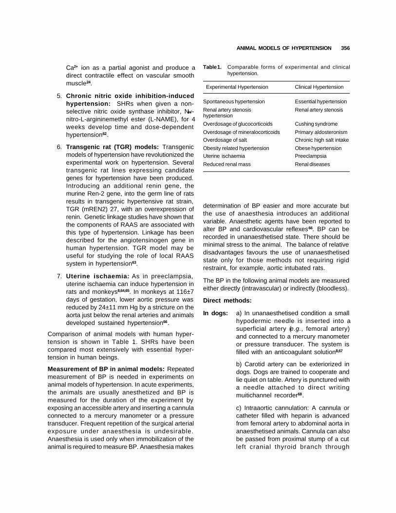
Comparison of animal models with human hyper-tension is shown in Table 1. SHRs have beencompared most extensively with essential hyper-tension in human beings.
Measurement of BP in animal models: Repeatedmeasurement of BP is needed in experiments onanimal models of hypertension. In acute experiments,the animals are usually anesthetized and BP ismeasured for the duration of the experiment byexposing an accessible artery and inserting a cannulaconnected to a mercury manometer or a pressuretransducer. Frequent repetition of the surgical arterialexposure under anaesthesia is undesirable.Anaesthesia is used only when immobilization of theanimal is required to measure BP. Anaesthesia makes
Table 1. Comparable forms of experimental and clinicalhypertension.
Experimental Hypertension Clinical Hypertension
Spontaneous hypertension Essential hypertension
Renal artery stenosis Renal artery stenosishypertension
Overdosage of glucocorticoids Cushing syndrome
Overdosage of mineralocorticoids Primary aldosteronismOverdosage of salt Chronic high salt intake
Obesity related hypertension Obese hypertensionUterine ischaemia Preeclampsia
Reduced renal mass Renal diseases
356
determination of BP easier and more accurate butthe use of anaesthesia introduces an additionalvariable. Anaesthetic agents have been reported toalter BP and cardiovascular reflexes66. BP can berecorded in unanaesthetised state. There should beminimal stress to the animal. The balance of relativedisadvantages favours the use of unanaesthetisedstate only for those methods not requiring rigidrestraint, for example, aortic intubated rats.
The BP in the following animal models are measuredeither directly (intravascular) or indirectly (bloodless).
Direct methods:
In dogs: a) In unanaesthetised condition a smallhypodermic needle is inserted into asuperficial artery (e.g., femoral artery)and connected to a mercury manometeror pressure transducer. The system isfilled with an anticoagulant solution8,67
b) Carotid artery can be exteriorized indogs. Dogs are trained to cooperate andlie quiet on table. Artery is punctured witha needle attached to direct writingmuitichannel recorder68.
c) Intraaortic cannulation: A cannula orcatheter filled with heparin is advancedfrom femoral artery to abdominal aorta inanaesthetised animals. Cannula can alsobe passed from proximal stump of a cutleft cranial thyroid branch through

D.K. BADYAL et al.
common carotid artery down todescending thoracic aorta and BP ismeasured8.
In rabbits: a) In an unanaesthetised animal, centralartery of ear is cannulated. The disadvan-tages of this method include developmentof hematoma and arterial spasm until totaldenervation of ear is carried out andanimal needs to be closely restrained8,57.Alternatively, carotid artery can becannulated after infusion with 1%lignocaine. Cannulas made from vinyltubing (outside diameter 1.52 mm andinternal diameter 0.86 mm) flushed withheparinised (50 u/ml) 0.9% saline isused16. An exteriorised arterial loop canbe used as in dogs57.
In rats: a) A week before the experiment,each rat is anaesthetised with 40 mg/kgpentobarbital57. Left or right carotid arteryor femoral artery (for recording BP) iscannulated under aseptic conditions withpolyethylene cannula filled with 1%heparin in normal saline69. Free end ofthe cannula is passed under the skin andallowed to protrude 3-4 cm from the skinbehind the ears of the rat. The skinincisions are sutured and a plastic skindressing is applied5. After recovery fromanaesthesia (2-2.5 h) each rat is placedin an individual cage for 24 h habituationperiod70.
On the day of the experiment, a pressuretube filled with 200 U/ml heparin in salineis tied to the implanted catheter andconnected to a pressure transducer andthen to the pre-amplifier and recordedon the polygraph or physiograph 57,69.Alternatively, Condon’s mercury mano-meter can also be used for recording BPin rats22,67. Popovic et al (1960) usedanimals for 40 days before cannula wasblocked69.
b) Abdominal aorta is cannulated with asmall polyethylene tube and led beneaththe skin to an exit at the back of the neckfor recording arterial BP. But a large
operation like laparotomy is required. Inother methods, catheters are inserted intothe inferior vena cava via the left femoralvein for intravenous injections and theabdominal aorta via the left femoral arteryfor the continuous recording of mean BPand heart rate on a polygraph via apressure transducer71.
Indirect methods: These following methods areused in unanaesthetised animals.
In dogs: a) Tibial artery is occluded with a cuff onthe anteromedial surface of hindlimb orradial artery on forelimb and BP isrecorded by auscultation or condensermicrophone. The problem in this methodis inherent elasticity of tissue surroundingthe artery8.
b) Exteriorised artery in a skin flap canbe used conveniently for long periods.The problem in this model is carotid sinusreflex. Femoral artery loop has been usedin dogs and cats8.
In rabbits: a) The central artery of ear is com-pressedby a transparent membrane applied toear, which permits visualisation of thecentral artery. The pneumatic pressurerequired just to break the column of bloodin this portion of central artery is takenas the systolic pressure. This is a cheapand simple method. Disadvantage is thatBP in rabbits is highly labile, BP in centralartery is lower than carotid artery andpressure varies with the tone of arterialwall, which is affected by heat9.
b) BP is recorded by a cuff around radialartery in forelimb8,9.
In rats: a) Tail cuff is a common and convenientmethod to measure systolic pressure inrats. Tail cuff is inflated and then deflated.Pulsations disappear when cuff is inflated.When cuff is deflated pulsations startappearing when pressure in the cuffequals systolic pressure. Various devicesare used. The cuff is attached to a tailcuff sphygmomano-meter or morecommonly to pressure transducer and BP
357
ANIMAL MODELS OF HYPERTENSION
is recorded on a chart. Training the animaland warming the tail are required for thismethod 61,72,73. Computerised tail cuffinstrument can also be used61.
b) Tail swelling: Pressure is applied tothe tail with a cuff. This results in swellingof the tail. When the pressure is loweredswelling starts decreasing indicatingsystolic pressure8.
c) Foot swelling: Pressure is applied onthe proximal part of the foot. Swellingappears on the foot. Photocell is used tomeasure the light obstructed by the foot.Swelling decreases the light reaching thephotocell8.
Effects of antihypertensive agents: Today, eachnew antihypertensive drug will be studied in morethan one hypertension model before it undergoesclinical trials. In the assessment of antihypertensiveactivity, it is essential to make sure that the drug isadministered according to a fixed schedule, whichcorresponds to its duration of action. The BP shouldbe measured at various intervals after administrationof the drug. It is necessary to prepare a protocol forsuch a study, which should be strictly adhered to andmonitored. Usually the drug is given afterestablishment of hypertension to obtain a therapeuticeffect. In SHRs drugs are administered at an earlystage to prevent an increase in BP. Complications ofhypertension can be abolished or delayed by effectivecontrol of hypertension.
Antihypertensive drugs, according to their mode ofaction, may affect the blood pressure in certain typesof experimental hypertension, and not in others.Diuretics and β-adrenergic blockers have no or littleeffect in renal hypertension. In contrast, pharma-cological agents that interrupt the functioning of theRAAS to prevent angiotensin II production or bindingto target receptors are highly effective in thesemodels. These drugs e.g. , ACE inhibitors,angiotensin-II type-I (AT-1) receptor antagonists andrenin inhibitors are effective in initial renin orangiotensin dependant phase of 2K1C hypertensionmodel and other renovascular models4,6,60,74-76. ACEinhibitors and AT-1 antagonists decrease BP in initialphase of 2K1C hypertension. In 1K1C model, ACEinhibitors and AT-1 antagonists are not effective
except in very early phase4,6. All known AT-1 receptorantagonists and ACE inhibitors induce a marked andsustained hypotension in high renin-dependent hyper-tensive models such as 2K1C renal hypertensive ratsand renal artery-ligated hypertensive rats77. They aremore effective in renal hypertensive rats than inSHRs. Both ACE inhibitor and AT-1 antagonists havenegligible effect in DOCA-salt hypertensive rats, alow-renin model of hypertension77,78. This indicatesthat normal levels of plasma renin activity arenecessary to demonstrate the antihypertensive actionof these drugs. However, ACE inhibitors and AT-1antagonists are effective in salt induced hypertensionin reduced renal mass rats and in transgenic rats,TGR (mREN2) 2779,80. Furthermore, these drugs havebeen reported to decrease complications andmortality of the salt-loaded SHRSP81,82.
Vasodilators like minoxidil, hydralazine and diazoxideare effective in renal hypertensive rats17,33,83. Calciumchannel blockers (CCB), ACE inhibitors and AT-1antagonists decrease BP in 5/6 nephrectomised SHRand are also effective at reducing renal injury in theserats84,78. Diuretics, are active in mineralocorticoid orsalt induced hypertension4-6.
Endocrine hypertension is the preferred model forscreening antihypertensive activity of diuretics e.g.chlorthiazide and hydrochlorthiazide8. Both endothelinreceptor antagonists and CCBs are effective inendocrine hypertension23,24. Drugs targetingsympathetic nervous system like phenoxybenzamine,guanethidine, α-methyldopa and clonidine decreaseBP in both endocrine and neurogenic hypertension7,8.β-adrenergic blockers decrease BP neither in renalnor in endocrine hypertension, but show some effectin SHR at a later stage when sympathetic activity isincreased2. SHR shows inconsistent BP responsesto β-blockers, diuretics and sodium restriction, whichare common and effective antihypertensive drugclasses in essential hypertension79,85,86.
In conclusion, it may be stated that the various animalmodels of experimental hypertension are tools in thestudy of the pathophysiology of sustained hyper-tension and its complications. These animal modelsare increasingly being used for testing new chemicalentities. Similarity of SHR to essential hypertensionand its complications and its easy availability hasmade SHR, the main animal model of hypertension.
358

D.K. BADYAL et al.
SHR is widely used because of its reliablespontaneous development not only of hypertensionbut also of hypertensive complications. Thus, until abetter experimental model is available, we feeljustified in taking the affirmative position that the SHRis indeed an excellent laboratory counterpart ofessential hypertension. Development of new modelsincorporating recent advances in pathophysiology ofhypertension can accelerate the research inunderstanding the pathophysiology and developmentof new therapeutic agents for hypertension.
REFERENCES
1. Trippodo NC, Frohlich ED. Similarities of genetic(spontaneous) hypertension. Circ Res 1981;48:309-19.
2. Gross F. Experimental models of hypertension and their usein the evaluation of antihypertensive drugs. In: Gross F,Robertson JIS, editors. Arterial hypertension: the gestationand birth of a WHO expert committee report. Boston: GKHall Medical publishers; 1980. p. 198-208.
3. Ferrario CM. Importance of rennin-angiotensin-aldosterone-system (RAS) in the physiology and pathology of hyper-tension. Drugs 1990;39:1-8.
4. Ganong WF. Review of medical physiology. 19th ed. London:Prentice Hall International Inc; 1999.
5. Mok JSL, Kong ML, Hutchinson JS. Cardiovascular effectsof central and peripheral administration of dopamine inhypertensive and normotensive rats. Indian J Pharmacol1985:17:192-6.
6. Guyton AC, Hall JE, editors. Textbook of Medical Physiology.9th ed. Noida: WB Saunders Company; 1998.
7. Goldblatt H, Lynch J, Hanzel RF, Summerville WW. Studieson experimental hypertension-II: The production of persistentelevation of systolic blood pressure by means of renalischaemia. J Exp Med 1934;59:347-79.
8. Boura ALA, Green AF. Antihypertensive agents. In: LaurenceDR, Bacharach AL, editors. Evaluation of drug activities-pharmacometrics. Vol.1. London: Acaedemic Press; 1964.p. 431-53.
9. Goldblatt H. Direct determination of systemic blood pressureand production of hypertension in rabbit. Proc Soc Exp BioMed 1960;105:213-6.
10. Hutchinson JS, Rambadram K, Kong ML, Chan JYH.Investigating a possible relationship between pain sensitivityand hypertension. Indian J Pharmacol 1986;18:68-72.
11. Cangiano JL, Rodriguez-Sargent C, Martinez-Maldonado
M. Effects of antihypertensive treatment on systolic bloodpressure and renin in experimental hypertension in rats. JPharmacol Exp Ther 1979;208:310-3.
12. Schaffenburg CA. Device to control constriction of main renalartery for production of hypertension in small animals. ProcSoc Exp Bio Med 1959;101:676-7.
13. DeNicola L, Keiser J, Blantz RC, Gabbai FB. Angiotensin-IIand renal functional reserve in rats with Goldblatthypertension. Hypertension 1992;19:790-4.
14. Page IH. The production of persistent arterial hypertensionby cellophane perinephritis. J Am Med Ass 1939;113:2046.
15. Roberts-Thomson P, McRitchie RJ, Chalmers JP. Experi-mental hypertension produces diverse changes in theregional vascular responses to endothelin-1 in the rabbitand the rat. J Hypertens 1994;12:1225-34.
16. Grollman A. The effect of various hypotensive agents onthe arterial blood pressure of hypertensive rats and dogs. JPharmacol Exp Ther 1955;174:263-70.
17. Thiedemann KU, Holubarsch C, Medugarac I, Jacob R.Connective tissue contraction and myocardial stiffness inpressure overload hypertrophy: a combined study ofmorphologic, morphometric, biochemical and mechanicalparameters. Basic Res Cardio 1983;78:140-55.
18. Brilla CG, Pick P, tan LB, Janicki JS, Weber KT. Remodellingof rat right and left ventricle in experimental hypertension.Cir Res 1990;67:1355-64.
19. Gabel RA, Kivlighn SD, Siegl PK. The effect of chronicallyadministered L-158,809 on the development of hypertensionin subtotally nephrectomised Munich Wistar rats. FASEB J1992;6:A982.
20. Anderson S, Meyer TW, Rennke HG, Brenner BM. Controlof glomerular hypertension limits glomerular injury in ratswith reduced renal mass. J Clin Inves 1985;76:612-9.
21. Carroll EP, Janicki JS, Pick P, Weber KT. Myocardial stiffnessand reparative fibrosis following coronary embolisation inthe rat. Cardiovas Res 1989;23:655-61.
22. Sharma ML. Antihypertensive activity of scoparone. IndianJ Pharmacol 1985;17:219-22.
23. Dahl LK. Possible role of salt intake in the development ofessential hypertension. In: Pork KD, Cottier PT, editors.Essential hypertension-an international symposium. Berlin:Springer-Verlag; 1960. p. 53-65.
24. Rathod SP, Shah N, Balaraman R. Antihypertensive effectof dietary calcium and diltiazem, a calcium channel blockeron experimentally induced hypertensive rats. Indian JPharmacol 1997;29:99-104.
359

ANIMAL MODELS OF HYPERTENSION
25. Coleman TG, Guyton AC, Young DB, De Clue JW, NormanRA Jr, Manning Rd Jr. The role of kidney in essentialhypertension. Clin Exp Pharm Physiol 1975;2:571-81.
26. Seyle H, Bois P. The hormonal production of nephrosclerosisand periarteritis nodosa in the primate. Br Med J 1957;1:183-6.
27. Hakim ZS, Goyal RK. Comparative evaluation of differentrat models with coexisting diabetes mellitus andhypertension. Indian J Physiol Pharmacol 2000;44:125-35.
28. Terris JM, Berecek KH, Cohen EL, Stanley JC, WhitehouseWM, Bohr DF. Deoxycorticosterone hypertension in the pig.Clin Sci Mol Biol 1976;51:303-5.
29. Knowlton AI, Loeb EN, Stoerk HC, White JP, Heffernan JF.Induction of arterial hypertension in normal and adrena-lectomised rats given cortisone acetate. J Exp Med 1952;96:187-205.
30. Santhoshkumari KS, Devi KS. Pharmacological and bio-chemical effects of few indigenous drugs. Indian J Pharmacol1991;23:160-3.
31. Katholi RE, Naftilon AJ. Importance of renal sympathetictone in development of DOCA-Salt hypertension in rat.Hypertension 1980;2:266-72.
32. Ammarguellat FA, Larouche I, Schiffrin F. Myocardial fibrosisin DOCA-salt hypertensive rats: effect of endothelin ETAreceptor antagonism. Circulation 2001;103:391-404.
33. Skelton FR. Adrenal regeneration and adrenal-regenerationhypertension. Physiol Rev 1959;39:162-82.
34. Foulkes R, Gardiner SM, Bennett T. Models of adrenalregeneration hypertension in the rat. J Hypertens 1988;6:117-22.
35. Cowley AW, Liard JF, Guyton AC. Role of baroreceptorreflexes in daily control of arterial pressure and othervariables in dog. Cir Res 1973;32:564-78.
36. Kreiger EM. Neurogenic hypertension in the rat. Cir Res1964;15:511-21.
37. Reis DJ, Doba N, Nathan MA. Neurogenic arterialhypertension produced by brainstem lesion. Onesti G,Fernandes M, Kim KE, editors. Regulation of blood pressureby the central nervous system. New York: Grune andStratton; 1976. p. 35-51.
38. Juskevich JC, Robinson DS, Whitehorn D. Effect ofhypothalamic stimulation in spontaneously hypertensive andWistar-Kyoto rats. Eur J Pharmacol 1978;51:429-39.
39. Machado BH, Brody MJ. Role of the nucleus ambiguus in
the regulation of heart rate and arterial pressure.Hypertension 1988;11:602-7.
40. Hatton DC, DeMerritt J, Coste SC, McCarron DA. Stress-induced hypertension in the borderline hypertensive rat:stimulus duration. Physiol Behav 1993;53:635-41.
41. Henry JP, Liu YY, Nadra WE, Qian CG, Mormede P, LemaireV, et al . Psychosocial stress can induce chronic hypertensionin normotensive strains of rats. Hypertension 1993;21:714-23.
42. Lawler JE, Barker GF, Hubbard JW, Cox RH, Randall GW.Blood pressure and plasma renin activity responses tochronic stress in the borderline hypertensive rat. PhysiolBehav 1984;32:101-5.
43. Okamoto K, Aoki K. Development of a strain of sponta-neously hypertensive rats. Jap Circ J 1963;27:282-93.
44. Kurtz TW, Castro R, Simonet L, Printz MP. Biometric geneticanalysis of blood pressure in the spontaneously hypertensiverat. Hypertension 1990;16:718-24.
45. Buccafusco JJ. The role of central cholinergic neurons inthe regulation of blood pressure and in experimentalhypertension. Pharmacol Rev 1996;48:179-211.
46. Horan MJ, Lovenberg W. Genetic rat models forhypertension: guidelines for breeding, care and use. JHypertens 1986;4:7-9.
47. Smirk FH, Hall WH. Inherited hypertension in rats. Nature1958;182:727-8.
48. Bianchi G, Baer PG, Fox U, Dazzi L, Pagetti D, GiovannettiHM. Changes in renin, water balance and sodium balancein genetically hypertensive rats. Cir Res 1975;36:153-61.
49. Dahl LK, Heine M, Tassinari L. Effects of chronic excess saltingestion: evidence that genetic factors play an importantrole in susceptibility to experimental hypertension. J ExpMed 1962;115:1173-90.
50. Zamir N, Gutman Y, Ben-Ishay D. Hypertension and braincatecholamines distribution in the Hebrew University SabraH and N rats. Clin Sci Mol Med 1978;55:105-7.
51. Vincent M, Bornet H, Berthezene F, Dupont J, Sassard J.Thyroid function and blood pressure in two new strains ofspontaneously hypertensive and normotensive rats. Clin SciMol Med 1980;54:391-5.
52. Okamoto K, Aoki K. Establishment of stroke pronespontaneously hypertensive rats (SHR). Cir Res 1973;34:143-53
53. Koletsky S. Spontaneous hypertension. Tokyo: Igaku shoin;1972.
360
D.K. BADYAL et al.
54. Yamori Y. Selection of arteriolipidosis-prone rats (ALR). JapHeart J 1977;18:602.
55. Yamori Y, Matsuanga M, Yamabe H, Okamoto K.Augmentation of spontaneous hypertension by chronicstress in rats. Jap Cir J 1969;33:399.
56. Aoki K, Yamori Y, Ooshima A, Okamoto K. Effects of high orlow sodium intake in spontaneously hypertensive rats.Stroke 1972;36:539.
57. Yamakawa T, Tanaka S, Tamura K, Isoda F, Ukawa K,Yamakura Y, et al. Wistar fatty rat is obese and sponta-neously hypertensive. Hypertension 1995;25:146-50.
58. Ozkutlu U, Onat F, Aslan AN, Oktay S. Central muscarinicM2 cholinoceptors involved in cholinergic hypertension. EurJ Pharmacol 1993;250:349-54.
59. Smith EC, Padnos B, Cordon CJ. Peripheral versus centralmuscarinic effects on blood pressure, cardiac contractility,heart rate, and body temperature in the rat monitored byradiotelemetry. Pharmacol Toxicol 2001;89:35-42.
60. Gorbea-Oppliger C, Kanagy NL, Fink GD. Losartan(DuP753) reverses angiotensin-induced hypertension inconscious rats. FASEB J 1992;6:1810.
61. Krege SH, Hodgin JB, Hagaman JR, Smithies O. Anoninvasive computerized tail-cuff system for measuringblood pressure in mice. Hypertension 1995;25:1111-5.
62. Li J, Deng LY, Grove K, Deschepper CF, Schiffrin EL.Comparison of effect of endothelin antagonism andangiotensin-converting enzyme inhibition on blood andvascular structure in spontaneous hypertensive rats treatedwith N omega-nitro-L-arginine methyl ester. Hypertension1996;28:188-95.
63. Paul M, Jurgen W. Transgenic rats: new experimental modelfor the study of candidate genes in hypertension research.Ann Rev physiol 1994;56:811-29.
64. Combs CA, Katz MA, Kitzmiller JL, Brescia RJ. Experimentalpreeclampsia produced by chronic constriction of the loweraorta: validation with longitudinal blood pressuremeasurements in conscious rhesus monkeys. Am J ObstetGynecol 1993;169:215-23.
65. Davisson RL, Hoffmann DS, Butz GM, Aldape G, SchlagerG, Merrill DC, et al. Discovery of a spontaneous geneticmouse model of preeclampsia. Hypertension 2002;39:337-42.
66. Kolatkar SB, Kulkarni SD, Joglekar GV. Quantitative evalu-ation of blood pressure responses in dogs to variousvasoactive agents under the influence of commonly used
anaesthetics. Indian J Pharmacol 1973;5:378-83.
67. Ghosh MN. Fundamentals of experimental pharmacology.2nd ed. Calcutta: Scientific Book Agency;1984.
68. Hall LW, Stevenson DE. Effect of ataractic drugs on the bloodpressure and heart rate of dogs. Nature 1960;187:696-7.
69. Popovic V, Popovic P. Permanent cannulation of aorta andvena cava in rats and ground squirrels. J Appl Physiol1960;15:727-8.
70. Wong PC, Price WA, Reilly TM, Dunica JU, TimmermansPBMWM. Antihypertensive mechanism of captopril in renalhypertensive rats: studies with a nonpeptide angiotensin-IIantagonist and angiotensin-II monoclonal antibodies. J ExpTher 1989;250:515-22.
71. Weeks JR, Jones JA. Routine direct measurement of arterialpressure in unanaesthetized rats. Proc Soc Exp Biol Med1960;104:646.
72. Pfeffer JM, pfeffer MP, Frohlich ED. Validity of an indirect tailcuff method for determining systolic arterial pressure inunanaesthetised normotensive and hypertensive rats. J LabClin Inv 1971;78:957-62.
73. Lovenberg W. Techniques for measuring blood pressure.Hypertension 1987;9:I5-6.
74. Wong PC, Price WA, Chiu AT. Nonpeptide angiotensin IIreceptor antagonists IX. Antihypertensive activity in rats ofDuP753, an orally active antihypertensive agent. JPharmacol Exp Ther 1990;252:726-32.
75. Mantlo NB, Chakravarty PK, Ondeyka D, Chen A, CamaraVJ, Greenlee WJ. Potent, orally active imidazole[4,5-b]pyridine angiotensin-II receptor antagonists. J Med Chem1991;34:2919-22.
76. Kanagy NL, Fink GD. Losartan (DuP 753) prevents saltinduced hypertension in reduced renal mass rats. FASEB J1992;6:1810.
77. Lacour C, Canals F, Galindo G, Cazaubon C, Segondy D,Nisato D. Efficacy of SR 47436 (BMS-186295), a non-peptide angiotensin AT1 receptor antagonist in hypertensiverat models. Eur J Pharmacol 1994;264:307-16.
78. Ots M, Mackenzie HS, Troy JL, Rennke HG, Brenner BM.Effects of combination therapy with enalapril and losartanon the rate of progression of renal injury in rats with 5/6renal mass ablation. J Am Soc Nephrol 1998;9:224-30.
79. Kanagy NL, Fink GD. Losartan prevents salt-inducedhypertension in reduced renal mass rats. J Pharmacol Exp
361

ANIMAL MODELS OF HYPERTENSION
Ther 1993;265:1131-6.
80. Richer C, Bruneval P, Menard J, Giudicelli JF. Additive effectsof enalapril and losartan in (mREN-2) 27 transgenic rats.Hypertension 1998;31:692-8.
81. Nagura J, Yamamoto M, Hui C, Yasuda S, Hachisu M, KonnoF. Protective effects of ME3221 on hypertensivecomplications and lifespan in salt-loaded stroke-pronespontaneously hypertensive rats. Clin Exp PharmacolPhysiol 1996;23:229-35.
82. Ogiku N, Sumikawa H, Hashimoto Y, Ishida R. Prophylacticeffect of imidapril on stroke in stroke-prone spontaneouslyhypertensive rats. Stroke 1993;24:245-52.
83. Freis ED, Regan DO. Relative effectiveness of chlorthiazide,reserpine and hydralazine in spontaneously hypertensiverats. Clin Sci Mol Med 1976;51:635-7.
84. Saruta T, Kanno Y, Hayashi K, Konishi K. Antihypertensiveagents and renal protection: calcium channel blockers.Kidney Int Suppl 1996;55:52-6.
85. Takeda K, Buang RD. Chronic propranolol treatment inhibitsnerve activity and keeps blood pressure from rising inspontaneously hypertensive rats. Hypertension 1980;2:228-35.
86. Aoki K, Yamori Y, Oshima A, Okamoto K. Effects of high orlow sodium intake in spontaneously hypertensive rats. JapCir J 1972;36:539.
362
IPS-2003
XXXVI ANNUAL CONFERENCE OFTHE INDIAN PHARMACOLOGICAL SOCIETY
5 - 7 DECEMBER, 2003
PRE-CONFERENCE WORKSHOP4 December, 2003
PHARMACOLOGY TODAYPROGRESSING ACADEMIA - INDUSTRY INTERACTIONS
Organized by : Vallabhbhai Patel Chest Institute, University of Delhi, Delhi-110007 (India)
Venue : India Habitat Centre, New Delhi
Please contact:
Prof. Arunabha RayHead, Dept. of Pharmacology, V.P. Chest Institute, Delhi-110 007.Phone: 011-27662155; Fax: 011-27667420E-mail: [email protected]
The brochure and registration forms are available at www.ijp-online.com
A copy of the abstract must be submitted (by email only as an attached MS-Word file) to the ChiefEditor, IJP ([email protected]) for publication in the journal. The abstract must conform to the IJP formati.e. Abstract structured as Objective, Methods, Results and Conclusion.