Animal Diversity Part Irlee/biol103/animal1.pdf1 Animal Diversity Part I Introduction One of the...
11
1 Animal Diversity Part I Introduction One of the primary goals of the second half of Biol 106 is to understand evolutionary relationships among animals and to gain an appreciation for the diversity of animal form and function. The huge diversity of animals requires us to divide our survey of different animals into a number of labs. Because of time limitations, we will consider only the major groups of animals, but your textbook can provide information about other groups represented by few or little known species. The study of animal phylogeny is an important and ongoing scientific investigation. Because there are differing hypotheses regarding the evolutionary relationships between animals, we will use a simplified phylogeny (Figure 1) to help us organize and understand the enormous diversity among animals. It is helpful to group animals according to certain unifying characteristics. The largest grouping of animals is the phylum (plural phyla). As you have learned in lecture, there are a few simple questions one can ask about animals to put them into different phyla. The first question is, “What type of symmetry does the animal exhibit?” Animals can be asymmetrical, that is, possessing no organized body plan. Only the Sponges fall into this category. Animals can also be radially symmetrical, where the body is arranged around a central point at all stages of life. Many in the phylum Cnidaria (pronounced “knee dare ya”) are radially symmetrical. All other animals are bilaterally symmetrical, that is, their bodies can be bisected into two identical, but mirror image halves.
Transcript of Animal Diversity Part Irlee/biol103/animal1.pdf1 Animal Diversity Part I Introduction One of the...

1
Animal Diversity Part I
Introduction
One of the primary goals of the second half of Biol 106 is to understand evolutionary
relationships among animals and to gain an appreciation for the diversity of animal form and
function. The huge diversity of animals requires us to divide our survey of different animals into
a number of labs. Because of time limitations, we will consider only the major groups of
animals, but your textbook can provide information about other groups represented by few or
little known species.
The study of animal phylogeny is an important and ongoing scientific investigation. Because
there are differing hypotheses regarding the evolutionary relationships between animals, we will
use a simplified phylogeny (Figure 1) to help us organize and understand the enormous diversity
among animals. It is helpful to group animals according to certain unifying characteristics. The
largest grouping of animals is the phylum (plural phyla). As you have learned in lecture, there
are a few simple questions one can ask about animals to put them into different phyla. The first
question is, “What type of symmetry does the animal exhibit?” Animals can be asymmetrical,
that is, possessing no organized body plan. Only the Sponges fall into this category. Animals can
also be radially symmetrical, where the body is arranged around a central point at all stages of
life. Many in the phylum Cnidaria (pronounced “knee dare ya”) are radially symmetrical. All
other animals are bilaterally symmetrical, that is, their bodies can be bisected into two
identical, but mirror image halves.

2
Figure 1. Simplified phylogeny of animals based on symmetry, absence or presence and number
of tissue layers, absence or presence of body cavity, and type of development characterize
different groups.
Another important consideration is whether tissues are present, and how many tissue types are
present. Sponges possess no true tissues. Cnidarians, such as jellyfish and sea anemones, have
only two tissue layers, ectoderm and endoderm. For the rest of the animals, three tissue types are
present: the ectoderm forms the outer layer and eventually gives rise to skin and nervous tissue;
the endoderm forms the interior of the embryo and gives rise to the lining of the digestive,
respiratory and reproductive tracts; the mesoderm layer, sandwiched between the other two,
eventually gives rise to muscle, organs, and supportive tissues.
Triploblastic animals, those possessing three tissue layers, are further classified by whether or
not they have a body cavity called a coelom (pronounced “sea loam”). The coelom, entirely
surrounded by the mesoderm, provides space in the body for specialized organs, an efficient
circulatory system, and reproductive structures. It separates the muscles of the digestive tract
from the muscles of the body wall, allowing for more variation in movement. This fluid-filled
cavity also provides hydrostatic force against which muscles can act. In the following figure
(Fig. 2), note the arrangement of the three tissue types in acoelomates, pseudocoelomates, and
coelomates.
Acoelomates – no body cavity;
Members of the Platyhelminthes are
acoelomate
Pseudocoelomates – fluid-filled
cavity is located between the
endoderm and mesoderm; Nematodes and
some others are in this group
Coelomates – body cavity is entirely enclosed
by mesoderm; mesenteries connect the
mesoderm layers; organs are suspended in the
coelom; Annelids, Arthropods, Echinoderms,
and Chordates are coelomates.
Images from Campbell, 8th Edition
Figure 2: Body cavities of triploblastic animals

3
Phylum Porifera: Sponges
Sponges are the simplest of the multicellular animals. They are the only animals that do not
exhibit obvious symmetry in their body organization. They have aggregations of different cell
types but do not have true tissues. It is possible to disassemble a sponge into a pile of individual
cells and within weeks, the various cell types will aggregate into their former structure. They are
characterized by numerous canals and chambers that open to the outside via pores. Sponges are
supported by a skeleton of secreted collagen or spongin protein and may have embedded
structures called spicules, composed of calcium carbonate or silica.
Sponges come in a variety of shapes and sizes, from large branched structures or giant cup
shapes to flattened inconspicuous forms. They feed by drawing water through numerous pores in
their bodies and trapping detritus, bacteria, and plankton carried in the current. The name
Porifera refers to this porous structure. Sponges do not have nervous, circulatory, or digestive
systems. Digestion takes place in individual cells, that is, intracellular digestion. For most of
their lives, sponges are also sessile, attached to a substrate such as rocks. Only when reproducing
sexually or asexually might they become planktonic and drift in the current until eventually
attaching to a suitable substrate.
Exercise 1 - Sponge Internal Structure, Grantia—prepared slide
1. Obtain a prepared slide of Grantia. Focus on the longitudinal section. Under low power,
identify the spongocoel, the open central cavity. Note the multiple chambers radiating to
the spongocoel. Each chamber is a radial canal that opens to the spongocoel. On the
outside of the body, openings called ostia allow water to move in between adjacent radial
canals. Entry into the radial canal is gained via multiple openings, seen in slide sections
as small breaks in the wall of the radial chamber.
The radial canals are lined by specialized cells unique to sponges, called choanocytes
(collar cells), each of which has a beating flagellum. The combined action of the flagella
draws water from outside through the ostia, into the radial canal, into the spongocoel, and
out through the large opening. You will not be able to see the cool amoebocytes, but
these are a class of wandering sponge cells that are involved in various functions
including producing spicules and delivering sperm cells to eggs. Use Figure 3 to help
identify some of the structures you see.
We have placed most images at the end of this lab so that you can choose to
print or not print them. They will be available in the lab.

4
2. Observe a preserved specimen of Grantia under a dissecting microscope. Note the
flattened base where the sponge attaches to the substrate. Look for needle-like calcerous
spicules. You may also see loose spicules on the bottom of the container.
3. Observe the sponges on display in lab. Please just lightly touch these fragile specimens.
Notice the familiar bath sponges on display. These soft resilient skeletons are made up of
spongin protein and lack spicules.
Phylum Cnidaria (c is silent): sea anemones, corals, and jellyfish
Most cnidarians are predators. You may be familiar with some cnidarians, such as sea anemones,
corals, and jellyfish. All cnidarians are diploblastic, meaning they have two tissue layers:
ectoderm and endoderm. Between the two tissue layers is a gel-like layer, the mesoglea. Within
the body is the gastrovascular cavity which functions both in digestion (hence, gastro-) and in
the distribution of digested food to the parts of the body (hence, vascular). A single opening
serves as the entrance to the gastrovascular cavity, within which prey is digested. Cnidarians
lack an anus, therefore, undigested material exits through the mouth. The phylum name comes
from the cnidocytes, specialized cells containing stinging structures called nematocysts.
Cnidocytes are primarily concentrated on the tentacles, but also occur all over the epidermis.
They are used for defense and for capturing prey. If you have ever been stung by a jellyfish,
then you were stung by nematocysts. In some species of jellyfish, the sting can be lethal to
humans.
No central nervous system is present in cnidarians, but a nerve net of interconnecting nerves
coordinates movement. Cnidarians have radial symmetry. Reproduction is both asexual, through
budding, and sexual. In sexual reproduction, each fertilized egg develops into a ciliated larva
called a planula larva. Generally, cnidarians have two body forms through which they progress
during their lifetimes: a sessile polyp stage and a free-floating medusa stage. As you examine
cnidarians more closely, ask yourself how the features of the body design are adaptive for a
predatory life style.
Exercise 2 – Hydra prepared slides
1. Examine a prepared cross section (C. S.) of Hydra, a common freshwater cnidarian.
The cavity within is the gastrovascular cavity, lined by the endoderm. The outer layer is
the ectoderm, termed the epidermis. Between the two tissue layers is the gel-like
mesoglea, seen here as a thin dark line. Nerve cells, impractical to see here, reach
between the layers.
a. What is the symmetry of Hydra?
b. Sketch and label the Hydra cross-section based on what you observe from the
slide.
5
2. Examine a prepared whole mount slide of Hydra budding. WARNING! This is a thick
slide so be sure to start on low power and remember to use only the fine focus adjustment
on 40X (please avoid crunching the lens into the slide!)
a. Is the hydra in a polyp or medusa stage?
b. What type of reproduction was this hydra undergoing?
c. Can you determine the hydra’s symmetry from this slide?
d. Make a sketch of this slide based on your observations. Use the poster of the
hydra life cycle on the side bench to help you label your drawing.
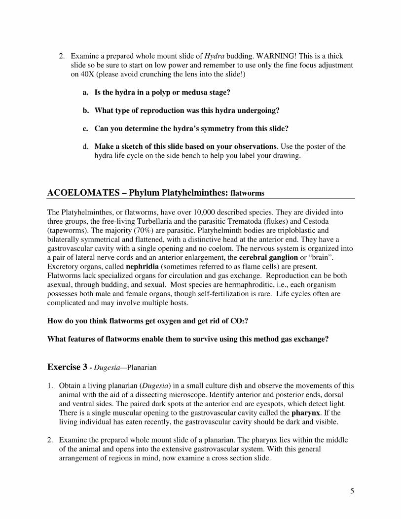
ACOELOMATES – Phylum Platyhelminthes: flatworms
The Platyhelminthes, or flatworms, have over 10,000 described species. They are divided into
three groups, the free-living Turbellaria and the parasitic Trematoda (flukes) and Cestoda
(tapeworms). The majority (70%) are parasitic. Platyhelminth bodies are triploblastic and
bilaterally symmetrical and flattened, with a distinctive head at the anterior end. They have a
gastrovascular cavity with a single opening and no coelom. The nervous system is organized into
a pair of lateral nerve cords and an anterior enlargement, the cerebral ganglion or “brain”.
Excretory organs, called nephridia (sometimes referred to as flame cells) are present.
Flatworms lack specialized organs for circulation and gas exchange. Reproduction can be both
asexual, through budding, and sexual. Most species are hermaphroditic, i.e., each organism
possesses both male and female organs, though self-fertilization is rare. Life cycles often are
complicated and may involve multiple hosts.
How do you think flatworms get oxygen and get rid of CO2?
What features of flatworms enable them to survive using this method gas exchange?
Exercise 3 - Dugesia—Planarian
1. Obtain a living planarian (Dugesia) in a small culture dish and observe the movements of this
animal with the aid of a dissecting microscope. Identify anterior and posterior ends, dorsal
and ventral sides. The paired dark spots at the anterior end are eyespots, which detect light.
There is a single muscular opening to the gastrovascular cavity called the pharynx. If the
living individual has eaten recently, the gastrovascular cavity should be dark and visible.
2. Examine the prepared whole mount slide of a planarian. The pharynx lies within the middle
of the animal and opens into the extensive gastrovascular system. With this general
arrangement of regions in mind, now examine a cross section slide.

6
3. Study the slide of the planarian in cross section. Sections are present from various points
along the body so different internal structures will be present. (Don’t be fooled by air
bubbles or open spaces that may have been produced during preparation of the slides.)
Identify the gastrovascular cavity. The lining of the gastrovascular cavity arises from the
endoderm. The outer surface of the planaria arises from ectoderm. The mesoderm gives rise
to structures lying between. Under 40X power, find a fringe of cilia. Based on this and your
preceding observation of live movement, what is the basis for locomotion in this
planarian?
4. Sketch a whole planarian and next to it sketch a representative cross-section based on
your observation of the slides.
PSEUDOCOELOMATES – Phylum Nematoda: round worms
Phylum Rotifera: rotifers
A body cavity formed between mesoderm and endoderm is termed a pseudocoelom. Nematodes
(round worms) and rotifers are in this group. Rotifers are microscopically small aquatic animals.
They have a complete digestive tract and move rapidly by use of cilia. Nematodes are an
extremely successful group, occupying marine, freshwater, and terrestrial environments. Over
15,000 species of Nematodes have been described. They are both free-living and parasitic.
Many nematodes cause diseases in domestic animals, humans, and agricultural crops. A free-
living nematode, Caenorhabditis elegans, is a model genetic organism that scientists study.
Exercise 4 – Ascaris prepared slides
1. Obtain a prepared slide of a cross-section of Ascaris. This nematode is a common intestinal
parasite of humans and domestic animals.
Surrounding the outer surface of the body is the translucent cuticle secreted by the epidermis.
Just under the epidermis is the frayed-appearing body musculature which includes only
longitudinally oriented muscle fibers. In the center of the cross-section is the intestine whose
wall is derived from endoderm. The space between muscles and intestine is the
pseudocoelom. Interspersed in the pseudocoelom, between intestine and body wall, are the
gonads: oviducts/uterus or testis/vas deferens. These animals are either female or male.
Reproduction is sexual with internal fertilization. The musculature is interrupted at 3 and 9
o’clock by the lateral line (the lateral lines are visible as two lines running along the length of
the body; in nematodes they contain nerves and excretory vessels). The small hole within the
lateral line is the cross section of the excretory canal. Seen in cross section, the dorsal and

7
ventral nerves are at 12 and 6 o’clock. As in the flatworms, circulatory and respiratory
systems are absent.
2. Draw a cross-section of the Ascaris based on your observations from the slide. Use the
diagrams provided in the photo atlases available in lab to help you label your sketch.
COELOMATES
Coelomates have a fluid-filled body cavity, called a coelom, formed within the mesoderm. There
are two major groups of coelomates: protostomes and deuterostomes. Each group includes
distinctive features of embryology, reviewed on pages 660-661 in your textbook. Among the
most distinctive features is one related to embryonic development and adult body orientation. In
early development of animals, cells undergo dramatic movement, called gastrulation, that
involves in-folding of cell layers which produces an opening called a blastopore. In
protostomes, the embryonic blastopore eventually becomes the mouth; in deuterostomes the
embryonic blastopore eventually becomes the anus.
Phylum Mollusca
The first group of protostomes we will examine are the Molluscs, a huge phylum comprising
over 110,000 living species. Molluscs include gastropods (snails and slugs), bivalves (clams),
cephalopods (squid, and octopi), and chitons. The mollusc body is composed of a muscular foot,
visceral mass, containing the major organs, and a mantle that covers the visceral mass. The
nervous system is comprised of ganglia connected by nerve cords. All molluscs have a shell
secreted by the mantle, although in some groups, like the octopi, the shell is inconspicuous. The
coelom is usually reduced to a sac surrounding the heart, gonad, and nephridia. Many molluscs
possess a specialized feeding structure called a radula that contains rows of teeth for rasping
(Figure 4). In some classes, the head/foot region develops a set of tentacles. The foot is usually
involved in locomotion, digging, or attachment.
We will concentrate on representative species from two groups: the bivalves and cephalopods.
Exercise 5 - Bivalves, clam
1. Select a clam to examine. The two shells, or valves, are joined at a hinge ligament. Open
the valves by carefully inserting a spatula and prying the valves apart. Be careful not to
hurt yourself.

8
The fleshy tissue found on the inside walls of the valves is the mantle. (Refer to the
figures 4 and 5, and to the preserved dissected clam on display in lab.) Identify the foot,
the tongue-like tissue near the open borders of the valves. Two sets of valve adductor
muscles are present at opposite ends of the valve. When they contract, they pull the
valves closed. Some bi-valves, such as scallops, swim by rapidly opening and closing the
valves.
In about the middle of the soft tissue, are two gills (called ctneidium), the respiratory
organs. To determine the anterior end of the clam, follow the gills to one end and locate
two small flaps, the labial palps. These are at the anterior end of the animal. The labial
palps cover the mouth. At the posterior side of the visceral mass is the siphon with two
openings. Water is drawn into the incurrent opening by the beating action of cilia on the
gills. It then flows across the gills and finally flows out through the excurrent opening.
Food particles are sieved out by the gills, caught up in strings of mucus, and carried by
cilia to the labial palps which direct the food into the mouth and pass it into the
esophagus. Then the food particles go into the stomach. If your clam is fresh, you may
notice a gelatinous rod, called a crystalline style, inside a sac. This rod contains enzymes
and rotates within the sac to help digestion.
Trace the route of the water and food in your clam.
A view through the smaller oval window in the soft tissue just next to the hinge shows
the heart. (Note where it says ventricle, anterior and posterior aorta in Figure 5).
2. Make a sketch of the soft tissues you have identified, based on what you actually see
on the clam.
Exercise 6 – Cephalopoda, Squid (see Figure 6)
1. Place a squid in a dissecting pan. The head/foot region includes the head, tentacles, and arms.
There are a pair of long tentacles and eight shorter arms. Notice the suction cups on the tentacles.
Between the eyes, is the siphon. The head is also equipped with a sharp beak and radula. The
mantle covers the tube-shaped body. Squid are one of the fastest swimming invertebrates. They
can open the mantle around their heads, trapping water underneath, and then force water out
through a small opening to propel them through the water. The lateral fins at the posterior end
aid in steering.
What direction does the squid move in the water?
2. Place the squid on its dorsal surface, siphon facing you. With scissors, make a long cut along
the body from the siphon to the base of the fins, and lay open the flaps of the body. Feathery gills
are present in the midline. At the base of each gill is the branchial heart. The squid has a closed
circulatory system.

9
What route does water take to reach the gills?
3. If the squid is female, the ovary may occupy most of the apex. If your squid is a male, small
testis are to be found near the apex of the body, along the midline. The gut extends to the apex.
In both sexes, a silvery ink sac lies close behind the siphon.
4. Trace the digestive system. Begin by inserting a blunt probe into the mouth, noticing the jaws
that must be parted. The tip of the probe enters the esophagus. Keeping the probe in place, try to
follow the esophagus further where it enters the stomach (sometimes difficult to distinguish) and
into the cecum (Figure 6). From the cecum, the digestive tract turns toward the head and exits
near the base of the siphon.
Why would this location, near the base of the siphon, be a beneficial place for the anus?
5. Push the head to one side to discover a pair of large ganglia. Several large nerve cords radiate
out from these ganglia to the body.
6. Squid have excellent eyesight. Remove the eye and examine the cornea, the film-like
substance, and the hard lens.
7. Dissect out the pen that lies dorsal to the visceral organs and extends from the free edge of the
collar to the apex of the mantle. This is the “shell” of the squid.
Make a labeled sketch of the internal anatomy of the squid based on what you actually see
in the dissection
An unusual squid: The Hawaiian Bobtail Squid has a kidney-shaped lobe in its mantle cavity.
This organ is filled with bioluminescent bacteria that act like a light bulb. Behind the light organ
is reflective tissue that reflects the light down through another tissue that functions as a lens. The
effect is very similar to a flashlight. The bobtail squid feeds at night, but does not need light to
spot its prey. What do you think is the purpose of the light organ?

10

11
Note: Images of Sponge, Clam and Squid from: The invertebrates: Function and form. A laboratory guide by IW
Sherman & UG Sherman, 2 Ed 1976.

![Chapter 32 animal diversity[1]](https://static.fdocuments.net/doc/165x107/555a8170d8b42abb628b4b61/chapter-32-animal-diversity1.jpg)