Anillos de Ferrara y ón a del Queratocono en rasil
213
A A D Anil Progra Espec Análisi Anillos D. Guilh llos de Ferra Te ama de D cialidade is de R Intrac con Trabaj herme H para D ra y Correcci esis Doctora Doctorad es Médic Resulta corneal Quera o de inve Hermet a optar po Director: Dr. Oviedo, 1 ión Quirúrgic l de Guilherm do de Inv o-Quirúr ados de les de F atocono estigació to Ferra or el grad Jesús M M 10 de Abri ca del Quera me H. Ferrar estigació rgicas (R e la Im Ferrara o en B ón realiza ara de A do de Do Merayo Llov il de 2017 atocono en B a ón en Cir RD 1393/2 mplanta a en Pa rasil ado por Almeid octor. ves 7 Brasil rugía y 2007) ación d aciente da Cunh 1 de es ha
Transcript of Anillos de Ferrara y ón a del Queratocono en rasil

AA
D
Anil
PrograEspec
AnálisiAnillos
D. Guilh
llos de Ferra
Te
ama de Dcialidade
is de RIntrac
con
Trabaj
herme H
para
D
ra y Correcci
esis Doctora
Doctorades Médic
ResultacornealQuera
o de inve
Hermet
a optar po
Director: Dr.
Oviedo, 1
ión Quirúrgic
l de Guilherm
do de Invo-Quirúr
ados deles de Fatocono
estigació
to Ferra
or el grad
Jesús M M
10 de Abri
ca del Quera
me H. Ferrar
estigaciórgicas (R
e la ImFerrarao en B
ón realiza
ara de A
do de Do
Merayo Llov
il de 2017
atocono en B
a
ón en CirRD 1393/2
mplantaa en Parasil
ado por
Almeid
octor.
ves
7
Brasil
rugía y 2007)
ación daciente
da Cunh
1
de es
ha


FOR
-MA
T-V
OA
-010
-BIS
RESUMEN DEL CONTENIDO DE TESIS DOCTORAL
1.- Título de la Tesis
Español: Análisis de Resultados de la Implantación de Anillos Intracorneales de Ferrara en Pacientes con Queratocono en Brasil
Inglés: Outcome Analysis of Ferrara Intrastromal Ring Segments in Keratoconus Patients in Brazil
2.- Autor
Nombre: Guilherme Hermeto Ferrara de Almeida Cunha
DNI/Pasaporte/NIE:
Programa de Doctorado: Programa de Doctorado de Investigación en Cirugía y Especialidades Médico-Quirúrgicas (RD 1393/2007)
Órgano responsable: Universidad de Oviedo
RESUMEN (en español)
Los segmentos de anillos intracorneales de Ferrara (ICRS) se llevan aplicando desde hace más de 30 años para la corrección del queratocono y como alternativa al trasplante de cornea o queratoplastia y su empleo se basa en la experiencia clínica de los cirujanos. Esta tesis doctoral evalúa los resultados de la aplicación de ICRS en pacientes con queratocono con el volumen y tiempo de seguimiento más amplio publicado tanto en la población general como en niños y evalúa los resultados de la aplicación de ICRS para la corrección del astigmatismo en las queratoplastias. Los ICRS en los pacientes estudiados mejoran la agueza visual con y sin corrección, remodelan la cornea mediante la reducción de los valores queratométricos y la regularización de valores topográficos con un procedimiento que preserva el eje visual. Además, influyen sobre la progresión de la enfermedad retrasando o evitando la queratoplastia. Se precisan estudios prospectivos y multicéntricos (ensayos clínicos) para estandarizar y personalizar la estrategia de implante y analizar los resultados con mayor fuerza que en los estudios observacionales.

RESUMEN (en Inglés)
Ferrara Intrastromal Ring Segments (ICRS) has been successfully used in the correction of keratoconus based on surgeon clinical practice for more than 30 years as an alternative to corneal transplant. The research work presented as a doctoral thesis develop an outcome analysis of ICRS in patients with keratoconus (general population and children) and also evaluate the role of ICRS in the correction of astigmatism after keratoplasty. The cohort of patients with ICRS analyzed improves visual acuity (expontaneus and corrected). ICRS is a procedure that, preserving the central part of the cornea, makes a reduction of the keratometric values and improves the corneal topography pattern. ICRS alter the progression of the disease delaying, or avoiding, the indication of keratoplasty. There are necessary clinical trials (multicentric and prospective studies) to Asses the customization and standardization of the implant protocols and to study the outcome of ICRS with more power than in observational studies SR. PRESIDENTE DE LA COMISIÓN ACADÉMICA DEL Programa de Doctorado de Investigación en Cirugía y Especialidades Médico-Quirúrgicas (RD 1393/2007)


Anillos de Ferrara y Corrección Quirúrgica del Queratocono en Brasil
Tesis Doctoral de Guilherme H. Ferrara 2
Agradecimientos
Agradezco al Dr. Merayo, director de mi tesis, por la amistad, y por me haber acogido
en cuanto estuve lejos de casa. Muchas gracias por toda la ayuda, por la trasmisión de
sus conocimientos y por fomentar mi crecimiento personal y profesional.
Al Dr Alfonso, por las enseñanzas, la inversión de tiempo en mi formación y por la
oportunidad y confianza.
A Don Luis Fernandez-Vega por me haber permitido tener una experiencia única de
aprendizaje en el Instituto durante mi estada en Oviedo.
A toda la equipo del Departamento de Segmento Anterior del IOFV, con especial
mención al Dr. Lisa y al Dr. Abdel por los momentos juntos en la clínica.
A todos los compañeros del Grupo de Cirugía Refractiva y Calidad Visual del IOBA y
después a los miembros del Grupo de Superficie Ocular de la FIO, por la recepción y
intercambio de conocimientos; a Nacho, por la acogida, apoyo y amistad.
A los compañeros del Instituto y de la FIO, por la amistad y por la acogida; a Carlos por
su amistad.
A Leo Torquetti, por todo su apoyo y ayuda, por toda dedicación a los estudios de los
anillos de Ferrara y a todo el equipo que me ha ayudado en Brasil
A mi madre, Beatriz por su apoyo incondicional, siempre presente en los momentos
difíciles; a mi padre, Paulo, ejemplo de dedicación y ética, por las oportunidades que
tengo. Por toda experiencia y conocimiento aportados en el día a día en la clínica,
además de nuestras charlas.
A mi mujer, Camila, siempre presente, amiga y compañera, siempre me apoyando en
mis decisiones; a mis dos hijos, Bernardo y Arthur, por su amor y inspiración.


Anillos de Ferrara y Corrección Quirúrgica del Queratocono en Brasil
Tesis Doctoral de Guilherme H. Ferrara 3
ÍNDICE
1. MOTIVACIÓN, PREGUNTA DE INVESTIGACIÓN Y ESTRUCTURA 5
2. RESUMEN 6
3. ACRÓNIMOS 7
4. INTRODUCCIÓN 8
4.1. CÓRNEA 8
4.1.1. Anatomía 8
4.1.2. Concepto de Asfericidad Corneal 9
4.2. QUEROTOCONO 11
4.2.1. Epidemiología 12
4.2.2. Patogenia 12
4.2.3. Diagnóstico Clínico y de Imagen 14
4.2.3.1. Síntomas 14
4.2.3.2. Signos Clínicos 14
4.2.3.3. Criterios de Diagnóstico 16
4.2.3.3.1. Topografía de córnea 16
4.2.3.3.2. Tomografía de córnea 17
4.2.4. Prevención y Tratamiento Médico 18
4.2.5. Corrección con lentes de contacto 19
4.2.6. Tratamiento Quirúrgico 19
4.3. CORRECCIÓN DEL QUERATOCONO CON CIRUGÍA ADITIVA DE LA
CÓRNEA: LAS BASES DE LOS ANILLOS DE FERRARA 23
4.3.1. Primeros estudios: JI Barraquer y Blavatskaya 23
4.3.2. Anillos de Ferrara 25
4.3.2.1. Evolución de los anillos de Ferrara 28
4.3.2.2. Evolución del Nomograma de los Anillos de Ferrara 32
4.3.2.2.1. Primera Generación 32
4.3.2.2.2. Segunda Generación 33
4.3.2.2.3. Tercera Generación 34
4.3.2.2.4. Cuarta Generación 34
4.3.2.3. Características de los Anillos de Ferrara 36
4.3.2.4. Mecanismo de Acción de los Anillos de Ferrara 36
4.3.2.5. Indicaciones y Contra-indicaciones de los Anillos de Ferrara 38
4.3.2.5.1. Indicaciones de los Anillos de Ferrara 38
4.3.2.5.2. Contra-indicaciones de los Anillos de Ferrara 39

Anillos de Ferrara y Corrección Quirúrgica del Queratocono en Brasil
Tesis Doctoral de Guilherme H. Ferrara 4
4.3.2.6. Cirugía de Implante de los Anillos 39
4.3.2.6.1. Instrumental 39
4.3.2.6.2. Técnica Quirúrgica y estrategia de implante 40
4.3.2.7. Complicaciones 41
4.3.2.7.1. Infección 41
4.3.2.7.2. Migración 42
4.3.2.7.3. Extrusión 43
4.3.2.7.4. Descentración 43
4.3.2.7.5. Halos y Reflejos 43
4.3.2.7.6. Hipo e Hipercorrecciones 43
4.3.2.7.7. Opacidades Peri-anulares 44
5. JUSTIFICACIÓN 45
6. HIPÓTESIS DE TRABAJO 46
7. OBJETIVOS 47
8. PACIENTES, MATERIAL Y MÉTODO 48
9. CAPITULO I. Resultados a largo plazo de la implantación de anillos de Ferrara en
pacientes con queratocono 52
10. CAPITULO II. Progresión del queratocono con o sin anillos de Ferrara. 64
11. CAPITULO III. Otras Aplicaciones de Anillos de Ferrara: Corrección del
astigmatismo tras queratoplastia 70
12. TRABAJOS EN REALIZACIÓN 79
12.1. Resultados de la implantación de anillos de Ferrara en niños 79
13. DISCUSIÓN 86
14. FUTUROS PROYECTOS DE INVESTIGACIÓN 95
15. CONCLUSIONES 96
16. REFERENCIAS 97
17. ARTICULOS CIENTIFICOS DE LA TESIS 104
17.1. Anexo I Publicaciones del doctorando en relación con el proyecto de tesis
17.2. Anexo II. Permiso de los coautores para la presentación de las publicaciones
como soporte para el proyecto de tesis doctoral

Anillos de Ferrara y Corrección Quirúrgica del Queratocono en Brasil
Tesis Doctoral de Guilherme H. Ferrara 5
1. Motivación, Pregunta de Investigación y Estructura de la Tesis. El queratocono es una patología de la estructura transparente del ojo, la cornea, que cursa con un progresivo adelgazamiento y aumento irregular de curvatura con resultado de perdida de la agudeza visual. Si la enfermedad avanza, la cornea pierde la trasparencia y se corre el riesgo de destrucción del tejido y perforación. Para su corrección se han empleado diversos tratamientos que se revisan en la introducción entre los que destacan los segmentos de anillos intracorneales (ICRS) desarrollados por el padre del autor. Si bien se tiene experiencia clínica con este tipo de prótesis desde hace más de 30 años y se han realizado trabajos en modelos experimentales, el desarrollo de la técnica y el análisis de resultados no ha tenido apenas soporte en publicaciones científicas y se ha basado fundamentalmente en datos empíricos de la experiencia clínica. Este hecho motivó el realizar un trabajo de investigación que revisara los resultados del implante de ICRS en la clínica del Dr. Paulo Ferrara para poder conocer el comportamiento de los ICRS a largo plazo con gran volumen de pacientes operados. El doctorando después de terminar la especialidad de oftalmología en Brasil se vino a España a realizar un largo periodo de formación postgraduada en investigación traslacional en ciencias de la visión (Universidad de Valladolid), master en superficie ocular, cornea, catarata y cirugía refractiva y el programa de doctorado (Universidad de Oviedo) y empezó la tarea de analizar los resultados del implante de los ICRS, fruto del cual se han realizado las publicaciones que soportan esta tesis. La Tesis se presenta como un compendio de artículos con una parte común (Introducción, Justificación, Hipótesis de Trabajo, Objetivos, Discusión y Conclusiones) y los capítulos correspondientes a los diferentes trabajos y publicaciones: Capitulo I dedicado al análisis de resultados de la implantación de segmentos de anillos a largo plazo, Capitulo II dedicado al estudio de la progresión del queratocono, Capitulo III dedicado al análisis del efecto de los ICRS en el astigmatismo en los pacientes con queratoplastias y el Capitulo IV dedicado al análisis de resultados del implante de ICRS en niños con queratocono. Como en el momento de escribir esta memoria el artículo que soporta este capítulo está en fase de revisión se ha de considerar como un “trabajo en realización” hasta que esté aceptada su publicación.
A Mis Padres Beatriz e Paulo, Mi mujer Camila
e mis hijos, Bernardo e Arthur

Anillos de Ferrara y Corrección Quirúrgica del Queratocono en Brasil
Tesis Doctoral de Guilherme H. Ferrara 6
2. Resumen.
Los segmentos de anillos intracorneales de Ferrara (ICRS) se llevan aplicando desde hace más de 30 años para la corrección del queratocono y como alternativa al trasplante de cornea o queratoplastia y su empleo se basa en la experiencia clínica de los cirujanos. Esta tesis doctoral evalúa los resultados de la aplicación de ICRS en pacientes con queratocono con el volumen y tiempo de seguimiento más amplio publicado tanto en la población general como en niños y evalúa los resultados de la aplicación de ICRS para la corrección del astigmatismo en las queratoplastias. Los ICRS en los pacientes estudiados mejoran la agueza visual con y sin corrección, remodelan la cornea mediante la reducción de los valores queratométricos y la regularización de valores topográficos con un procedimiento que preserva el eje visual. Además, influyen sobre la progresión de la enfermedad retrasando o evitando la queratoplastia. Se precisan estudios prospectivos y multicéntricos (ensayos clínicos) para estandarizar y personalizar la estrategia de implante y analizar los resultados con mayor fuerza que en los estudios observacionales. Ferrara Intrastromal Ring Segments (ICRS) has been successfully used in the correction of keratoconus based on surgeon clinical practice for more than 30 years as an alternative to corneal transplant. The research work presented as a doctoral thesis develop an outcome analysis of ICRS in patients with keratoconus (general population and children) and also evaluate the role of ICRS in the correction of astigmatism after keratoplasty. The cohort of patients with ICRS analyzed improves visual acuity (expontaneus and corrected). ICRS is a procedure that, preserving the central part of the cornea, makes a reduction of the keratometric values and improves the corneal topography pattern. ICRS alter the progression of the disease delaying, or avoiding, the indication of keratoplasty. There are necessary clinical trials (multicentric and prospective studies) to Asses the customization and standardization of the implant protocols and to study the outcome of ICRS with more power than in observational studies
Anillos de Ferrara y Corrección Quirúrgica del Queratocono en Brasil
Tesis Doctoral de Guilherme H. Ferrara 7
3. Acrónimos.
AV: Agudeza visual
CDVA: Agudeza visual con corrección
CXL: Crosslinking
DALK: Deep anterior lamellar keratoplkasty
ICRS: Intracorneal ring segments
IL-1: Interleucina 1
IL-6: Interleucina 6
K: Queratometría
Lasik: Laser in situ keratomileusis
LCRGP: Lentes de contacto semirrígidas permeables al gas
MMP: metaloproteinasas
PKP: Trasplante penetrante de córnea
PRK: keratectomia foto refractiva
Q: Asfericidad corneal
TIMP 1: Tissue Inhibitors of metalloproteinases 1
TNF- : Factor de necrosis tumoral
UDVA: Agudeza visual sin corrección
RK: Queratotomía radial
SD: Standard Deviation
SE: equivalente esférico

Anillos de Ferrara y Corrección Quirúrgica del Queratocono en Brasil
Tesis Doctoral de Guilherme H. Ferrara 8
4. Introducción.
4.1. Córnea
La córnea constituye la primera lente del ojo, responsable por el 70% del poder
refractivo del sistema óptico humano (entre 40 y 44 Dioptrías) (ALDER, 1950).1 Se
trata de una estructura transparente, avascular, ricamente inervada, y que funciona como
barrera química y mecánica entre el interior del ojo y su exterior. (WARING, 1884 &
KLYCE & BEUERMAN, 1988).2,3 Su forma y transparencia dependen de la
organización de las fibras de colágeno del estroma, que se mantienen ordenadas y
paralelas entre ellas (MEEK ET AL, 2005 & MEEK ET AL, 2003).4,5 Tiene su espesor
más delgado en el centro (520 y 550 µm), y que aumenta hacia el limbo. Alteraciones
en su geometría pueden alterar de manera significativa su poder óptico (NEJAD;
FOSTER; GONDAL, 2014).6
4.1.1. Anatomía
La córnea está estructurada clásicamente en 5 capas: el epitelio, con un grosor de
aproximadamente 50 µm, está formado por 5 a 7 capas de células. Posee la función de
barrera frente al medio exterior y, juntamente con la lágrima, constituye una superficie
homogénea para la refracción. La capa de Bowman está situada entre el estroma corneal
y la membrana basal. Está constituida por colágenos tipo I, III, V, VII y XIII y
proteoglicanos. El estroma corneal constituye el 90% del espesor corneal, formado por
78% de hidratación, 15% de fibras de colágeno entrelazadas, 7% de proteoglicanos,
glicoproteínas y sales inorgánicos (MAURICE, 1984 & KRACHMER ET AL, 1984).7,8
La capa de DUA ha sido descrita recientemente (DUA ET AL, 2013)9 como una capa
acelular en la región pre Descemet. Su reconocimiento puede tener un impacto
considerable sobre el entendimiento de la biomecánica corneal. El endotelio es una
estructura de gran importancia para el metabolismo corneal, responsable por la nutrición
de la córnea, así como por la excreción de los productos finales del metabolismo y de su
deturgescencia.

Figura
Actu
descr
AL,
toda
Pode
oblad
más
super
posit
Cuán
más
cero
el ce
coinc
caso.
hacia
los ra
aberr
con
(PHI
Anil
a 1: Histologí
4.1.2. Co
ualmente se
rita por la a
2007).10 Se
la vida.
emos aborda
da. La supe
curva en e
rficie asfér
tivo).
ndo un haz
posteriores,
y la aberra
entro y más
ciden con lo
. Eso hace q
a un único f
ayos perifér
ración esfér
que la cal
ILIPPINE JO
llos de Ferra
Te
a de la córnea
oncepto d
e cree que
asfericidad (
e cree que l
ar la forma
erficie esféri
el centro y m
ica oblada
de luz para
, mientras l
ación es disc
s plana en l
os rayos cen
que la calida
foco. En una
ricos enfoca
rica serán p
lidad visua
OURNAL O
ra y Correcci
esis Doctora
a
e Asfericid
el contorno
(Q) y por el
la forma asf
de una sup
ica es redon
más plana
es más pla
alelo pasa p
os rayos pe
cretamente
la periferia,
ntrales. El v
ad de la vis
a superficie
an más ante
positivos. E
l sea degra
OF OPHTH
ión Quirúrgic
l de Guilherm
dad Corne
o de la cór
l radio de c
férica de la
perficie com
nda (Q=0) F
hacia la pe
ana en el c
por una lent
eriféricos en
positiva. En
, los rayos
valor de Q y
ión sea bue
e oblada, má
eriores que
En este caso
adada y la
HALMOLO
ca del Quera
me H. Ferrar
eal
rnea se ace
curvatura (G
a córnea se
mo esférica,
FIGURA A
eriferia (Q n
centro y má
te esférica,
nfocan más
n una super
periféricos
y la asfericid
na, pues tod
ás plano en
los rayos c
o, la aberrac
a sensibilid
OGY, 2011).
atocono en B
a
erca a una
GONZÁLEZ
mantiene c
, asférica pr
A. Una supe
negativo) F
ás curva en
los rayos c
anteriores.
rficie prolad
enfocan m
dad serán n
dos los rayo
el centro qu
centrales. El
ción esféric
dad al cont
.11
Brasil
sección có
Z-MÉIJOM
onstante du
rolada o asf
rficie asféri
FIGURA B.
n la perifer
centrales en
El valor de
da, más curv
más posterio
negativos, en
os luminoso
ue en la per
l valor de Q
ca inducida
traste dismi
9
ónica,
ME ET
urante
férica
ica es
. Una
ia (Q
focan
e Q es
va en
ores y
n este
os van
riferia,
Q y la
hace
inuya

La m
varía
valor
0,08
Figura
Fuent
La as
En la
cuan
una
dege
Figura
Anil
mayoría de
a de -0,01 a
r más comú
en la zona ó
a 2: Córnea es
te: Robert Edw
sfericidad c
a córnea ect
ndo se la com
córnea norm
neración m
a 3: córnea ob
llos de Ferra
Te
los estudios
a -0,80 (AS
únmente ac
óptica de 4,
sférica (A) y a
ward T Ang, 2
corneal es di
tásica, la cu
mpara a una
mal (oblada
marginal pelú
blata
ra y Correcci
esis Doctora
s está de ac
SBELL; UC
eptado en a
,5 mm (SIG
asférica (B).
2011
istinta cuan
urvatura pue
a córnea no
a), situació
úcida.
ión Quirúrgic
l de Guilherm
cuerdo de q
CAKHAN, 2
adultos jóve
GANOS, C.
ndo compara
ede present
ormal (hiper
n que ocur
ca del Quera
me H. Ferrar
que la asfer
2001; COL
enes es de,
S. et al, 200
amos córne
tarse bastan
rprolada), o
rre en el qu
atocono en B
a
ricidad corn
LIN; VELOU
aproximad
02).14
as sanas y c
nte más elev
o más plana
ueratocono
Brasil
neal en hum
U, 2003).12
damente, -0
córneas ectá
vada en el c
en el centro
inferior y
10
manos 2,13 El
,23 ±
ásicas.
centro
o que
en la

Anillos de Ferrara y Corrección Quirúrgica del Queratocono en Brasil
Tesis Doctoral de Guilherme H. Ferrara 11
4.2. Queratocono
El queratocono es la patología ectásica más frecuente en el paciente joven. Cursa con
adelgazamiento progresivo de la córnea, seguida por el aumento progresivo de su
curvatura y protrusión, que resultan en astigmatismos irregulares, miopía, además de
elevados índices de aberraciones de alto orden y, consecuentemente, a un
comprometimiento importante de la agudeza visual (TORQUETTI ET AL 2009).15
Aunque sea considerada como una patología no inflamatoria, diversos estudios han
demostrado marcadores de la inflamación elevados en esta patología (LEMA ET AL
2004; LEMA ET AL 2008; GALVIS ET AL 2015).16,17,18
Los síntomas del queratocono pueden variar, dependiendo de su etapa evolutiva
(RABINOWITZ, 1998).19 En grados iniciales, puede que sea difícil diferenciar el
queratocono de otras condiciones refraccionales, como el astigmatismo, y su
diagnóstico puede pasar desapercibido. Con la evolución del queratocono, el
desenfoque visual creciente y la dificultad de la realización de la refracción nos hace
sospechar de la patología.
La forma frustra de queratocono fue primeramente descrita por Amsler en 1961, y luego
citada por otros autores (Klyce et al, 2009).20 Esta forma de la patología puede aparecer
en cualquier momento de la vida, con patrones queratométricos extremamente sutiles y
sin presentar los signos clínicos de la enfermedad. Lo que diferencia la forma frustra de
queratocono del queratocono manifiesto es que la primera no progresa.
La incidencia de queratocono se hace más frecuente en los centros de cirugía refractiva,
cuando se la compara con la población en general. Su detección precoz es de gran
preocupación en el “screening” de pacientes candidatos a la cirugía refractiva, dado que
el procedimiento causa debilidad de la estructura de la córnea y predispone a la
aparición de la ectasia corneal (Kleyce, 2009).20 La forma frustra de queratocono es la
principal causa de ectasia post Lasik (Randleman et al, 2008).21

Anillos de Ferrara y Corrección Quirúrgica del Queratocono en Brasil
Tesis Doctoral de Guilherme H. Ferrara 12
4.2.1. Epidemiología
El queratocono comienza, en general, en la segunda década de vida y puede progresar
hasta la tercera o cuarta década (RABINOWITZ 1998).19 Se ha observado que el
queratocono progresa más rápidamente en personas más jóvenes cuanto más temprana
la edad de aparición (OWEN ET AL 2003).22 La incidencia del queratocono depende de
otros factores, como el grupo étnico analizado y los criterios de diagnóstico utilizados.
El frotamiento ocular y la presencia de enfermedades sistémicas como síndrome de los
parpados laxos, alergias, eczema e historia familiar también fueron relacionados con la
aparición del queratocono (PIÑERO ET AL, 2012)23, aunque su aparición no esté
relacionada con otro factor de riesgo en la mayoría de los casos.
La incidencia del queratocono, según diferentes autores, oscila entre 50-230 para cada
100.000 habitantes (1 de cada 2000 en la población general) (PIÑERO ET AL, 2012).23
Se ha aceptado al queratocono como una patología con igual distribución en ambos
sexos.
4.2.2. Patogenia
La presentación más frecuente del queratocono ocurre en su forma aislada, sin otra
patología sistémica u ocular detectable a la evaluación clínica. Patologías como el
síndrome de Down y amaurosis congénita de Leber presentan alta incidencia con el
queratocono. Su asociación con el queratocono se hace por la alta incidencia de
frotación de los ojos en estos pacientes.
La disposición de las fibras de colágeno en córneas con queratocono no es la misma que
en córneas normales (Radner et al, 1998).24 La reducción del número de queratocitos y
del número de lamelas, con degradación de fibroblastos en córneas con queratocono, ha
sido descrita por Romero-Jiménez en 2010, así como las alteraciones en la organización
de las lamelas de colágeno, con distribución desigual de las lamelas del colágeno por
Meek, en 2005. Estas alteraciones estructurales pueden resultar en debilidad estromal,
con subsecuente adelgazamiento y protrusión de la córnea.
Anillos de Ferrara y Corrección Quirúrgica del Queratocono en Brasil
Tesis Doctoral de Guilherme H. Ferrara 13
Los aspectos bioquímicos subyacentes al desarrollo del queratocono parecen
desempeñar un papel importante en su patogénesis, aunque no sean totalmente
conocidos, a pesar de la intensa investigación realizada en este campo.
Los estudios bioquímicos e inmunohistoquímicos de córneas con queratocono sugieren
que existe una degradación de la matriz extracelular debido al aumento de enzimas
proteolíticas y otras enzimas catalíticas: enzimas lisosomales como la gelatinasa, la
fosfatasa ácida, las catepsinas B y G y varias metaloproteasas como la MMP-2; y/o
disminución de los niveles de las enzimas inhibidoras de las proteasas, como los TIMP-
1 (Inhibidores tisulares de las metaloproteinasas-1), inhibidoras de la alpha 1
antiproteasa y la alpha 2 macroglobulina.25
Los hallazgos histológicos han demostrado una disminución del número de queratocitos
en el estroma anterior , probablemente causada por un aumento de la apoptosis celular
en córneas con queratocono (Wilson et al, 1996).26 Los queratocitos de las córneas con
queratocono presentan un número 4 veces superior de receptores para interleuquina-1,
una citocina relacionada con la apoptosis de queratocitos y la liberación de otras
citocinas.
Los microtraumas corneales decurrentes del frotamiento ocular y/o por el uso de lentes
de contacto, permiten que la IL-1 producido por el epitelio y endotelio corneal tenga
contacto con los queratocitos hipersensibles, resultando en su apoptosis y gradual
disminución del colágeno.
En lágrimas de pacientes con queratocono y portadores de lentes de contacto se han
encontrado sobreexpresados marcadores proinflamatorios como IL-1, IL-6, TNF y
otras metaloproteasas. La hipótesis de que factores externos, como las lentes de
contacto, la atopia y el frotamiento crónico desencadenarían una inflamación crónica a
nivel molecular, con progresiva degradación del estroma corneal, quedaría justificada
(Lema et al, 2008; Lema et al, 2009).17,27
Los niveles bajos de antioxidantes, como aldehído deshidrogenasa y superóxido
dismutasa, enzimas responsables de la eliminación de especies reactivas de oxígeno,

Anillos de Ferrara y Corrección Quirúrgica del Queratocono en Brasil
Tesis Doctoral de Guilherme H. Ferrara 14
pueden resultar en elevados niveles de radicales libres y peróxido de hidrógeno, con el
aumento del estrés oxidativo y la consiguiente toxicidad celular secundaria.
4.2.3. Diagnóstico Clínico y de Imagen
El queratocono es la patología ectásica más común de la córnea y se manifiesta, en
general, en la segunda o tercera década de vida. El signo más precoz es el
adelgazamiento paracentral del estroma, con protrusión apical y astigmatismo miópico
asimétrico (Duke Elder y Leigh, 1965).28 Las alteraciones inducidas en la córnea
resultan en disminución de la calidad visual, además de perdida de líneas de visión.
Factores ambientales pueden estar asociados con el desarrollo de la patología, como
atopia, frotamiento de los ojos y lentes de contacto semirrígidas permeables al gas mal
adaptadas. Además, las ectasias pueden tener un carácter progresivo.
4.2.3.1. Síntomas
En general, los primeros síntomas ocurren en la pubertad. Atopia y frotamiento de los
ojos son frecuentes en estos pacientes. Empeora de la visión, cambios frecuentes de la
graduación de las gafas por el aumento de la miopía y del astigmatismo, y por cambios
el eje del astigmatismo, ocurren en visitas rutineras al oftalmólogo.
Con la progresión de la ectasia, el paciente puede referir imágenes muy distorsionadas,
deslumbramientos, halos y poliopia, con importante compromiso de la agudeza visual.
La adaptación de lentes de contacto en genera, es compleja en estos pacientes, incluso
en los grados iniciales.
4.2.3.2. Signos Clínicos
En las fases iniciales, es posible observar sombras en tijera a la esquiascopia, además
del signo de la gota de aceite de “Charleux” por retroiluminación, con la pupila dilatada.
El aspecto de la córnea a la lámpara de hendidura suele ser normal en grados iniciales
del queratocono.

Anillos de Ferrara y Corrección Quirúrgica del Queratocono en Brasil
Tesis Doctoral de Guilherme H. Ferrara 15
Con la progresión de la ectasia, los nervios se vuelven visibles, seguido de la
visualización del anillo de Fleischer, una línea color ocre-marronácea en forma de arco
o anillo que rodea la base del cono, y las estrías de Vogt, líneas finas verticales en el
estroma profundo y la membrana de Descemet, paralelas al eje del cono. Suelen suceder
opacidades superficiales en el vértice del cono relacionadas con roturas en la capa de
Bowman y cicatrización secundaria y las opacidades secundarias a la cicatrización en
respuesta al estrés.
Figura 4: Anillo de Fleischer (Flecha)
En fases más avanzadas, el adelgazamiento y la ectasia se hacen más severas,
provocando la pérdida de la visión. En este momento es posible observar el signo de
Munson, que consiste en la deformación en V del párpado inferior cuando el paciente
mira hacia abajo, y el signo de Rizzuti, que consiste en la aparición de un reflejo
luminoso en el limbo nasal cuando se ilumina desde el lado temporal. La hidropsia
aguda consiste en el aumento abrupto del espesor corneal (edema) resultante de roturas
en la membrana de Descemet, con entrada de humor acuoso hacia el interior de la
misma. Dolor y opacidad corneal secundaria al edema, con disminución de la agudeza
visual, ocurren como consecuencia de este fenómeno. Al cabo de semanas, el cuadro se
resuelve con la absorción del edema y la restauración de la trasparencia corneal. Ocurre
cicatrización de la rotura y se forma un leucoma central.

Anillos de Ferrara y Corrección Quirúrgica del Queratocono en Brasil
Tesis Doctoral de Guilherme H. Ferrara 16
Figura 5: Queratocono
4.2.3.3. Criterios de diagnóstico
4.2.3.3.1. Topografía de córnea
Aunque el queratocono sea una patología que cursa con adelgazamiento e incremento
de la curvatura corneal, ninguno de estos parámetros, ni la curvatura, ni la paquimetría o
la regularidad de la superficie corneal es, de forma aislada, útil para el diagnóstico de
queratocono.
La topografía de córnea basada en el disco de Placido proporciona abundante
información sobre el contorno corneal. En ella se pueden reconocer los patrones de
regularidad corneal, bien como los índices cuantitativos generados por este sistema. Las
alteraciones de estos parámetros, asociadas a los datos clínicos del paciente, nos llevan a
la sospecha del diagnóstico del queratocono.
Los patrones considerados sospechosos son aquellos que presentan áreas aisladas de
encorvamiento; asimetría inferior-superior; desviación de eje entre los hemimeridianos
de la córnea, o bien la asociación de estos patrones. También se han desarrollado varios
índices destinados a auxiliar en el diagnóstico del queratocono, como el Rabinowitz-
McDonnell, el índice KISA% y el Klyce-Maeda-Smolek, con elevada sensibilidad y
especificidad para su diagnóstico.

Figura
La to
cáma
el to
difer
(map
La u
Germ
por e
evalu
de Fe
En c
entre
altera
al (2
prece
del e
pacie
visua
Anil
a 6: Topograf
4.2.3.3.2.
omografía c
ara anterior
opógrafo d
renciales, ad
pa paquimét
utilización
man) posibil
el queratoco
uar los cam
errara.
corneas nor
e sí. Las a
aciones en
2008)29 sugi
eden las alt
epitelio corn
entes sospe
ales.
llos de Ferra
Te
fía de córnea d
Tomogra
corneal hac
del ojo par
de córnea―
demás de l
trico).
del tomóg
lita un estud
ono. Los da
mbios induci
rmales, el m
alteraciones
la superfici
iere que las
teraciones d
neal que enm
chosos de
ra y Correcci
esis Doctora
de un paciente
afía de córn
ce la recon
ra proveer d
―, de elev
la variación
grafo de có
dio profund
atos propor
idos en córn
mapa de ele
s de la ele
ie anterior d
s alteracion
del mapa an
mascara o d
queratocon
ión Quirúrgic
l de Guilherm
e con queratoc
nea
nstrucción tr
datos de la s
vación ant
n del espes
órnea (Pent
dizado de la
rcionados po
neas ectásic
evación ant
evación pos
de la córnea
nes del map
nterior, prob
disminuye la
no, incluso
ca del Quera
me H. Ferrar
cono
ridimension
superficie c
erior y po
or en toda
tacam; Ocu
a córnea y d
or el Pentac
cas después
terior y pos
sterior pue
a. Un estudi
pa de eleva
bablemente
as alteracion
antes de la
atocono en B
a
nal de las e
corneal ―ta
osterior, as
la superfic
ulus Optikg
de las altera
cam® nos
del implan
sterior guar
eden ocurrir
io realizado
ción poster
e debido al
nes topográ
a aparición
Brasil
estructuras
al como fun
sí como m
cie de la có
geräte, We
ciones indu
permite tam
nte de los an
rda una rel
r antes qu
o por Schleg
rior de la có
remodelam
ficas inicial
de los sínt
17
de la
nciona
mapas
órnea
etzlar,
ucidas
mbién
nillos
ación
ue las
gel et
órnea
miento
les en
tomas

Anillos de Ferrara y Corrección Quirúrgica del Queratocono en Brasil
Tesis Doctoral de Guilherme H. Ferrara 18
El espesor corneal empieza a tener especial importancia en los pacientes que serán
sometidos a la cirugía refractiva (Ambrosio et al; 2003),30 como también la
caracterización de la distribución del espesor corneal (Ambrosio et al, 2011).31 La
combinación de la caracterización del espesor corneal juntamente con los mapas de
elevación nos permite evaluar e identificar anormalidades en los patrones corneales.
En córneas ectásicas, la protrusión anterior genera un aumento de la prolacidad asociada
a la negativización del valor de Q (asfericidad) (Piñero et al. 2012).23 La asfericidad
marcadamente más negativa para la superficie anterior de la córnea fue relatada en un
estudio por Piñero et al. (2010)32 comparando algunas características topográficas en
una muestra de ojos normales con aquellos encontrados en córneas con queratocono
grados I y II (Amsler-Krumeich).
4.2.4. Prevención y Tratamiento Médico
La presencia de alergias y del frotamiento ocular son frecuentemente asociados a la
aparición y a la progresión del queratocono, además de cuadros variables de ojo seco.
Esas condiciones afectan no solo el curso de la enfermedad, sino también a la
adaptación de lentes de contacto en estos pacientes. El tratamiento clínico de estos
síntomas es el primer paso para lograr rehabilitar al paciente desde el punto de vista de
su corrección visual, como también para frenar la evolución del queratocono.
El objetivo mayor en el tratamiento del queratocono es la rehabilitación visual. La
elección del tratamiento puede variar de acuerdo con el estadio de la patología y de los
síntomas que presenta el paciente.
En casos iniciales, la corrección con gafas suele ser satisfactoria. Con la progresión del
cuadro clínico, las gafas no son capaces de proporcionar una visión adecuada, y las
lentes de contacto semirrígidas al gas (LCRGP) pasan a desempeñar un papel
importante para el mantenimiento de la visión de estos pacientes. Las LCRGP no

Anillos de Ferrara y Corrección Quirúrgica del Queratocono en Brasil
Tesis Doctoral de Guilherme H. Ferrara 19
detienen la progresión de la ectasia e incluso, en algunos casos, pueden estar
relacionadas a ella.
El uso de las lentes de contacto semirrígidas permeables al gas constituye una de las
modalidades más eficaces para la mejora de la agudeza visual en estos pacientes
(MAGUIRE, 1988).33 Las LCRGP neutralizan las aberraciones ópticas y las
distorsiones de la superficie anterior de la córnea, mejorando la agudeza visual, incluso
en queratoconos más avanzados. Algunos casos difíciles también pueden beneficiarse
de lentes especiales con diseño multicurva, como ocurre en las lentes Rose K® y
Menicom KRC® .
Hay pacientes que se vuelven intolerantes a las LCRGP. Una alternativa de tratamiento
para estos pacientes es la técnica de Piggy Back, que consiste en la adaptación de una
LCRGP encima de una lente de contacto blanda. Esa modalidad de adaptación mejora la
tolerancia del paciente al uso de las lentes y proporciona una buena agudeza visual.
Nuevos materiales con mayor permeabilidad al oxígeno amplían aún más la gama de
opciones para el tratamiento de la visión en el paciente portador de queratocono con
lentes de contacto. Las lentes de apoyo escleral nos permiten la adaptación incluso en
pacientes con queratoconos muy avanzados. El hecho de que esas lentes no tocan la
córnea, además de ser muy estables, proporcionan gran comodidad y buena calidad
visual.
4.2.6. Tratamiento Quirúrgico
El queratocono es una patología del paciente joven, y su diagnóstico se lleva a cabo
cada vez más temprano en los días actuales. Se ha observado que su progresión ocurre
más rápidamente en personas más jóvenes, y que más rápido puede progresar cuanto
más temprana sea la edad de aparición. Por eso resulta imprescindible el seguimiento
cercano de estos pacientes, así como la pronta actuación del oftalmólogo frente a la
progresión de la ectasia es fundamental para su desenlace.
El crosslinking (CXL) para el tratamiento del queratocono fue primeramente propuesto
por Theo Seiler y Wollensak en 2003 (Wollensak G, Spoerl E, Seiler T.).34

Anillos de Ferrara y Corrección Quirúrgica del Queratocono en Brasil
Tesis Doctoral de Guilherme H. Ferrara 20
Muchos trabajos en la literatura también preconizan su utilización para el tratamiento de
las ectasias de córnea, como en queratocono (Padmanabhan et al, 2017)35 y ectasias de
córnea post lasik (Hafezi et al, 2007).36
La técnica de Crosslinking altera la biomecánica corneal, haciéndola más dura. Este
tratamiento induce el entrecruzamiento de las fibras de colágeno del estroma y, para que
esto ocurra, se aplica riboblavina sobre la córnea desepitelizada, seguido de irradiación
con luz ultravioleta como mediadores (Sporel et al, 1999).37 Su potencial terapéutico fue
establecido en un estudio previo publicado por Wollensak et al.34
La mejor recomendación del CXL es en queratoconos iniciales, dado que su principal
objetivo es el endurecimiento de la córnea, con poca influencia en su forma. Algunos
estudios preconizan su utilización en pacientes jóvenes después de diagnosticado el
queratocono, ya que su progresión sucede más rápidamente en personas más jóvenes y
más rápido cuanto más temprana sea la edad de aparición.
La asociación del implante de los anillos, a efectos de mejorar la agudeza visual y
disminuir los valores de la queratometria, con el crosslinking, para aumentar la
estabilidad del tratamiento, ha sido propuesto por muchos (Ibrahim et al, 2016; Xuan-
Li Liu, 2014)38,39 con buenos resultados.
Las limitaciones de la técnica de CXL son los casos avanzados, la baja agudeza visual
corregida con gafas y los elevados valores queratométricos, además de las corneas
delgadas, con un espesor inferior a 400 µm.
Los implantes de segmentos intracorneales (ICRS) para la corrección de altas miopías
fue primeramente descrito en la década de 1950 por Barraquer (Barraquer 1966)40.
Ferrara empezó en 1986 sus estudios para la corrección de altos grados de miopía con
los anillos intraestromales (Anillos de Ferrara) y en 1991 hizo el primer implante en un
paciente humano. En 1996 empezó sus estudios en pacientes con queratocono.
El implante de segmentos intracorneales, propuesto para la corrección del queratocono,
es un tratamiento menos invasivo y ha sido utilizado con éxito para disminuir la

Anillos de Ferrara y Corrección Quirúrgica del Queratocono en Brasil
Tesis Doctoral de Guilherme H. Ferrara 21
queratometria, además de regularizar la superficie corneal y mejorar la agudeza visual
(Siganos, 2002; Colin, 2003).14,13
Para nuestro grupo de investigación, los anillos intracorneales son la primera alternativa
de tratamiento destinada a pacientes que presentan progresión del queratocono, bien
como aquellos que presentan agudeza visual por debajo de 20/30 usando gafas. En
nuestra experiencia, un 5% de los pacientes terminan en queratoplastia, a pesar de los
implantes.
Se han llevado a cabo implantes de segmentos intracorneales exitosos para el
tratamiento de las ectasias corneales como el queratocono (Ferrara 1995; Siganos
2002)41,14, con el objetivo de disminuir las deformidades corneales, la curvatura corneal,
el astigmatismo irregular y la mejora de la agudeza visual, con un índice bajo de
complicaciones (Ferrara 2011).42 Estudios han demostrado la reducción de la miopía y
del astigmatismo irregular con el implante de los anillos intracorneales en queratoconos
iniciales y moderados (Rodrigues 2014)43, como también en queratoconos moderados y
avanzados (Fahd 2012).44 La seguridad del procedimiento, la estabilidad, la
reversibilidad y el hecho de que el implante de los segmentos intracorneales no afecta el
eje visual son las principales ventajas de su utilización (Torquetti 2014).45 El mejor
momento para el implante de los anillos intracorneales es todavía un tema polémico.
Las contraindicaciones para el implante de los anillos intracorneales son: opacidades
corneales importantes, hidropesía, atopia severa, además de cualquier proceso
infeccioso, local o sistémico (Ferrara; Torquetti 2011).42 El espesor corneal inferior a
300 µm en el trayecto del anillo también es una contraindicación absoluta para la
implantación de los segmentos.
El trasplante de córnea es la última alternativa de tratamiento para pacientes portadores
de queratocono. El trasplante consiste en la sustitución de la porción central de la córnea
enferma, parcial (Deep anterior lamellar keratoplasty – DALK) o en su totalidad
(trasplante penetrante), por otra córnea donante sana. Se trata de un procedimiento más
invasivo y con mayor índice de complicaciones.

Anillos de Ferrara y Corrección Quirúrgica del Queratocono en Brasil
Tesis Doctoral de Guilherme H. Ferrara 22
Las complicaciones del trasplante penetrante pueden ser preoperatorias, como la
hemorragia expulsiva y los daños intraoculares (iris y lente) o bien postoperatorias,
como rechazo agudo y tardío, infecciones, glaucoma, catarata, además de recidiva del
queratocono en el injerto trasplantado. Sin embargo, las complicaciones de esta técnica
han disminuido en los últimos años, probablemente por la mejora en la técnica
empleada (Mascaro et al, 2007).46 Aunque los resultados visuales sean satisfactorios, la
rehabilitación puede ser lenta, y el astigmatismo irregular generado por la intervención
puede restringir el pronóstico visual (Marcomini et al, 2011).47
La técnica de trasplante parcial (DALK) consiste en la retirada del estroma corneal,
preservando la capa de Descemet y el endotelio, los principales responsables de la
falencia del trasplante penetrante, el rechazo endotelial. Las complicaciones
relacionadas a esta técnica son: perforación de la córnea en el preoperatorio,
pseudocámara anterior y proliferación en el interface del trasplante (Fernandez;
Albertazzi, 2010).48

Anillos de Ferrara y Corrección Quirúrgica del Queratocono en Brasil
Tesis Doctoral de Guilherme H. Ferrara 23
4.3. Corrección del queratocono con cirugía aditiva de la córnea: las bases de los anillos de Ferrara
4.3.1. Primeros estudios: JI Barraquer y Blavatskaya
Barraquer, en 1949,49 describió la “Ley de los Espesores de Barraquer” que determinaba
el comportamiento de la córnea cuando era sometida a la cirugía refractiva. Su
postulado dice que cuando se añade tejido a la periferia de la córnea, o se retira tejido de
su centro, ocurre el aplanamiento de la misma. De manera reversa, cuando se añade
tejido al centro de la córnea o se retira tejido de su periferia, ocurre su encorvamiento.
La primera citación de implantes de anillo corneal en la literatura es referida en el libro
publicado por Barraquer en 1989.50
Bock y Maumenee51 estudiaron el mecanismo de transporte de nutrientes a través de la
córnea, utilizándose de membranas impermeables de polietileno. Krwawiscs,51 en 1960,
realizó implantes intraestromales a través de la creación de una lamela estromal, con su
remoción después de diez días, con mantenimiento de los resultados refractivos por
algún tiempo. Belau et al.53 estudiaron la biocompatibilidad de materiales plásticos,
como la silicona y el PMMA, observando su tolerancia durante largos periodos de
tiempo. También describieron la correlación linear de las alteraciones refractivas y la
dimensión de los implantes.
Choyce54,55 empleó implantes corneales de acrílico PERSPEX CQ con 8 mm de
diámetro y 0,2 mm de espesor para el tratamiento de distrofia endotelial,
proporcionando alivio de los síntomas de la queratopatía bullosa.
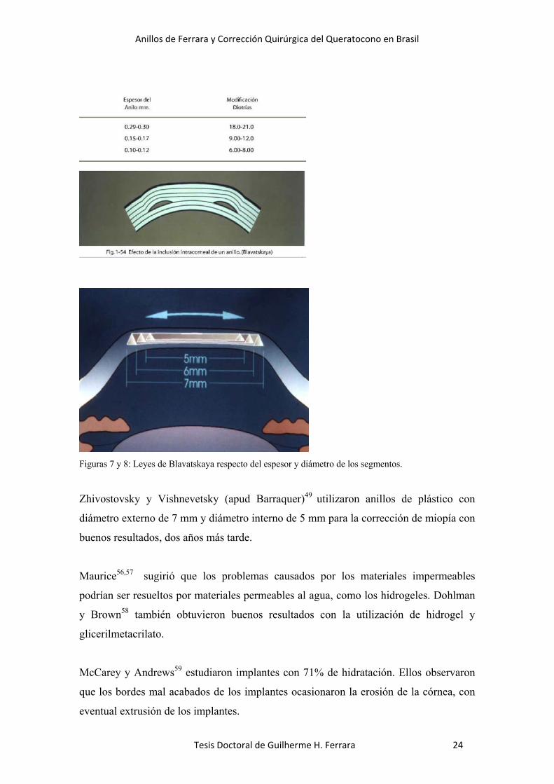
En 1966, Blavatskaya (apud Barraquer)49, realizó diversos experimentos en conejos con
el objetivo de estudiar los efectos refractivos inducidos por implantes de discos,
pequeñas lentes y tejido corneal implantados en la córnea, a través de la técnica de
disección manual. Con el implante de anillos de tejido corneal, Blavatskaya logró
corregir hasta 21 dioptrías de miopía. En aquel momento se demostró que la corrección
obtenida a través del implante de los anillos era directamente relacionada a su espesor e
inversamente relacionada a su diámetro. Así, los anillos de diámetro más pequeño y de
mayor espesor producen mayor corrección.
Anillos de Ferrara y Corrección Quirúrgica del Queratocono en Brasil
Tesis Doctoral de Guilherme H. Ferrara 24
Figuras 7 y 8: Leyes de Blavatskaya respecto del espesor y diámetro de los segmentos.
Zhivostovsky y Vishnevetsky (apud Barraquer)49 utilizaron anillos de plástico con
diámetro externo de 7 mm y diámetro interno de 5 mm para la corrección de miopía con
buenos resultados, dos años más tarde.
Maurice56,57 sugirió que los problemas causados por los materiales impermeables
podrían ser resueltos por materiales permeables al agua, como los hidrogeles. Dohlman
y Brown58 también obtuvieron buenos resultados con la utilización de hidrogel y
glicerilmetacrilato.
McCarey y Andrews59 estudiaron implantes con 71% de hidratación. Ellos observaron
que los bordes mal acabados de los implantes ocasionaron la erosión de la córnea, con
eventual extrusión de los implantes.

Anillos de Ferrara y Corrección Quirúrgica del Queratocono en Brasil
Tesis Doctoral de Guilherme H. Ferrara 25
Maurice56 en 1969, determinó que el estroma anterior al implante recibe nutrientes por
difusión, y que la nutrición es proporcional al diámetro y a la profundidad del implante.
De esta manera, ortesis con diámetro mayor que 5 mm implantada a menos de un 50%
de espesor corneal seguramente será extruida.
La utilización de polisulfonas en ojos humanos demostró su poca tolerancia, a pesar de
los resultados refractivos positivos (Choyce).55
4.3.2. Anillos de Ferrara
Los estudios con los implantes intracorneales se iniciaron en 1986, teniendo como
objetivo la corrección de miopías moderadas y elevadas, una vez que las técnicas
existentes hasta este momento permitían apenas la corrección de pequeñas miopías,
hasta 6,00 dioptrías. En dicho momento no se disponía de una estructura fabril para
producir los anillos y, por lo tanto, las dimensiones de las ortesis no eran precisas.
Otros estudios realizados por Burris T.E. (1993,1994),60,61 Schanzlin (1997),62 Nose W
(1996)63 y Fleming (1998)64 también confirmaron la eficacia de los anillos para la
corrección de la miopía baja o moderada.
El objetivo de la investigación, en aquella época, era definir la tolerancia de la córnea a
la ortesis, sus dimensiones, forma y la profundidad de implantación, ya que los datos de
la literatura disponibles hacían referencia sólo a los implantes lenticulares de hidrogel y
polisulfona.55,59
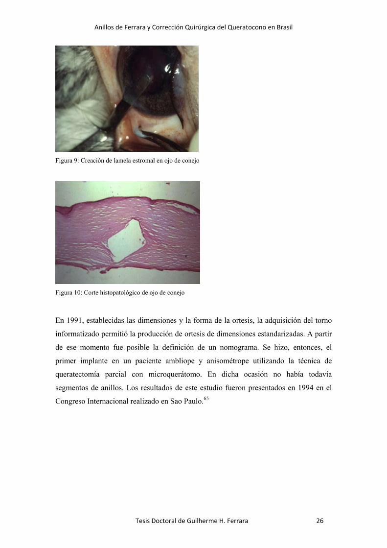
Se han realizado estudios histopatológicos de varios segmentos implantados en ojos de
conejos albinos, a los 3, 6, 9 y 12 meses. Los resultados obtenidos demostraron la
ausencia de reacción inflamatoria perianular importante, así como ausencia de
alteraciones de las estructuras adyacentes, como epitelio y endotelio.
Anillos de Ferrara y Corrección Quirúrgica del Queratocono en Brasil
Tesis Doctoral de Guilherme H. Ferrara 26
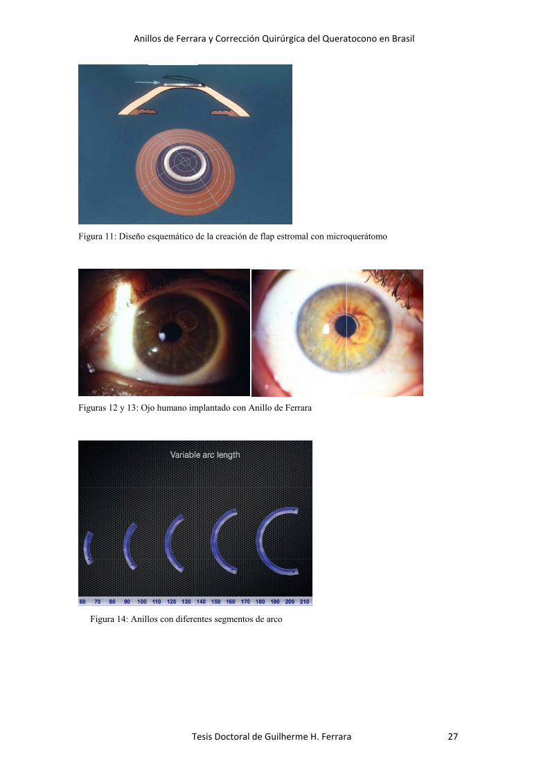
Figura 9: Creación de lamela estromal en ojo de conejo
Figura 10: Corte histopatológico de ojo de conejo
En 1991, establecidas las dimensiones y la forma de la ortesis, la adquisición del torno
informatizado permitió la producción de ortesis de dimensiones estandarizadas. A partir
de ese momento fue posible la definición de un nomograma. Se hizo, entonces, el
primer implante en un paciente ambliope y anisométrope utilizando la técnica de
queratectomía parcial con microquerátomo. En dicha ocasión no había todavía
segmentos de anillos. Los resultados de este estudio fueron presentados en 1994 en el
Congreso Internacional realizado en Sao Paulo.65
Figura
Figura
Fig
Anil
a 11: Diseño e
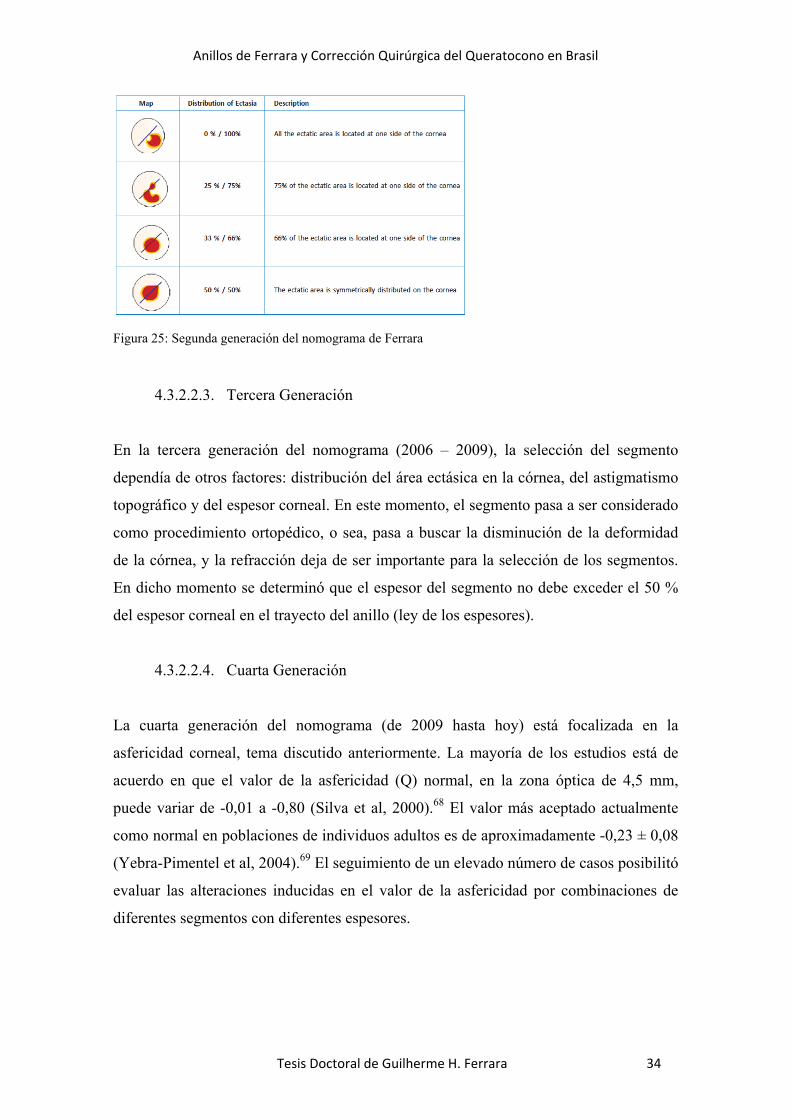
as 12 y 13: Oj
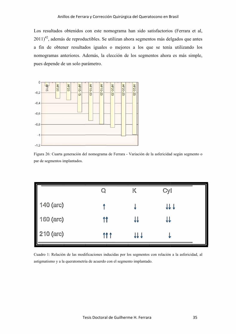
gura 14: Anill
llos de Ferra
Te
esquemático d
jo humano im
los con diferen
ra y Correcci
esis Doctora
de la creación
mplantado con
ntes segmento
ión Quirúrgic
l de Guilherm
de flap estrom
Anillo de Fer
os de arco
ca del Quera
me H. Ferrar
mal con micro
rrara
atocono en B
a
oquerátomo
Brasil
27

Anillos de Ferrara y Corrección Quirúrgica del Queratocono en Brasil
Tesis Doctoral de Guilherme H. Ferrara 28
4.3.2.1. Evolución de los anillos de Ferrara
Con el objetivo de evitar el eje visual se hizo necesario el desarrollo de una nueva
técnica, la técnica de tunelización (1994), la misma que se utiliza en la actualidad. Con
ella se desarrolló también la primera generación de segmentos de anillos. Estos tenían
350 grados de arco.
Figura 15: Primeros instrumentales para el implante de los Anillos de Ferrara, hechos en oro, por un
orives.
Figura 16: Primera generación de los anillos de Ferrara

Anillos de Ferrara y Corrección Quirúrgica del Queratocono en Brasil
Tesis Doctoral de Guilherme H. Ferrara 29
Figura 17: Foto de un ojo de conejo implantado con la primera generación de los anillos de Ferrara
El primer anillo utilizado en paciente post queratoplastia de córnea y queratotomía
radial fue implantado en 1995. Esta paciente fue referida al Servicio de Córneas del
Hospital São Geraldo de la Universidad Federal de Minas Gerais (UFMG), para un
retrasplante. Se intentó el implante del segmento, como alternativa al trasplante, tras la
firma del consentimiento informado de la cirugía. El resultado fue satisfactorio, con
corrección de la ametropía y perfecta tolerancia de la ortesis por parte del tejido corneal.
Después de 6 años de seguimiento, la paciente se presentaba bien, con la córnea
compensada y la refracción estable.
Figura 18: Paciente post queratoplastia y queratotomía radial implantado con anillo de Ferrara (1995)
El implante de estos segmentos de primera generación era difícil. La proximidad de las
puntas del segmento de anillo a la incisión impedía su cicatrización, así como causaba la
extrusión de los segmentos, en muchos casos. Estos problemas llevaron al desarrollo de
la segunda generación de segmentos de anillos: los de 160 grados de arco. Estos

Anillos de Ferrara y Corrección Quirúrgica del Queratocono en Brasil
Tesis Doctoral de Guilherme H. Ferrara 30
segmentos eran implantados a partir de la creación de 2 incisiones opuestas entre sí. El
acabado de estos segmentos era pobre, además de no disponer de los agujeros en la
extremidad de los segmentos, presentes en los segmentos actuales.
Figura 19: Segunda generación de los Anillos de Ferrara
El implante de 2 segmentos simétricos, además de causar el aplanamiento de la córnea,
también resultaba en la reducción del astigmatismo corneal y refraccional.
Los anillos con objetivo refractivo se mostraron poco predecibles, a pesar de presentar
resultados estables y un buen rendimiento visual.

Anillos de Ferrara y Corrección Quirúrgica del Queratocono en Brasil
Tesis Doctoral de Guilherme H. Ferrara 31
Figuras 20 y 21: Baja predictibilidad de los implantes para la corrección de la miopía y del astigmatismo
corneal.
Sin embargo, los resultados de este implante fueron animadores, y la tolerabilidad del
órgano al implante dio la seguridad necesaria para aplicar la técnica en córneas con
queratocono. De esa manera, a partir de 1996, se decidió implantar los anillos en
pacientes con queratocono, intolerantes a lentes de contacto y con indicación para
trasplante de córnea.
Colin, en 1997,66 publicó los primeros estudios de anillos intracorneales de gran
diámetro en paciente con queratocono inicial.
La mejora de la técnica quirúrgica y de los conocimientos sobre el mecanismo de acción
de los anillos permitió el desarrollo de nomogramas basados en el análisis estadístico.
De esa manera se crea la tercera generación de los segmentos, dotada de mejoras en la
superficie y la inclusión de un agujero en la punta de la extremidad del segmento.

Anillos de Ferrara y Corrección Quirúrgica del Queratocono en Brasil
Tesis Doctoral de Guilherme H. Ferrara 32
Figuras 22 y 23: Tercera generación de los Anillos de Ferrara
La utilización de los segmentos intracorneales para la corrección de ectasias de córnea
post Excimer Laser se inició en 1999. A partir de 2000 es posible encontrar trabajos en
la literatura haciendo referencia al uso de estos implantes para la misma finalidad
(Lovisolo).67
4.3.2.2. Evolución del nomograma de los anillos de Ferrara
4.3.2.2.1. Primera Generación
Al principio (1997 – 2002), la indicación de los segmentos ocurría de acuerdo con el
grado del queratocono, y siempre se implantaban 2 segmentos (tabla I). En
queratoconos avanzados, en los cuales las córneas son más delgadas, y que se utilizaban
segmentos más gruesos, su extrusión ocurría con alguna frecuencia.

Anillos de Ferrara y Corrección Quirúrgica del Queratocono en Brasil
Tesis Doctoral de Guilherme H. Ferrara 33
Nomograma
La selección del anillo depende de la ametropía y, tratándose del queratocono, del grado evolutivo del
mismo.
Diámetro 5,00 mm Grosor Dioptría a ser corregida
0,150 mm-2,00 a -4,00
Cono I 0,200 mm -4,25 a -6,00
Cono II 0,250 mm -6,25 a -8,00
Cono III 0,300 mm -8,25 a -10,00
Cono IV 0,350 mm -10,25 a -12,00
El uso de este nomograma permite asociar a ambos: el grado evolutivo del queratocono y la ametropía
existente. De esa forma, si tenemos un paciente con cono incipiente y alta miopía, utilizaremos un anillo
de 350 micras, y así sucesivamente.
Figura 24: Primera generación del nomograma de Ferrara
4.3.2.2.2. Segunda Generación
La segunda generación del nomograma (2002 – 2006) llevaba en consideración la
distribución del área ectásica, además del equivalente esférico para la selección de los
anillos. Los pacientes con elevado equivalente esférico tenían implantados segmentos
más gruesos. Sin embargo, en muchos pacientes con queratocono, la miopía no era
inducida por la ectasia, sino por el crecimiento anteroposterior del ojo. Por eso, en
muchos casos se observaba una hipercorrección debido al implante de segmentos
gruesos en queratoconos iniciales.
Anillos de Ferrara y Corrección Quirúrgica del Queratocono en Brasil
Tesis Doctoral de Guilherme H. Ferrara 34
Figura 25: Segunda generación del nomograma de Ferrara
4.3.2.2.3. Tercera Generación
En la tercera generación del nomograma (2006 – 2009), la selección del segmento
dependía de otros factores: distribución del área ectásica en la córnea, del astigmatismo
topográfico y del espesor corneal. En este momento, el segmento pasa a ser considerado
como procedimiento ortopédico, o sea, pasa a buscar la disminución de la deformidad
de la córnea, y la refracción deja de ser importante para la selección de los segmentos.
En dicho momento se determinó que el espesor del segmento no debe exceder el 50 %
del espesor corneal en el trayecto del anillo (ley de los espesores).
4.3.2.2.4. Cuarta Generación
La cuarta generación del nomograma (de 2009 hasta hoy) está focalizada en la
asfericidad corneal, tema discutido anteriormente. La mayoría de los estudios está de
acuerdo en que el valor de la asfericidad (Q) normal, en la zona óptica de 4,5 mm,
puede variar de -0,01 a -0,80 (Silva et al, 2000).68 El valor más aceptado actualmente
como normal en poblaciones de individuos adultos es de aproximadamente -0,23 ± 0,08
(Yebra-Pimentel et al, 2004).69 El seguimiento de un elevado número de casos posibilitó
evaluar las alteraciones inducidas en el valor de la asfericidad por combinaciones de
diferentes segmentos con diferentes espesores.
Los
2011
a fin
nomo
pues
Figura
par de
Cuadr
astigm
Anil
resultados
1)42, además
n de obten
ogramas an
depende de
a 26: Cuarta g
e segmentos im
ro 1: Relación
matismo y a la
llos de Ferra
Te
obtenidos c
s de reprodu
ner resultad
nteriores. A
e un solo pa
generación de
mplantados.
n de las modi
a queratometrí
ra y Correcci
esis Doctora
con este no
uctibles. Se
dos iguales
Además, la e
arámetro.
el nomograma
ificaciones in
ía de acuerdo
ión Quirúrgic
l de Guilherm
omograma
utilizan ah
s o mejore
elección de
a de Ferrara -
ducidas por l
con el segmen
ca del Quera
me H. Ferrar
han sido s
hora segmen
es a los qu
e los segme
Variación de
os segmentos
nto implantad
atocono en B
a
atisfactorio
ntos más de
ue se tenía
entos ahora
la asfericidad
s con relación
do.
Brasil
os (Ferrara
lgados que
a utilizando
a es más sim
d según segm
n a la asfericid
35
et al,
antes
o los
mple,
mento o
dad, al

Anillos de Ferrara y Corrección Quirúrgica del Queratocono en Brasil
Tesis Doctoral de Guilherme H. Ferrara 36
4.3.2.3. Características de los Anillos de Ferrara
El anillo de Ferrara presenta las siguientes características:
confeccionado en Acrílico CQ
diámetro total (externo) de 6,2 mm
sección triangular
base de 600 micras
espesuras variables
segmentos de 140, 160, 210 y 320 grados de arco
1 orificio en cada extremidad
4.3.2.4. Mecanismo de acción de los Anillos de Ferrara
El anillo corneal obedece a los postulados de Barraquer y Blavatskaya, según los cuales
la adición en la periferia de la córnea resulta en aplanamiento de la misma, y el diámetro
del anillo determina cuánto la córnea será aplanada. De esa manera, cuanto más tejido
adicionado (espesor del anillo) y cuanto menor el diámetro, mayor será la corrección
miópica obtenida.49,50
De sus estudios resultan observaciones adicionales:
• Aplanamiento central y periférico de la córnea, preservando su asfericidad;
Figura 27: aplanamiento de la córnea después del implante del Anillo de Ferrara

Anillos de Ferrara y Corrección Quirúrgica del Queratocono en Brasil
Tesis Doctoral de Guilherme H. Ferrara 37
• Disminución de la altura de la cámara anterior, demostrado por la biomicroscopía
ultrasónica (UBM);
Figura 28: Disminución de la altura de la cámara anterior después del implante de los Anillo de Ferrara.
• Regularización de la superficie de la córnea a través de un movimiento de báscula
de los segmentos, provocado por la superficie plana de la base del anillo. Ese
movimiento resulta en un aplanamiento de la córnea en las puntas de los segmentos
y en un encorvamiento de la misma en la región correspondiente al cuerpo del anillo;
Figura 29: Mapas de elevación anterior y posterior en 3D. Las flechas en rojo indican la región de la
incisión (inferior) y las flechas en azul muestran la elevación de las puntas de los anillos.
• Paralización de la evolución del queratocono, disminución de las opacidades
presentes en el ápice del cono, reducción de síntomas como prurito, fotofobia, dolor
ocular y/o incomodidad;
Anillos de Ferrara y Corrección Quirúrgica del Queratocono en Brasil
Tesis Doctoral de Guilherme H. Ferrara 38
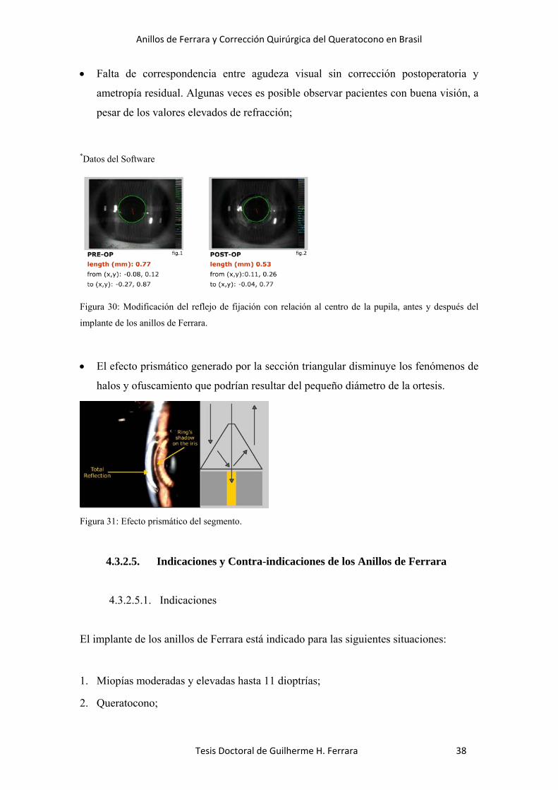
• Falta de correspondencia entre agudeza visual sin corrección postoperatoria y
ametropía residual. Algunas veces es posible observar pacientes con buena visión, a
pesar de los valores elevados de refracción;
*Datos del Software
Figura 30: Modificación del reflejo de fijación con relación al centro de la pupila, antes y después del
implante de los anillos de Ferrara.
• El efecto prismático generado por la sección triangular disminuye los fenómenos de
halos y ofuscamiento que podrían resultar del pequeño diámetro de la ortesis.
Figura 31: Efecto prismático del segmento.
4.3.2.5. Indicaciones y Contra-indicaciones de los Anillos de Ferrara
4.3.2.5.1. Indicaciones
El implante de los anillos de Ferrara está indicado para las siguientes situaciones:
1. Miopías moderadas y elevadas hasta 11 dioptrías;
2. Queratocono;

Anillos de Ferrara y Corrección Quirúrgica del Queratocono en Brasil
Tesis Doctoral de Guilherme H. Ferrara 39
3. Degeneración Marginal Pelúcida
4. Astigmatismos irregulares elevados post trasplante de córnea;
5. Astigmatismos irregulares post queratotomía radial;
6. Ectasia corneal post Excimer Laser
El pequeño diámetro del anillo, además de permitir correcciones bastante elevadas,
tanto de miopía como de astigmatismo, lo hace adecuado para la corrección de
patologías oculares como queratocono, astigmatismos irregulares de cualquier etiología
y ectasias corneales post Excimer Laser. Con respecto al queratocono, debe ser
considerado el grado evolutivo, su tolerancia, o no, a lentes de contacto semirrígidas
permeables al gas, y la estabilidad, o no, del queratocono.
4.3.2.5.2. Contraindicaciones
El implante de los anillos de Ferrara es contraindicado en las siguientes situaciones:
1. Conos muy avanzados, con curvaturas superiores a 75 dioptrías y opacidades
apicales importantes;
2. Hidropsía;
3. En los casos de astigmatismos elevados post trasplante de córnea, el anillo no
deberá ser implantado si la córnea donada se encuentra muy descentrada;
4. Pacientes con atopia intensa deberán ser tratados previamente;
5. Cualquier proceso infeccioso en actividad, local o sistémico.
4.3.2.6. Cirugía del implante de los anillos de Ferrara
4.3.2.6.1. Instrumental
Para la realización de la cirugía son necesarios los siguientes instrumentos:
espátula doble y simples de Ferrara,
Suarez spreader,
marcador de zona óptica de 3,5,7 mm,

Figura
Anes
2%.
Se ha
micro
que,
anillo
Figura
Se de
de la
Anil
marca
ganch
micró
a 32: Instrume
4.3.2.6.2.
stesia: la cir
ace la marca
oscopio en
en el quera
o deberá qu
a 33: Marcaci
elimita la zo
a córnea, do
llos de Ferra
Te
ador de inci
ho de Sinsk
ómetro de d
entales para el
Técnica Q
rugía se hac
ación del ej
la córnea, i
atocono, el
uedar situad
ión del eje visu
ona óptica d
onde será re
ra y Correcci
esis Doctora
isiones radi
ey de 0.20 m
diamante con
l implante de
Quirúrgica
ce con anest
je visual con
ndependien
ápice suel
o en la base
ual en el prim
de 5,00 mm
ealizada la
ión Quirúrgic
l de Guilherm
ales de 8 m
mm,
n lámina de
los Anillos de
tesia tópica
n base en el
ntemente de
le estar a m
e del cono, d
mero reflejo de
m con violeta
incisión rad
ca del Quera
me H. Ferrar
mm,
e Ferrara.
e Ferrara
después de
l reflejo del
la pupila. E
menudo desp
donde sea q
e Purkinje en l
a genciana y
dial, con bi
atocono en B
a
e la miosis c
filamento d
Es necesario
plazado inf
que se encue
la córnea
y se marca
isturí aprop
Brasil
con pilocarp
de la lámpar
o tener en c
feriormente,
entre.
el eje más c
iado y calib
40
pina a
ra del
uenta
, y el
curvo
brado

Anillos de Ferrara y Corrección Quirúrgica del Queratocono en Brasil
Tesis Doctoral de Guilherme H. Ferrara 41
para un 80% de la espesura de la córnea en el local de la incisión. A seguir, se aplica el
delaminador de córnea para realizar el bolso por donde será introducida la espátula de
Ferrara para la confección del túnel. La introducción de los segmentos es simples y la
cirugía termina con el posicionamiento adecuado de estos segmentos dentro de los
túneles.
Figura 34: Marcación de la zona óptica de 5 mm.
Figura 35: Creación del túnel estromal.
4.3.2.7. Complicaciones
La incidencia de complicaciones después de la curva de aprendizaje es muy baja. Las
principales complicaciones son:
4.3.2.7.1. Infección: pueden ocurrir en dos situaciones: en el postoperatorio
inmediato, o tardíamente asociado al uso de lentes de contacto
blandas. En el primer caso, la conducta consiste en remover el

Anillos de Ferrara y Corrección Quirúrgica del Queratocono en Brasil
Tesis Doctoral de Guilherme H. Ferrara 42
segmento del túnel acometido e instituir terapia intensiva con
antibióticos. En las infecciones tardías puede o no haber necesidad de
remover el segmento, dependiendo de la pérdida o no de sustancia
corneal y de la intensidad de la infección.
Figura 36: Infección en la punta del Anillo.
4.3.2.7.2. Migración: habitualmente los pacientes portadores de queratocono son
atópicos y presentan prurito intenso. El acto de rascarse los ojos puede
desplazar los segmentos, llevándolos hacia cerca de las incisiones y
posibilitando la extrusión de los mismos;
Figura 37: Migración de la punta del Anillo por debajo de la incisión.

Anillos de Ferrara y Corrección Quirúrgica del Queratocono en Brasil
Tesis Doctoral de Guilherme H. Ferrara 43
4.3.2.7.3. Extrusión: resulta de una implantación superficial o de la migración
de los segmentos. Esta situación se puede prevenir a través de los
exámenes de rutina, removiendo el segmento antes que el mismo se
exponga y reimplantándolo después de algún tiempo.
Figura 38: Extrusión del segmento, secundario a la confección de túnel superficial.
4.3.2.7.4. Descentralización: el anillo deberá estar situado, obligatoriamente, en
la base del cono. Por lo tanto, la centralización del procedimiento
deberá ser realizada siempre llevando en cuenta el reflejo.
4.3.2.7.5. Halos y Reflejos: pueden estar presentes en los primeros meses, mas
raramente son referidos por los pacientes. Cuando y sí es necesario, se
pueden prescribir mióticos suaves. La incidencia de pacientes con esta
queja es muy pequeña. En la mayoría de los caso, el paciente sólo
relata este fenómeno cuando se le pregunta;
4.3.2.7.6. Hipo e Hipercorrección: son complicaciones relativas si consideramos
que el objetivo principal de la cirugía es ortopédico y que la
corrección visual final deberá ser realizada utilizando los métodos
convencionales. La mayoría de los casos queda hipo corregido, si es
analizado el componente esférico. Los astigmatismos, en general, son
híper corregidos con la inversión del eje del astigmatismo;

Anillos de Ferrara y Corrección Quirúrgica del Queratocono en Brasil
Tesis Doctoral de Guilherme H. Ferrara 44
4.3.2.7.7. Opacidades perianulares; son pequeños depósitos blancos, opacos, que
se depositan a lo largo de la fase interna del anillo. No tienden a crecer
y no perjudican el desempeño visual, siendo sólo antiestéticos al
examen a la lámpara de hendidura.
Figura 38: opacidades perianelares

Anillos de Ferrara y Corrección Quirúrgica del Queratocono en Brasil
Tesis Doctoral de Guilherme H. Ferrara 45
5. Justificación
El queratocono es una enfermedad que puede llevar a la ceguera en las
edades más productivas de la vida. Aunque cada vez se conoce mejor su
fisiopatología, no tiene etiología conocida y los tratamientos se basan en
prevenir los factores de riesgo y en medidas de corrección quirúrgica. Respecto
a la corrección con cirugía, el queratocono sigue siendo una de las principales
indicaciones de trasplante de cornea y se están evaluando alternativas al
trasplante, como el entrecruzamiento del colágeno corneal con luz ultravioleta y
el implante de ICRS. La clínica del Dr. Paulo Ferrara tiene una larga
experiencia de más de 20 años en el implante de ICRS y cuenta con las
historias clínicas y bases de datos que pueden ser soporte para el análisis de
resultados con masa crítica suficiente de pacientes y seguimiento adecuado.
Por ello, queda justificado el intentar analizar los resultados del implante de
ICRS con objeto de conocer los efectos de los mismos sobre la agudeza visual,
cambios en las medidas corneales, con especial atención al astigmatismo y en
la progresión del queratocono.

Anillos de Ferrara y Corrección Quirúrgica del Queratocono en Brasil
Tesis Doctoral de Guilherme H. Ferrara 46
6. Hipótesis de Trabajo
El implante de segmentos de anillos intraestromales en los pacientes con queratocono
(adultos y niños) permite mejorar la agudeza visual, cambiar los valores de medidas
corneales y alterar la evolución de la enfermedad. Además, como aplicación práctica en
queratoplastias, pueden ser empleados para el manejo del astigmtismo.

Anillos de Ferrara y Corrección Quirúrgica del Queratocono en Brasil
Tesis Doctoral de Guilherme H. Ferrara 47
7. Objetivos
1. Evaluar los resultados a largo plazo del implante de anillos de Ferrara
en cuanto a los cambios en la agudeza visual y los cambios en
valores de las medidas corneales con especial atención al
astigmatismo y los valores queratométicos;
2. Evaluar el efecto de los ICRS en la evolución del queratocono;
3. Evaluar el efecto de los ICRS en la corrección del astigmatismo tras
queratoplastia;
4. Evaluar los resultados de los ICRS implantados en niños con
queratocono.

Anillos de Ferrara y Corrección Quirúrgica del Queratocono en Brasil
Tesis Doctoral de Guilherme H. Ferrara 48
8. Pacientes, Material de Trabajo y Método
8.1. Diseño del Estudio
Estudio observacional, retrospectivo, de análisis de resultados de los pacientes
con queratocono intervenidos de cirugía de ICRS y de pacientes con
queratoplastia a los que se ha implantado ICRS para el control del
astigmatismo
8.2. Pacientes
Las cirugías de los pacientes con queratocono fueron realizadas en los
pacientes que acudieron a la clínica de Ojos Dr. Paulo Ferrara, en Belo
Horizonte, Minas Gerais, Brasil, y se incluyeron pacientes desde julio de 1996 a
Abril de 2014.
El estudio sobre la corrección del astigmatismo post trasplante de córnea en
pacientes implantados con los ICRS fue realizado en los pacientes que
acudieron a la clínica de Ojos Dr. Enio Coscarelli, en Belo Horizonte, Minas
Gerais, Brasil, en el periodo de mayo de 2005 a septiembre de 2009
8.3. Clasificación del Queratocono
El queratocono fue clasificado de acuerdo con la clasificación de Amsler-
Krumeich,70 que combina efecto refractivo inducido por la ectasia, lecturas
queratométricas , signos clínicos y paquimetría.
8.4. Indicación del implante de ICRS en queratocono
8.4.1. Criterios de inclusión La principal indicación para el implante de los ICRS fue intolerancia a las lentes
de contacto semirrígidas y/o progresión de la ectasia , definida por empeora de
la agudeza visual con y sin corrección, intolerancia progresiva a las lentes de
contacto y empeora progresiva de la curvatura corneal documentada por
topografía de córnea (aumento del promedio de la queratometría de 1 D en un
año)

Anillos de Ferrara y Corrección Quirúrgica del Queratocono en Brasil
Tesis Doctoral de Guilherme H. Ferrara 49
8.4.2. Criterios de Exclusión
Los criterios de exclusión fueron queratoconos avanzados con la queratometría
mayor que 60 D, opacidades apicales significantes, cicatrices, hidropsía,
córneas delgadas con paquimetría inferior a 300 µm en el trayecto del anillo,
atopia intensa o proceso infeccioso activo, local o sistémico.
8.5. Implante de ICRS en queratoplastias 8.5.1. Criterios de inclusión
Se incluyeron en el estudio pacientes sometidos al implante de los ICRS para
la corrección del astigmatismo post trasplante de córnea.
Los criterios de inclusión fueron enjerto trasparente, con astigmatismos entre
2,5 y 8D, intolerantes a lentes de contacto. Todos los pacientes tenían mas de
dos años de post operatorio de trasplante de córnea en el momento del
implante de los anillos intracorneales, y por los menos 1 año de seguimiento de
pos operatorio después de haber implantado los anillos
8.5.2. Criterios de Exclusión
Los criterios de exclusión fueron queratometría mayor que 60D, opacidades
apicales significantes, cicatrices, córneas delgadas con paquimetría inferior a
300µm en el trayecto del anillo, atopia intensa o proceso infeccioso activo, local
o sistémico.
8.6. Técnica Quirúrgica y estrategia de implante de ICRS Todos los pacientes fueros operados por la técnica manual y por el mismo
cirujano, Dr. Paulo Ferrara, con excepción de los pacientes del estudio de
queratoplastia y astigmatismo, que fueron operados por el mismo cirujano, Dr
Sandro Coscarelli, por la técnica manual.
La cirugía se hace con anestesia tópica después de la miosis con pilocarpina a
2%.
Se hace la marcación del eje visual con base en el reflejo del filamento de la
lámpara del microscopio en la córnea, independientemente de la pupila
(primero reflejo de Purkinje), utilizándose del Sinkey hook. Se delimita la zona
óptica de 5,00 mm con violeta genciana y marcamos el eje más curvo de la

Anillos de Ferrara y Corrección Quirúrgica del Queratocono en Brasil
Tesis Doctoral de Guilherme H. Ferrara 50
córnea, donde será realizada la incisión radial, con bisturí apropiado y calibrado
para 80% de la espesura de la córnea en el local de la incisión. A seguir, se
aplica el delaminador de córnea para realizar el bolso por donde será
introducida la espátula de Ferrara para confección del túnel. La introducción de
los segmentos es simples y la cirugía termina con el posicionamiento adecuado
de estos segmentos dentro de los túneles. La técnica fue la misma en todos los
pacientes operados.
La selección del espesor y del tamaño del arco de los anillos se hizo de
acuerdo con el nomograma de Ferrara vigente en el momento de la cirugía tal y
como se describe en la introducción. El nomograma tenía en cuenta la posición
del área de la ectasia en la córnea, el astigmatismo topográfico y la
paquimetría corneal en el trayecto del anillo.
8.7. Análisis de los resultados: Los parámetros evaluados fueron agudeza visual con corrección, sin corrección,
queratometría, paquimetría central. La asfericidad y volumen corneal también
fueron estudiados. Los datos fueron obtenidos a través del topógrafo de córnea
y del Pentacam®.
Los parámetros evaluados en el estudio astigmatismo y queratoplastia fueron
agudeza visual con corrección, sin corrección, queratometría, equivalente
esférico, astigmatismo topográfico y valores de k mínimo y máximo. Los datos
fueron obtenidos a través del topógrafo de córnea (CT4000 Corneal
Topographer, Eyetech, Inc.).
El análisis estadístico fue realizado con la utilización del GraphPad Instat 3
para Macintosh (versión 3,1ª; GraphPad Software, Inc., La Jolla, CA). El test t
de Student para pares pareados fue utilizado para comparar los datos pre y
post operatorios.
El análisis estadístico para el estudio de queratoplastia y astigmatismo incluyó
el test t de Student, transformación de Welch, testes no paramétricos de de
Mann-Whitney, utilizándose del Instat 3 para Macintosh (versión 3,1ª;

Anillos de Ferrara y Corrección Quirúrgica del Queratocono en Brasil
Tesis Doctoral de Guilherme H. Ferrara 51
GraphPad Software, Inc., La Jolla, CA). El análisis vectorial fue realizado
utilizándose del SigmaPlot software (SSP Inc.) el análisis del seguimiento
clínico fue realizado con el software basado en internet de análisis refractiva
(Zubisoft GmbH).
En cada capitulo y en las publicaciones se detalla la metodología empleada en
en cada caso.

Anillos de Ferrara y Corrección Quirúrgica del Queratocono en Brasil
Tesis Doctoral de Guilherme H. Ferrara 52
9. CAPITULO I. Resultados a largo plazo de la implantación de anillos de Ferrara en pacientes con queratocono
Se resumen a continuación los trabajos de investigación clínica relacionados con el
análisis de resultados a largo plazo del implante de ICRS y que han sido objeto de
publicación en las siguientes publicaciones que se adjuntan:
1. Ferrara G, Torquetti L, Ferrara P, Merayo-Lloves J. Intrastromal cornal ring
segments: visual outcomes from a large case series. Clinical Experiment
Ophthalmol 2012;40(5):433-439
2. Torquetti L, Ferrara G, Almeida F, Ferrara P, Merayo-Lloves J. Clinical
outcomes after intrastromal corneal ring segments reoperation in keratoconus
patients. Int Ophthalmol 2013;6(6):796-800
3. Torquetti L, Ferrara G, Ferrara P. Predictors of Clinical Outcomes after
Intraestromal corneal Ring Segments Implatation. Int Keratoco Ectatic
Corneal Dis 2012;1(1):26-30
4. Torquetti L, Ferrara G, Almeida F, Cunha L, Araujo LPN, Machado AP, Lyra
JM, Merayo-Lloves J, Ferrara P. Intrastromal Corneal Ring Segment
Implantation in Patients with Keratoconus: 10-Year Follow-Up. J Refract Surg
2014;30(1)22-6
El queratocono es una patología bilateral y asimétrica, no inflamatoria por definición,
que cursa con adelgazamiento progresivo de la córnea, seguido por el aumento de su
curvatura y protrusión, resultando en elevados grados de astigmatismo irregular y
miopía y, consecuentemente, baja de la agudeza visual. Su presentación es variable, y
ocurre en general en la segunda década de vida, pudiendo progresar hasta la tercera o
cuarta década de vida (Rabinowitz 1998).71
Las primeras indicaciones de los anillos intracorneales fueron para la corrección de
miopías moderadas y elevadas (FERRARA, 1991, NOSE W, 1996)41,63, pero como el
objetivo refractivo resultó poco predecible, en 1996 (FERRARA, 1996) se cambió la
indicación de los anillos por la corrección de córneas irregulares. Se ha utilizado, desde
entonces, el implante de los anillos intracorneales para el tratamiento de las ectasias
corneales, principalmente en el queratocono. Los ICRS actúan disminuyendo las

Anillos de Ferrara y Corrección Quirúrgica del Queratocono en Brasil
Tesis Doctoral de Guilherme H. Ferrara 53
irregularidades corneales y los valores de queratometría, además de aumentar los
valores de asfericidad y del espesor corneal. Es un procedimiento seguro y poco
invasivo que preserva el eje visual.
Los resultados de las alteraciones inducidas por los implantes intracorneales en la
córnea enferma son la mejora de la calidad visual, añadida de la mejora de la agudeza
visual con y sin corrección. Otro efecto benéfico que se pudo observar en los pacientes
portadores de queratocono implantados con los anillos intracorenales fue la
estabilización de la patología, además de la mejora de los síntomas del paciente, como
la fotofobia, el prurito y el lagrimeo.
Muchos estudios fueron realizados con la finalidad de se evaluar a largo plazo los
resultados del implante de los anillos de Ferrara. El estudio Intrastromal cornal ring
segments: visual outcomes from a large case series42 presentó los resultados visuales y
topográficos de 1073 ojos de 810 pacientes implantados con los anillos de Ferrara para
la corrección de queratocono. Los criterios de inclusión fueron la evidencia de
progresión de la ectasia e intolerancia a lentes de contacto semirrígidas permeables al
gas. Se excluyeron del estudio a los pacientes con queratoconos muy avanzados, con
curvaturas mayores que 62 dioptrías, opacidades apicales o bien espesor corneal por
debajo de 300 µm.
Los pacientes fueron separados en 2 grupos, de acuerdo con el fenotipo del queratocono:
paracentral (oval) (grupo I) o central (pezón o nipple) (grupo II). El número, tamaño de
arco y espesor de los anillos fue determinado por el nomograma previamente
descrito.41,72
El promedio de seguimiento de los grupos I y II fue de 23,8 y 22,9 meses,
respectivamente. (Tabla I). La agudeza visual ―con y sin corrección― de los dos
grupos mostró una mejora estadísticamente significativa. Se observó un aumento del
espesor de la córnea, como también de la asfericidad en todos los pacientes,
estadísticamente significativo. El equivalente esférico también disminuyó en el
postoperatorio. (Tabla 2)

Anillos de Ferrara y Corrección Quirúrgica del Queratocono en Brasil
Tesis Doctoral de Guilherme H. Ferrara 54
Tabla 1: Grupo I de pacientes implantados con segmentos de 160˚ de arco, y Grupo II
de pacientes implantados con segmentos de 210˚ de arco
Tabla 2: Preoperatorio y ultimo postoperatorio de pacientes implantados con los anillos
de Ferrara
Las complicaciones observadas en el postoperatorio fueron pocas (3,82%), y la
principal fue la hipo corrección (16 ojos). (Tabla 3).
Tabla 3: Complicaciones después del implante de anillos de Ferrara
Los pacientes que presentaron complicaciones fueron sometidos a una nueva cirugía
para el implante de un segmento adicional (16 ojos) en hipo correcciones, la remoción
de segmentos en caso de hipercorrección (6 ojos), sustitución de segmentos (11 ojos) y
reposicionamiento (4 ojos). Se observó una mejora de estos pacientes recuperados
estadísticamente significativa (Tabla 4).

Anillos de Ferrara y Corrección Quirúrgica del Queratocono en Brasil
Tesis Doctoral de Guilherme H. Ferrara 55
Tabla 4: Preoperatorio y último postoperatorio de pacientes sometidos a reimplante,
retiro o cambio de ICRS.
El estudio Clinical outcomes after intrastromal corneal ring segments reoperation in
keratoconus patients73 fue realizado con la finalidad de evaluar los resultados de estos
pacientes sometidos a la reoperación. El promedio de intervalo de tiempo entre la
primera cirugía y la reoperacion fue de 8,4 meses, y el promedio de tiempo de
seguimiento tras la reoperación fue de 30,5 meses.
Se observó una mejora del promedio de la agudeza visual sin corrección post operatoria
de 20/300 para 20/80 (p=0.005), una mejora del promedio de la agudeza visual con
corrección de 20/160 para 20/50 (p=0.0002), el promedio de la queratometría
disminuyo de 49.33 D para 46.16 D (p=0.0001), el aumento del promedio de la
paquimetría en el punto más delgado de 450 µm para 469 µm (p=0.0001). Se observó
una mejora de los valores de asfericidad y de equivalente esférico , pero esas
alteraciones no fueron estadísticamente significativas.
En el estudio Predictors of Clinical Outcomes after Intraestromal corneal Ring
Segments Implatation, Torquetti et al,74 con el mismo grupo de pacientes, hizo otras dos
evaluaciones independientes del comportamiento del implante de los ICRS, de acuerdo
con la edad de los pacientes y con el grado evolutivo del queratocono.
Los pacientes fueron divididos en 4 grupos de acuerdo con la edad: < de 20 años, de 21
a 30 años, de 31 a 40 años y mayores de 40 años. (Tabla 5). Los pacientes también
fueron divididos en 4 grupos de acuerdo con el grado evolutivo del queratocono, de
acuerdo con el promedio de K: Grado I (Km < 46D), grado II (46 < Km < 52D), grado
III (52 < Km < 60D) o grado IV (Km > 60 D). Los mismos criterios fueron utilizados
para este estudio.

Anillos de Ferrara y Corrección Quirúrgica del Queratocono en Brasil
Tesis Doctoral de Guilherme H. Ferrara 56
Tabla 5: Datos de los pacientes estudiados
El promedio del seguimiento de los pacientes fue de 23,8 ± 12,2 meses. La mayoría de
los pacientes tenían entre 21 y 30 años. Los pacientes más jóvenes presentaban valores
más negativos de asfericidad, aunque los de queratometría fueran semejantes en todos
los grupos. El aplanamiento de la queratometría, así como el aumento de los valores de
la asfericidad fueron más acentuados en los pacientes con menos de 20 años. (Tabla 6).
La reducción de la queratometría fue estadísticamente significativa en todos los grupos.
Tabla 6: Datos pre y postoperatorios de acuerdo con los valores de K y Q
El promedio de la agudeza visual con y sin corrección mejoró en todos los grupos
(p<0,001, Tabla 7). La mejora de la AV con corrección fue más acentuada en los
pacientes entre 21 y 30 años de edad y en los pacientes con queratocono grado I.
Tabla 7: Datos pre y postoperatorios de la UCVA y CDVA, de acuerdo con la edad y el
grado de queratocono

Anillos de Ferrara y Corrección Quirúrgica del Queratocono en Brasil
Tesis Doctoral de Guilherme H. Ferrara 57
Torquetti et al15 (2009) publicaron un estudio con pacientes operados con ICRS para el
tratamiento de queratocono. Los pacientes operados presentaban evidencias de
progresión de la ectasia y/o intolerancia a lentes de contacto semirrígidas permeables al
gas. Se excluyeron del estudio los casos de queratoconos muy avanzados, con
curvaturas mayores que 62 dioptrías, opacidades apicales o bien espesor corneal por
debajo de 300 µm.
Se estudió en este trabajo la agudeza visual con y sin corrección y la queratometría pre
y postoperatoria. Las cirugías fueron realizadas por el mismo cirujano (Paulo Ferrara), y
la selección de los anillos fue determinada por el nomograma previamente descrito. 41,72
El seguimiento de los pacientes varió entre 5 y 12 años de postoperatorio e incluyó 35
ojos de 28 pacientes. Se observó un aplanamiento de todos los ojos a la topografía de
córnea, estadísticamente significativa. (Figura 1) La agudeza visual sin corrección
mejoró hasta el cuarto año postoperatorio y después permaneció estable hasta el quinto
año postoperatorio. La agudeza visual mejoró hasta el tercer año postoperatorio,
permaneciendo estable hasta en quinto año postoperatorio. (Figura 2).
Figura 1: Agudeza visual con corrección a lo largo de los 5 años de seguimiento

Anillos de Ferrara y Corrección Quirúrgica del Queratocono en Brasil
Tesis Doctoral de Guilherme H. Ferrara 58
Figura 2: Promedio de K máximo a lo largo de los 5 años de seguimiento
La estabilidad a largo plazo encontrada en este trabajo puede ser comparada a otros
estudios de implante de ICRS en la literatura. (Tabla 8)
Tabla 8: Comparación de resultados de implante de anillos de Ferrara e Intacs a largo
plazo
En 2014, Torquetti et al45 publicó Intrastromal Corneal Ring Segment Implantation in
Patients with Keratoconus: 10-Year Follow-Up, con seguimiento a los 5 y 10 años de
post operatorio de pacientes implantados con ICRS para el tratamiento de queratocono.
Los pacientes operados tenían intolerancia a lentes de contacto semirrígidas permeables
al gas y/o presentaban progresión de la ectasia. Se excluyeron del estudio los casos con
curvaturas mayores que 60 dioptrías, opacidades apicales o bien espesor corneal por
debajo de 300 µm.

Anillos de Ferrara y Corrección Quirúrgica del Queratocono en Brasil
Tesis Doctoral de Guilherme H. Ferrara 59
Fueron evaluados en este trabajo la agudeza visual con y sin corrección, la
queratometría pre y postoperatoria, así como la variación del espesor corneal después
del implante de los ICRS. Las cirugías fueron realizadas por el mismo cirujano (Paulo
Ferrara), y la selección de los anillos fue determinada por el nomograma previamente
descrito. 41,72
Treinta y seis ojos de 30 pacientes operados entre junio de 1996 y enero de 2002 fueron
evaluados. Se observó una disminución de los valores de la queratometría y del espesor
corneal a los 5 años de seguimiento, además de una mejora de la agudeza visual
estadísticamente significativa. Sin embargo, cuando comparamos los resultados a los 5
años de seguimiento con los resultados a los 10 años, observamos que las variaciones de
los valores estudiados no fueron estadísticamente significativos. (Tabla 9) (Figura 3). El
56,5% de los pacientes ganó 2 o más líneas de visión a los 5 años, mientras que el 66,7 %
de los pacientes ganó 2 o más líneas de visión a los 10 años de seguimiento.
Tabla 9: Comparación de resultados de implante de anillos de Ferrara a los 5 y 10 años
de seguimiento.
Figura 3: Valores preoperatorios de K a los 5 y 10 años de seguimiento

Anillos de Ferrara y Corrección Quirúrgica del Queratocono en Brasil
Tesis Doctoral de Guilherme H. Ferrara 60
De acuerdo con el nomograma de Ferrara, la escoja del arco de anillo se hace de
acuerdo con la morfología del queratocono a ser tratado. Para queratoconos centrales,
con astigmatismo bajo, el segmento de elección es el de 210 grados de arco, pues induce
importante aplanamiento corneal, disminución de los valores de queratometría y
aumento de la asfericidad, sin alterar de manera significativa el astigmatismo. Para
queratoconos paracentrales, los segmentos de elección son los de 160 grados de arco.
Estos segmentos aplanan la córnea y disminuyen su queratometría con menor intensidad,
pero actúan de manera más importante en la reducción del astigmatismo corneal En el
estudio Intrastromal cornal ring segments: visual outcomes from a large case series, 2
grupos fueron separados según su morfología, si central (Nipple), o paracentral (Oval).
Los resultados en los 2 grupos demostraron mejora significativa de la UDVA y en la
CDVA, con aumento significativo del espesor corneal. El valor de la asfericidad
aumentó, de manera significativa, después del implante de los ICRS. El promedio del
valor en el pos operatorios del grupo I (tipo Oval) fue de -0.35, y en el grupo II (tipo
Nipple) fue de -0.56. La asfericidad es considerada como uno de los marcadores de la
calidad de la visión. La mayoría de los estudios esta de acuerdo que los valores de
asfericidad en la córnea humana varia de -0.01 a -0.80.68
Para todos los parámetros mensurados, los resultados fueron mejor en el grupo I. Una
posible explicación es que el grupo de queratocono central presentaba grados más
avanzados de queratocono, con valores pre operatorios más alterados.
La incidencia de complicaciones fue baja en el estudio (3,82% de los ojos tratados). Eso
puede ser explicado por la pericia del cirujano y por la evolución del nomograma. La
principal complicación del estudio fue la hipo corrección.
Además de pocas las complicaciones observadas en el post operatorio del implante de
los anillos, la mayoría de ellas puede ser fácilmente resuelta, debido a la reversibilidad y
ajustabilidad de la técnica. Los pacientes que necesitaron ser reoperados presentaron
mejora de la AV y de los valores topográficos tras la reintervención en el periodo
estudiado.

Anillos de Ferrara y Corrección Quirúrgica del Queratocono en Brasil
Tesis Doctoral de Guilherme H. Ferrara 61
Hipo e hipercorrección fueron los principales motivos de reoperación en el grupo
estudiado. Las causas no son totalmente conocidas, probablemente estén relacionadas a
las propiedades biomecánicas de la córnea. Córneas con patrones topográficos
semejantes pueden tener distintas respuestas al implante de los anillos, debido a que la
rigidez y la viscoelasticidad pueden variar entre los pacientes.
Chan y Khan75 evaluaron resultado de pacientes que tuvieron los segmentos sustituidos,
con mejora de visión (mejora de la AV sin corrección de por lo menos 1 línea de visión)
en todos los pacientes. Los autores de los ensayos clínicos fase II y fase III realizados
por el FDA (Food and Drug Administration)76 en los Estados Unidos concluyeron que
la remoción de los ICRS (Intacs) es segura, efectiva y fácil de ser realizada, con retorno
los parámetros pre operatorios después de 3 meses de la retirada del segmento.
Las alteraciones corneales inducidas por los segmentos pueden ocurrir hasta el sexto
mes post operatorio, motivo por lo cual las reintervenciones solamente ocurrieron
después de ese periodo. La remoción de los implantes ocurrió en 6 pacientes. El grupo
de pacientes que presentaron peor agudeza visual, con y sin corrección, fueron aquellos
que presentaron híper corrección en el post operatorio. El aplanamiento excesivo de la
córnea inducida por los segmentos es una de las causas de insatisfacción del paciente en
el post operatorio. Por lo tanto, la selección correcta de los anillos en el pre operatorio
es de suma importancia.
En el estudio Predictors of Clinical Outcomes after Intraestromal corneal Ring
Segments Implatation,74 la mayoría de los pacientes tenían entre 21 y 40 años de edad.
Los grupos fueron divididos de acuerdo con la edad. No presentaron diferencias
estadísticamente significativas de la variación del promedio de la queratometría, del
volumen corneal y del espesor corneal entre ellos. La asfericidad presentó mayor
variación en los pacientes menores de 21 años, cuando comparamos los valores pre y
post operatorios. La asfericidad es el parámetro considerado como más confiable para
demostrar el remodelamiento corneal. Pacientes más jóvenes, por el hecho de que tienen
la cornea menos rígida, presentan mayor efecto tras el implante de los anillos.
No fue observado diferencia estadísticamente significativa entre los grupo, con relación
a la agudeza visual con y sin corrección, de acuerdo con la edad. Entretanto, la agudeza
Anillos de Ferrara y Corrección Quirúrgica del Queratocono en Brasil
Tesis Doctoral de Guilherme H. Ferrara 62
visual con corrección fue mejor en pacientes entre 21 y 30 años de edad. Con relación al
estadio del queratocono, cuanto más inicial el queratocono, mejor la respuesta al
tratamiento, con mejora de la agudeza visual final.
Nuestros resultados están de acuerdo con otros estudios en la literatura. Alió et al. 77
relataron haber tenido buenos resultados en un grupo de estudio con un promedio de
edad de 29,5 años de edad. Los factores relacionados con los buenos resultados fueron
queratocono inicial, valores de K bajos (< 53D), buena agudeza visual preoperatoria y
bajos valores de miopía y astigmatismo.
La variación de la biomecánica corneal con la edad puede desempeñar un papel en los
diferentes resultados clínicos tras el implante de los ICRS, cuando comparamos grupos
de diferentes edades. Elsheikh et al.78 realizaron un estudio experimental para se
determinar el comportamiento del tejido corneal sometido al estrese, y como varia de
acuerdo con la edad. Demostraron con ese estudio que hay una fuerte relación de la
edad con la rigidez corneal.
Otro estudio relacionando el espesor y biomecánica corneal con la edad de los pacientes,
demuestra que no hay cambios del espesor corneal con la edad, pero hay alteraciones de
los parámetros biomecánicos, independientes del espesor corneal central, lo que sugiere
cambios estructurales resultantes del crosslinking del colágeno relacionados a la edad.79
Hay pocos trabajos que evalúen resultados a largo plazo del implante de los anillos
intracorneales en la literatura. Intrastromal Corneal Ring Segment Implantation in
Patients with Keratoconus: 10-Year Follow-Up45 es el estudio con mayor tiempo de
seguimiento publicado hasta los días de hoy. Los resultados del estudio refuerzan la
reproductibilidad y eficacia de la técnica.
En un estudio de 2 años de seguimiento, Colin y Malet80 describieron una mejora de la
agudeza de pacientes con queratocono implantados con Intacs de 1 línea de visión el
primer año de postoperatorio en el 61% de los ojos operados, mientas que la mejora, al
segundo año de postoperatorio, fue en el 68,3% de los ojos.

Anillos de Ferrara y Corrección Quirúrgica del Queratocono en Brasil
Tesis Doctoral de Guilherme H. Ferrara 63
Los ICRS aplanan la córnea y su efecto persiste por un largo periodo de tiempo. Alio et
al.78 realizaron un estudio retrospectivo con más de 2 años de seguimiento en pacientes
implantados con Intacs para el tratamiento del queratocono. La agudeza visual mejoró
de manera significativa (p<.01), de 0.46 (20/50) en el preoperatorio para 0.66 (20/30) a
los 6 meses de postoperatorio. El promedio de K redujo 3,13D (p<.01). La comparación
a los 6 meses y a los 36 meses demostró una estabilidad en los resultados.
El implante de los ICRS es un tratamiento mínimamente invasivo para el tratamiento
del queratocono. Observamos una mejora importante de los valores de la agudeza visual
en los pacientes de nuestros estudios, sobre todo en aquellos operados precozmente. Los
valores de queratometría han bajado de manera significativa, así como han aumentado
los valores de la asfericidad y del espesor corneal de manera significativa en la
evaluación postoperatoria.
Las principales ventajas del procedimiento son la seguridad, la reversibilidad y la
estabilidad de los resultados que proporciona a los pacientes con queratocono. El éxito
de la técnica depende de muchos factores, como la técnica quirúrgica, el implante
centrado en el centro óptico del ojo, la simetría de los implantes y la profundidad
adecuada.
Nuestros estudios denostaron que los implantes de ICRS inducen una mejora de la
regularidad corneal y de la visión en pacientes con queratocono. Se puede observar que,
a los diez años de seguimiento, la mayoría de los pacientes evaluados mantuvieron
estables las medidas corneales de la K, de la Q y del espesor corneal, además de la
agudeza visual, confirmando en el grupo estudiado que los anillos fueron capaces de
retrasar, o incluso frenar, la progresión del queratocono.

Anillos de Ferrara y Corrección Quirúrgica del Queratocono en Brasil
Tesis Doctoral de Guilherme H. Ferrara 64
10. CAPITULO II. Progresión del queratocono con o sin los anillos de
Ferrara.
Se resumen a continuación los trabajos de investigación clínica relacionados con la
progresión del queratocono con y sin anillos de Ferrara y que han sido objeto de
publicación en las siguientes publicaciones que se adjuntan:
1. Torquetti L, Arce C, Merayo‐Lloves J, Ferrara G, Ferrara P, Signorelli B,
Signorelli A. Evaluation of anterior and posterior surfaces of the cornea using
a dual Scheimpflug analyser in keratoconus patients implanted with
intrastromal corneal rings. Int Ophthalmol 2016;9(9):1283‐1288
2. Torquetti L, Ferrara G, Almeida F, Cunha L, Araujo LPN, Machado AP, Lyra JM,
Merayo‐Lloves J, Ferrara P. Intrastromal Corneal Ring Segment Implantation
in Patients with Keratoconus: 10‐Year Follow‐Up. J Refract Surg 2014;30(1)22‐
6
El queratocono es una patología caracterizada por el adelgazamiento progresivo de la
córnea, de causa indeterminada, en la cual la córnea asume una forma cónica. El
implante de los anillos intracorneales proporciona buenos resultados visuales, con
mejora de los patrones de la regularidad corneal, de la visión con y sin corrección,
además de frenar, o incluso evitar, la progresión del queratocono.45,81 El procedimiento
para el implante de los segmentos es mínimamente invasivo, además de reversible.45
El volumen de la córnea fue recientemente correlacionado como factor adicional en el
“screening” del queratocono, y se relataron diferencias entre córneas normales y córnea
de pacientes con queratocono. “Intrastromal Corneal Ring Segments Implantations in
Patients With Mild Keratoconus”43 describe los resultados de 50 pacientes que fueron
sometidos al implante de anillos intracorneales para el tratamiento de queratocono leve
a moderado. El estudio describe que hay un remodelamiento del estroma corneal con
aumento de su volumen en un 60% de los casos implantados con los anillos
intracorneales. Estos hallazgos sugieren una reorganización de las lamelas del estroma
corneal por parte de los ICRS, aportando a la mejora de la queratometría, la asfericidad
y la agudeza visual. El mismo hecho fue descrito por otro estudio, que demostró la

Anillos de Ferrara y Corrección Quirúrgica del Queratocono en Brasil
Tesis Doctoral de Guilherme H. Ferrara 65
formación de colágeno y otros componentes de la matriz extracelular después de la
implantación de los ICRS.82
En el estudio “Evaluation of anterior and posterior surfaces of the cornea using a dual
Scheimpflug analyser in keratoconus patients implanted with intrastromal corneal rings”
Torquetti et al.81 describió el aumento del grosor de la córnea de pacientes con
queratocono después del implante de los anillos intracorneales. Este hecho también
puede ser explicado por la teoría del remodelamiento del colágeno de la córnea inducido
por el implante de los ICRS. Actuando como espaciadores, los arcos de los anillos
pueden interferir en la renovación del colágeno corneal, con el consecuente aumento del
espesor corneal.83 La redistribución de la curvatura corneal resulta en la redistribución
del estrés corneal, interrumpiendo el ciclo biomecánico de progresión del
queratocono.84
Hemos realizado un estudio para evaluar los resultados a largo plazo de la estabilidad,
así como de la agudeza visual, de pacientes jóvenes implantados con los ICRS. Se
evaluaron 58 ojos de 37 niños con queratocono tratados con los ICRS,
retrospectivamente, por un periodo mínimo de 6 meses. Catorce ojos permanecieron sin
tratamiento y 2 ojos fueron tratados con trasplante de córnea debido a las alteraciones
del queratocono avanzado antes del inicio del estudio. El promedio de edad de los
pacientes tratados fue de 13 años de edad. (unpublished data).
De los pacientes operados, 16 pacientes tuvieron un ojo operado, mientras 21 pacientes
tuvieron los dos ojos operados. De los 21 pacientes con los dos ojos operados, 18 de
ellos tuvieron la cirugía de los dos ojos realizada con un intervalo de tiempo entre uno y
3 meses, mientras en 3 pacientes, el tiempo entre la cirugía del primer ojo y del segundo
varió entre 6 y 22 meses.
La principal indicación para el implante de los anillos en este estudio fue la intolerancia
a las LCRGP y/o progresión de la ectasia. Todas las cirugías fueron realizadas por el
mismo cirujano (Dr. Paulo Ferrara), como previamente descrito. 15,42,43,73,85 Los
segmentos fueron implantados de acuerdo con el nomograma de Ferrara, descrito
anteriormente. 68,72

Anillos de Ferrara y Corrección Quirúrgica del Queratocono en Brasil
Tesis Doctoral de Guilherme H. Ferrara 66
Los datos pre y postoperatorios de la AV con y sin corrección, de la asfericidad, la
queratometría y del espesor corneal fueron colectados de todos los pacientes. El
promedio de la AV sin corrección preoperatoria fue de 0,45 LogMAR, y de la AV con
corrección fue de 0,41 LogMAR. En el primer mes de postoperatorio hubo una mejora
de la AV sin corrección para 0,31 LogMAR y de la AV con corrección para 0,20
LogMAR. En el primer año de postoperatorio, el promedio de la AV sin corrección
permaneció estable, mientras que el promedio de la AV con corrección mejoró para
0,17 LogMAR. La topografía evidenció el aplanamiento de la córnea en todos los ojos.
El promedio del K mínimo y del K máximo disminuyó en todos los pacientes, de la
misma manera que los valores de Q y del espesor corneal aumentaron en todos los
pacientes. Un paciente tuvo que ser sometido al CXL y un paciente fue sometido al
trasplante de córnea, debido al empeoramiento de su queratocono, a pesar del implante
del ICRS. La k mínima disminuyó entre el periodo preoperatorio y el periodo
postoperatorio, y sus valores se mantuvieron estables a los 2 años de seguimiento. La K
máxima disminuyó entre el periodo preoperatorio y el periodo postoperatorio, pero sus
valores aumentaron en un promedio de 0,3 dioptrías al año. Los valores de la
asfericidad y del espesor corneal aumentaron entre el periodo preoperatorio y el primer
mes postoperatorio, y se mantuvieron constantes a los 2 años de seguimiento.
En otro estudio retrospectivo con 5 años de seguimiento realizado por Torquetti et al,15
se evaluó el implante de los anillos intraestromales para la corrección del queratocono
en 37 ojos de 28 pacientes. Todos los pacientes fueron operados por el mismo cirujano
(Dr. Paulo Ferrara). Siete de los 28 pacientes operados tuvieron el ojo contra lateral
operado entre el primer día y el tercer año de post operatorio, según la evolución de la
patología.
La principal indicación para el implante de los ICRS fue la progresión de la ectasia,
además de intolerancia a las lentes de contacto semirrígidas permeables al gas. Se
observó en el estudio una mejora de la agudeza visual sin corrección hasta el cuarto año
postoperatorio, con estabilidad del resultado en el quinto año de postoperatorio. La
agudeza visual con corrección mejoró hasta el tercero año postoperatorio,
permaneciendo estable hasta el 5 año postoperatorio. Los valores de la queratometría
postoperatoria disminuyeron en todos los pacientes operados (0=.000) (tabla 1)

Anillos de Ferrara y Corrección Quirúrgica del Queratocono en Brasil
Tesis Doctoral de Guilherme H. Ferrara 67
Tabla 1: Datos pre y postoperatorio
En “Intrastromal Corneal Ring Segment Implantation in Patients with Keratoconus: 10-
Year Follow-Up”,45 36 pacientes implantados con los ICRS para el tratamiento de
queratocono, a los 5 años y a los 10 años de seguimiento, fueron evaluados. De éstos,
15 pacientes han tenido los 2 ojos tratados. Los resultados postoperatorios fueron
satisfactorios, el 70% de los pacientes ganó 2 o más líneas de visión sin corrección y el
56,5% de los pacientes ganó 2 o más líneas de visión corregida a los 5 años de
seguimiento, mientras que el 66,7% de los pacientes ganó 2 o más líneas de visión
corregida a los 10 años de seguimiento. (Tabla 2)
Tabla 2: Datos postoperatorios a los 5 y 10 años de seguimiento
Cuando comparamos los datos preoperatorios con los resultados a los 5 años de cirugía,
observamos que las variaciones de los valores de K, de la agudeza visual con y sin
corrección y del espesor corneal fueron estadísticamente significativas. Sin embargo,
cuando comparamos los resultados a los 5 años de seguimiento con los resultados a los
10 años, observamos que las variaciones de los valores estudiados no fueron
estadísticamente significativos.

Anillos de Ferrara y Corrección Quirúrgica del Queratocono en Brasil
Tesis Doctoral de Guilherme H. Ferrara 68
El queratocono en una patología bilateral, aunque en muchos pacientes la manifestación
de los signos y de los síntomas ocurra en un ojo solamente. En los estudios realizados
con nuestros pacientes, muchos tuvieron los dos ojos operados en un corto periodo de
tiempo, mientras que otros tuvieron el ojo contralateral operado hasta 3 años después
del primer ojo. La progresión del queratocono en el ojo contralateral, con estabilidad de
la ectasia en el ojo operado con el anillo, sugiere que el ICRS tiene influencia en la
estabilidad de la esctasia.15 También, en el análisis de los resultados de anillos
implantados en niños, tres de los 37 niños operados necesitaron que se les implantara el
ICRS en el ojo contralateral después de, por lo menos, 6 meses, lo que indica un
progresión del queratocono en el ojo no tratado, mientras que, en el ojo tratado, el
queratocono se mantuvo estable.
En la evaluación a los diez años de seguimiento, donde se estudió el comportamiento
del queratocono después de haberse implantado los anillos intracorneales, se observó a
los 5 años de postoperatorio una mejora de la regularidad de la superficie corneal, la
reducción de los valores de los valores de queratometría y la mejora de la agudeza
visual con y sin corrección, además de un incremento del espesor corneal entre el
periodo preoperatorio y el periodo postoperatorio. Sin embargo, la comparación de los
resultados de K, de la agudeza visual y del espesor corneal entre el quinto año y el
décimo año de seguimiento no fueron estadísticamente significativos, lo que significa
que los pacientes evaluados permanecieron estables entre el quinto y el decimo año post
operatorio. 45
Nuestros estudios están de acuerdo con otros estudios. Alió et al77 publicaron los
resultados de un estudio retrospectivo con 48 meses de seguimiento en el que pacientes
portadores de queratocono fueron sometidos al implante de anillos intraestromales para
la corrección de queratocono, con mejora de la agudeza visual y reducción de los
valores de queratometría estadísticamente significativa (P<,01). La comparación de los
resultados a los 6 meses con los resultados a los 36 meses de evolución demostró una
estabilidad refractiva y topográfica de los pacientes tratados con los ICRS.
Anillos de Ferrara y Corrección Quirúrgica del Queratocono en Brasil
Tesis Doctoral de Guilherme H. Ferrara 69
Los anillos intraestromales tienen como objetivo disminuir las irregularidades corneales
y mejorar la visión del paciente además de frenar, o incluso evitar, la progresión del
queratocono.15,42,73 Beniz et al86 realizaron un estudio retrospectivo de 18 pacientes
portadores de queratocono avanzado con indicación de trasplante de córnea,
implantados con anillos intraestromales como alternativa al trasplante. En su estudio, el
84,2% de los pacientes mantuvo la agudeza visual corregida a los 2 años de seguimiento,
el 52,6% con gafas y el 31,6% de los pacientes con LCRGP. Tres de los 18 pacientes
necesitaron el trasplante de córnea, a pesar del implante de los ICRS. De acuerdo con
Beniz,86 el implante de los anillos intraestromales puede efectivamente frenar, o incluso
eliminar, la necesidad del trasplante de córnea en un grupo de pacientes con indicación
para esta modalidad de tratamiento.
En el estudio de 10 años de seguimiento, el promedio de la edad de los pacientes fue de
39 años. El hecho de que el queratocono tiende a mantenerse estable después de los 30
años de edad puede influir en los resultados del estudio. Por tanto, no podemos asegurar
que la estabilidad de los resultados no hayan sido influenciados por la edad de los
pacientes.45
De modo contrario, el estudio retrospectivo realizado con niños entre 8 y 16 años de
edad (en proceso de realización) demostró que el implante de los anillos logró frenar la
evolución de queratocono en pacientes jóvenes. De acuerdo con nuestra experiencia, el
5% de los pacientes requiere de trasplante de córnea debido a progresión del
queratocono a pesar del implante de los anillos intracorneales.
En conclusión, los anillos intracorneales disminuyen la irregularidad de la superficie
corneal, disminuyen los valores de las queratometrías, aumentan los valores de la
asfericidad y del espesor corneal y, consecuentemente, mejoran la agudeza visual en
pacientes con queratocono. Otro efecto benéfico de los anillos es el de frenar la
evolución de la patología, impidiendo el empeoramiento de la visión del paciente.
Futuros estudios con un seguimiento más largo y con un número mayor de pacientes es
necesario para confirmar estos resultados.

Anillos de Ferrara y Corrección Quirúrgica del Queratocono en Brasil
Tesis Doctoral de Guilherme H. Ferrara 70
11. CAPITULO III. Otras Aplicaciones de los Anillos de Ferrara: Corrección del astigmatismo tras queratoplastia.
Se resume a continuación el trabajo de investigación clínica sobre astigmatismo y
queratoplastia
1. Coscarelli S, Ferrara G, Alfonso SJ, Ferrara P, Merayo-Lloves J, Araujo LPN,
Machado AP, Lyra JM, Torquetti L. Intrastromal Corneal Ring Segment
Implantation to correct astigmatism after penetrating keratoplasty. J Cataract
Surg 2012;38:1006-1013.
El astigmatismo postoperatorio asociado al trasplante de córnea es una condición
frecuente en la clínica medica.87 La causa de esta condición se debe a factores
relacionados a la córnea receptora, como ocurre por ejemplo en traumas corneales
previos o en pacientes con queratocono.88 Otras causas pueden estar relacionadas a la
técnica quirúrgica, edad del paciente y remoción precoz de las suturas.89
El astigmatismo irregular postoperatorio es el responsable de la disminución de la
agudeza visual. Aunque las gafas puedan corregir la ametropía del paciente, en general
las lentes de contacto semirrígidas permeables al gas son la mejor opción para esos
pacientes, pues les permite tener una mejor agudeza visual.87
Las alternativas quirúrgicas para el tratamiento del astigmatismo post trasplante de
córnea, como la resección en cuña,90-92 LASIK,93,94 bien como el implante de lentes
intraoculares fáquicas95,96 han sido propuestas con buenos resultados.
El implante de segmentos intracorneales fue primeramente propuesto para la corrección
de la miopía, pero debido a la baja previsibilidad de los resultados obtenidos, dejaron de
ser utilizados con este propósito. El implante de los anillos se mostró una técnica poco
invasiva, reversible y ajustable. Es un tratamiento que ha sido utilizado con éxito para
disminuir la queratometría, regularizar la superficie corneal y mejorar la agudeza visual
(Siganos, 2002; Colin, 2003).13,14

Anillos de Ferrara y Corrección Quirúrgica del Queratocono en Brasil
Tesis Doctoral de Guilherme H. Ferrara 71
El objetivo del presente estudio es evaluar los resultados de la CDVA, el equivalente
esférico (SE) y la queratometría mínima y máxima en pacientes con astigmatismo
residual postrasplante de córnea, sometidos al tratamiento con implante de los anillos
intracorneales.
En el estudio Intrastromal Corneal Ring Segment Implantation to correct astigmatism
after penetrating keratoplasty97 fue realizado una análisis retrospectiva con pacientes
consecutivos que fueron implantados con ICRS para llevar a cabo la corrección de
astigmatismo residual post queratoplastia penetrante de córnea, entre los meses de mayo
de 2005 y septiembre de 2009.
En los criterios de inclusión constaron el injerto trasparente, con astigmatismos que
variaban entre 2,5 y 8,0 dioptrías, e intolerancia a lentes de contacto rígidas permeables
al gas. Todos los pacientes completaron, por lo menos, 2 años de postoperatorio del
trasplante de córnea antes del implante de los segmentos de anillos intracorneales,
además de por lo menos 6 meses después de haber removido las suturas. Todas las
cirugías fueron realizadas por el mismo cirujano, Dr. Sandro Coscarelli, por la técnica
manual, como descrito previamente.15
El seguimiento de los pacientes se realizó de la siguiente forma: primero y séptimo día
postoperatorio, primero y sexto mes postoperatorio, cada año sucesivamente. En cada
visita postoperatoria se evaluó a la lámpara de hendidura y se efectuó la refracción de
los pacientes, la medición de la AV y la topografía de córnea, además de tonometría y
fundoscopia. Se realizó el análisis aritmético del astigmatismo y de su eje a través de un
análisis vectorial.98
El análisis estadístico incluyó el test t de Student, la transformación de Welch y el test
non paramétrico de Mann-Whitney, utilizándose del Instat para Macintosh software
(versión 3.1ª, Graphpad Software). Asimismo, se realizó el análisis vectorial a través del
Sigmaplot software (SPSS Inc.). A efectos de realizar el “outcome” y el análisis de los
resultados se utilizó el Software de análisis refractivo (Zubisoft GmbH).
El estudio incluyó 59 ojos de 54 pacientes implantados con ICRS. El promedio de edad
fue 36,01 ± 11,02 años (SD) (19 a 72 años). (Tabla1). Todos los pacientes completaron

Anillos de Ferrara y Corrección Quirúrgica del Queratocono en Brasil
Tesis Doctoral de Guilherme H. Ferrara 72
1 año de seguimiento. El astigmatismo corneal preoperatorio ha variado de 3 a 5
dioptrías. Las figuras 1 y 2 muestran la CDVA pre y postoperatoria. No hubo pérdida de
líneas de visión en ninguno de los pacientes. La agudeza visual, el equivalente esférico
y el error refractivo fue estadísticamente significativo (p<.0001).
Tabla 1: Datos del preoperatorio y el último examen postoperatorio.

Figu
Figu
pacie
En
hipoc
esfér
astig
Se ob
para
Anil
ura 1: CDVA
ura 2: 28 (
ente perdió
relación a
corrección,
rico (SE). So
gmatismo en
bservó una
el post oper
llos de Ferra
Te
A antes y d
(47,4%) de
líneas de vi
a la previ
y 9 (15,2%
olamente 7
n el postope
disminució
ratorio (p<.
ra y Correcci
esis Doctora
después del i
e los pacien
isión.
isibilidad d
%) de los o
ojos (13%)
eratorio fue
ón del prom
0001).
ión Quirúrgic
l de Guilherm
implante de
ntes ganaro
de la ciru
ojos presen
) tuvieron la
estadísticam
medio del as
ca del Quera
me H. Ferrar
el ICRS (dat
on 2 o más
ugía, 43 o
ntaron hiper
a corrección
mente signif
tigmatismo
atocono en B
a
tos no parea
s líneas de
ojos (72,8
rcorrección
n esperada. L
ficativo (p<
topográfico
Brasil
ados)
visión. Ni
%) presen
del equiva
La reducció
<.001)
o de 3 D de
73
ingún
ntaron
alente
ón del
el pre

Figu
hipoc
hiper
La to
todos
ojos
signi
Anil
ura 3: Prev
corrección,
rcorrección
opografía ev
s los ojos (
ha tenido
ificativo (Fi
llos de Ferra
Te
visibilidad
los puntos
.
videnció un
p<.0001). (
un reducc
igura 4).
ra y Correcci
esis Doctora
de la corr
s verdes, c
n aplanamie
(Tabla 1). E
ción del er
ión Quirúrgic
l de Guilherm
rección del
corrección
ento de la có
El análisis v
rror refracti
ca del Quera
me H. Ferrar
l SE. Los
total, y los
órnea estad
vectorial mo
ivo esfero
atocono en B
a
puntos azu
s puntos ro
dísticamente
ostró que la
cilíndrico
Brasil
ules repres
ojos repres
e significativ
a mayoría d
estadísticam
74
entan
entan
vo en
de los
mente

Figu
Los p
astig
El D
cilind
astig
Figu
contr
Ning
(5%)
injert
impl
Anil
ura 4: Vect
puntos rojo
gmatismo en
ouble-angle
dro y de
gmatismo re
ura 5: La
racción de l
guno de los
), la cirugía
to y la cór
ante del ani
llos de Ferra
Te
tores astigm
s más cerca
n el postope
ed Plot nos
su eje (gr
fractivo se
reubicación
la elipse (en
pacientes p
a ha tenido
rnea recepto
illo (por lo m
ra y Correcci
esis Doctora
máticos (J0 y
anos al cent
eratorio del i
proporciona
rado). Desp
situó 1,0 di
n del centr
n rojo) en el
presentaron
o que ser in
ora, a pesa
menos 2 año
ión Quirúrgic
l de Guilherm
y J45) antes
tro (postope
implante de
a una visión
pués del im
optría más c
roide más
Double-an
n complicac
nterrumpida
ar del perio
os).
ca del Quera
me H. Ferrar
s y después
eratorio) rep
e los anillos
n general de
mplante de
cercano al c
cercano al
gled Plot m
ciones post
a por dehis
odo entre el
atocono en B
a
del implan
presentan un
.
e la magnitu
el ICRS, e
cero. (Figur
l origen (p
muestran el g
operatorias
scencia de j
l trasplante
Brasil
nte de los I
na reducció
ud (dioptría
el centroide
ra 5).
punto rojo)
grado de me
. En 3 paci
junción ent
e de córnea
75
ICRS.
ón del
s) del
e del
y la
ejora.
ientes
tre el
a y el

Anillos de Ferrara y Corrección Quirúrgica del Queratocono en Brasil
Tesis Doctoral de Guilherme H. Ferrara 76
El astigmatismo postoperatorio es una condición frecuente en pacientes sometidos al
trasplante de córnea, y su corrección sigue siendo un reto para los clínicos. Muchas son
las causas del astigmatismo residual post trasplante de córnea, y factores relacionados al
paciente, a la córnea receptora, a la técnica quirúrgica y a la conducción del
postoperatorio pueden influir en el astigmatismo residual. Los trastornos periféricos,
como el queratocono, pueden persistir en la córnea receptora, resultando en
astigmatismos irregulares. Este hecho puede explicar la alta prevalencia de pacientes
con queratocono en nuestro estudio.
Entre las alternativas para la corrección del astigmatismo postoperatorio están las lentes
de contacto y la corrección refractiva con el láser de excímeros. Sin embargo, deben ser
considerados los frecuentes problemas en estos pacientes, como síndrome de ojo seco,
neovascularización corneal y rechazo del injerto,99 así como también las
contraindicaciones para el PRK y el LASIK, como altas ametropías, córneas delgadas y
la edad de los pacientes, en general, jóvenes.
La utilización de lentes fáquicas para la corrección de los errores refractivos fue
evaluado por algunos autores (Alfonso et al.100 y Morshirfar et al. 101) demostrando ser
un procedimiento seguro, con resultados previsibles y estables. Alfonso et al.100
describió la seguridad, previsibilidad y eficacia de los implantes de Implantable
Collamer Lens (ICL) para la corrección del error refractivo en 15 ojos de 15 pacientes
post queratoplastia. Se observó una mejora importante del error refractivo, con mejora
de la agudeza visual corregida. Morshirfar et al.101 evaluó el resultado del implante de
Artisan para la corrección de alta miopía en pacientes post queratoplastia, con mejora de
la agudeza visual – con y sin corrección. Después de 6 meses de seguimiento, no fue
observado disminución significativa de la densidad de células endoteliales.
El estudio “Intrastromal Corneal Ring Segment Implantation to correct astigmatism
after penetrating keratoplasty”97 describe los resultados de 59 ojos de 49 pacientes
sometidos al implante de los ICRS para corrección de astigmatismos irregulares en
pacientes trasplantados de córnea. Los astigmatismos irregulares y elevados pueden
perjudicar los resultados de la cirugía, a pesar de una cicatrización adecuada.

Anillos de Ferrara y Corrección Quirúrgica del Queratocono en Brasil
Tesis Doctoral de Guilherme H. Ferrara 77
El “outcome” en nuestro estudio fue satisfactorio. La CDVA no se alteró en el
postoperatorio de 16 ojos (27,2%), mientras que 43 ojos (72,8%) mejoraron, por lo
menos, una línea de visión. El promedio del equivalente esférico disminuyó de -6,34 ±
3,40 D a -2,66 ± 2,52 D, y los valores de K disminuyeron, mejorando la regularidad
corneal.
Nuestro estudio está de acuerdo con los resultados de otros trabajos. Coskunseven et
al.102 defiende el implante de los ICRS para la corrección de altos astigmatismos post
queratoplastia. Para Coskunseven, córneas delgadas y con queratocono recurrente son
inadecuadas para la realización de corrección con laser de excímeros. Arriola-Villalobos
et al.103 publicó un estudio con 9 ojos demostrando la mejora de la agudeza visual
corregida en todos los ojos, además de la disminución de los valores de la queratometría
máxima y mínima, concluyendo que los anillos son una buena opción para la correcion
de altos astigmatismos post trasplante de córnea. Chang y Hardten104 sugiere que no se
haga el implante de los anillos por, por lo menos, un año después de la realización del
trasplante de córnea y por lo menos 3 meses después de la remoción de la sutura.
En nuestro estudio, la realización del implantes ocurrió después de, por lo menos, 2
años de la queratoplastia y, por lo menos, 6 meses después de la remoción de la sutura
de córnea a fin de se evitar daño en la junción de la córnea donante con la córnea
receptora, por la tracción inducida por el tunelizador durante la realización de la cirugía
por la técnica manual. El implante de los segmentos tuvo que ser postergado en un 5%
de los casos (3/59 pacientes) por dehiscencia en la región de la cicatriz entre el injerto y
la córnea receptora durante la confección del túnel estromal. En estos casos está
indicada la utilización del láser de Femtosegundo pues facilita la confección del túnel
estromal y previene ese tipo de complicación.105,106
La utilización de anillos de 5 mm de diámetro es preferible en pacientes post PKP. Su
diámetro menor promueve un mayor aplanamiento de la córnea central, dado que el
resultado refractivo es inversamente proporcional a su diámetro. El segundo beneficio
de estos segmentos es que su diámetro pequeño asegura una distancia mayor de la
junción del injerto con la córnea receptora, lo que reduce el riesgo de dehiscencia del
injerto y de la neovascularización del túnel por neovasos provenientes del limbo o de la

Anillos de Ferrara y Corrección Quirúrgica del Queratocono en Brasil
Tesis Doctoral de Guilherme H. Ferrara 78
cornea receptora. La desventaja de los segmentos de zona óptica pequeña son los halos
y el deslumbramiento, sobre todo en condiciones de baja luminosidad.
Los resultados del implante de los segmentos intraestromales en pacientes con
trasplante de córnea presentaron resultados diferentes cuando se los comparó con los
resultados de anillos implantados para el tratamiento del queratocono. Las córneas con
queratocono presentan biomecánica distinta a las córneas trasplantadas, pues las
primeras son más delgadas y elásticas que las últimas, más rígidas, con espesor y
elasticidad normales. En la teoría, ese hecho puede explicar la hipo corrección en
nuestros resultados.
El objetivo principal de la indicación del implante de los anillos intracorneales es la
regularización de la superficie corneal, con mejora de la AV. La reducción de la
refracción debe ser considerada como un efecto secundario de los anillos.
Muchas son las ventajas del tratamiento del astigmatismo corneal con los segmentos
intracorenales. En primero lugar, los ICRS evitan el centro de la córnea, y no causan
alteraciones de trasparencia en el eje visual, como puede ocurrir con el láser de
excímeros, lo que podría comprometer nuestro resultado. Por tratarse de una técnica
reversible, se puede volver atrás cuando no se obtiene el resultado esperado. De la
misma manera, se puede ajustar el resultado mediante el cambio de los segmentos por
otros más gruesos, o delgados, con el objetivo de mejorar los resultados visuales. Por
último, los segmentos corneales evitan las complicaciones de un procedimiento
intraocular.
Los resultados de nuestro estudio sugieren que los ICRS constituyen un tratamiento
promisor para pacientes con astigmatismo residual post PK, sobre todo en aquellos con
irregularidades corneales, pues resulta en reducción de la refracción, mejora de la AV y
de los patrones topográficos. Es necesario llevar a cabo unos estudios en el futuro con
miras a evaluar mejor los resultados de esta técnica destinada a la corrección de
astigmatismos irregulares post PKP.

Anillos de Ferrara y Corrección Quirúrgica del Queratocono en Brasil
Tesis Doctoral de Guilherme H. Ferrara 79
12. Trabajos en realización 12.1. Resultados de la implantación de anillos de Ferrara en niños
Se resume a continuación el trabajo de investigación clínica relacionado con
los resultados de la implantación de anillos de Ferrara en niños. El artículo está
enviado en fase de revisión y pendiente de aceptación.
Ferrara G1, Merayo-Lloves J2, Ferrara P1. Torquetti L1,3.Intrastromal
corneal ring segments in children with keratoconus. Int J Kerat Ect Cor Dis
Submitted April 5 2017. 1. Departament of Ophthalmolgoy, Paulo Ferrara Eye Clinic, Minas Gerais, Brazil 2. Instituto Universitario Fernández-Vega. FIO Universidad de Oviedo. Spain 3. Departament of Ophthalmolgoy, Center for Excellence in Ophthalmology, Para de Minas, Brazil
El queratocono es la patología ectásica más frecuente en el paciente joven. Se inicia, en
general, en la segunda década de vida, pudiendo progresar hasta la tercera o cuarta
década (RABINOWITZ 1998).19 Se ha observado que el queratocono progresa más
rápidamente en personas más jóvenes, y más rápido cuanto más temprana la edad de
aparición (OWEN ET AL 2003).22 Hay una fuerte correlación entre pacientes atópicos,
frotamiento ocular y el queratocono.
Se ha empleado los ICRS para la corrección de las ectasias con el objetivo de disminuir
las irregularidades corneales y mejorar la agudeza visual. 12,13,14,66,107,108 Además de
disminuir el astigmatismo y los valores de queratometría, los ICRS pueden ser, también,
una alternativa quirúrgica para frenar la evolución del queratocono, si no, eliminar la
necesidad de un trasplante de córnea.
Hemos realizado el estudio “Intrastromal Corneal Ring Segments in Children with
Keratoconus” donde se evaluó los resultados del implante de los ICRS en 58 ojos de 37
niños entre 8 a 16 años, con diagnostico de queratocono, con 2 años de seguimiento.
Todos los pacientes tenían por lo menos 6 meses de seguimiento postoperatorio. Fueran
estudiados la variación de la agudeza visual, de los valores de queratometría,
paquinetría y de la asfericidad corneal tras el implante de los ICRS.
La principal indicación para el implante de los anillos fue intolerancia a las LCRGP y/o
progresión de la ectasia. La progresión de la ectasia fue definida por el empeoramiento

Anillos de Ferrara y Corrección Quirúrgica del Queratocono en Brasil
Tesis Doctoral de Guilherme H. Ferrara 80
de la AV sin y con corrección y encorvamiento progresivo de la córnea documentada
por la topografía de córnea. Se excluyó del análisis a los pacientes con queratocono muy
avanzado, con opacidades apicales importantes y/o cicatrices, hidropsía, córneas
delgadas con el espesor corneal inferior a 300 µm en el trayecto del anillo y pacientes
con atopia, así como también a los pacientes con procesos infecciosos de carácter local
o sistémico.
El análisis estadístico incluyó la agudeza visual con y sin corrección, queratometría,
paquimetría y asfericidad corneal. Todos los datos fueron extraídos del Pentacam
(Oculus Pentacam®, Germany). El análisis estadístico fue realizado por el MINITAB
software (versión 3.3.1). El test t de Student´s para datos pareados fue utilizado para
comparar los valores del pre y del postoperatorio.
Todas las cirugías fueron realizadas por el mismo cirujano (Dr. Paulo Ferrara), como
previamente descrito.15 Los segmentos fueron implantados de acuerdo con el
nomograma de Ferrara descrito anteriormente. 68,72
Fueron estudiados 58 ojos de 37 pacientes implantados con ICRS. 14 ojos
permanecieron sin tratamiento y 2 ojos fueron tratados con trasplante de córnea debido
a las alteraciones del queratocono avanzado antes del inicio del estudio. El promedio de
edad de los pacientes tratados fue de 13 ± 2.1 años de edad (8 a 16 años). No hubo
ninguna complicación peri ni post operatoria.
Los datos pre y post operatorios de la AV con y sin corrección, de la asfericidad, y de la
queratometría fueron colectados de todos los pacientes. El promedio de la AV sin
corrección preoperatoria fue de 0,41 LogMAR, y de la AV con corrección fue de 0,36
LogMAR. En el primer mes de postoperatorio hubo una mejora de la AV sin corrección
para 0,29 LogMAR y de la AV con corrección para 0,20 LogMAR. En el segundo año
de postoperatorio, el promedio de la AV sin corrección fue 0,25, mientras el promedio
de la AV con corrección fue 0,16 LogMAR. (Tabla 1)
Pre and Post operative data Post operative
Pre operative 1st month p 2nd year p

Anillos de Ferrara y Corrección Quirúrgica del Queratocono en Brasil
Tesis Doctoral de Guilherme H. Ferrara 81
UCVA 0,41 0,29 0,004 0,25 0,262 CDVA 0,36 0,2 0,16 0,983
Tabla 1. Reducción de los valores de la AV (LogMAR) con y sin corrección tras el
implante de los ICRS
La topografía evidenció el aplanamiento de la córnea en todos los ojos. El promedio del
K mínimo y del K máximo disminuyeron en todos los pacientes, de la misma manera
que los valores de Q y paquimetría aumentaron en todos ellos. Un paciente tuvo que ser
sometido al CXL y uno al trasplante de córnea debido al empeoramiento de su
queratocono, a pesar del implante del ICRS.
La queratometría mínima disminuyó en un promedio de 4 unidades entre el periodo
preoperatorio y el postoperatorio (p-value <0.001). Los resultados se mantuvieron
estables a los 2 años de seguimiento (p-value 0,412). (Grafico 1)
Grafico 1. Reducción de los valores de la queratometría mínima tras el implante de los
ICRS
La queratometría máxima disminuyó en un promedio de 6 unidades entre el periodo
preoperatorio y el postoperatorio (p-value <0.001). Los valores de K máximo
aumentaron en un promedio de 0,7 dioptrías al año (p-value = 0,002). (Grafico 2)
Tempo
Cera
tom
etria
mín
ima
2 an
os1 a
no
6 me
ses
1 mê
sPr
é
70
65
60
55
50
45
40
35

Anillos de Ferrara y Corrección Quirúrgica del Queratocono en Brasil
Tesis Doctoral de Guilherme H. Ferrara 82
Grafico 2. Reducción de los valores de la queratometría máxima tras el implante de los
ICRS
Los valores de la asfericidad aumentaron en un promedio de 0,61 unidades (0,44 a 0,79)
entre el periodo preoperatorio y el primer mes postoperatorio (p-value <0,001), y se
mantuvieron constantes a los 2 años de seguimiento (p-value = 0,275). (Grafico 3)
Grafico 3. Aumento de los valores de la asfericidad corneal tras el implante de los ICRS
Los valores de la paquimetría aumentaron un promedio de 8.5 unidades (p-values =
0.05) entre el periodo preoperatorio y el primer mes postoperatorio, y se mantuvieron
estables a los 2 años de seguimiento (p-value = 0.112). (Grafico 4)
Tempo
Cera
tom
etria
máx
ima
2 an
os1 a
no
6 mes
es
1 mêsPr
é
75
70
65
60
55
50
45
40
Tempo
Valo
r da
asf
erici
dade
2 an
os1 a
no
6 mes
es
1 mêsPr
é
1
0
-1
-2
-3
-4
-5

Anillos de Ferrara y Corrección Quirúrgica del Queratocono en Brasil
Tesis Doctoral de Guilherme H. Ferrara 83
Grafico 4. Variación de la paquimetría a lo largo del tiempo, tras el implante de los
ICRS
Estudios anteriores han demostrado que los ICRS son eficaces para corregir los
astigmatismos y los astigmatismos miópicos49, con preservación de la AV, y estables a
lo largo del tiempo41. El objetivo de la técnica de adicción es reforzar la córnea,
disminuyendo su irregularidad y mejorando la AV de los pacientes afectados.
En el presente estudio, concluimos que los valores de queratometría - mínima y máxima
- han reducido entre la evaluación pre operatoria y la evaluación en el primer mes
postoperatorio, mientras que los valores de la paquimetría y de la asfericidad
aumentaron en ese mismo periodo.
Entre el primer mes y el segundo año postoperatorio, observamos que la K mínima no
se alteró, y que la K máxima sufrió pequeños cambios a lo largo del tiempo. Aunque
hubo un ligero aumento de la queratometría máxima durante el periodo estudiado (un
promedio de 0,7 dioptrías al año), ese aumento no tuvo significancia clínica. Los
valores de asfericidad y de paquimetría no variaron entre el primero mes y el segundo
año de seguimiento, de manera significativa.
Estudios similares han demostrado resultados parecidos al nuestro. Miranda et al. 109
observaron una reducción del promedio de la curvatura corneal central en el
postoperatorio en sus estudios. La agudeza visual con y sin corrección mejoraron en el
80,56% y en el 77,78% de los pacientes, respectivamente. Siganos et al 14 presentaron
una mejora del promedio de la agudeza visual sin corrección de 0.07 + 0.08 en el pre
Tempo
Varia
ção
da e
spes
sura
2 an
os1 a
no
6 mes
es
1 mêsPr
é
550
500
450
400
350
300

Anillos de Ferrara y Corrección Quirúrgica del Queratocono en Brasil
Tesis Doctoral de Guilherme H. Ferrara 84
operatorio a 0.20 + 0.13 y 0.30 + 0.21 después de uno y seis meses de post operatorio,
respectivamente, y una mejora del promedio de la agudeza visual con corrección de
0.37 + 0.25 en el pre operatorio a 0.50 + 0.43 en el post operatorio. Kuitko y Severo 16
reportaron una mejora de la agudeza visual con corrección del 86,4% de los ojos
operados, mantenimiento de la visión en el 1,9% y el 11,7%, empeoramiento de la
visión corregida en el periodo postoperatorio, después de haber implantado los ICRS.
Con relación al agudeza visual sin corrección, el 86,4% de los ojos mejoraron, el 7,8%
se mantuvo sin alteraciones y el 5,8% de los ojos empeoró. El promedio de la curvatura
corneal central disminuyó de 48.76 ± 3.97 a 43.17 ± 4.79.
Alió et al.77 realizaron un estudio retrospectivo para evaluar los resultados a largo plazo
(hasta 48 meses) tras la implantación de Intacs en pacientes con queratocono. Se
encontró que el promedio de la agudeza visual con corrección aumenta
significativamente (p <0,01) de 0,46 (20/50) preoperatoriamente a 0,66 (20/30) 6 meses
después de la implantación. También la media del valor de K disminuyó
significativamente (p <0,01) por 3,13 D. La comparación de los resultados 6 meses y 36
meses después de la implantación mostró refracción y topografía estables. Kymionis et
al.110 estudiaron 17 ojos de pacientes con queratocono que tenían implante de Intacs
para aplanar la córnea. Encontraron que el agudeza visual sin corrección pre-Intacs era
20/50 o peor en todos los ojos, mientras que en el último examen de seguimiento el 59%
tenía agudeza visual sin corrección de 20/50 o mejor. La mayoría de los ojos (59%)
experimentó una ganancia de una a 8 líneas de agudeza visual. Estudios anteriores
demostraron que el anillo intracorneal aplana la córnea y mantiene este efecto durante
un largo período de tiempo. No hay en adultos un re-encorvamiento significativo de la
córnea con el tiempo.
Encontramos que los valores de queratometría pre operatoria fueron mayores en nuestro
estudio en comparación con estudios similares77,80,110. Además, la agudeza visual - con
y sin corrección - pre operatorios fueron peores en nuestro estudio. Esto puede
explicarse por el comportamiento más agresivo del queratocono en niños. De esta
manera, cuando la cirugía fue indicada, la enfermedad se encontraba en un estadio
evolutivo más avanzado.

Anillos de Ferrara y Corrección Quirúrgica del Queratocono en Brasil
Tesis Doctoral de Guilherme H. Ferrara 85
En nuestro estudio, dos pacientes (5,4%) se sometieron a queratoplastia, y un paciente
(2,7%) se sometió a crosslinking corneal debido a la progresión del queratocono. De
acuerdo con nuestra experiencia personal, aproximadamente el 5% de los pacientes
tiene que someterse a la queratoplastia penetrante o lamelar, debido progresión del
queratocono, a pesar de la implantación adecuada del ICRS. Es importante destacar que
la implantación de los anillos en estos pacientes usualmente tuvo lugar en fases muy
avanzadas de la enfermedad, lo que no significa necesariamente una evolución del
queratocono, sino un resultado visual insatisfactorio.
Hubo mejoría de todos los parámetros en nuestro estudio. El patrón de reducción de los
parámetros de la queratometría fue similar a otros estudios. En cuanto a la agudeza
visual, hubo mejora en la agudeza visual sin corrección y en la agudeza visual con
corrección, pero no tan acentuada como ocurre en adultos.
El implante de anillo intracorneal juega un papel importante con miras a retrasar la
progresión del queratocono y pospone una cirugía de trasplante corneal. El presente
estudio demostró que los anillos de Ferrara, a pesar de la pequeña muestra de pacientes,
podría ser una valiosa herramienta para proporcionar mejora de la topografía corneal y
visual en niños con queratocono. La realización de estudios adicionales, con muestras
más grandes y períodos de seguimiento más largos, se hacen necesarios para confirmar
los resultados presentados.
Anillos de Ferrara y Corrección Quirúrgica del Queratocono en Brasil
Tesis Doctoral de Guilherme H. Ferrara 86
13. Discusión
Las primeras indicaciones de los anillos intracorneales de Ferrara fueron para la corrección de
miopías moderadas y severas (1991). Como el objetivo refractivo resultó poco predecible, en
1996 se cambió la indicación del implante de los anillos por la corrección de córneas irregulares.
Se han utilizado, desde entonces, los ICRS para el tratamiento de las deformidades corneales,
como en los casos de queratocono, ectasia post Lasik, ectasia post queratotomía radial,
astigmatismos irregulares y miopía, obteniéndose una mejora de la agudeza visual con y sin
corrección.
La principal indicación de los ICRS es para la corrección del queratocono. El objetivo de la
técnica aditiva de la córnea es reforzar su estructura y disminuir las deformidades, mejorando
así la visión funcional de los pacientes y, de esta manera, postergar o incluso eliminar la
necesidad del trasplante de córnea. Las principales ventajas de los ICRS son la seguridad y la
reversibilidad, dado que la cirugía no afecta el eje visual ni la estabilidad de los resultados
obtenidos.
Las alteraciones inducidas por los implantes de anillos intracorneales pueden ser previstas por la
ley de los espesores de Barraquer, es decir, cuando el material es adicionado a la periferia de la
córnea, o igual cantidad de material es sustraído del centro de la córnea, inducimos el
aplanamiento de la misma. Los resultados de la corrección varían en relación directa con el
espesor de los anillos y en relación inversa al diámetro de los anillos. Así, cuanto más espeso el
anillo y cuanto más pequeño su diámetro, mayor será el resultado obtenido.
Los ICRS inducen el aplanamiento central y periférico de la córnea, preservando su asfericidad,
además de disminuir la altura de la cámara anterior. El movimiento de báscula provocado por
los segmentos resulta en un aplanamiento de la córnea en las puntas de los segmentos y en un
encorvamiento de la misma en la región correspondiente al cuerpo del anillo. Además, se
observa la reducción de síntomas de los pacientes, como el prurito, la fotofobia, y el dolor
ocular, probablemente por la regularización de la superficie corneal.
El queratocono es la patología ectásica más frecuente del paciente joven, y su diagnóstico tiene
lugar, en general, en la segunda década de vida. La baja calidad de la visión de estos pacientes
fue demostrada por Aydin et al. Su progresión puede causar una debilidad visual importante, lo
cual ocasiona un importante impacto en la calidad de vida de los pacientes, ya que afecta sus

Anillos de Ferrara y Corrección Quirúrgica del Queratocono en Brasil
Tesis Doctoral de Guilherme H. Ferrara 87
actividades laborales en las edades más productivas de la vida. Por esa razón se hace necesaria
la intervención precoz de estos pacientes a fin de restaurar la visión y evitar su progresión.
El empleo de los ICRS para el tratamiento de estos pacientes proporciona una rápida
rehabilitación visual, además de ser poco invasivo, posibilitando un rápido regreso a sus
actividades habituales. Los anillos disminuyen las irregularidades corneales, el astigmatismo
irregular y los valores de queratometría, mejorando de esta manera la agudeza visual de los
pacientes. En su estudio, Freitas Santos Paranhos J et al.111 demostraron la mejora de la calidad
de vida de los pacientes tras el implante de los ICRS. Esta mejora también fue relatada por
Sahebjada S et al.112
La rigidez de la córnea aumenta con la edad, hecho que puede interferir en los resultados de los
implantes de ICRS. Además de la edad del paciente, el estadio evolutivo de la enfermedad
también interfiere en el resultado final, donde esperamos resultados menos efectivos en
queratoconos avanzados cuando los comparamos con los casos iniciales.
Demostramos en nuestros estudios que la mejora de la AV con corrección fue más acentuada en
los pacientes entre 21 y 30 años de edad y en los pacientes con queratocono grado I.74 Alió et
al.113 relataron que, en pacientes mayores, los resultados fueron peores que en pacientes más
jóvenes, con mayor índice de complicaciones. Los factores relacionados con buenos resultados
en su estudio fueron la edad joven, queratocono inicial, valores de K bajos (< 53D), buena
agudeza visual preoperatoria, bajos valores de miopía y astigmatismo. Rodrigues P et al.43
demostraron la eficiencia del implante de los ICRS en pacientes con queratoconos iniciales y
moderados.
Cuanto más joven es el paciente en el momento del diagnóstico de queratocono, mayor es la
probabilidad de un mal pronóstico. En estos casos, se espera una rápida progresión de la
enfermedad, con comprometimiento importante de la visión. La intervención precoz se hace
necesaria, con objeto de evitarse, o postergar, la necesidad del trasplante de córnea.
La evaluación de los resultados del implante de los ICRS en 58 ojos de 37 niños con
queratocono (en realización), demostró que los pacientes operados presentaron una disminución
de los valores de la queratometría y un aumento de los valores del espesor corneal y asfericidad,
además de mejorar la AV en el postoperatorio de manera estadísticamente significativa. A lo
largo de dos años de seguimiento, la AV con y sin corrección, los valores del K mínimo, del
espesor corneal y asfericidad se mantuvieron estables, mientras que el promedio del K máximo
aumentó de manera significativa (0.3D al año). A pesar del aumento del promedio del K

Anillos de Ferrara y Corrección Quirúrgica del Queratocono en Brasil
Tesis Doctoral de Guilherme H. Ferrara 88
máximo de manera estadísticamente significativa, éste no lo fue clínicamente. En este estudio, 2
pacientes (5,4%) se sometieron a queratoplastia, y un paciente (2,7%) se sometió a crosslinking
corneal, debido a la progresión del queratocono, al final de 5 años de seguimiento.
La evaluación de 1073 ojos de 810 pacientes operados consecutivamente entre enero de 2006 y
julio de 2008, realizada por nuestro grupo de investigación, demostró la mejora de la AV con y
sin corrección, el aumento del volumen corneal y el incremento de la asfericidad tras el
implante de los ICRS.42 Los pacientes que alcanzaron los mejores resultados fueron aquellos
que presentaban queratocono tipo oval, con menores valores de K y córneas menos proladas.
Torquetti et al.15 observaron en su estudio con 35 ojos de 28 pacientes y un seguimiento mínimo
de 5 años (5 y 12 años) de postoperatorio, la disminución de la queratometría en todos los ojos,
de manera estadísticamente significativa. La mejora gradual de la agudeza visual sin corrección
ocurrió hasta el cuarto año postoperatorio permaneciendo estable hasta el quinto año
postoperatorio. La agudeza visual con corrección mejoró hasta el tercer año postoperatorio y
permaneció estable hasta el quinto año postoperatorio. En lo que respecta al “follow-up” de 10
años de pacientes implantados con los anillos de Ferrara, se evaluaron 36 ojos de 30 pacientes
operados entre junio de 1996 y enero de 2002.45 Se realizó el seguimiento de estos pacientes
implantados con ICRS a los 5 años y a los 10 años de postoperatorio. Se observó una
disminución de los valores de la queratometría a los 5 años de seguimiento, además de una
mejora de la agudeza visual estadísticamente significativa. El 70% de los pacientes ganó 2 o
más líneas de visión sin corrección, mientras que un 10% de ellos perdió 2 o más líneas de
visión sin corrección en el postoperatorio. Todos los pacientes que perdieron líneas de visión
presentaban queratocono grado III y grado IV, o habían sido sometidos a una reoperación para
efectuar el reposicionamiento, la remoción o el cambio de segmentos de ICRS, demostrando
como descrito anteriormente, que los queratoconos más avanzados presentan un pronóstico
visual peor después de la cirugía. (Rodrigues P, Torquetti L y Alió JI).15,45,113 El 56,5% de los
pacientes ganó 2 o más líneas de visión corregida a los 5 años, mientras que el 66,7% de los
pacientes ganó 2 o más líneas de visión a los 10 años de seguimiento. Sin embargo, cuando
comparamos los resultados a los 5 años de seguimiento con los resultados a los 10 años,
observamos que las variaciones de la queratometría, AV con y sin corrección y espesor corneal
no fueron estadísticamente significativas. El implante de los anillos genera una redistribución de
la curvatura corneal, lo que resulta en redistribución del estrés, interrumpiendo el ciclo de
progresión del queratocono.84
Las alteraciones estructurales relacionadas con la edad del paciente resultan en el
entrecruzamiento del colágeno y pueden ocasionar alteraciones en la biomecánica corneal,79
desacelerando o incluso frenando la evolución del queratocono. Estas variaciones pueden influir
Anillos de Ferrara y Corrección Quirúrgica del Queratocono en Brasil
Tesis Doctoral de Guilherme H. Ferrara 89
en algunos de nuestros resultados, ya que parte de nuestros pacientes tenía más de 30 años en el
momento de la cirugía.
Otros estudios ya habían demostrado que el aplanamiento corneal y el efecto de los implantes de
ICRS se mantienen estables en la mayoría de los pacientes. Alió et al.77 realizaron un estudio
retrospectivo con más de 2 años de seguimiento, en el cual evaluaron los resultados del implante
de ICRS (Intacs) en pacientes con queratocono. Después de 6 meses de follow-up, hubo una
mejora de la agudeza visual sin corrección, además de una disminución de los valores
queratométricos estadísticamente significativos. En un estudio de 2 años de seguimiento, Colin
y Malet80 describieron los resultados del implante de Intacs: mejora de la agudeza visual con
corrección del 61% de los ojos con un año de follow-up, y del 68,3% de los ojos a los 2 años de
seguimiento. El 14,63% de los ojos implantados perdieron una o más líneas de visión a los 2
años de seguimiento.
En muchos de los pacientes la manifestación del queratocono ocurre en solamente un ojo,
mientras que, en otros, la manifestación ocurre en los 2 ojos. En la evaluación de niños con
diagnóstico de queratocono tratados con los ICRS en nuestro servicio, observamos que 21 de
los 37 niños operados necesitaron del implante del ICRS en el ojo contralateral. De éstos, 3 de
los pacientes tuvieron el ojo contralateral operado después de por lo menos 6 meses del
implante del primer ojo, lo que indica una progresión del queratocono en el ojo no tratado,
mientras que en el ojo tratado el queratocono se mantuvo estable. En otro estudio con 5 años de
seguimiento (Torqueti et al, 2009),15 7 de los 28 pacientes tuvieron el ojo contralateral operado
hasta 3 años después del primer ojo.
En estos estudios, se indicaron los ICRS en los casos con evidencia de progresión del
queratocono y/o intolerancia a las lentes de contacto. El hecho de que los ojos operados en el
primero momento del diagnóstico no evidenciaron progresión después del implante, y la
comprobación de la progresión en el ojo contralateral, con indicación de tratamiento quirúrgico,
sugiere que los anillos intracorneales fueron efectivos para la paralización de la evolución de la
ectasia durante el periodo estudiado.
El implante de los anillos de Ferrara es mínimamente invasivo, además de reversible. El
resultado del implante de los ICRS depende de muchos factores, entre ellos, el implante
correcto de los segmentos, centralizando el tratamiento en el eje visual (primer reflejo de
Purkinge), en la zona óptica adecuada, y en la profundidad correcta del 80% del espesor corneal
en el sitio de la incisión (técnica manual) o en una profundidad del 80% del espesor corneal en
el punto más delgado de la córnea en el trayecto del anillo (técnica de Femtosegundo). La

Anillos de Ferrara y Corrección Quirúrgica del Queratocono en Brasil
Tesis Doctoral de Guilherme H. Ferrara 90
incidencia de complicaciones es pequeña con el dominio de la técnica quirúrgica.
Las complicaciones, en general, están relacionadas a la técnica quirúrgica, al nomograma y a los
anillos intracorneales. Las patologías sistémicas, como las enfermedades del colágeno, también
pueden influir en los resultados y deben ser diagnosticadas antes de la indicación de la cirugía.
Inicialmente se utilizaron dos segmentos simétricos en todos los casos, y la indicación de los
segmentos ocurría de acuerdo con el grado del queratocono. Se confeccionaba la incisión en el
meridiano más curvo para sacar provecho del “efecto coupling” de los segmentos.
La segunda generación del nomograma tenía en cuenta la refracción del paciente para la
selección de los anillos, además de la distribución del área de ectasia en la córnea. Se
escogieron segmentos más gruesos y, como la miopía no era inducida por la ectasia, sino por el
crecimiento anteroposterior del ojo, en muchos casos se observaba una híper corrección debido
al implante de segmentos gruesos en queratoconos iniciales.
La tercera generación del nomograma pasó a considerar el procedimiento como tratamiento
ortopédico, y la refracción dejó de tener importancia en la selección de los segmentos. Este
nomograma tiene en cuenta la distribución del área ectásica en la córnea, el astigmatismo
topográfico y el espesor corneal. Se determinó, en ese momento, que el espesor del segmento no
debe exceder el 50% del espesor corneal en el trayecto del anillo.
El seguimiento de un número elevado de casos posibilitó llevar a cabo la evaluación de las
alteraciones inducidas en el valor de la asfericidad (Q) por combinaciones de diferentes
segmentos con diferentes espesores. La cuarta generación del nomograma fue desarrollada a
partir de estos datos. Los resultados obtenidos con este nomograma han sido satisfactorios
(Ferrara et al, 2011),42 además de reproducibles. El valor de Q normal, en la zona óptica de 4,5
mm, puede variar de -0,01 a -0,80 (Silva et al, 2000),68 y el valor más aceptado actualmente
como normal en poblaciones de individuos adultos es de aproximadamente -0,23 ± 0,08 (Yebra-
Pimentel et al, 2004).69
Actualmente se utilizan segmentos más delgados que antes para lograr resultados iguales o
mejores a los que se obtenía utilizando los nomogramas anteriores. Además, la elección de los
segmentos resulta ahora mucho más sencilla, pues depende de un solo parámetro.
En el estudio realizado con 1073 ojos de 810 pacientes (Ferrara G et al.),42 las complicaciones
observadas en el postoperatorio fueron hipo e hipercorrección, extrusión, descentramiento, neo
Anillos de Ferrara y Corrección Quirúrgica del Queratocono en Brasil
Tesis Doctoral de Guilherme H. Ferrara 91
vascularización y progresión del queratocono, totalizando 41 ojos (3,82%). La principal
complicación observada fue la hipocorrección, que ocurrió en 16 ojos, seguida de una
hipercorrección ocurrida en 11 ojos.
La mejora de la técnica quirúrgica y de los conocimientos sobre el mecanismo de acción de los
anillos permitieron el desarrollo de nomogramas basados en el análisis estadístico, mejorando la
previsibilidad de los resultados. Incluso con la evolución del nomograma, todavía ocurren casos
de hipo e hipercorrección, relacionados probablemente a alteraciones de la biomecánica corneal.
La respuesta al implante de los anillos puede ser diferente entre pacientes con estándares
topográficos similares, posiblemente debido a diferencias de viscoelasticidad y rigidez. Las
córneas más elásticas tienden a responder mejor al implante de los anillos.
De las complicaciones relacionadas con la técnica quirúrgica, la extrusión es la más temida. Ella
está relacionada al implante superficial de los anillos y al implante de segmentos muy gruesos
(300µm), segmentos excluidos del nomograma actual. La paquimetría en el trayecto del anillo
debe ser, por lo menos, el doble del espesor del segmento que se pretende implantar.
Los casos de infección pueden ocurrir en dos situaciones: en el postoperatorio inmediato o en el
tardío asociado al uso de lentes de contacto blandas. Las infecciones en el postoperatorio
inmediato requieren el explante de los segmentos, además de la administración de antibióticos
tópicos, mientras que, en los casos tardíos, la utilización de los antibióticos suele ser suficiente.
La migración de los segmentos está relacionada con el prurito intenso en pacientes atópicos y el
acto de rascarse el ojo puede desplazar los segmentos. La descentración y asimetría de los
segmentos están relacionadas a la técnica quirúrgica. Todas las etapas de la cirugía deben ser
seguidas a fin de evitar las complicaciones. Las incisiones superficiales pueden dificultar la
confección del túnel escleral, generando implantes descentrados y/o asimétricos. Además, los
implantes superficiales corren más riesgo de extrusión.
Los halos y los reflejos son complicaciones relacionadas a los anillos y pueden estarlo al
tamaño de la pupila. Cuando son muy sintomáticos, los pacientes pueden beneficiarse de
pilocarpina o tartarato de brimonidina, con el objetivo de cerrar la pupila y disminuir los reflejos
no deseados. Se desarrollaron también los anillos amarillos, que filtran la luz azul, lo que
mejora de manera significativa la queja de los halos y del reflejo.
Las opacidades perianulares son pequeños depósitos blancos, opacos, que se depositan a lo
largo de la fase interna del anillo. Pueden ser observados en casos de trauma quirúrgico
significativo, pero no perjudican el desempeño visual. La neovascularización del túnel es raro, y
Anillos de Ferrara y Corrección Quirúrgica del Queratocono en Brasil
Tesis Doctoral de Guilherme H. Ferrara 92
puede ser tratada con inyección de bevacizumab subconjuntival.
Las complicaciones tales como descentración, extrusión e infección bacteriana fueron descritas
por Kwitko y Severo.114 Para ellos, la curva de aprendizaje del cirujano y los diferentes procesos
de cicatrización en las córneas con queratocono pueden causar la mayoría de las complicaciones
relacionadas a la técnica quirúrgica. Una vez dominado el procedimiento quirúrgico, la tasa de
complicaciones es muy baja.
La incidencia de complicaciones en nuestros estudios ha sido prácticamente nula, lo que puede
ser explicado por el dominio de la técnica quirúrgica, dado que todos los pacientes fueron
operados por el Dr. Paulo Ferrara, experto cirujano del segmento anterior, y por los
conocimientos adquiridos a lo largo del tiempo, además de la evolución del nomograma.
Para nuestro grupo de estudios, los anillos intracorneales son la primera alternativa de
tratamiento para los pacientes que presentan una progresión del queratocono. La técnica puede
asociarse, en casos de progresión, a la realización del CXL. En nuestra experiencia, un 5% de
los pacientes terminan en queratoplastia, a pesar de los implantes. El trasplante de córnea sigue
siendo la última alternativa de tratamiento para pacientes portadores de queratocono.
Además del queratocono, los implantes intracorneales han sido indicados para el tratamiento de
astigmatismos irregulares secundarios a trasplante de córnea y ectasia post Lasik, con el
objetivo de disminuir la queratometría, regularizar la superficie corneal y mejorar la agudeza
visual
El astigmatismo irregular, asociado al trasplante de córnea, son responsables de la disminución
de la agudeza visual en el postoperatorio de estos pacientes. El implante de segmentos
intracorneales ha sido propuesto para el tratamiento de estos astigmatismos, con buenos
resultados.
Un estudio retrospectivo de Coscarelli et al.97 con 59 ojos de 54 pacientes implantados con
ICRS para la corrección de astigmatismo residual postrasplante penetrante de córnea, evidenció
el aplanamiento de la córnea y una reducción del error refractivo esfero cilíndrico
estadísticamente significativo. La CDVA no se alteró en el postoperatorio de 16 ojos (27,2%),
mientras que 43 ojos (72,8%) mejoraron, por lo menos, una línea de visión.

Anillos de Ferrara y Corrección Quirúrgica del Queratocono en Brasil
Tesis Doctoral de Guilherme H. Ferrara 93
Otros estudios también han demostrado buenos resultados para la corrección del astigmatismo
post PKP, con reducción de la refracción, mejora de la AV y de los resultados topográficos.
(Coskunseven et al., Arriola-Villalobos et al., Chang y Hardten).102,103,104
En estos pacientes, es preferible el uso de segmentos de pequeño diámetro, pues tienen más
efecto y promueven un mayor aplanamiento de la córnea, además de quedar lejos de la junción
del injerto con la córnea receptora, lo que reduce el riesgo de dehiscencia del injerto y de
neovascularización del túnel por neovasos provenientes del limbo o de la córnea receptora.
Las complicaciones del estudio fueron hipocorrección y dehiscencia de sutura. El 72,8% de los
ojos (43 ojos) del estudio presentó hipocorrección. La causa de la misma se debe,
probablemente, a diferencias en la biomecánica corneal, que es distinta entre córneas normales y
córneas de pacientes con queratocono, pues presentan rigidez, espesor y elasticidad normales y,
por consiguiente, menos responsivas al efecto de los implantes. El 5% de los casos (3/59
pacientes) han tenido que ser reoperados por dehiscencia en la región de la cicatriz entre el
injerto y la córnea receptora durante la confección del túnel estromal. El advenimiento del láser
de femtosegundo facilita la confección del túnel estromal y previene ese tipo de
complicación.105,106
La ectasia de la córnea es una complicación infrecuente en la cirugía refractiva y ocurre más
frecuentemente en pacientes sometidos al Lasik. Los tratamientos para la ectasia post Lasik
incluyen lentes esclerales, CXL, trasplante de córnea y el ICRS.
Torquetti et al.115 evaluaron 25 ojos de 20 pacientes que presentaron ectasia de la córnea tras la
cirugía refractiva, de los cuales 20 ojos habían sido sometidos a la técnica de Lasik, 4 ojos
habían sido sometidos a queratotomía radial y un ojo había sido sometido a PRK. El promedio
de seguimiento fue de 39,8 meses. Se observó en el estudio una mejora del promedio de la
agudeza visual con y sin corrección, un aumento del promedio de la asfericidad y de la
paquimetría, además de la reducción de los valores del promedio de la queratometría,
estadísticamente significativos.
Los pacientes que desarrollan ectasia post Lasik, en general presentan valores preoperatorios de
Q superiores cuando se los compara con los pacientes portadores de queratocono. Al evaluar el
aumento de la asfericidad en los resultados de pacientes operados de anillos intracorneales para
el tratamiento de ectasia post lasik encontramos valores cercanos al de córneas normales. En

Anillos de Ferrara y Corrección Quirúrgica del Queratocono en Brasil
Tesis Doctoral de Guilherme H. Ferrara 94
este estudio el promedio de la asfericidad postoperatoria fue de -0,23, considerado como valor
normal de Q para la población en general.
Entre las ventajas de la técnica del implante de los ICRS sobre el trasplante de córnea se
destacan la reversibilidad de la técnica, el hecho de que la técnica puede ser ajustada al
resultado y, lo más importante, porque evita las complicaciones de una cirugía intraocular.
Las limitaciones del estudio de los segmentos en ectasia post Lasik fueron la pequeña muestra
de pacientes, además de no poseer un grupo de comparación. En eses casos de ectasia post
cirugía refractiva, los ICRS presentaron resultados satisfactorios.
Los anillos intracorneales se mostraron eficaces en el tratamiento del queratocono, así como
para la corrección del astigmatismo irregular post queratoplastia y ectasia post cirugía refractiva,
con mejora de la agudeza visual, valores queratométricos, aumento de la asfericidad corneal y
paquimetría. El implante de los anillos intracorenales es una técnica mínimamente invasiva,
segura y reversible. Los ICRS desempeñan un papel importante en la estabilización del
queratocono, pudiendo postergar, o mismo evitar, el trasplante de córnea.

Anillos de Ferrara y Corrección Quirúrgica del Queratocono en Brasil
Tesis Doctoral de Guilherme H. Ferrara 95
14. Futuros Proyectos de Investigación
i. Estudio a largo plazo del “outcome” del implante de los ICRS en niños.
ii. Seguimiento a largo plazo de un número mayor de pacientes implantados
con los ICRS.
iii. Desarrollo de nuevos materiales, buscando mayor biocompatibilidad corneal y
índices de refracción más cercanos a los índice corneales.
iv. Mejora del nomograma a partir de conocimientos de la biomecánica corneal

Anillos de Ferrara y Corrección Quirúrgica del Queratocono en Brasil
Tesis Doctoral de Guilherme H. Ferrara 96
15. Conclusiones
1. El implante de ICRS en pacientes con queratocono, con las
limitaciones de un estudio observacional y retrospectivo, regulariza la
superficie de la cornea, disminuye los valores queratométricos –
especialmente la queratometría máxima, disminuyendo el
astigmatismo irregular - y mejora la agudeza visual con y sin
corrección a los pacientes implantados.
2. Dentro de las limitaciones del estudio, el implante de ICRS estabiliza
la cornea y detiene la progresión de la enfermedad.
3. El implante de ICRS en pacientes con queratoplastia disminuye el
equivalente esférico, disminuye los valores queratométricos y el el
astigmatismo topográfico, además de mejorar la agudeza visual con y
sin corrección.
4. El análisis de resultados de implante de ICRS en niños muestra
resultados similares a los de adultos con reducción de valores
queratométricos, mejora de la agudeza visual y estabilidad refractiva a
los dos años de seguimiento.

Anillos de Ferrara y Corrección Quirúrgica del Queratocono en Brasil
Tesis Doctoral de Guilherme H. Ferrara 97
16. Referencias
1. Adler FH. Physiolgy of the eye: clinical application. St. Lois C.V. Mosby., p. 37-79. 1950.
2. Waring III GO. Corneal structure and pathophysiology. In: Leibowitz HM. Corneal disorders:
clinical diagnosis and managements. Philadelphia, WB. Saunders, p. 3-25, 1984.
3. Klyce SD, Beuerman RW. Structure and function of the cornea. In: Kaufman, H.E, Barronid A,
McDolnald MB, Waltman SR. The cornea. New York, Chuchill Livingstone, p. 3-48, 1988.
4. Meek KM, Boote C. The organization of collagen in the corneal stroma . Exp Eye Res. 2004
Mar;78(3):503-12.
5. Meek KM, Tuft SJ, Huang Y, Gill PS, Hayes S, Newton RH, Bron AJ. Changes in collagen
orientation and distribution in keratoconus corneas. Invest Ophthalmol Vis Sci. 2005;46:1948-
1956.
6. Nejad TM, Foster C, Gondal D. Finite element modeling of cornea mechanics: a review. Rev.
Bras. Oftalmol., v.77, n. 1, p. 60-65, 2014.
7. Maurice D. The cornea and sclera. In: Davson H. The Eye. Orlando: Academic Press, 1984. p. 1-
158.
8. Krachmer JH, Feder RS, Belin MW. Keratoconus and related noninflammatory corneal thinning
disorders. Surv Ophthalmol, v. 28, n. 4, p. 293-322, Jan-Fev. 1984.
9. Dua HS, Faraj LA, Said DG, Gray T, Lowe J. Human corneal anatomy redefined: a novel pre-
Descemet’s layer (Dua’s layer). Ophthalmology 2013; 120:1778-1785.
10. González-Méijome JM, Villa-Collar C, Montés-Micó R, Gomes A. Asphericity of the anterior
human cornea with different corneal diameters. J Cataract Surg 2007; 33:465-473.
11. Ang RET. What is the relevance of asphericity in today’s ophthalmic practice? Philippine
Journal of Ophthalmology 2011; 1:5-6.
12. Asbell PA, Ucakhan OO. Long-term follow-up of Intacs from a single center. J Cataract Refract
Surg 2001; 27:1456-68.
13. Colin J, Velou S. Implantation of Intacs and a refractive intraocular lens to correct keratoconus. J
Cataract Refract Surg 2003; 29:832-834.
14. Siganos D, Ferrara P, Chatzinikolas K, et al. Ferrara intrastromal corneal rings for the correction
of keratoconus. J Cataract Refract Surg 2002; 28:1947-1951.
15. Torquetti L, Berbel R, Ferrara P. Long-Term follow-up of intrastromal corneal ring segments in
keratoconus. J Cataract Refract Surg 2009; 35:1768-73.
16. Lema I, Durán JA. Inflammatory molecules in the tears of patients with keratoconus.
Ophthalmology. 2005;112(4):654-9.
17. Lema I, Durán JA; Ruiz C, Diez-Feijoo E, Acera A, Merayo J. Inflammatory response to contact
lenses in patients with Keratoconus compared with myopic subjects. Cornea 2008; 27:758-763.
18. Galvis V, Tello A, Barrera R, Niño CA. Inflammation in Keratoconus. Cornea 2015;34:22-23.
19. Rabinowitz YS. Keratoconus. Surv Ophthalmol, 1998; 42:297-319.
20. Klyce SD. Chasing the suspect: keratoconus. Br J Ophthalmol. 2009; 93:845-7.

Anillos de Ferrara y Corrección Quirúrgica del Queratocono en Brasil
Tesis Doctoral de Guilherme H. Ferrara 98
21. Randleman JB, Trattler WB, Stulting RD. Validation of the Ectasia Risk Score System for
preoperative laser in situ keratomileusis screening. Am J Ophthalmol. 2008; 145: 813-818.
22. Owens H, Gamble G. A profile of keratoconus in New Zealand. Cornea 2013;22:122-5.
23. Piñero DP, Nieto JC, Lopez-Miguel A. Caracterization of corneal structure in keratoconus. J
Cataract Refract Surg. 2012;38:2167-83.
24. Radner W, Zehetmayer M, Skorpik C, Mallinger R. Altered organization of collagen in the apex
of keratoconus corneas. Ophthalmic Res. 1998;30(5):327-32.
25. Swaguchi S, Twining SS, Yue BY, et al. Alpha 2 –macoglobulin levels in normal human and
keratoconus córneas. Invest Ophthalmol Vis Sci. 1994;35:4008-4014.
26. Wilson SE, He YG, Weng J, Li Q, McDowall AW, Vital M, Chwang EL. Epithelial injury
induces keratocyte apoptosis: hypothesized role for the interleukin-1 system in the modulation of
corneal tissue organization and wound healing. Exp Eye Res. 1996 Apr;62(4):325-7.
27. Lema I, Sobrino T, Durán JA, Brea D, Díez-Feijoo E. Subclinical keratoconus and inflammatory
molecules from tears. Br J Ophthalmol. 2009 Jun;93(6):820-4.
28. Duke-Elder S, Leigh AG. System of Ophthalmology. In: Disease of the outer eye.
London:Henry Kimpton,1965;8:964-067.
29. Schlegel Z, Hoang-Xuan T, Gatinel D. Comparison of and correlation between anterior and
posterior corneal elevation maps in normal eyes and keratoconus-suspect eyes. J Cataract
Refract Surg. 2008 May;34(5):789-95.
30. Ambrósio R Jr1, Klyce SD, Wilson SE. Corneal topographic and pachymetric screening of
keratorefractive patients. J Refract Surg. 2003 Jan-Feb;19(1):24-9.
31. Ambrósio R Jr, Caiado AL, Guerra FP, Louzada R, Roy AS, Luz A, Dupps WJ, Belin MW.
Novel pachymetric parameters based on corneal tomography for diagnosing keratoconus. J
Refract Surg. 2011 Oct;27(10):753-8.
32. Piñero DP, Alió JL, Alesón A, Escaf Vergara M, Miranda M. Corneal volume, pachymetry, and
correlation of anterior and posterior corneal shape in subclinical and different stages of clinical
keratoconus. J Cataract Refract Surg. 2010 May;36(5):814-25.
33. Maguire LJ, Meyer R. Ectatic corneal degenerations.In:Kaufman,H:Barron B, McDonnel M,
Watman S, The córnea, NY, Churchill, Livigstone,1988:495-510.
34. Wollensak G, Spoerl E, Seiler T. Riboflavin/ultraviolet-a-induced collagen crosslinking for the
treatment of keratoconus. Am J Ophthalmol. 2003 May;135(5):620-7.
35. Padmanabhan P1, Rachapalle Reddi S, Rajagopal R, Natarajan R, Iyer G, Srinivasan B,
Narayanan N, Lakshmipathy M, Agarwal S. Corneal Collagen Cross-Linking for Keratoconus in
Pediatric Patients-Long-Term Results. Cornea. 2017 Feb;36(2):138-143.
36. Hafezi F1, Kanellopoulos J, Wiltfang R, Seiler T. Corneal collagen crosslinking with riboflavin
and ultraviolet A to treat induced keratectasia after laser in situ keratomileusis. J Cataract
Refract Surg. 2007 Dec;33(12):2035-40.
37. Spoerl E1, Seiler T. Techniques for stiffening the cornea. J Refract Surg. 1999 Nov-
Dec;15(6):711-3.

Anillos de Ferrara y Corrección Quirúrgica del Queratocono en Brasil
Tesis Doctoral de Guilherme H. Ferrara 99
38. Ibrahim O1, Elmassry A1, Said A2, Abdalla M3, El Hennawi H1, Osman I1. Combined
femtosecond laser-assisted intracorneal ring segment implantation and corneal collagen cross-
linking for correction of keratoconus. Clin Ophthalmol. 2016 Mar 22;10:521-6.
39. Liu XL1, Li PH1, Fournie P2, Malecaze F2. Investigation of the efficiency of intrastromal ring
segments with cross-linking using different sequence and timing for keratoconus. Int J
Ophthalmol. 2015 Aug 18;8(4):703-8.
40. Barraquer JI. Modification of refraction by means of intracorneal inclusions. Int Ophthalmol
Clin, 1966; 6(1)53-78.
41. Ferrara de A, Cunha P. Técnica cirurgica para correção de miopía; Anel corneano intra-
estromal. (Myopia correction with intrastromal corneal ring). Rev Bras Oftalmol 1995;
54:577-88.
42. Ferrara G, Torquetti L, Ferrara P, Merayo-Lloves J. Intrastromal cornal ring segments: visual
outcomes from a large case series. Clinical Experiment Ophthalmol 2012;40(5):433-439.
43. Rodrigues PF, Ferrara G, Ferrara P, Dias WG, Torquetti L. Intrastromal Corenal Ring
Segments Implantation in Patients with Mild Keratocnous. Int Kerat Ect Cor Dis
2014;3(3):122-126.
44. Fahd DC, Jabbur NS, Awwad ST. Intrastromal corneal ring segment SK for moderate to
severe keratoconus: a case series. J Refract Surg. 2012 Oct;28(10):701-5.
45. Torquetti L, Ferrara G, Almeida F, Cunha L, Araujo LPN, Machado AP, Lyra JM, Merayo-
Lloves J, Ferrara P. Intrastromal Corneal Ring Segment Implantation in Patients with
Keratoconus: 10-Year Follow-Up. J Refract Surg 2014;30(1)22-6.
46. Mascaro VL1, Scarpi MJ, Hofling-Lima AL, de Sousa LB. Corneal transplantation in
keratoconus: evaluation of results and complications obtained by skillful and surgeons in
training. Arq Bras Oftalmol. 2007 May-Jun;70(3):395-405.
47. Marcomini LAG et al. Corenal Selection for transplants. Revista Brasileira de
Oftalmologia.2011:70;430-436.
48. Fernandez M, Albertazzi R. Epidemiologia del queratocono. Queratocono:pautas para su
diagnostico y tratamento. Buenos Aires:Ediciones Cientificas Argentinas para la Keratoconus
Society,2010:92-5.
49. Barraquer JI: Modification of refraction by means of intracorneal inclusion. Int Ophth Clin 6: 53,
1966.
50. Barraquer JL: Cirurgia Refractiva de la Córnea - Instituto Barraquer de América – Bogotá –
Tomo I, 1989.
51. Bock RH , Maumenee AE : Corneal fluid metabolism . Arch Ophtalmol 50: 282, 1953.
52. Krwawicz T: New plastic operation for correcting refractive error of aphakic eves bychanging
corneal curvature. Preliminary report. Brint Ophthalmol 45: 59, 1961.
53. Belau PG , Dyer JA , Ogle KN, Henderson JW: Correction of ametropia with intra-corneal
lenses: An experimental study. Arch Ophthalmol 72: 541, 1964.
54. Choyce P : Management of endothelial corneal dystropy with acrylic corneal inlays .Brit J
Ophthalmol 49: 432, 1965.

Anillos de Ferrara y Corrección Quirúrgica del Queratocono en Brasil
Tesis Doctoral de Guilherme H. Ferrara 100
55. Choyce P: The present status of intracorneal implants. J. Canad Ophth 3: 295, 1968.
56. Maurice DM : Nutritional aspects of corneal grafts and prosthesis. In Corneo- Plastic Surgery
Pergamon Press , Oxford and New York , 1969, pp 197.
57. Maurice DM : The Cornea and Sclera. In Hugh Davison (ed) The Eye . New York Academic
Press , 1984, pp 95.
58. Dohlman CM, Brown S: Treatment of corneal edema with a buried implant. Trans Amer Acad
Ophthal Otol 70: 1966.
59. McCarey BE, Andrews DM: Refractive Keratoplasy with intrastoma hydrogel lenticular
implants . Invest Ophthal Vis Sei 21: 107, 1981.
60. Burris TE, Ayer CT, Evensen DA, Davenport JM,: Effects of Instrastromal corneal ring size and
thickness on corneal flattening in human eyes, Refract Corneal Surg, 7: 45-50,1994.
61. Burris TE, Baker PC, Ayer CT, Loomas BE, Mathis L, Silvestrini T,: Flattening of central
conreal curvature with intrastromal corenal ring of increasing thickness: an eye bank eye study, J
Cataract Refract Surg 19, Suppl. 182-187, 1993.
62. Schanzlin D, Verity SM: Intrastromal corneal ring, Principles and practice of Refractive
Surgery, Elander R., Rich LF, Robin JB, W.B Saunders Editora, 415-419,1997.
63. Nose W, Neves RA, Burris TE, Schanzlin DJ, Belfort Jr R,: Intrastromal corneal ring: 12
months sighted myopic eyes, J. Refract Surg 12: 20-28,1996.
64. Flemming JF, Wan WL, Schanzlin D: The Theory of Corneal Curvature Change with the ICR,
CLAO J.15,2: 146-150,1989.
65. Cunha PFA: Técnica Cirúrgica para Correção de Miopia com Implante de Anel Corneano
Intraestromal, II Congresso Internacional da Sociedade Brasileira de Cirurgia Refrativa, São
Paulo, 1994.
66. Colin J, Cochener B, Savary G, et al. Correcting keratoconus with intracorneal rings. J Cataract
Refract Surg. 2000;26:1117–1122.
67. Lovisolo, C. et cols., Intrastromal Corneal Ring Segments in a Patient with Previous Laser in
Situ Keratomileusis. J. Refract. Surg. 16,3:
68. Silva FBD, Alves EAF, Cunha PFA. Utilização do Anel de Ferrara na estabilização e correção
da ectasia corneana pós PRK. Arq Bras Oftalmol. 2000;63:215–218.
69. Yebra-Pimentel E, González-Méijome JM, Cerviño A, ET AL. Asfericidad corneal en una
poblácion de adultos jóvenes. Implicaciones clínicas. Arch Soc Esp Oftalmol 2004;79:385-392.
70. Kamiya K, Ichii R, Shimizu K, Igarashi A. Evaluation of corneal elevation, pachymetry and
keratometry in keratoconic eyes with respect to the stage of Amsler-Krumeich classification. Br
J Ophthalmol 2014;98:459463.
71. Rabinowitz YS. Keratoconus. Surv Ophthalmol;1998:42(4)297-319.
72. Siganos D, Ferrara P, Chatznikolas K et al. Ferrara intrastromal corenal rings for the
correction of keratoconus. J Cataract Refract Surg 2002;28:1947-51.
73. Torquetti L, Ferrara G, Almeida F, Ferrara P, Merayo-Lloves J. Clinical outcomes after
intrastromal corneal ring segments reoperation in keratoconus patients. Int Ophthalmol
2013;6(6):796-800.
Anillos de Ferrara y Corrección Quirúrgica del Queratocono en Brasil
Tesis Doctoral de Guilherme H. Ferrara 101
74. Torquetti L, Ferrara G, Ferrara P. Predictors of Clinical Outcomes after Intraestromal corneal
Ring Segments Implatation. Int Keratoco Ectatic Corneal Dis 2012;1(1):26-30.
75. Chan SM, Khan HN. Reversibility and exchangeability of intrastromal corneal ring segments.
J Cataract Refract Surg. 2002;28(4):676-681.
76. Asbell PA, Ucakhan OO, Abbot RL, Assil KA, Burris TE, Durrie DS,Lindstrom RL,
Schanzlin DJ, Verity SM, Waring GO 3rd. Intrastromalcorneal ring segments: reversibility of
refractive effect. J Refract Sur. 2001;17(1):25-31.
77. Alió JI, Shabayek MH, Artola A. Intracorneal ring segments for keratoconus correction: long
term Follow up. J Cataract Refract Surg 2006;32:978-985.
78. Elsheikh A, Geraghty B, Rama P, Campanelli M, Meek KM. Characterization of age-related
variation in corneal biomechanical properties. J R Soc Interface.2010 Oct 6;7(51):1475-85
79. Kamiya K, Shimizu K, Ohomoto F. Effect of aging on corneal biomechanical parameters
using the ocular response analyzer. J Refract Surg 2009;25(10)888-93.
80. Colin J, Malet FJ. Intacs for the correction of keratoconus: two-year follow-up. J Cataract
Refract Surg. 2007;33:69-74.
81. Torquetti L, Arce C, Merayo-Lloves J, Ferrara G, Ferrara P, Signorelli B, Signorelli A.
Evaluation of anterior and posterior surfaces of the cornea using a dual Scheimpflug analyser
in keratoconus patients implanted with intrastromal corneal rings. Int Ophthalmol
2016;9(9):1283-1288.
82. Maguen E, Rabinowitz YS, Regev L, Saghizadeh M, Sasaki T, Ljubimov AV. Alterations of
extracellular matrix components and proteinases in human corneal buttons with INTACS for
post-laser in situ keratomileusis keratectasia and keratoconus. Cornea 2008;27(5)565-573.
83. Brendan P. Flynn, Amit P. Bhole, Nima Saeidi, Melody Liles, Charles A. DiMarzio, Jeffrey
W. Mechanical Strain stabilizes reconstituted collagen fibrils against enzymatic degradation
by mammalian collagenase matrixmetalloproteinase 8 (mmp-8). PLoS One
2010;5(8):e12337.
84. Ertan A, Kamburoglu G. Intacs implantation using a femtosecond laser for management of
keratoconus: comparsion of 306 cases in different stages. J Cataract Surg. 2003;19(6):645-53.
85. Ambrosio R Jr, Alonso RS, Luz A, Coca Velarde LG. Corneal-thickness spatial profile and
corneal-volume distribution: tomographic índices to detect keratoconus. J Cataract Refrac
Surg 2006;32(11):1851-1869.
86. Beniz LA, Queiroz GH, Queiroz CF, LOpes WL, Moraes LF, Beniz J. Intrastromal corneal
ring segments delay corenal grafting in patients qith keratoconus. Arq Bras
Oftalmol.2016;79(1):30-2.
87. Lim L, Pesudovs K, Goggin M, Coster DJ. Late onset postkeratoplastyastigmatism in patients
with keratoconus. BrJ Ophthalmol 2004; 88:371–376.
88. Barraquer RI, _Alvarez de Toledo JP, _Alvarez Fisher M, Mart_ınezGrau G. Prevenci_on y
tratamiento del astigmatismo en queratoplastiapenetrante. Ann Oftalmol 2002; 10:69–80.
89. Landau D, Siganos CS, Mechoulam H, Solomon A, Frucht-Pery J. Astigmatism after
mersilene and nylon suture use for penetrating keratoplasty. Cornea 2006; 25:691–694.

Anillos de Ferrara y Corrección Quirúrgica del Queratocono en Brasil
Tesis Doctoral de Guilherme H. Ferrara 102
90. Burillon C, Durand L, Hachmanian KF. La resection cuneiforme, traitement correctif des
astigmatismes corneens geants [Wedge resection, corrective treatment of giant corneal
astigmatism]. J Fr Ophtalmol 1989; 12:447–453.
91. Saragoussi JJ, Abenhaim A, Pouliquen Y. Resultats des incisions transverses dans la
correction chirurgicale des forts astigmatismes post-keratoplastie [Results of transverse
incisions in surgical correction of severe post-keratoplasty astigmatism]. J Fr Ophtalmol1990;
13:492–499.
92. Coscarelli SA, Figueiredo P, Ramos Miranda MDF. Delaminacao lımbica: uma nova tecnica
para correcao de astigmatismo pos-transplante [Limbal delamination: a new technique for a
new technique for postkeratoplasty astigmatism correction]. Arq Bras Oftalmol 2003;
66:189–192 postkeratoplasty astigmatism correction]. Arq Bras Oftalmol 2003; 66:189–192.
93. Aguilar-Montes G, Perez-Balbuena L, Naranjo-Tackman R. Manejo quirurgico con tecnica
de lasik en la correcci_on del astigmatismo mi_opico secundario a transplante corneal [Lasik
technique for myopic astigmatism correction after corneal transplantation]. Rev Mex
Oftalmol 2000; 74:221–236.
94. Campos M, Hertzog L, Garbus J, Lee M, McDonnell PJ. Photorefractive keratectomy for
severe postkeratoplasty astigmatism. Am J Ophthalmol 1992; 114:429–436.
95. Moshirfar M, Barsam CA, Parker JW. Implantation of an Artisan phakic intraocular lens for
the correction of high myopia after penetrating keratoplasty. J Cataract Refract Surg
2004;30:1578–1581.
96. Moshirfar M, Gr_egoire FJ, Mirzaian G, Whitehead GF, Kang PC. Use of Verisyse iris-
supported phakic intraocular lens for myopia in keratoconic patients. J Cataract Refract Surg
2006; 32:1227–1232.
97. Coscarelli S, Ferrara G, Alfonso SJ, Ferrara P, Merayo-Lloves J, Araujo LPN, Machado AP,
Lyra JM, Torquetti L. Intrastromal Corneal Ring Segment Implantation to correct
astigmatism after penetrating keratoplasty. J Cataract Surg 2012;38:1006-1013.
98. Holladay JT, Dudeja DR, Koch DD. Evaluating and reportingastigmatism for individual and
aggregate data. J Cataract Refract Surg 1998; 24:57–65.
99. Genvert GI, Cohen EJ, Arentsen JJ, Laibson PR. Fittinggas-permeable contact lenses after
penetrating keratoplasty. Am J Ophthalmol 1985; 99:511–514.
100. Alfonso JF, Lisa C, Abdelhamid A, Montes-Mico R, Poo-Lopez A, Ferrer-Blasco T.
Posterior chamber phakic intraocularlenses after penetrating keratoplasty. J Cataract Refract
Surg2009; 35:1166–1173.
101. Moshirfar M, Barsam CA, Parker JW. Implantation of an Artisanphakic intraocular lens for
the correction of high myopia afterpenetrating keratoplasty. J Cataract Refract Surg
2004;30:1578–1581.
102. Coskunseven E, Kymionis GD, Talu H, Aslan E, Diakonis VF, Bouzoukis DI, Pallikaris I.
Intrastromal corneal ring implantation with the femtosecond laser in a post-keratoplasty
patient with recurrent keratoconus. J Cataract Refract Surg 2007;33:1808–1810.

Anillos de Ferrara y Corrección Quirúrgica del Queratocono en Brasil
Tesis Doctoral de Guilherme H. Ferrara 103
103. Arriola-Villalobos P, Dıas-Valle D, Guel JL, Iradier-Urrutia MT, Jimenez-Alfaro I, Cuina-
Sardina R, Ben_ıtez-del-Castillo JM. Intrastromal corneal ring segment implantation for high
astigmatismo after penetrating keratoplasty. J Cataract Refract Surg 2009; 35:1878–1884.
104. Chang DH, Hardten DR. Refractive surgery after corneal transplantation. Curr Opin
Ophthalmol 2005; 16:251–255.
105. Prazeres TMB, Cezar da Luz Souza A, Pereira NC, Ursulino F, Grupenmacher L, Barbosa de
Souza L. Intrastromal corneal ringsegment implantation by femtosecond laser for the
correction ofresidual astigmatism after penetrating keratoplasty. Cornea 2011; 30:1293–1297
106. Coskunseven E, Kymionis GD, Jankov MR II. Complications ofintra corneal ring segment
implantation with femtosecond laserchannel creation in patients with keratoconus
(explanations and solutions). J Emmetropia 2010; 1:221–228.
107. Siganos CS, Kymionis GD, Kartakis N, et al. Management of keratoconus with Intacs. Am J
Ophthalmol 2003; 135:64–70.
108. Colin J, Cochener B, SavaryG, et al. INTACS inserts for treating keratoconus; one-year results.
Ophthalmology 2001; 108:1409–1414.
109. Miranda D, Sartori M, Francesconi C, Allemann N, Ferrara P, Campos M. Ferrara intrastromal
corneal ring segments for severe keratoconus. J Refract Surg. 2003 Nov-Dec;19(6):645-53.
110. Kymionis GD, Siganos CS, Tsiklis NS, Anastasakis A, Yoo SH, Pallikaris AI, Astyrakakis N,
Pallikaris I. Long-term follow-up of Intacs in Keratoconus. Am J Ophthalmol 2007; 143:236-
244.
111 De Freitas Santos Paranhos J, Avila MP, Paranhos A Jr, Schor P. Evaluation of the impact of
intracorneal ring segments implantation on the quality of life of patients with keratoconus
using the National Eye Institute Refractive Error Quality of life (NEI-RQL) instrument. Br J
Ophthalmol 2010;94(1):101-105.
112 Sahebjada S, Fenwick EK, Xie J, Snibson GR, Daniell MD, Baird PN. Impact of
keratoconus in the better eye and the worse eye on vision-related quality of life. Investigative
Ophthalmology and Visual Science 2014;55(1):412-416.
113 Alió JL, Shabayek MH, Belda JI, et al. Analysis of the result related to good and bad
outcomes of Intacs implantation for keratoconus correction. J Cataract Refract Surg
2006;32:756-61.
114 Kwitko S, Severo N. Ferrara intracorneal ring segmentsfor keratoconus. J Cataract Refract
Surg 2004; 30:812–20.
115 Torquetti L, Ferrara P. Intrastromal corneal ring segment implantation for ectasia after
refractive surgery. J Cataract Refract Surg. 2010 Jun;36(6):986-90.

Anillos de Ferrara y Corrección Quirúrgica del Queratocono en Brasil
Tesis Doctoral de Guilherme H. Ferrara 104
17. Publicaciones Científicas que avalan la presente tesis doctoral como “compendio de artículos”
“…Copia completa de los trabajos en la que conste el nombre y adscripción del autor y de todos los coautores, si los hubiere, así como la referencia completa de la revista o editorial en la que los trabajos hayan sido publicados o aceptados para su publicación, en cuyo caso se aportará justificante de la aceptación por parte de la revista o editorial. Informe con el factor de impacto de las publicaciones presentadas. En aquellas áreas en las que no sea aplicable este criterio se sustituirá por las bases relacionadas por la Comisión Nacional Evaluadora de la Actividad Investigadora (CNEAI) para estos campos científicos…” 1. AUTOR: Ferrara G1, Torquetti L1, Ferrara P1, Merayo-Lloves J2. TITULO: Intrastromal corneal ring segments: visual outcomes from a large case series REF. REVISTA / LIBRO: Clin Experiment Ophthalmol. 2012; 40:433-439. Editorial: Wiley-Blackwwell. DOI: 10.1111/j.1442-9071.2011.02698.x. CLAVE: A Categoría A (Oftalmología). Índice de Impacto: 1,964. Índice Citación: 11. Posición que ocupa la revista en el área: 21/56. Cuartil: Q2 (oftalmología) ISSN: 1442-6404. [ 1 ] Paulo Ferrara Eye Clinic, Belo Horizonte, MG, Brazil [ 2 ] Fernandez Vega Eye Institute, Oviedo, Spain 2. AUTOR: Coscarelli S1, Ferrara G2, Alfonso JF4, Ferrara P2, Merayo-Lloves J4, Araújo LP3, Machado AP3, Lyra JM3, Torquetti L 2,3. TITULO: Intrastromal corneal ring segment implantation to correct astigmatism after penetrating keratoplasty. REF. REVISTA / LIBRO: Journal of Cataract & Refractive Surgery 2012; 38:1006-1013. Editorial: Elsevier Science Inc. DOI: 10.1016/j.jcrs.2011.12.037 CLAVE: A Categoría A (Oftalmología). Índice de Impacto: 2,527. Índice Citación: 5 Posición que ocupa la revista en el área: 8/56 Cuartil: Q2 (oftalmología) Q1 (cirugía). ISSN: 0886- 3350 [ 1 ] Ennio Coscarelli Eye Clin, Belo Horizonte, MG, Brazil [ 2 ] Paulo Ferrara Eye Clin, Belo Horizonte, MG, Brazil [ 3 ] Brazilian Study Grp Artificial Intelligence & Cor, Maceio, Brazil [ 4 ] Fernandez Vega Eye Inst, Oviedo, Spain

Anillos de Ferrara y Corrección Quirúrgica del Queratocono en Brasil
Tesis Doctoral de Guilherme H. Ferrara 105
3. AUTOR: Torquetti L1, Ferrara G1,2, Almeida F3, Cunha L3, Ferrara P1, Merayo-Lloves J2 TÍTULO: Clinical outcomes after intraestromal corneal ring segments reoperation in keratoconus patients. REF. REVISTA/LIBRO: International Journal of Ophthalmology. 2013; 6: 796-800. Editorial: SCImagolmago Journal & Country Rank. IJO Press. DOI: 10.3980/J.ISSN.2222-3959.2013.06.10 CLAVE: A Categoría A (Oftalmología) Índice de impacto: 0,500. Índice de Citación: 6. Posición que ocupa la revista en el área: 54/58. Cuartil: Q4 (oftalmología). ISSN: 2222-3959 [ 1 ] Paulo Ferrara Eye Clin, BR-30110031 Belo Horizonte, MG, Brazil [ 2 ] Fernandez Vega Eye Inst, Oviedo 33012, Spain [ 3 ] Hilton Rocha Fdn, BR-30210090 Belo Horizonte, MG, Brazil 4. AUTOR: Torquetti L1,3, Ferrara G1, Almeida F2, Cunha L2, Araujo LP3, Machado A3, Marcelo Lyra J3, Merayo-Lloves J4, Ferrara P1. TITULO: Intrastromal corneal ring segments implantation in patients with keratoconus: 10-year follow-up. REF. REVISTA/LIBRO: Journal of Refractive Surgery. 2014; 30:22-U23. Editorial: SLACK Inc. DOI: 10.39281/1081597x-20131217-02 CLAVE: A Categoría (Oftalmología). Índice de impacto: 2,781. Índice de Citación: 7. Posición que ocupa la revista en el área: 11/58 (oftalmología), 37/204 (cirugía). Cuartil: Q1 (oftalmología) Q1 (cirugía). ISSN: 1081-597X. [ 1 ] Paulo Ferrara Eye Clin, BR-30110031 Belo Horizonte, MG, Brazil [ 2 ] Hilton Rocha Fdn, Belo Horizonte, MG, Brazil [ 3 ] Brazilian Study Grp Artificial Intelligence & Cor, Maceio, Brazil [ 4 ] Fernandez Vega Eye Inst, Oviedo, Spain 5. AUTOR: Torquetti L1,2, Arce C2, Merayo-Lloves J3, Ferrara G4, Ferrara P4, Signorelli B5, Signorelli A5. TITULO: Evaluation of anterior and posterior surface of the cornea using a dual scheimpflug analyzer in Keratotocuns patients implanted with intracorneal ring segments. REF. REVISTA: Int. J. Ophthalmol. 2016; 9:1283-1288. Editorial:IJO Press Co., Limited. DOI:10.18240/ijo.2016.09.08 CLAVE: A Categoría A (Oftalmología) Índice de Impacto: 0,939. Índice Citación: 0. Posición que ocupa la revista en el área: 49/56. Cuartil Q4 (oftalmología). ISSN 2222-3959 print, ISSN 2227-4898 online. [ 1 ] Ctr Excellence Ophthalmol, BR-35660051 Para De Minas, Brazil [ 2 ] Arce Clin, BR-13106028 Campinas, SP, Brazil

Anillos de Ferrara y Corrección Quirúrgica del Queratocono en Brasil
Tesis Doctoral de Guilherme H. Ferrara 106
[ 3 ] Fernandez Vega Eye Inst, Oviedo 33012, Spain [ 4 ] Paulo Ferrara Eye Clin, BR-30110921 Belo Horizonte, MG, Brazil [ 5 ] Campineiro Microsurg Eye Ctr, BR-13013161 Campinas, SP, Brazil 6. Artículos publicados en el International Journal of Keratoconus and Ectatic Corneal Diseases. ISSN: 2277-3800. Indesada en : IndexCopernicus, EBSCO, Genamics JournalSeek, HINARI, Google Scholar, Open J Gate, UlrichsWeb, proquest, SIS, Cite Factor, OAJI, Infobase, COSMOS, ESJI, SJIF, IIJIF, DRJI, SIF.
6.1. Torquetti L1, Ferrara G1, Ferrara P1. Predictors of Clinical Outcomes
after Intraestromal corneal Ring Segments Implatation. Int Keratoco Ectatic
Corneal Dis 2012;1(1):26-30 1. Ferrara Department of Cornea, Paulo Ferrara Eye Clinic, Minas Gerais, Brazil
Paulo Ferrara
6.2. Torquetti L1, Ferrara G1, Dias WG2, Ferrara P1. The influence of Corneal
Volume in Surgical Planning: Insights for a New Parameter for the Ferrara
Ring Nomogram. Int J Kerat Ect Dis 2013;2(1):00-00 1. Ferrara Department of Cornea, Paulo Ferrara Eye Clinic, Minas Gerais, Brazil
Paulo Ferrara
2. Hilton Rocha Foundation, MG, Brasi
Nota: Publicación referida en la tesis.
6.3. Rodrigues PF1, Ferrara G2, Ferrara P3, Dias WG4, Torquetti L5.
Intrastromal Corenal Ring Segments Implantation in Patients with Mild
Keratocnous. Int Kerat Ect Cor Dis 2014; 3(3):122-126 1. Departamento Ophthalmology and Visual Science, Federal University Of Sao
Paulo, SP, Brasil
2. Ferrara Department of Cornea, Paulo Ferrara Eye Clinic, Minas
Gerais, Brazil
3,4. Sector Of Cornea and Caratact Hilton Rocha Foundation, Belo
Horizonte, MG, Brasil
5. Departament of Ophthalmology, Center of Excellent in Ophthalmology,
Belo Horizonte, MG, Brasil

Anillos de Ferrara y Corrección Quirúrgica del Queratocono en Brasil
Tesis Doctoral de Guilherme H. Ferrara 107
Nota: Publicación referida en la tesis.
6.4. Torquetti L1, Sandes J2, Ferrara G3, Ferrara P4. Ferrara Intrastromal
Corenal Ring Segments. Int J Kerat Ect Cor Dis 2016:5(3):114-127 1. Departament of Ophthalmolgoy, Center for Excellence in Ophthalmology, Para
de Minas, Brasil 2. Departament of Ophthalmolgoy, Center of Reference in Ophthalmology, Goiania,
Brasil 3, Departament of Ophthalmolgoy, Paulo Ferrara Eye Clinic, Minas Gerais, Brazil
6.5. Ferrara G1, Merayo-Lloves J2, Ferrara P1. Torquetti L1,3.Intrastromal
corneal ring segments in children with keratoconus. Int J Kerat Ect Cor Dis
Submitted April 5 2017. (articulo enviado y pendiente de aceptación en
el momento de depositar la tesis)
1. Departament of Ophthalmolgoy, Paulo Ferrara Eye Clinic, Minas Gerais, Brazil 2. Instituto Universitario Fernández-Vega. FIO Universidad de Oviedo. Spain 3. Departament of Ophthalmolgoy, Center for Excellence in Ophthalmology, Para de Minas, Brazil

JOBNAME: No Job Name PAGE: 1 SESS: 10 OUTPUT: Tue Sep 20 18:05:41 2011 SUM: BA7682B4/v2503/blackwell/journals/ceo_v0_i0_new_style/ceo_2698
Original Article
Intrastromal corneal ring segments: visualoutcomes from a large case seriesGuilherme Ferrara MD,1,2 Leonardo Torquetti MD PhD,1 Paulo Ferrara MD PhD1 andJesús Merayo-Lloves MD PhD2
1Paulo Ferrara Eye Clinic, Belo Horizonte, Brazil; and 2Fernandez-Vega Eye Institute, Oviedo, Spain
ABSTRACT
Background: To evaluate the clinical safety and effi-cacy of implanted Ferrara intrastromal corneal ringsegments in a large sample of patients with ectaticcorneal disease.
Design: Retrospective, consecutive case series.
Samples: A total of 1073 eyes of 810 patients con-secutively operated from January 2006 to July 2008were evaluated.
Methods: Two groups were created according to thetype of ring implanted: Group 1 – patients implantedwith the 160° of arc ring – and Group 2 – patientsimplanted with the 210° of arc ring.
Main Outcome Measures: Uncorrected visual acuity,best-corrected visual acuity, keratometry, asphericityand pachymetry at the thinnest point of the cornea.All patients were evaluated using a corneal tom-ography (Pentacam, Oculus, Inc., Lynnwood, WA,USA).
Results: For Group 1 patients, uncorrected visualacuity increased to 20/80, best-corrected visualacuity increased to 20/40, asphericity decreased to-0.35, spherical equivalent decreased to -2.26 D andkeratometry decreased to 45.72 D (P < 0.001 foreach compared with preoperative values). For Group2 patients, uncorrected visual acuity increased to20/130, best-corrected visual acuity increased to
20/60, asphericity decreased to -0.56, sphericalequivalent decreased to -4.14 D and keratometrydecreased to 48.10 D (P < 0.001 for each comparedwith preoperative values). The 210° intrastromalcorneal ring segments reduced keratometry andasphericity more than the 160° intrastromal cor-neal ring segments did. The complication rate was3.82%.
Conclusions: Ferrara intrastromal corneal ring seg-ments implantation is safe and effective and has alow complication rate. It can effectively reduce thecorneal steepening and improve uncorrected visualacuity and best-corrected visual acuity in patientswith keratoconus.
Key words: cornea, corneal topography, keratoconus.
INTRODUCTION
Ferrara pioneered the technique of intrastromalcorneal ring segment (ICRS) implantation inkeratoconus.1 ICRS are polymethylmethacrylatedevices that have now been successfully used for themanagement of keratoconus,2–6 pellucid marginaldegeneration,7 postoperative corneal ectasia,8,9 myo-pia10,11 and high postkeratoplasty astigmatism.12
ICRS implantation is a safe, reversible alternative tokeratoplasty and does not affect the central visualaxis of the cornea. The goal of ring segment implan-tation is to improve visual acuity and to delay oravoid corneal grafts in patients with keratoconus.
� Correspondence: Dr Guilherme Ferrara, Contorno Avenue 4747, Suite 615, Lifecenter, Funcionários, Belo Horizonte, MG 30110-031, Brasil.
Email: [email protected]
Received 9 July 2011; accepted 15 August 2011.
Competing/conflicts of interest: No stated conflict of interest.
Funding sources: Dr Paulo Ferrara, Dr Guilherme Ferrara and Dr Jesús Merayo-Lloves have financial interest in the Ferrara Ring. Dr Leonardo
Torquetti has no financial interest.
1
2
3
4
5
6
7
89
10
11
12
13
14
15
16
17
18
19
20
21
22
23
24
25
26
27
28
29
30
31
32
33
34
35
36
37
38
3940
41
42
43
44
45
46
47
48
49
50
51
52
53
54
55
56
57
58
59
60
61
62
63
64
65
66
67
68
69
70
71
72
73
74
Clinical and Experimental Ophthalmology 2011; ••: ••–•• doi: 10.1111/j.1442-9071.2011.02698.x
© 2011 The AuthorsClinical and Experimental Ophthalmology © 2011 Royal Australian and New Zealand College of Ophthalmologists

JOBNAME: No Job Name PAGE: 2 SESS: 10 OUTPUT: Tue Sep 20 18:05:41 2011 SUM: 932146A0/v2503/blackwell/journals/ceo_v0_i0_new_style/ceo_2698
The changes in corneal structure induced by addi-tive technologies can be roughly predicted by theBarraquer thickness law.13,14 This law states thatwhen material is added to the periphery of thecornea or an equal amount of material is removedfrom the central area, a flattening effect is achieved.In contrast, when material is added to the centre orremoved from the corneal periphery, the surface cur-vature is steepened. The corrective result varies indirect proportion to the thickness of the implant andin inverse proportion to its diameter. The thicker andthe smaller the diameter of the device, the higher thecorrective result.13,14
The purpose of this study was to evaluate thevisual and topographic outcomes of the Ferrara ICRSfor the treatment of keratoconus and keratectasia in alarge sample of patients.
METHODS
This study was approved by the institutional reviewboard of Dr Paulo Ferrara Eye Clinic, Belo Horizonte,MG, Brazil and followed the tenets of the Declarationof Helsinki. The procedures were fully explained toeach patient, and each provided written informedconsent.
In the present study, 1073 eyes of 810 consecutivesurgical patients from January 2006 to July 2008were retrospectively evaluated. The patients weredivided into two groups according to the type ofkeratectasia and ring implanted. Patients with kera-toconus and keratectasias of the oval- or bowtie-type15,16 were designated as Group 1 (n = 972 eyes,Table 1) and implanted with ICRS with 160° of arc(160-ICRS, Ferrara e Hijos, Boecillo, Spain). Patientswith the nipple-type keratectasia15,16 were desig-nated as Group 2 (n = 101 eyes) and were implantedwith ICRS with 210° of arc (210-ICRS). Only casesof primary ectasias were included in this study.
Inclusion criteria were contact lens intoleranceand/or evidence of ectasia progression as measuredby worsening of uncorrected visual acuity (UCVA)and best-corrected visual acuity (BCVA), progressiveintolerance to contact lens wear and progressivecorneal steepening documented by topographicalchanges. Two or more lines of UCVA and/or BCVAworsening and at least 2 diopters (D) of increase inmean keratometry as measured with a Pentacam(Pentacam HR, Oculus, Inc., Lynnwood, WA, USA)were required to define progression of the disease.Exclusion criteria included any of the followingdiscovered during the preoperative examination:advanced keratoconus with curvatures over 62 D,significant apical opacity and scarring, hydrops,corneas with thickness below 300 mm in the ringtrack as evaluated by Pentacam pachymetry, andintense unresolved atopia, which is more appro-
priately treated before implantation. Pregnant ornursing women and patients with evidence of anysystemic disease that would increase the risk ofsurgery were also excluded from the study.
Clinical measurements
A complete ophthalmologic examination wasperformed before surgery and included UCVAand BCVA assessment, biomicroscopy, fundoscopy,tonometry, corneal topography, pachymetric mapand asphericity measurement using the Penta-cam HR. All clinical examinations were performedin a standardized manner by an experiencedexaminer (PF).
On the first postoperative day, slit-lamp biomicro-scopic examination was performed (Fig. 1). Healingof the wound and migration of the segments wereevaluated. At the last follow-up examination, mani-fest refraction, UCVA, BCVA, slit-lamp and topo-graphic examinations were performed.
Surgical technique
All surgeries were performed by the same surgeon(PF) using the manual technique. The arc and thick-
Table 1. Demographic data for Groups 1 and 2
Group 1 Group 2
Eyes (n) 972 101Age (years) 29.4 � 9.4
(range 17 to 59)30.2 � 8.7
(range 14 to 64)Sex (male/female) 57/43 51/49Follow-up (months) 23.8 � 12.2 22.9 � 15.1
Group 1 patients were implanted with the 160° arc ring (160-instrastromal corneal ring segments [ICRS]), Group 2 patientswere implanted with the 210° arc ring (210-ICRS). P-values >0.05for all variables.
Figure 1. Day 1 postoperative slit-lamp examination.
1
2
3
4
5
6
7
8
9
10
11
12
13
14
15
16
17
18
19
20
21
22
23
24
25
26
27
28
29
30
31
32
33
34
35
36
37
38
39
40
41
42
43
44
45
46
47
48
49
50
51
52
53
54
55
56
57
58
596061626364
65666768
69
70
71
72
73
74
75
76
77
78
79
80
81
82
83
84
85
86
87
88
89
90
91
92
93
94
2 Ferrara et al.
© 2011 The AuthorsClinical and Experimental Ophthalmology © 2011 Royal Australian and New Zealand College of Ophthalmologists
1313
m101
colour
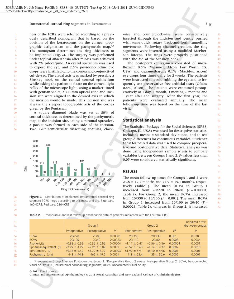
JOBNAME: No Job Name PAGE: 3 SESS: 10 OUTPUT: Tue Sep 20 18:05:41 2011 SUM: 98D0FE63/v2503/blackwell/journals/ceo_v0_i0_new_style/ceo_2698
ness of the ICRS were selected according to a previ-ously described nomogram that is based on theposition of the keratoconus on the cornea, topo-graphic astigmatism and the pachymetric map.4,5
The nomogram determines the ring thickness tobe implanted (Fig. 2). The surgery was performedunder topical anaesthesia after miosis was achievedwith 2% pilocarpine. An eyelid speculum was usedto expose the eye, and 2.5% povidone-iodine eyedrops were instilled onto the cornea and conjunctivalcul-de-sac. The visual axis was marked by pressing aSinskey hook on the central corneal epitheliumwhile asking the patient to fixate on the corneal lightreflex of the microscope light. Using a marker tintedwith gentian violet, a 5.0-mm optical zone and inci-sion site were aligned to the desired axis in whichthe incision would be made. This incision site wasalways the steepest topographic axis of the corneagiven by the Pentacam.
A square diamond blade was set at 80% ofcorneal thickness as determined by the pachymetricmap at the incision site. Using a ‘stromal spreader’,a pocket was formed in each side of the incision.Two 270° semicircular dissecting spatulas, clock-
wise and counterclockwise, were consecutivelyinserted through the incision and gently pushedwith some quick, rotary ‘back-and-forth’ tunnellingmovements. Following channel creation, the ringsegments were inserted using a modified McPher-son forceps. The rings were properly positionedwith the aid of the Sinskey hook.
The postoperative regimen consisted of moxi-floxacin 0.5% (Vigamox, Alcon, Fort Worth, TX,USA) and dexamethasone 0.1% (Maxidex, Alcon)eye drops four times daily for 2 weeks. The patientswere instructed to avoid rubbing the eye and to fre-quently use preservative-free artificial tears (Oftane0.4%, Alcon). The patients were examined postop-eratively at 1 day, 1 month, 3 months, 6 months and1 year after the surgery. After the first year, thepatients were evaluated annually. The meanfollow-up time was based on the time of the lastvisit.
Statistical analysis
The Statistical Package for the Social Sciences (SPSS,Chicago, IL, USA) was used for descriptive statistics,including means � standard deviations, and to testgroup differences for continuous variables. Student’st-test for paired data was used to compare preopera-tive and postoperative data. Statistical analysis wasdone using independent sample t-tests to comparevariables between Groups 1 and 2. P-values less than0.05 were considered statistically significant.
RESULTS
The mean follow-up times for Groups 1 and 2 were23.8 � 12.2 months and 22.9 � 15.1 months, respec-tively (Table 1). The mean UCVA in Group 1increased from 20/220 to 20/80 (P = 0.00001,Table 2). For Group 2, the mean UCVA increasedfrom 20/350 to 20/130 (P = 0.001). The mean BCVAin Group 1 increased from 20/100 to 20/40 (P =0.00023, Table 2), whereas in Group 2, it increased
150
200
250
150–
150
150–
200
150–
250
200–
200
200–
250
250–
250
250–
300
300–
300
Ring thickness (micra)
113 119
350
300
250
200
150
n
100
50
0
289
20
60
2156 56 63
1 543
96131
Figure 2. Distribution of implanted instrastromal corneal ringsegment (ICRS) rings according to thickness and arc. Blue bars,160–ICRS; Red bars, 210–ICRS.
Table 2. Preoperative and last follow-up examination data of patients implanted with the Ferrrara ICRS
Group 1 Group 2Unpaired t-test
(between groups)
Preoperative Postoperative P† Preoperative Postoperative P‡ P
UCVA 20/220 20/80 0.00001 20/350 20/130 0.001 0.038BCVA 20/100 20/40 0.00023 20/110 20/60 0.0003 0.0034Asphericity -0.88 � 0.52 -0.35 � 0.55 0.00004 -1.17 � 0.47 -0.56 � 0.56 0.00004 0.0031Spherical equivalent (D) -3.99 � 4.22 -2.26 � 3.09 0.0002 -8.52 � 5.63 -4.14 � 4.37 0.0002 0.0010Keratometry (D) 49.18 � 4.42 45.72 � 3.72 0.00003 51.92 � 5.91 48.10 � 4.96 0.0001 0.0001Pachymetry (mm) 448 � 44.8 465 � 49.2 0.0001 418 � 53.4 435 � 56.6 0.0002 0.0001
†Preoperative Group 1 versus Postoperative Group 1. ‡Preoperative Group 2 versus Postoperative Group 2. BCVA, best-correctedvisual acuity; ICRS, intrastromal corneal ring segments; UCVA, uncorrected visual acuity.
1
2
3
4
5
6
7
8
9
10
11
12
13
14
15
16
17
18
19
20
21
22
23
24
25
262728
29
30
3132
33
34353637383940
4142
43
44
45
46
47
48
49
50
51
52
53
54
55
56
57
58
59
60
61
62
63
64
65
66
67
68
69
70
71
72
73
74
75
76
77
78
79
80
81
82
Intrastromal corneal ring segments in keratoconus 3
© 2011 The AuthorsClinical and Experimental Ophthalmology © 2011 Royal Australian and New Zealand College of Ophthalmologists
m101
colour
m101
New Stamp1

JOBNAME: No Job Name PAGE: 4 SESS: 10 OUTPUT: Tue Sep 20 18:05:41 2011 SUM: 732A7E57/v2503/blackwell/journals/ceo_v0_i0_new_style/ceo_2698
from 20/110 to 20/60 (P = 0.0003). Asphericitychanged in Group 1 from -0.88 to -0.35 (P = 0.001)and from -1.17 to -0.56 in Group 2 (P = 0.000).
For Group 1 patients, the preoperative sphericalequivalent, -3.99 D, was reduced to -2.26 D at thelast postoperative examination and the keratometrydecreased from 49.18 to 45.72 D (both P < 0.001,Table 2). Simultaneously, the pachymetry at the thin-nest point increased from 448 to 465 mm (P < 0.001).For Group 2 patients, the spherical equivalentdecreased from -8.52 to -4.14 D and the keratometricvalues decreased from 51.92 to 48.10 D (bothP < 0.001, Table 2). Simultaneously, the pachymetryat the thinnest point increased from 418 to 435 mm(P < 0.001).
The mean keratometry values decreased betweenthe preoperative and postoperative periods (Fig. 3),and the thicker rings induced larger reductions. The
210-ICRS caused larger changes in mean keratom-etry than did the 160-ICRS (Table 2). Asphericityvalues changed between the preoperative and post-operative periods (Fig. 4), and the thicker the ring,the larger the asphericity change. Also, the 210-ICRScaused larger changes in asphericity than the 160-ICRS did (Table 2).
Patients in Group 1 had better preoperative andpostoperative UCVAs and BCVAs than patients inGroup 2 (Table 2). The changes in keratometry,asphericity, spherical equivalent and pachymetrywere larger in Group 2 than in Group 1 (Table 2).Regarding lines gain/loss, 81% of patients of Group1 gained at least two lines of BCVA. In Group 2, 49%of patients gained at least two lines of BCVA.(Figs 5,6).
Complications
The complication rate after Ferrara ICRS implan-tation was low, 3.82% (Table 3). The main com-
54.5
43.5
32.5
21.5
10.5
015–160
0.78
2.65
1.82
3.79
2.74
4.52
15–210 20–160 20–210 25–160 25–210
Figure 3. Effect of ring thickness on mean keratometry. Themean decrease in keratometry from preoperative to postopera-tive values at the last follow-up visit was greater for thicker rings.Blue bars, 160–instrastromal corneal ring segments (ICRS); Redbars, 210–ICRS. The numbers 15, 20 and 25 on the bottom of thechart, before the numbers 160 and 210, refer to ring thickness:15 = 150 micra, 20 = 200 micra and 250 = 250 micra.
0–0.1–0.2–0.3–0.4–0.5–0.6–0.7–0.8–0.9
–0.07
–0.36–0.31
–0.6
–0.34
–0.82
15–160 15–210 20–160 20–210 25–160 25–210
Figure 4. Effect of ring thickness on mean asphericity. Themean decrease in asphericity from preoperative to postoperativevalues at the last follow-up visit was greater for thicker rings. Bluebars, 160 – instrastromal corneal ring segments (ICRS); Red bars,210 – ICRS and thickness implanted. The numbers15, 20 and 25on the bottom of the chart, before the numbers 160 and 210,refer to ring thickness: 15 = 150 micra, 20 = 200 micra and250 = 250 micra.
>–4
0.9
30.0
25.0
20.0
15.0
10.0
5.0
0.0
1.6 1.8
14.716.3 16.1
11.8 12.5
24.3
–3 –2 –1 0 1 2 3 4 >4
Figure 5. Best-corrected visual acuity lines gain/lost inGroup 1 (%).
25
20
15
10
5
0>–4 –3
4.4 4.4 4.4
17.8 17.8
20.0
11.1
6.7
13.3
–2 –1 0 1 2 3 4 >4
Figure 6. Best-corrected visual acuity lines gain/lost inGroup 2 (%).
1234567
8
910111213141516
17
18
19
20
21
22
23
24
25
26
27
28
29
30
31
32
33
34
35
3637
38
3940
41
42
43
44
45
46
47
48
49
50
51
52
53
54
55
56
57
58
59
60
61
4 Ferrara et al.
© 2011 The AuthorsClinical and Experimental Ophthalmology © 2011 Royal Australian and New Zealand College of Ophthalmologists
11 11
12 12
m101
colour
m101
colour
m101
colour
m101
colour

JOBNAME: No Job Name PAGE: 5 SESS: 11 OUTPUT: Tue Sep 20 18:05:41 2011 SUM: AB41F193/v2503/blackwell/journals/ceo_v0_i0_new_style/ceo_2698
plication, 16 cases, was undercorrection, requiringimplantation of an additional segment. One eye eachof 37 patients (34 in Group 1 and 3 in Group 2)underwent follow-up surgery (Table 4) to remove(n = 6), exchange (n = 11), reposition (n = 4) or insertan additional ICRS (n = 16). For those patients, therewere significant improvement between the preop-erative values and the final follow-up values forUCVA, BCVA, keratometry and pachymetry. Asphe-ricity and spherical equivalent for these patients didnot improve significantly.
DISCUSSION
Modern treatment of keratoconus and keratectasiaincludes the implantation of ICRS that can effec-tively reduce corneal steepening and improve UCVAand BCVA. The Ferrara ring nomogram requires thekeratoconus type, oval, bowtie or nipple, to deter-mine the arc segment, 160° or 210°, which willbe implanted. Longer arc ring segments providemore keratometry reduction and less astigmatismreduction. In the nipple type of keratoconus, Group2 in this study, the cornea is usually very steepwith relatively low astigmatism. Therefore, for this
type of keratoconus, the 210° ring segments are themost appropriate. They provide significant flat-tening without a large concomitant induction ofastigmatism.3
Our postoperative results showed a significantimprovement in UCVA and BCVA. These results arein concordance with most similar papers;1,2,4,17–19
however, this is the first study to describe the clinicaloutcomes in a large sample of consecutive surgicalpatients. To the best of our knowledge, this study hasthe largest sample of patients implanted with ICRSever published. Our data reinforce the reproducibil-ity and efficacy of the technique.20,21
Miranda et al. obtained a significant reduction inthe postoperative central corneal curvature, and theBCVA and UCVA improved in 87.1 and 80.6% of theeyes, respectively.22 Siganos et al. showed an increaseof the UCVA from 20/285 preoperatively to 20/100and 20/60 after 1 and 6 months, respectively.2 TheBCVA improved from 20/55 preoperatively to 20/40and 20/33 after 1 and 6 months, respectively. Kwitkoand Severo reported that after implantation ofFerrara rings in keratoconus eyes, the BCVAimproved in 86.4% of eyes, was unchanged in 1.9%and worsened in 11.7%.3 The UCVA improved in86.4% of eyes, was unchanged in 7.8% and wors-ened in 5.8%. The mean corneal curvature wasreduced from 48.76 D to 43.17 D.
When comparing our results with studies usingother types of ICRS (e.g. Intacs and Keraring), wefound similar outcomes. Alio’ et al. performed a ret-rospective study to evaluate the long-term (up to48 months) results after Intacs implantation inpatients with keratoconus.23 After 6 months, themean UCVA increased significantly (P < 0.01) from0.46 (20/50) preoperatively to 0.66 (20/30), and theaverage keratometry decreased by 3.13 D. Coskun-seven et al. evaluated the results Keraring ICRS in 50eyes of patients with keratoconus.20 Of these, 47 hadUCVA of 20/40 (range: counting fingers to 20/30). Atthe last follow-up examination, 14 of the 50 eyes hada UCVA of 20/40 or better (range: counting fingers to20/25). Nine eyes maintained the preoperativeBCVA, whereas 39 eyes experienced a BCVA gain ofone to four lines.
We found a significant increase in corneal thick-ness in both groups. In theory, this can be explainedby corneal collagen remodelling induced by theimplantation of the ICRS.24,25 By acting as ‘spacers’,the ring segments could interfere with corneal col-lagen turnover, with consequent increases in thecorneal pachymetry.
We found a significant decrease in asphericityvalues after implantation of the ICRS. The postop-erative value was -0.35 for Group 1 and -0.56 forGroup 2. Most studies agree that human corneaasphericity values range from -0.01 to -0.80.26–28
Table 3. Complication rate after ICRS implantation
Complication Treatment Eyes (%)
Undercorrection Implantation of additionalsegment
16 (1.49)
Overcorrection Segment removal andreimplantation
11 (1.02)
Extrusion Segment removal 6 (0.56)Malposition Segment repositioning 4 (0.37)Progressive corneal
steepeningKeratoplasty 2 (0.18)
Ring neovascularization Bevacizumab 2 (0.18)Total 41 (3.82)
ICRS, intrastromal corneal ring segments.
Table 4. Preoperative and last follow-up examination data ofpatients who underwent follow-up surgery for removal,exchange or additional ICRS implantation
Preoperative Postoperative P
UCVA 20/300 20/80 0.005BCVA 20/160 20/50 0.0002Asphericity -0.84 � 0.74 -0.35 � 0.81 0.15Spherical
equivalent (D)-4.64 � 4.87 -3.04 � 3.45 0.137
Keratometry (D) 49.33 � 4.19 46.16 � 3.90 0.0001Pachymetry (mm) 450 � 42.9 469 � 40.8 0.0001
n = 37 eyes, 34 from Groups 1 and 3 from Group 2. BCVA,best-corrected visual acuity; ICRS, intrastromal corneal ring seg-ments; UCVA, uncorrected visual acuity.
Intrastromal corneal ring segments in keratoconus 5
© 2011 The AuthorsClinical and Experimental Ophthalmology © 2011 Royal Australian and New Zealand College of Ophthalmologists

JOBNAME: No Job Name PAGE: 6 SESS: 11 OUTPUT: Tue Sep 20 18:05:41 2011 SUM: CC9310F9/v2503/blackwell/journals/ceo_v0_i0_new_style/ceo_2698
Currently, the most commonly accepted value in ayoung adult population is approximately -0.23.29
The asphericity can be considered as one of markersof visual quality. Thus, returning it closer to ‘normal’or at least reducing the excess prolateness usuallyfound in keratoconus could be a predictor ofimproved visual quality.
For all the measured parameters, the results werebetter for Group 1 than Group 2. The type of kerato-conus can explain the differences. The Group 2patients had the nipple-type keratoconus that tendsto be more aggressive and respond to the ‘conven-tional’ 160° ring segments with less efficacy than theoval-type keratoconus. Nipple-type keratoconus isbetter treated with long-arc ring segments, such asthe 210° ring segments. Given the same thickness ofICRS, the 210° ring segments can provide greaterchanges in keratometry and asphericity than the 160°ring segments. The efficacy and safety of the 210°ring has been demonstrated.3
The incidence of complications found in this studywas extremely low. This can be explained bytwo factors: (i) mastery of the technique; and (ii)nomogram evolution. After mastering the surgicaltechnique, especially the deep incision and thewell-constructed intrastromal tunnel, the technique-related complications become very infrequent. Thenomogram has evolved based on the knowledge thatthinner segments achieve the same or better resultsthan the thicker segments used in the past.4,22 Insome cases, an undercorrection or overcorrection wasfound; the cause for these changes are not wellunderstood but probably are related to cornealbiomechanics. The reason for insertion of additionalICRS was usually undercorrection, that is, a subop-timal reduction of corneal steepening after implanta-tion of a single ICRS. One of the most fearedcomplications of ICRS, ring extrusion, is now rarebecause the 350-mm thick rings are no longerimplanted. The pachymetry at the ring track must beat least double the ring thickness to be implanted.
We showed that the outcome of patients requir-ing follow-up surgery because of overcorrectionor undercorrection, 3.4%, is acceptable. For thesepatients, there was improvement of UCVA, BCVA,keratometry and pachymetry. However, asphericityand spherical equivalent did not improve in thesepatients undergoing subsequent surgery, perhapsbecause of the scarring of corneal tissue and/orstroma secondary to the first procedure.
Kwitko and Severo reported Ferrara ICRS decen-tration in 3.9% of cases, segment extrusion in 19.6%and bacterial keratitis in 1.9%.2 As the authors men-tioned in their paper, the surgeon’s learning curveand different healing processes in keratoconiccorneas can cause the majority of complicationsrelated to the surgical technique. Once the surgical
procedure is mastered, the complications rate relatedto the surgery itself is very low, as demonstrated inthe present study. To avoid surgery-related compli-cations, the steps must be followed carefully, includ-ing constructing the stromal tunnel with theadjustable diamond knife set at 80% of local cornealthickness. This reduces the chance of a shallowtunnel and subsequent ring extrusion.
Extrusion of the ICRS usually occurs in patientswith little stroma, overlying the implanted segmentsand when the ring is located close to the incision. Asa general rule, it must be assumed that the thickestportion of a pair of segments in the stromal bedcannot exceed half the thickness of the cornea. If thedesired ICRS thickness exceeds half the thickness ofthe cornea, then a thinner diameter ICRS must beselected even if the correction is likely to be smallerthan desired. This should be considered as the‘pachymetry law’ for ICRS implantation. Since thisrule began to be followed, the incidence of extrusionhas decreased significantly.30
Kubaloglu et al. evaluated the clinical outcomes ofkeratoconus patients that had ICRS implantation inwhich the intrastromal tunnel was created manually,as we did in our study, and by femtosecond laser.31
After 1 year, there was significant improvement inUCVA, BCVA, keratometry, spherical equivalent,manifest sphere and cylinder in both groups. Impor-tantly, there were no significant differences betweenthe two groups regarding the visual or refractiveresults. After mastering the manual technique, theincidence of perioperative complications is extremelylow, and this technique is both very safe and effective.
In conclusion, Ferrara ICRS implantation is aneffective treatment for keratoconus and keratectasia.The procedure is minimally invasive and yields goodvisual, refractive and keratometric outcomes. More-over, it is a safe technique and does not preclude anyfuture additional treatment if necessary.
REFERENCES
1. Ferrara de A, Cunha P. Técnica cirúrgica para correçãode miopia; Anel corneano intra-estromal. (Myopia cor-rection with intrastromal corneal ring). Rev Bras Oftal-mol 1995; 54: 577–88.
2. Siganos D, Ferrara P, Chatzinikolas K et al. Ferraraintrastromal corneal rings for the correction ofkeratoconus. J Cataract Refract Surg 2002; 28: 1947–51.
3. Kwitko S, Severo N. Ferrara intracorneal ring seg-ments for keratoconus. J Cataract Refract Surg 2004; 30:812–20.
4. Ferrara P, Torquetti L. Clinical outcomes after implan-tation of a new intrastromal ring with a 210-degree ofarch. J Cataract Refract Surg 2009; 35: 1604–8.
5. Torquetti L, Ferrara P. Long term follow-up of intras-tromal corneal ring segments in keratoconus. J CataractRefract Surg 2009; 35: 1768–73.
2 2
3 3
44
55
6 Ferrara et al.
© 2011 The AuthorsClinical and Experimental Ophthalmology © 2011 Royal Australian and New Zealand College of Ophthalmologists

JOBNAME: No Job Name PAGE: 7 SESS: 10 OUTPUT: Tue Sep 20 18:05:41 2011 SUM: 92A5F427/v2503/blackwell/journals/ceo_v0_i0_new_style/ceo_2698
6. Ferrara P, Torquetti L. Ferrara ring. An overview. Cata-ract Refract Surg Today Eur 2009; ••: 27–30.
7. Rodrigues-Prats J, Galal A, Garcia-Lledo M et al. Intra-corneal rings for the correction of pellucid marginaldegeneration. J Cataract Refract Surg 2003; 29: 1421–4.
8. Siganos CS, Kymionis GD, Astyrakakis N et al. Man-agement of corneal ectasia after laser in situ keratom-ileusis with INTACS. J Refract Surg 2002; 18: 43–6.
9. Silva FBD, Alves EAF, Cunha PFA. Utilização do Anelde Ferrara na estabilização e correção da ectasia cor-neana pós PRK. Arq Bras Oftalmol 2000; 63: 215–18.
10. Nose W, Neves RA, Burris TE et al. Intrastromalcorneal ring: 12-month sighted myopic eyes. J RefractSurg 1996; 12: 20–8.
11. Schanzlin DJ, Asbell PA, Burris TE, Durrie DS. Theintrastromal ring segments; phase II results for thecorrection of myopia. Ophthalmology 1997; 104: 1067–78.
12. Ruckhofer J, Stoiber J, Twa MD et al. Correction ofastigmatism with short arc-length intrastromal cornealring segments: preliminary results. Ophthalmology2003; 110: 516–24.
13. Barraquer JI. Queratoplastia refractiva, estudios einformaciones. Oftalmologicas 1949; 2: 10–30.
14. Barraquer JI. Modification of refraction by means ofintracorneal inclusion. Int Ophthalmol Clin 1966; 6:53–78.
15. Bogan SJ, Waring GO III, Ibrahim O et al. Classifica-tion of normal corneal topography based on computer-assisted videokeratography. Arch Ophthalmol 1990; 108:945–9.
16. Rabinowitz YS, McDonnell PJ. Computer-assistedcorneal topography in keratoconus. Refract Corneal Surg1989; 5: 400–8.
17. Colin J, Cochener B, Savary G, Malet F. Correctingkeratoconus with intracorneal rings. J Cataract RefractSurg 2000; 26: 1117–22.
18. Colin J, Cochener B, Savary G et al. INTACS inserts fortreating keratoconus; one-year results. Ophthalmology2001; 108: 1409–14.
19. Rodriguez-Prats J, Galal A, Garcia-Lledo M et al. Intra-corneal rings for the correction of pellucid marginaldegeneration. J Cataract Refract Surg 2003; 29: 1421–4.
20. Coskunseven E, Kymionis GD, Tsiklis NS et al. One-year results of intrastromal corneal ring segmentimplantation (KeraRing) using femtosecond laser in
patients with keratoconus. Am J Ophthalmol 2008; 145:775–9.
21. Zare MA, Hashemi H, Salari MR. Intracorneal ringsegment implantation for the management of kerato-conus: safety and efficacy. J Cataract Refract Surg 2007;33: 1886–91.
22. Miranda D, Sartori M, Francesconi C et al. Ferraraintrastromal corneal ring segments for severekeratoconus. J Refract Surg 2003; 19: 645–53.
23. Alio’ JI, Shabayek MH, Artola A. Intracorneal ringsegments for keratoconus correction: long-termfollow-up. J Cataract Refract Surg 2006; 32: 978–85.
24. Flynn BP, Bhole AP, Saeidi N et al. Mechanical strainstabilizes reconstituted collagen fibrils against enzy-matic degradation by mammalian collagenase matrixmetalloproteinase 8 (MMP-8). PLoS ONE 2010; 5:e12337.
25. Mackiewicz Z, Määttä M, Stenman M et al. Col-lagenolytic proteinases in keratoconus. Cornea 2006;25: 603–10.
26. Davis WR, Raasch TW, Mitchell GL et al. Cornealasphericity and apical curvature in children: a cross-sectional and longitudinal evaluation. Invest OphthalmolVis Sci 2005; 46: 1899–906.
27. Holmes-Higgin DK, Baker PC, Burris TE, SilvestriniTA. Characterization of the aspheric corneal surfacewith intrastromal corneal ring segments. J Refract Surg1999; 15: 520–8.
28. Eghbali F, Yeung KK, Maloney RK. Topographicdetermination of corneal asphericity and its lack ofeffect on the refractive outcome of radial keratotomy.Am J Ophthalmol 1995; ••: 275–80.
29. Yebra-Pimentel E, González-Méijome JM, Cerviño Aet al. Asfericidad corneal en una poblácion de adultosjóvenes. Implicaciones clínicas. Arch Soc Esp Oftalmol2004; 79: 385–92.
30. Torquetti L, Ferrara P. Reasons for intrastromal cornealring segment explantation. J Cataract Refract Surg 2010;36: 2014; author reply 2014–15.
31. Kubaloglu A, Sari ES, Cinar Y et al. Comparison ofmechanical and femtosecond laser tunnel creation forintrastromal corneal ring segment implantation inkeratoconus: prospective randomized clinical trial.J Cataract Refract Surg 2010; 36: 1556–61.
32. Shetty R, Kurian M, Anand D et al. Intacs in advancedkeratoconus. Cornea 2008; 27: 1022–9.
123456789
10111213141516171819202122232425262728293031323334353637383940414243444546
47484950515253545556575859606162636465666768697071727374757677787980818283848586878889909192
6 6
7 7
88
99
1010
Intrastromal corneal ring segments in keratoconus 7
© 2011 The AuthorsClinical and Experimental Ophthalmology © 2011 Royal Australian and New Zealand College of Ophthalmologists

JOBNAME: No Job Name PAGE: 8 SESS: 10 OUTPUT: Tue Sep 20 18:05:41 2011 SUM: 50830B40/v2503/blackwell/journals/ceo_v0_i0_new_style/ceo_2698
Toppan Best-set Premedia LimitedJournal Code: CEO Proofreader: JasonArticle No: 2698 Delivery date: 20 September 2011Page Extent: 7 Copyeditor: Cindy
AUTHOR QUERY FORM
Dear Author,During the preparation of your manuscript for publication, the questions listed below have arisen. Please attend to
these matters and return this form with your proof.Many thanks for your assistance.
QueryReferences
Query Remark
q1 AUTHOR: Please shorten the running head to no more than 40 charactersincluding spaces, to follow journal style.
q2 AUTHOR: ‘corenal’ has been changed to ‘corneal’. Please check and confirmthat this is correct.
q3 AUTHOR: ‘Kwitiko et al.’ has bee changed to ‘Kwitko and Severo’ so that thiscitation matches the entry in the Reference List. Please check and confirm thatthis is correct.
q4 AUTHOR: Should ‘complications rate related’ be changed to ‘complicationrates related’?
q5 AUTHOR: ‘Kubalgodlu et al.’ has been changed to ‘Kubaloglu et al.’ so thatthis citation matches the entry in the Reference List. Please check and confirmthat this is correct.
q6 AUTHOR: Please check and confirm if the journal title is abbreviated correctlyin Reference 6. Also, please supply the volume number for this reference.
q7 AUTHOR: Please check and confirm if the journal title in Reference 13 iscorrect.
q8 AUTHOR: If Reference 24 is not a one-page article, please supply the first andlast pages for this article.
q9 AUTHOR: Please supply the volume number for Reference 28.
q10 AUTHOR: Reference 32 in the original file has not been cited in the text.Please indicate where it should be cited; or delete it from the Reference List.
q11 AUTHOR: In the figures, commas that function as decimal points have beenchanged to full points. Please check and confirm if the changes made arecorrect.
q12 AUTHOR: Please check and confirm if all instances of the unit micra shouldbe changed to mm.
q13 AUTHOR: Please provide a heading for the first column of Tables 1, 2 and 4.

ARTICLE
Intrastromal corn
eal ring segmentimplantation to correct astigmatismafter penetrating keratoplastySandro Coscarelli, MD, Guilherme Ferrara, MD, Jose F. Alfonso, MD, PhD, Paulo Ferrara, MD, PhD,
Jes�us Merayo-Lloves, MD, PhD, Luana P.N. Ara�ujo, MD, Aydano P. Machado, PhD,Jo~ao Marcelo Lyra, MD, PhD, Leonardo Torquetti, MD, PhD
Q
P
1006
2012 Aublished
PURPOSE: To evaluate the clinical outcomes of implantation of Ferrara intrastromal corneal ringsegments (ICRS) in patients with astigmatism after penetrating keratoplasty (PKP).
SETTING: Private clinic, Belo Horizonte, Brazil.
DESIGN: Retrospective consecutive case series.
METHODS: Chart records of post-PKP patients who had ICRS implantation from May 2005 toSeptember 2009 were retrospectively reviewed. The following parameters were studied:corrected distance visual acuity (CDVA), keratometry (K) values, spherical equivalent (SE),spherical refractive error, corneal topographic astigmatism, minimum K, and maximum K.
RESULTS: The study evaluated 59 eyes (54 patients). The mean CDVA (logMAR) improved from0.45 G 0.17 (SD) (range 0.18 to 1.00) to 0.30 G 0.17 (range 0.00 to 1.00). The mean SE was�6.34 G 3.40 diopters (D) (range 0.37 to �16.50 D) preoperatively and �2.66 G 2.52 D (range0.87 to �10.50 D) postoperatively. The mean spherical refractive error decreased from �7.10 G3.07 D (range 2.15 to 16.68 D) preoperatively to �3.46G 2.05 D (range 0.88 to 10.79 D) postop-eratively. No patient lost visual acuity. The mean corneal topographic astigmatism decreasedfrom 3.37 G 1.51 D preoperatively to 1.69 G 1.04 D postoperatively. The mean maximumK value decreased from 48.09 G 2.56 D to 44.17 G 2.67 D and the mean minimum K value,from 44.90 G 2.54 D to 42.46 G 2.63 D. All changes were statistically significant (P<.0001).
CONCLUSION: Intrastromal corneal ring segments effectively reduced corneal cylinder in patientswith astigmatism after PKP.
Financial Disclosure: Drs. Ferrara and Merayo-Lloves have proprietary interest in the Ferrara ring.Drs. Coscarelli, Torquetti, and Alfonso have no financial or proprietary interest in any material ormethod mentioned.
J Cataract Refract Surg 2012; 38:1006–1013 Q 2012 ASCRS and ESCRS
The postoperative astigmatism associated with pene-trating keratoplasty (PKP) is a common condition inclinical practice. The reason for this could be relatedto factors inherent to the receptor, such as previouscorneal trauma or keratoconus.1 Additional contrib-uting factors may include the trephination technique,inadequate fixation of the eye during surgery withcompression or deformation of the ocular globe,the suture technique, and postoperative issues suchas the patient’s age and receptor corneal disease,time on topical corticosteroids, and early sutureremoval.2
SCRS and ESCRS
by Elsevier Inc.
Previous studies2–4 report several risk factors thatmay increase the incidence of post-PKP astigmatismand its management. Contact lenses are a better choicethan spectacles to correct astigmatism because theyprovide better quality of vision.5,6 Moreover, thecorrected distance visual acuity (CDVA) with contactlenses is usually better than with spectacles in thistype of astigmatism. When optical methods fail toachieve satisfactory visual rehabilitation, surgicaltreatment may be necessary. Whereas some authorshave published that incisions and wedge resection ofthe cornea could be useful to correct the
0886-3350/$ - see front matter
doi:10.1016/j.jcrs.2011.12.037

1007ICRS FOR ASTIGMATISM CORRECTION AFTER PKP
astigmatism,7–9 other authors report that laser in situkeratomileusis (LASIK)10,11 or the implantation oftoric phakic intraocular lenses (pIOLs) could achievebetter and more predictable results.12,13
In the present study, we evaluated the use of intra-stromal corneal ring segments (ICRS) as an alternativesurgical option for the treatment of astigmatism inpatients who had PKP for keratoconus, bullous kerat-opathy, radial keratotomy (RK), post-LASIK ectasia,or stromal scarring. The outcome analysis comprisedthe CDVA, spherical equivalent (SE), refractive error,corneal topographic astigmatism, and minimum andmaximum keratometry (K) values. To our knowledge,this study has the largest sample of patients with ICRSimplantation to correct post-PKP astigmatism in theliterature.
PATIENTS AND METHODS
This retrospective study included consecutive post-PKPpatients who had ICRS implantation (Ferrara ring, Ferrarae Hijos) fromMay 2005 to September 2009 to correct residualastigmatism. All patients were informed about the possibleintraoperative and postoperative complications and gavewritten informed consent in accordance with institutionalguidelines and the Declaration of Helsinki.
Inclusion criteria were a clear and transparent cornealgraft, a minimum of 2.50 diopters (D) and a maximum of8.00 D of astigmatism, and contact lens intolerance. All pa-tients had at least 2 years of follow-up after PKP beforeICRS implantation. Patients who did not meet the inclusioncriteria were not evaluated in this study.
Surgical Technique
The same surgeon (S.C.) performed all surgeries usinga manual technique as previously described.14 The surgerywas performed using topical anesthesia after miosis wasachieved with pilocarpine 2.0%. The visual axis was markedby pressing a Sinskey hook on the central corneal epitheliumwhile asking the patient to fixate on the corneal light reflex ofthe microscope light. Using a marker tinted with gentianviolet, a 5.0 mm optical zone and incision site were alignedto the desired axis in which the incision would be made.This incision site was always at the steepest topographicaxis of the cornea given by the topographer.
Submitted: May 23, 2011.Final revision submitted: November 18, 2011.Accepted: December 5, 2011.
From Ennio Coscarelli Eye Clinic (Coscarelli), Paulo Ferrara EyeClinic (G. Ferrara, P. Ferrara, Torquetti), Belo Horizonte andBrazilian Study Group of Artificial Intelligence and Cornea (Ara�ujo,Machado, Lyra, Torquetti), Macei�o, Brazil; Fernandez-Vega EyeInstitute (Alfonso, Merayo-Lloves), Oviedo, Spain.
Corresponding author: Guilherme Ferrara, MD, Avenida Brasil,1312 – Santa Efigenia – Belo Horizonte, MG 30140-001, Brazil.E-mail: [email protected].
J CATARACT REFRACT SURG
A square diamond blade was set at 80% of corneal thick-ness at the incision site, and this blade was used to makethe incision. A pocket was formed in each side of the incisionusing a stromal spreader. Two (clockwise and counterclock-wise) 270-degree semicircular dissecting spatulas wereconsecutively inserted through the incision and gentlypushed with quick rotary back-and-forth tunneling move-ments. After channel creation, the ring segments wereinserted using a modified McPherson forceps. The ringswere properly positioned with the aid of a Sinskey hook.
Postoperative Regimen and Assessment
The postoperative regimen consisted of tobramycin 0.3%and dexamethasone 0.1% eyedrops 4 times a day for1 week, after which the dose was tapered over 3 weeks. Inaddition, patients received topical lubricants 4 times a dayfor at least 3 months.
Postoperative examinations were performed at 1 and7 days, after 1 and 6 months, and then every year. Measure-ment of CDVA, slitlamp evaluation, refraction, cornealtopography, fundoscopy, and tonometry were performedat each control visit. Visual acuity was determined ona Snellen chart and then converted to logMAR notation.The K values were obtained by corneal topography(CT4000 Corneal Topographer, Eyetech, Inc.).
To evaluate the CDVA, refractive error, and corneal topo-graphic astigmatism, the nonparametric Mann-Whitney testwas used because at least 1 datum from the sample did nothave aGaussian distribution. The SEwas corrected byWelchtransformation because of significant difference between2 standard deviations (SDs).
Analysis of Astigmatism
The astigmatism results were analyzed arithmetically(nonvector analysis) and with regard to the cylindrical axisusing vector analysis. Although empirical changes in cylin-ders are commonly reported, they do not accurately reflectthe true nature of the change in cylinder. Cylinders havea magnitude and an axis, which are related to the sphericalpower.15 To take into account all 3 components, the data inthis study were transformed into Cartesian coordinates (ie,x and y coordinates) to allow mathematic analyses. The re-sult in the Cartesian coordinate form was then reconvertedinto polar coordinates (sphere, cylinder, axis). To distinguishthe mean value of the cylinder calculated in this manner, theterm centroid has been proposed.16
Refractions before and 12 months after ICRS insertionwere assessed for astigmatism using the power vectormethod.17 Any spherocylindrical refractive error was ex-pressed by 3 dioptric powers: M, J0, J45, with M being theaspheric lens equal to the SE of the given refractive errorand J0 and J45 being 2 Jackson cross-cylinders equivalentto the conventional cylinders. These numbers are the coordi-nates of a point in a 3-dimensional dioptric space, being thepower vector from the origin of this space to the point (M, J0,J45). Thus, the length of the vector is a measure of the overallblurring strength of the spherocylindrical refractive error.Changes in refractive error induced by the surgery werecomputed by the ordinary rules of vector extraction.
The target astigmatism is the intended astigmatic correc-tion in each individual eye. The ideal target astigmatismwas zero (ie, intention to correct the full magnitude of thecylinder).
- VOL 38, JUNE 2012

1008 ICRS FOR ASTIGMATISM CORRECTION AFTER PKP
Statistical Analysis
Data reported here are from the 12-month examinationafter ICRS implantation. Statistical analysis included theStudent t test, Welch transformation, and Mann-Whitneynonparametric test and was performed using Instat for Mac-intosh software (version 3.1a, Graphpad Software). Vectorialanalysis was performed using SigmaPlot software (SPSSInc.). Internet-Based Refractive Analysis software (ZubisoftGmbH) was used for clinical outcomes analysis.
Figure 1. The CDVA (logMAR) before and after the ICRS implanta-tion (unpaired t test) (CDVA Z corrected distance visual acuity).
RESULTS
This study included 59 eyes of 54 patients. The meanage of the 28 women (51.85%) and 26 men (48.14%)was 36.01 years G 11.02 (SD) (range 19 to 72 years).The indications for PKP were keratoconus in 49 eyes,post-LASIK ectasia in 5 eyes, progressive hyperopiasecondary to RK in 2 eyes, stromal scarring in 2 eyes,and bullous keratopathy in 1 eye. Forty-nine patientshad a single eye treatment, whereas 5 patients hadboth eyes treated.
All patients completed at least 1 year of follow-up.The mean follow-up was 14 months.
Table 1 shows the preoperative and last follow-upexamination data. The preoperative corneal astigma-tism ranged from 3.00 to 5.00 D. Figures 1 and 2show the preoperative and postoperative CDVA. Nopatient lost visual acuity. Of the patients, 28 (47.4%)gained 2 or more lines of CDVA (Figure 3). The
Table 1. Preoperative and last follow-up examination data.
Parameter Preoperative PostoperativeP
Value
Eyes (n) 59 d d
Patients (n) 54 d d
Sex (n)Male 26 d d
Female 28 d d
Mean age (y) 36.01 G 11.02 d d
Mean follow-up (mo) 14 d d
CDVA (logMAR) .001Mean G SD 0.45 G 0.17 0.30 G 0.17Range 0.18, 1.00 0.00, 1.00
SE (D) .001Mean G SD �6.34 G 3.40 �2.66 G 2.52Range 0.37, �16.50 0.87, �10.50
Spherical refractiveerror (D)
.001
Mean G SD �7.10 G 3.07 �3.46 G 2.05Range 2.15 to 16.68 0.88 to 10.79
Mean TA at 3.0 mm (D) 3.37 G 1.51 1.69 G 1.04 .001Mean maximum K (D) 48.09 G 2.56 44.17 G 2.67 .001Mean minimum K (D) 44.90 G 2.54 42.46 G 2.63 .001
CDVAZ corrected distance visual acuity; KZ keratometry; SEZ spher-ical equivalent; TA Z topographic astigmatism
J CATARACT REFRACT SURG
improvements in CDVA, SE, and refractive errorwere statistically significant (P!.0001).
Intended Correction
Regarding the predictability of the postoperative SE,43 eyes (72.8%) presented with undercorrection and9 (15.2%) with overcorrection. There was concordancebetween the attempted refraction and achieved refrac-tion in 7 eyes (13.0%) (Figure 4).
The decrease in the mean corneal topographicastigmatism at 3.0 mm from preoperatively to post-operatively was statistically significant (P!.0001).Most eyes had more than 3.0 D of refractive astigma-tism preoperatively (Figure 5). Approximately halfthe eyes remained with more than 3.00 D of ref-ractive astigmatism postoperatively; the rest hadless than 3.00 D (Figure 6). The decrease in K valuesfrom preoperatively to postoperatively was
Figure 2. Preoperative and postoperative CDVA (logMAR) (CDVAZ corrected distance visual acuity).
- VOL 38, JUNE 2012

Figure 3. Lines of CDVA lost and gained. Figure 4. Predictability of SE correction. The blue dots representundercorrection, the green dots represent full correction, and thered dots represent overcorrection (Cor.Z correlation; Coeff.Z coef-ficient; Lin. Z linear; SE Z spherical equivalent; Res. Var. Z re-sponse variable).
1009ICRS FOR ASTIGMATISM CORRECTION AFTER PKP
statistically significant (P!.0001) (Table 1). Vectorialanalysis showed that most eyes had a statisticallysignificant reduction in spherocylinder refractiveerror (Figure 7).
Double-Angle Plot
Figure 8 shows the double-angle plots of the indi-vidual cylinders, providing an overview of the cylin-der magnitude (diopter) and axis (degree) of eachdata point. The radius from the center of the plot toeach individual point represents the magnitude ofthe cylinder. After ICRS implantation, the refractiveastigmatism centroid was 1.00 D closer to zero andthe SD of the astigmatism was reduced by a factor of
Figure 5. Preoperative refractive astigmatism.
J CATARACT REFRACT SURG
1.66 (3.83 D/2.31 D). The relocation of the centroidcloser to the origin and the contraction of the ellipseon the doubled-angle plots show the amount ofimprovement.
Figure 9 shows the doubled-angle plot of the preop-erative and postoperative keratometric astigmatism.Although there was a reduction in the mean kerato-metric astigmatism, it was considerably less than thereduction in refractive astigmatism.
There were no vision-threatening complications.The ICRS were deeply implanted in all eyes(Figure 10). In 3 eyes (5%), the surgical procedurewas interrupted due to dehiscence of the inner layersof corneal transplantation, even 2 years aftertransplantation.
Figure 6. Postoperative refractive astigmatism.
- VOL 38, JUNE 2012

Figure 7. Astigmatic vectors (J0 and J45) before and 12 months afterICRS implantation. The more central location (0,0) of postoperativedata represents the reduction of preoperative astigmatism by the im-plantation of the ICRS.
Figure 8. Double-angle plot of refractive astigmatism. Individualcylinders demonstrate the cylinder magnitude (D) and axis (de-grees). The radius from the center of the plot to each individual pointrepresents the magnitude of the cylinder.
1010 ICRS FOR ASTIGMATISM CORRECTION AFTER PKP
DISCUSSION
Post-PKP residual astigmatism and refractive error arefrequently observed,11,18 and their management maybe a challenge for anterior segment surgeons. In thisretrospective study, we evaluated 59 eyes of 49 pa-tients who had ICRS implantation to correct irregularastigmatism after previous PKP. Despite properwound healing and good anatomic results, highand/or irregular astigmatism can preclude satisfac-tory vision in these patients.
Figure 9. Double-angle plot of keratometric astigmatism.
J CATARACT REFRACT SURG
Many factors inherent to the patient, host cornea,surgical technique, and postoperative managementmay influence the astigmatism.2 Peripheral disorders,such as keratoconus,1 can persist on the corneal hostand cause irregular astigmatism. This cause possiblyexplains the high prevalence of keratoconus patientsin our study. Although contact lenses and excimer la-ser refractive surgery are viable options in this groupof eyes,19–21 contact lens–related problems, such asdry eye, neovascularization, and rejection of donorcornea, must be considered,22 as well as contraindica-tions to PRK or LASIK because of high ametropia, lowbaseline corneal thickness, and young age that makethese patients unsuitable for corneal laser refractivesurgery.23
The visual outcomes in our study were satisfactory.The CDVA was unchanged in 16 eyes (27.2%)
Figure 10. Anterior segment optical coherence tomography showsthe ICRS deeply implanted in the cornea stroma and graft–host inter-face (yellow arrow).
- VOL 38, JUNE 2012

1011ICRS FOR ASTIGMATISM CORRECTION AFTER PKP
postoperatively, whereas 43 eyes (72.8%) improved atleast 1 line. The mean SE value decreased from �6.34G 3.40 D to �2.66 G 2.52 D, and the K values alsodecreased, improving the corneal irregularity.
Alfonso et al.24 and Morshirfar et al.12 evaluated theresults of pIOL implantation in young patients for thecorrection of refractive errors after PKP and foundsafe, predictable, and stable visual and refractiveoutcomes. Alfonso et al.24 describe the efficacy, pre-dictability, and safety of Implantable Collamer Lensposterior chamber pIOL implantation for the correc-tion of post-PKP refractive error in 15 eyes of 15 pa-tients; there was a large reduction in the refractiveerror and CDVA improvement. The postoperativeCDVA was 20/40 or better in 12 eyes (80%) and20/25 in 6 eyes (40%). No eye lost more than 1 lineof acuity, 2 eyes gained 1 line, and 5 eyes gainedmore than 2 lines; 8 eyes were unchanged. Morshirfaret al.12 evaluated the Artisan iris-supported pIOL totreat high myopia after PKP in 2 patients; there wasimprovement in uncorrected distance visual acuityand CDVA and no significant endothelial cell densityloss 6 months postoperatively.
Our findings are in agreement with results in otherstudies.25–27 In a case report, Coskunseven et al.25
advocate the use of ICRS, a minimally invasive proce-dure, to correct high astigmatism after PKP. Accordingto Coskunseven et al., eyeswith thin corneal grafts andrecurrent keratoconus are unsuitable for laser refrac-tive corrections because of the possibility of postoper-ative complications. In addition, Arriola-Villaloboset al.,5 in a series of 9 patients, found that ICRS implan-tation improved the CDVA in all eyes and decreasedthe topographic mean, minimum, and maximumK values. They conclude that ICRS implantationmightbe a good surgical choice to correct high astigmatismafter PKP and yields good visual, refractive, andtopographic outcomes.
Chang and Hardten27 recommend that ICRS im-plantation after PKP not be performed until at least1 year after transplantation and at least 3 months aftersuture removal. We proceeded as Arriola-Villaloboset al.5 suggest; that is, we waited at least 2 years aftercorneal transplantation and a minimum of 6 monthsafter suture removal to avoid damaging the interfaceby the traction generated by the dissectors used duringsurgery in the manual technique.
The use of Ferrara ICRS with a 5.0 mm optical zonehas 2 advantages over the use of larger optical zoneICRS. First, the central cornea flattening is theoreticallygreater because the refractive outcome is inverselyproportional to the diameter of the segment.28 Thesecond benefit is that a small diameter ensures greaterdistance between the rings and the graft–host junction.This reduces the risk for interface dehiscence or
J CATARACT REFRACT SURG
vascularization of the stromal channel by vessels ex-tending from the limbus and host cornea. Thus, in pa-tients with a corneal transplant with a diameter of7.5 mm or smaller, the ICRS with larger optical zones(6.0 or 7.0 mm) should not be used because the seg-ments would be very close to the graft–host junction.Potential disadvantages of ICRS with a small opticalzone are halos and glare. Some patients, especiallythose with large pupil diameters in dim-light condi-tions, occasionally report halos and glare.
All patients had PKP performed by the samesurgeon (S.C.), who always used a discrepancy of0.50 mm in trephination between the donor graft(8.0 mm) and the host (7.5 mm). Because the FerraraICRS is placed at a 5.0 mm optical zone, it is alwayslocated far from the graft–host junction, which makesthe procedure safer for small-optical-zone ICRS. How-ever, in cases of small trephinations or decenteredgrafts, care must be taken to avoid excessive torque,which could open the previous keratoplasty wound,thus requiring sutures and postponing ICRS im-plantation. In our study, ICRS implantation had tobe postponed in 5% of cases (3/59 eyes) becausewound dehiscence occurred during dissection. Allpatients had at least 2 years of follow-up after PKP,which indicates that in some cases the graft–host inter-face strength can be permanently reduced. This maybe related to the depth (too shallow or too deep) ofthe passage of the 10-0 needle during the keratoplastyand early removal of sutures. Tunnel creation usingthe femtosecond laser in these cases could not onlyfacilitate the surgical procedure but also prevent thistype of complication.26,29
Implantation of ICRS after PKP may yield resultsdifferent than those when the technique is used forkeratoconus treatment. Keratoconic corneas are thin-ner and more elastic than healthy corneas, whereascorneas that had PKP are more rigid, with normalthickness and elasticity. Theoretically, this couldexplain why 73% of eyes presented with undercorrec-tion and there was a low concordance between theattempted refraction and the achieved refraction. Thenomogram of the Ferrara ICRS is designed for kerato-conus treatment; thus, the ring thickness should beadjusted when using ICRS for post-keratoplasty cases.This means that thicker segments should be implantedin post-PKP patients given the same K values as inkeratoconus patients. The main purpose of ICRSimplantation is to regularize the corneal surface andimprove visual acuity; the refraction reduction afterthe surgery could be considered a secondary goal.
There are several potential advantages of ICRSimplantation over other surgical techniques in eyeswith high astigmatism after PKP. First, ICRS implanta-tion avoids excimer laser treatment, eliminating
- VOL 38, JUNE 2012

1012 ICRS FOR ASTIGMATISM CORRECTION AFTER PKP
central corneal wound healing, which could be unsat-isfactory in post-PKP corneas. This leaves the opticalcenter of the cornea untouched, enhancing refractiveoutcomes. Second, the technique is reversible in casesof unsatisfactory refractive or clinical outcomes. Third,adjustment can be performed using thinner or thickerrings. In cases of unexpected corneal shape changes,1 segment can be removed or exchanged. Fourth, itavoids the complications of intraocular surgery.
The results in our study suggested that ICRSimplantation is a promising treatment for post-PKPastigmatism, especially in eyes with thin and irregularcorneas. Long-term randomized comparative prospec-tive studies are needed to better evaluate this tech-nique as a treatment for irregular astigmatism inpost-PKP patients.
WHAT WAS KNOWN
� Surgical treatments such as wedge resection of thecornea, LASIK, and pIOL implantation are often necessaryto manage high astigmatism after PKP when nonsurgicalmethods fail to achieve satisfactory visual acuity.
� Intrastromal corneal ring segment implantation for thetreatment of post-PKP astigmatism has been describedin a case report and a 9-patient case series.
WHAT THIS PAPER ADDS
� In a larger clinical series, ICRS implantation improvedCDVA in 73% of eyes and produced significant reductionin topographic astigmatism. Dehiscence of the graft–host junction with mechanical dissection occurred in 5%of eyes.
REFERENCES1. Lim L, Pesudovs K, Goggin M, Coster DJ. Late onset post-
keratoplasty astigmatism in patients with keratoconus. Br
J Ophthalmol 2004; 88:371–376. Available at: http://www.ncbi.
nlm.nih.gov/pmc/articles/PMC1772053/pdf/bjo08800371.pdf.
Accessed January 28, 2012
2. Barraquer RI, �Alvarez de Toledo JP, �Alvarez Fisher M, Mart�ınezGrau G. Prevenci�on y tratamiento del astigmatismo en querato-
plastia penetrante. Ann Oftalmol 2002; 10:69–80. Available at:
http://www.nexusediciones.com/pdf/ao2002_2/of-10-2-003.pdf.
Accessed January 28, 2012
3. Landau D, Siganos CS, Mechoulam H, Solomon A, Frucht-
Pery J. Astigmatism after mersilene and nylon suture use for
penetrating keratoplasty. Cornea 2006; 25:691–694
4. Alvarez de Toledo J, de la Paz MF, Barraquer RI, Barraquer J.
Long-term progression of astigmatism after penetrating
keratoplasty for keratoconus; evidence of late recurrence.
Cornea 2003; 22:317–323
5. Arriola-Villalobos P, D�ıas-Valle D, G€uel JL, Iradier-Urrutia MT,
Jim�enez-Alfaro I, Cui~na-Sardi~na R, Ben�ıtez-del-Castillo JM.
J CATARACT REFRACT SURG
Intrastromal corneal ring segment implantation for high astigma-
tism after penetrating keratoplasty. J Cataract Refract Surg
2009; 35:1878–1884
6. Price FW Jr, Whitson WE, Marks RG. Progression of visual
acuity after penetrating keratoplasty. Ophthalmology 1991;
98:1177–1185
7. Burillon C, Durand L, Hachmanian KF. La resection cuneiforme,
traitement correctif des astigmatismes corneens geants [Wedge
resection, corrective treatment of giant corneal astigmatism].
J Fr Ophtalmol 1989; 12:447–453
8. Saragoussi JJ, Abenhaim A, Pouliquen Y. Resultats des
incisions transverses dans la correction chirurgicale des forts
astigmatismes post-keratoplastie [Results of transverse
incisions in surgical correction of severe post-keratoplasty
astigmatism]. J Fr Ophtalmol 1990; 13:492–499
9. Coscarelli SA, Figueiredo P, Ramos Miranda MDF. Delam-
inac‚ ~ao l�ımbica: uma nova t�ecnica para correc‚ ~ao de astigmatis-
mo p�os-transplante [Limbal delamination: a new technique for
postkeratoplasty astigmatism correction]. Arq Bras Oftalmol
2003; 66:189–192. Available at: http://www.scielo.br/pdf/abo/
v66n2/15472.pdf. Accessed January 28, 2012
10. Aguilar-Montes G, P�erez-Balbuena L, Naranjo-Tackman R.
Manejo quir�urgico con t�ecnica de lasik en la correcci�on del
astigmatismo mi�opico secundario a transplante corneal [Lasik
technique for myopic astigmatism correction after corneal
transplantation]. Rev Mex Oftalmol 2000; 74:221–236
11. Campos M, Hertzog L, Garbus J, Lee M, McDonnell PJ. Photo-
refractive keratectomy for severe postkeratoplasty astigmatism.
Am J Ophthalmol 1992; 114:429–436
12. Moshirfar M, Barsam CA, Parker JW. Implantation of an Artisan
phakic intraocular lens for the correction of high myopia after
penetrating keratoplasty. J Cataract Refract Surg 2004;
30:1578–1581
13. Moshirfar M, Gr�egoire FJ, Mirzaian G, Whitehead GF,
Kang PC. Use of Verisyse iris-supported phakic intraocular
lens for myopia in keratoconic patients. J Cataract Refract
Surg 2006; 32:1227–1232
14. Torquetti L, Berbel RF, Ferrara P. Long-term follow-up of intra-
stromal corneal ring segments in keratoconus. J Cataract
Refract Surg 2009; 35:1768–1773
15. Bennett AG,RabbettsRB. Proposed newmethodof analysis. In:
Bennett AG, Rabbetts RB, eds, Clinical Visual Optics. London,
England, Butterworth, 1984; 437
16. Holladay JT, Dudeja DR, Koch DD. Evaluating and reporting
astigmatism for individual and aggregate data. J Cataract
Refract Surg 1998; 24:57–65
17. Thibos LN, Horner D. Power vector analysis of the optical out-
come of refractive surgery. J Cataract Refract Surg 2001;
27:80–85
18. Serdarevic ON, Renard GJ, Pouliquen Y. Randomized clinical
trial comparing astigmatism and visual rehabilitation after
penetrating keratoplasty with and without intraoperative suture
adjustment. Ophthalmology 1994; 101:990–999
19. Lipener C, Kwitko S, Uras R, Zamboni F, Lewinski R, Ara�ujo A,
Pereira Lima R Jr. Adaptac‚ €ao de lentes de contato p�os-trans-
plante de c�ornea [Fitting contact lenses after penetrating kerato-plasty]. Arq Bras Oftalmol 1990; 53:41–44
20. HuangPYC,HuangPT,AstleWF, IngramAD,H�ebert A,HuangJ,
Ruddell S. Laser-assisted subepithelial keratectomyandphotore-
fractive keratectomy for post-penetrating keratoplasty myopia
and astigmatism in adults. J Cataract Refract Surg 2011;
37:335–340
21. MalechaMA, Holland EJ. Correction of myopia and astigmatism
after penetrating keratoplasty with laser in situ keratomileusis.
Cornea 2002; 21:564–569
- VOL 38, JUNE 2012

1013ICRS FOR ASTIGMATISM CORRECTION AFTER PKP
22. Genvert GI, Cohen EJ, Arentsen JJ, Laibson PR. Fitting
gas-permeable contact lenses after penetrating keratoplasty.
Am J Ophthalmol 1985; 99:511–514
23. Lee HS, Kim MS. Factors related to the correction of astigma-
tism by LASIK after penetrating keratoplasty. J Refract Surg
2010; 26:960–965
24. Alfonso JF, Lisa C, Abdelhamid A, Mont�es-Mic�o R, Poo-
L�opez A, Ferrer-Blasco T. Posterior chamber phakic intraocular
lenses after penetrating keratoplasty. J Cataract Refract Surg
2009; 35:1166–1173
25. Coskunseven E, Kymionis GD, Talu H, Aslan E, Diakonis VF,
Bouzoukis DI, Pallikaris I. Intrastromal corneal ring implantation
with the femtosecond laser in a post-keratoplasty patient with
recurrent keratoconus. J Cataract Refract Surg 2007;
33:1808–1810
J CATARACT REFRACT SURG
26. Prazeres TMB, Cezar da Luz Souza A, Pereira NC, Ursulino F,
Grupenmacher L, Barbosa de Souza L. Intrastromal corneal ring
segment implantation by femtosecond laser for the correction of
residual astigmatism after penetrating keratoplasty. Cornea
2011; 30:1293–1297
27. Chang DH, Hardten DR. Refractive surgery after corneal
transplantation. Curr Opin Ophthalmol 2005; 16:251–255
28. Barraquer JI. Modification of refraction by means of intracorneal
inclusions. Int Ophthalmol Clin 1966; 6(1):53–78
29. Coskunseven E, Kymionis GD, Jankov MR II. Complications of
intra corneal ring segment implantation with femtosecond laser
channel creation in patients with keratoconus (explanations
and solutions). J Emmetropia 2010; 1:221–228. Available at:
http://www.journalofemmetropia.org/2171-4703/jemmetropia.
2010.1.221.228.pdf. Accessed January 28, 2012
- VOL 38, JUNE 2012

Clinical outcomes after intrastromal corneal ring
segments reoperation in keratoconus patients
Clinical Research
1Paulo Ferrara Eye Clinic, Belo Horizonte 30110-031, Brazil2Fernandez-Vega Eye Institute, Oviedo 33012, Spain3Hilton Rocha Foundation, Belo Horizonte 30210-090, BrazilCorrespondence to: Leonardo Torquetti. R. Capit oTeixeira, 415. B. Nossa Senhora das Gra!as, Par" deMinas-MG-35660-051, Brazil. [email protected]: 2013-07-13 Accepted: 2013-08-16
Abstract
AIM: To evaluate the clinical outcomes after Ferrara
intrastromal corneal ring segments (ICRS) reoperation inpatients with keratoconus.
METHODS: A total of 37 keratoconus eyes implantedwith intrastromal corneal ring segments, which had anICRS exchange, addition, reposition or removal wereevaluated. Uncorrected visual acuity (UCVA), bestcorrected visual acuity (BCVA), keratometry (K),asphericity (Q) and pachymetry at the thinnest point(PTP) of the cornea were evaluated using a cornealtomography (Oculus Pentacam, USA)
RESULTS: The mean follow -up time after the
reoperation was 30.5 9.7 months. The mean UCVAimproved from 20/300 to 20/80 ( =0.005); the meanBCVA improved from 20/160 to 20/50 ( =0.0002), themean keratometry reduced from 49.33 4.19D to 46.16 3.90D ( =0.0001), the mean pachymetry at the thinnestpoint increased from 450 42.9!m to 469 40.8!m ( =0.0001). The asphericity increased from -0.84 0.74 to-0.35 0.81 ( =0.15) and the spherical equivalent reducedfrom -4.64 4.87D to -3.04 3.45D ( =0.137). The changesin the asphericity and spherical equivalent were notstatistically significant.
CONCLUSION: Ferrara ICRS implantation showed to be
a reversible and readjustable surgical procedure forkeratoconus treatment. Good outcomes can be obtainedeven after removal, addition, reposition or exchange ofICRS.
KEYWORDS: keratoconus; intrastromal corneal ring
segments; reoperation
DOI:10.3980/j.issn.2222-3959.2013.06.10
Torquetti L, Ferrara G, Almeida F, Cunha L, Ferrara P,
Merayo-Lloves J. Clinical outcomes after intrastromal corneal ring
segments reoperation in keratoconus patients. 2013;
6(6):796-800
INTRODUCTION
I ntrastromal corneal ring segments (ICRS) implantation inkeratoconus provides favorable visual and refractive
results such as decreased corneal surface irregularity,improvement in spectacle corrected vision, and delay orelimination of the need for keratoplasty[1-3].The Ferrara ICRS are made of polymetilmetacrilate(PMMA)Perspex CQ acrylic segments. They vary in thickness, and areavailable in 0.15, 0.20, 0.25 and 0.30mm. The segmentcross-section is triangular, and the base for every thicknessand diameter is 0.60mm. The segments have 90, 120, 160 or210 degrees of arc.The reversibility and adjustability of the refractive effect ofintracorneal ring segments have been previously studied inmyopic eyes and in patients with keratoconus, however onlya very small sample of patients was evaluated in thesesstudies [4,5]. To our knowledge, no studies have demonstratedin detail the behavior of the abnormal keratoconic cornealtissue after different types of reoperation following ICRSimplantation.The purpose of this study is to evaluate the reversibility of thevisual, refractive, and topographic changes occurring inkeratoconic eyes after ICRS exchange, reposition, addition orremoval.SUBJECTS AND METHODSThis study was approved by the institutional review board ofDr. Paulo Ferrara Eye Clinic, Belo Horizonte, MG, Brazil,and followed the tenets of the Declaration of Helsinki. Theprocedures were fully explained to each patient, and eachprovided written informed consent.Subjects We retrospectively collected the data of 1 073 eyesof 810 consecutive surgical patients that had ICRSimplantation from January 2006 to July 2008. Of these eyes,
Intrastromal corneal ring segments reoperation in keratoconus
796

!" # $%&"&'()*(+ ,*(- 6+ .*- 6+ Dec.18, /013 www. IJO. cn12(3862945//6789/ 8629-82210956 :)';(3ijopress<8=>-?*)
37 needed a reoperation as follows: ICRS removal ( =6),exchange ( =11), repositioning ( =4), and insertion of anadditional ICRS ( =16). The mean age of patients was29#9.6 (range 17 to 57 years old). Twenty-four patients weremale and 13 were female. All patients had uneventfulintracorneal ring segment implantation for clear cornealkeratoconus; the reoperation was necessary during thefollow-up period.The main indications for the primary surgery were: contactlens intolerance and/or evidence of ectasia progression asmeasured by worsening of uncorrected distance visual acuity(UCVA) and corrected distance visual acuity (BCVA),progressive intolerance to contact lens wear, and progressivecorneal steepening documented by topographical changes.Two or more lines of UCVA and/or BCVA worsening and atleast 2 diopters (D) of increase in mean keratometry asmeasured with a Pentacam (Pentacam HR, OCULUS, Inc.,Lynnwood, WA, USA) were required to define progressionof the disease. Exclusion criteria included any of thefollowing discovered during the preoperative examination:advanced keratoconus with curvatures over 60D, significantapical opacity and scarring, hydrops, corneas with thicknessbelow 300$m in the ring track as evaluated by Pentacampachymetry, and intense unresolved atopia.The main indications for the reoperation were: 1) ICRSremoval: superficial segment and/or overcorrection; 2) ICRSexchange: overcorrection or undercorrection; 3) ICRSreposition: migration of segment and/or wrong placement ofsegment and 4) ICRS addition: undercorrection.Overcorrection was defined as excessive flattening (oblatecornea) after the primary ICRS implantation.Undercorrection was defined as a less than expected corneaflattening, with unsatisfactory UCVA and/or BCVA.MethodsClinical measurements A complete ophthalmologicexamination was performed before surgery and includedUCVA and BCVA assessment, biomicroscopy, fundoscopy,tonometry, corneal topography, pachymetric map, andasphericity measurement using the Pentacam HR. All clinicalexaminations were performed in a standardized manner by anexperienced examiner (PF).Surgical technique All primary surgeries and reoperationswere performed by the same surgeon (PF) using the manualtechnique, as previously described [3]. The surgery wasperformed under topical anesthesia after miosis was achievedwith 2% pilocarpine. An eyelid speculum was used to exposethe eye, and 2.5% povidone iodine eyedrops were instilledonto the cornea and conjunctival cul-de-sac. The visual axiswas marked by pressing a Sinskey hook on the central
corneal epithelium while asking the patient to fixate on thecorneal light reflex of the microscope light. Using a markertinted with gentian violet, a 5.0mm optical zone and incisionsite were aligned to the desired axis in which the incisionwould be made.The incision site was always the steepest topographic axis ofthe cornea given by the Pentacam. A square diamond bladewas set at 80% of corneal thickness as determined by thepachymetric map at the incision site. It was alwaysconsidered the pachymetric map obtained after the primarysurgery, to define the incision depth. For ICRS removal orexchange, an inverted Sinskey hook was used to engage thesegment hole, from beneath, and it was pulled out of thetunnel. For ICRS reposition, the segment was pushed forwardinto the tunnel after a new dissection. For ICRS addition, apocket was formed, using the Suarez spreader and thedissecting spatula was inserted through the incision andgently pushed with some, quick, rotary "back and forth"tunneling movements. Following channel creation, the ringsegment was inserted using a modified McPherson forceps.The segment was properly positioned with the aid of theSinskey hook.The postoperative regimen consisted of moxifloxacin 0.5%(Vigamox®, Alcon, Ft. Worth, TX, USA) and dexamethasone0.1% (Maxidex®, Alcon) eye drops four times daily for twoweeks. The patients were instructed to avoid rubbing the eyeand to frequently use preservative-free artificial tears(Systane ® 0.4% , Alcon). The patients were examinedpostoperatively at 1d, 1 month, 3 months, 6 months, and 1year after the surgery. After the first year, the patients wereevaluated annually. The mean follow-up time was based onthe time of the last visit.Statistical Analysis The Statistical Package for the SocialSciences (SPSS, Chicago, IL, USA) was used for descriptivestatistics , including means # standard deviations . Student's
-test for paired data was used to compare preoperative andpostoperative data. -values less than 0.05 were consideredstatistically significant.RESULTSOne eye each of 37 patients underwent follow-up surgery toremove ( =6), exchange ( =11), reposition ( =4), or insertan additional ICRS ( =16). No patient had reoperationbecause of subjective complaints ( discomfort, dryness,glare, halos). The mean follow-up time after the primarysurgery was 7.1 #2.8 months. The mean follow-up timebetween the primary surgery and the reoperation was 8.4#2.2months. The mean follow-up after the reoperation was 30.5#9.7 months. After the reoperation, the mean UCVA improvedfrom 20/300 to 20/80 ( =0.005); the mean BCVA improved
797

from 20/160 to 20/50 ( =0.0002), the mean keratometryreduced from 49.33#4.19D to 46.16#3.90D ( =0.0001), themean pachymetry at the thinnest point increased from 450#42.9$m to 469#40.8$m ( =0.0001). The changes in theasphericity and spherical equivalent were not statisticallysignificant. The asphericity increased from -0.84#0.74 to-0.35#0.81 ( =0.15) and the spherical equivalent reducedfrom -4.64#4.87D to -3.04#3.45D ( =0.137).The data evaluation according with the type of reoperationshowed similar results for the different types of surgery(Table 1).DISCUSSIONImplantation of ICRS is a minimally invasive and reversiblesurgical procedure that improves the visual acuity byreshaping of the cornea; moreover it prevents or postponeskeratoplasty [3]. Successful implantation of ICRS depends onseveral factors, including correct placement, optical zonediameter, and accurate depth of implantation.Our postoperative results showed a significant improvementin UCVA and BCVA in reoperation cases. These results arein concordance with most similar papers [1,3,5-10]. In the U.S.Food and Drug Administration phases II and III clinical trials,for Intacs, ICRS removal was required in 4.68% of eyes [11].The authors of this study concluded that intrastromal ringsegments were safely, effectively, and easily extracted, with areturn to preoperative refractive status within 3 months.Clinch [12] evaluated 684 myopic eyes, in which 6.87%needed ICRS removal. The authors concluded thatintrastromal corneal ring segment removal was not associatedwith loss of BCVA, induction of astigmatism, or myopia.After initial ICRS placement, undercorrection (requiring anadditional ICRS implantation, =16) occurred much morefrequently than overcorrection. The reason for insertion ofadditional ICRS was usually undercorrection, . a
suboptimal reduction of corneal steepening after implantationof a single ICRS. The causes for these changes(undercorrection and overcorrection) are not well understoodbut probably are related to corneal biomechanics [13-15]. Thecornea rigidity and viscoelasticity can vary in keratoconusand thereby could theoretically be implied in differentresponses to the ring in patients with similar preoperativetopographic patterns. The more elastic the corneal tissue, thegreater the ability of intrastromal corneal ring segments toflatten the corneal curvature and correct keratoconus. Studieshave shown no change in corneal hysteresis after ICRSimplantation, however the use of biomechanic parameters inthe nomograms could potentially reduce the risk ofundercorrection and overcorrection, thus improving thereproducibility of the technique[15,16]. Moreover, an inaccuratedepth of implantation of ICRS can lead to overcorrection andundercorrection.In all cases, the pre-reoperation UCVA and BCVA wasunsatisfactory, with limited improvement after the primarysurgery. The mean follow-up time after the primary surgery isrelatively short because there are progressive changes afterthe surgery usually until 6 months postoperatively. Therefore,in most cases we waited up to 6 months to check if anyimprovement could occur before to proceed to thereoperation. In cases of ICRS removal, the mean keratometrywas 43.06D (Table 1) before the reoperation, usually causedby overcorrection, with excessive flattening of the cornea.These patients usually have the worse UCVA and BCVA.This emphasizes the importance of a careful preoperative ringselection, always trying to avoid overcorrection.We showed that the outcome of patients requiring follow-upsurgery due to overcorrection or undercorrection isacceptable. For these patients, there was improvement ofUCVA, BCVA, keratometry, and pachymetry. However,
Table 1 Clinical data according to the type of reoperation (mean values for all data) Removal (n=6) Exchange (n=11) Reposition (n=4) Addition (n=16) All cases (n=37) Pre ICRS implantation
UCVA 20/730 20/700 20/415 20/445 20/570 BCVA 20/130 20/100 20/160 20/115 20/125 Km (D) 49.2 49.8 52.15 46.78 49.48 Post ICRS implantation UCVA 20/315 20/260 20/350 20/370 20/325 BCVA 20/110 20/60 20/60 20/60 20/70 Km (D) 43.06 44.93 48.6 45.98 45.64 Post ICRS reoperation UCVA 20/150 20/190 20/200 20/450 20/240 BCVA 20/65 20/40 20/50 20/45 20/50 Km (D) 47.63 47.28 49.35 45.12 47.34
Mean values for all data.
Intrastromal corneal ring segments reoperation in keratoconus
798

!" # $%&"&'()*(+ ,*(- 6+ .*- 6+ Dec.18, /013 www. IJO. cn12(3862945//6789/ 8629-82210956 :)';(3ijopress<8=>-?*)
asphericity and spherical equivalent did not improve in thesepatients undergoing subsequent surgery, perhaps due to thescarring of corneal tissue and/or stroma secondary to the firstprocedure.Chan and Khan [17] evaluated the results of ICRS exchangeprocedures and found satisfactory outcomes, withimprovement of at least 1 line of UCVA in all postexchangeeyes. As stated in their paper, this enables the surgeon tohave a significant "second chance" of obtaining a successfulresult in patients whose eyes do not have the expected changeafter the first ICRS placement.When comparing our reoperation results with studies ofprimary surgeries, we found slightly worse outcomes in thereoperation cases. Alio% [18] performed a retrospectivestudy to evaluate the long-term (up to 48 months) results afterIntacs implantation in patients with keratoconus. After 6months, the mean UCVA increased significantly ( <0.01),from 0.46 (20/50) preoperatively to 0.66 (20/30), and theaverage keratometry decreased by 3 .13D. Coskunseven
[19] evaluated the results Keraring ICRS in 50 eyes ofpatients with keratoconus. Of these, 47 had UCVA of 20/40(range: counting fingers to 20/30). At the last follow-upexamination, 14 of the 50 eyes had a UCVA of 20/40 orbetter (range: counting fingers to 20/25).Regarding the surgical technique for the reoperation, due tocorneal scarring, these procedures can be more demanding tothe surgeon. For the ICRS removal, when the primaryprocedure was recent (less than 10d), the incision can beeasily opened, and the segment retrieved using an invertedSinskey hook, which engages the ICRS hole from below. Inlongstanding cases, a new incision should be made, over thehole of the ring. For ICRS repositioning, which is usuallyrequired soon after the primary surgery because of migration,the segment should be pulled or pushed (according to thedesired position), using the Sinskey hook. In cases of ICRSaddition, a new incision and a new tunnel should be made, asin primary procedures. In cases of ICRS exchange, theprocedure can be made in a single step, removal of thesegment followed by immediate insertion of a new segment,or in two separate procedures, removal, followed by insertionin another time. When the reason for ICRS exchange isundercorrection or overcorrection, if the primary segment iswell positioned, deeply located in the stroma, the exchangecan be made in a single procedure. However, in cases ofstromal thinning over a thick segment, it is advisable toremove the segment first, wait at least 3 months, and thenproceed to a new ICRS implantation. The choice of thesegment to be implanted in a reoperation is based on thesurgeon experience, as there are no specific nomograms for
reoperations.In this series of cases, only the manual technique was usedfor the primary surgery and reoperation. However,reoperation after ICRS implantation assisted by thefemtosecond laser has been described [20-23]. The use of thefemtosecond laser in corneal tunnel creation makes theprocedure easier (especially for inexperienced surgeons). Themain advantages of femtosecond-assisted channel creationover the mechanical technique seem to be the precise depthof implantation and symmetry of segments [24,25]. However,these advantages are minimized when the procedure is doneby high-volume, experienced surgeons, as in our paper (PF).In conclusion, ICRS reoperation showed to provide favorableclinical outcomes in keratoconus. The procedure is minimallyinvasive and yields good visual and keratometric outcomes.Moreover it is a safe technique and does not preclude anyfuture additional treatment or adjustments, if necessary. Thisstudy, adds evidence to support the claim of reversibility,readjustability and exchangeability of the ICRS. Furtherrandomized, multi-centric studies, are needed to confirm theresults found in the current paper.REFERENCES1 Ferrara G, Torquetti L, Ferrara P, Merayo-Lloves J. Intrastromal corneal
ring segments: visual outcomes from a large case series.
2012;40(5):433-439
2 Alfonso JF, Fern"ndez-Vega Cueto L, Baamonde B, Merayo-Lloves J,
Madrid-Costa D, Mont&s-Mic% R. Inferior intrastromal corneal ring
segments in paracentral keratoconus with no coincident topographic and
coma axis. 2013;29(4):266-272
3 Torquetti L, Berbel RF, Ferrara P. Long-term follow-up of intrastromal
corneal ring segments in keratoconus. 2009;35(10):
1768-1773
4 G'ell JL, Morral M, Salinas C, Elies D, Gris O, Manero F. Intrastromal
corneal ring segments to correct low myopia in eyes with irregular or
abnormal topography including form frustre keratoconus: 4-year follow-up.
2010;36(7):1149-1155
5 Pesando PM, Ghiringhello MP, Di Meglio G, Romeo S. Treatment of
keratoconus with Ferrara ICRS and consideration of the efficacy of the
Ferrara nomogram in a 5-year follow-up. 2010;20 (5):
865-873
6 Bedi R, Touboul D, Pinsard L, Colin J. Refractive and topographic
stability of Intacs in eyes with progressive keratoconus: five year follow-up.
2012;28(6):392-396
7 Gharaibeh AM, Muhsen SM, AbuKhader IB, Ababneh OH, Abu-Ameerh
MA, Albdour MD. KeraRing intrastromal corneal ring segments for
correction of keratoconus. 2012;31(2):115-120
8 Coskunseven E, Kymionis GD, Tsiklis NS, Atun S, Arslan E, Jankov MR,
Pallikaris IG. One-year results of intrastromal corneal ring segment
implantation (KeraRing) using femtosecond laser in patients with
keratoconus. 2008;145(5):775-779
9 Ganesh S, Shetty R, D'Souza S, Ramachandran S, Kurian M. Intrastromal
corneal ring segments for management of keratoconus
799

? 013 Aug;61(8):451-455
10 Ancele E, Malecaze F, Arn( J, Fourni( P. Predictive factors for
successful Ferrara intracorneal ring segment implantation in keratoconus.
2011;34(8):513-520
11 Asbell PA, Ucakhan OO, Abbot RL, Assil KA, Burris TE, Durrie DS,
Lindstrom RL, Schanzlin DJ, Verity SM, Waring GO 3rd. Intrastromal
corneal ring segments: reversibility of refractive effect. .
2001;17(1):25-31
12 Clinch TE, Lemp MA, Foulks GN, Schanzlin DJ. Removal of INTACS
for myopia. 2002;109(8):1441-1446
13 Kirwan C, O'Malley D, O'Keefe M. Corneal hysteresis and corneal
resistance factor in keratoectasia: findings using the Reichert ocular
response analyzer. 2008;222(5):334-337
14 Fontes BM, Ambr)sio Jr R, Jardim D, Velarde GC, Nos* W. Corneal
biomechanic metrics and anterior segment parameters in mild keratoconus.
2010;117(4):673-679
15 Dauwe C, Touboul D, Roberts CJ, Mahmoud AM, K rautret J, Fournier
P, Malecaze F, Colin J. Biomechanical and morphological corneal response
to placement of intrastromal corneal ring segments for keratoconus.
2009;35(10):1761-1767
16 Gorgun E, Kucumen RB, Yenerel NM. Influence of intrastromal corneal
ring segment implantation on corneal biomechanical parameters in
keratoconic eyes. 2011;55(5):467-471.
17 Chan SM, Khan HN. Reversibility and exchangeability of intrastromal
corneal ring segments. 2002;28(4):676-681
18 Ali% JL, Shabayek MH, Artola A. Intracorneal ring segments for
keratoconus correction: long-term follow-up.
2006;32(6):978-985
19 Coskunseven E, Kymionis GD, Tsiklis NS, Atun S, Arslan E, Jankov
MR, Pallikaris IG. One-year results of intrastromal corneal ring segment
implantation (KeraRing) using femtosecond laser in patients with
keratoconus. 2008;145(5):775-779
20 Coimbra CC, Gomes MT, Campos M, Figueiroa Jr ES, Barbosa EP,
Santos MS. Femtosecond assisted intrastromal corneal ring (ISCR)
implantation for the treatment of corneal ectasia. 2012;
75(2):126-130
21 Prazeres TM, Souza AC, Pereira NC, Ursulino F, Grupenmacher L, de
Souza LB. Intrastromal corneal ring segment implantation by femtosecond
laser for the correction of residual astigmatism after penetrating
keratoplasty. 2011;30(12):1293-1297
22 Kubaloglu A, Sari ES, Cinar Y, Cingu K, Koytak A, Co+kun E, Ozert'rkY. Comparison of mechanical and femtosecond laser tunnel creation for
intrastromal corneal ring segment implantation in keratoconus: prospective
randomized clinical trial. 2010;36(9):1556-1561
23 Pi,ero DP, Alio JL, El Kady B, Coskunseven E, Morbelli H,
Uceda-Montanes A, Maldonado MJ, Cuevas D, Pascual I. Refractive and
aberrometric outcomes of intracorneal ring segments for keratoconus:
mechanical versus femtosecond assisted procedures. 2009;
116(9):1675-1687
24 Tunc Z, Helvacioglu F, Sencan S. Evaluation of intrastromal corneal ring
segments for treatment of keratoconus with a mechanical implantation
technique. 2013;61(5):218-225
25 Pi,ero DP, Alio JL . Intracorneal ring segments in ectatic corneal
disease - a review. 2010;38(2):154-167
Intrastromal corneal ring segments reoperation in keratoconus
800

22 Copyright © SLACK Incorporated
O R I G I N A L A R T I C L E
ntrastromal corneal ring segments (ICRS) have been used as an additive surgical procedure for keratoconus correction, which provides an alternative to delay if not
avoid corneal grafting in patients with keratoconus.1,2 ICRS act by an “arc-shortening effect” on the corneal lamellae and flatten the central cornea.3 The main advantages of ICRS are safety,4,5 reversibility,6,7 stability,8 and the fact that the surgi-cal process does not affect the central corneal visual axis.
There are some reports in the literature regarding the long-term follow-up of eyes with keratoconus implanted with ICRS. These reports, with a variable period of follow-up (2 to 5 years), evaluated a small sample of patients.2,6-8
In the current study, we evaluated the long-term safety and efficacy after implantation of the Ferrara ICRS (Ferrara Ring; AJL, Boecillo, Spain) in patients with keratoconus. To our knowledge, this study has the largest follow-up period for eyes implanted with ICRS for correction of keratoconus in the literature.
PATIENTS AND METHODSThis retrospective study included 36 eyes of 30 patients
with keratoconus implanted with the Ferrara ICRS, operated on between July 1996 and January 2002. Six patients had a single eye treatment, whereas 15 patients had both eyes treated. There were 18 women and 12 men with a mean age of 39.44 ± 10.18 years (range: 25 to 66 years). The evolutive grade of keratoconus was classified according to Amsler–Krumeich classification: 2 eyes were grade I, 13 eyes were grade II, 14 eyes were grade III, and 7 eyes were grade IV.
IABSTRACT
PURPOSE: To evaluate the long-term safety and effica-cy of Ferrara intrastromal corneal ring segments (ICRS) (Ferrara Ring; AJL, Boecillo, Spain) in patients with kera-toconus.
METHODS: The chart records of 36 eyes of 30 patients with keratoconus implanted with ICRS, operated on be-tween July 1996 and January 2002, were retrospec-tively reviewed. The following parameters were studied: uncorrected distance visual acuity (UDVA), corrected distance visual acuity (CDVA), keratometry (K), and cen-tral corneal thickness. The outcomes were evaluated at 5 and 10 years after ICRS implantation.
RESULTS: The mean UDVA (logMAR) improved from 1.01 ± 0.28 (20/200 Snellen) to 0.71 ± 0.38 (20/100 Snellen) at 5 years (P < .05) and 0.67 ± 0.25 (20/90 Snellen) at 10 years (P = .735). The mean CDVA (logMAR) improved from 0.45 ± 0.45 (20/55 Snellen) to 0.24 ± 0.19 (20/35 Snellen) at 5 years (P < .05) and 0.29 ± 0.09 (20/38 Snellen) at 10 years (P = .292). The mean maximum K value decreased from 54.99 ± 6.33 to 50.58 ± 5.11 D at 5 years (P < .05) and 50.65 ± 5.17 D at 10 years (P = .854). The mean minimum K value decreased from 48.85 ± 5.70 to 46.90 ± 5.08 D at 5 years (P < .05) and 47.12 ± 4.22 D at 10 years (P = .945). The central corneal thickness decreased from 457.42 ± 58.21 to 421.34 ± 74.12 μm at 5 years (P = .039) and 434.32 ± 77.65 μm at 10 years (P = .427).
CONCLUSIONS: Intrastromal corneal ring segments can effectively improve UDVA and CDVA 10 years after implantation in patients with keratoconus.
[J Refract Surg. 2014;30(1):22-26.]
From Paulo Ferrara Eye Clinic, Belo Horizonte, Brazil (LT, GF, PF); Hilton Rocha Foundation, Belo Horizonte, Brazil (FA, LC); Brazilian Study Group of Artificial Intelligence and Cornea (BRaIN), Maceió, Brazil (LT, LPNA, APM, JML); and Fernandez-Vega Eye Institute, Oviedo, Spain (JM-L).
Submitted: June 2, 2013; Accepted: September 18, 2013; Posted online: January 10, 2014
Drs. P. Ferrara, G. Ferrara, and Merayo-Lloves have a financial interest in the Ferrara intrastromal cornea ring. The remaining authors have no financial or proprietary interest in the materials presented herein.
Correspondence: Leonardo Torquetti, MD, PhD, Paulo Ferrara Eye Clinic, Av. Contorno 4747, Suite 615, Lifecenter, Funcionários, Belo Horizonte, MG-30110-031, Brasil. E-mail: [email protected]
doi:10.3928/1081597X-20131217-02
Intrastromal Corneal Ring Segments Implantation in Patients With Keratoconus: 10-Year Follow-UpLeonardo Torquetti, MD, PhD; Guilherme Ferrara, MD; Franklin Almeida, MD; Leandro Cunha, MD; Luana P. N. Araujo, MD; Aydano P. Machado, PhD; João Marcelo Lyra, MD, PhD; Jesús Merayo-Lloves, MD, PhD; Paulo Ferrara, MD, PhD

23Journal of Refractive Surgery • Vol. 30, No. 1, 2014
ICRS in Keratoconus/Torquetti et al
All patients were informed about the possible in-traoperative and postoperative complications and gave written informed consent in accordance with institu-tional guidelines and the tenets of the Declaration of Helsinki. The following parameters were studied: un-corrected distance visual acuity (UDVA), corrected distance visual acuity (CDVA), keratometry (K), and central corneal thickness. The outcomes were evalu-ated at 5 and 10 years after ICRS implantation.
The main indication for ICRS implantation was con-tact lens intolerance and/or progression of the ectasia. The progression of the disease was defined by wors-ening of UDVA and CDVA, progressive intolerance to contact lens wear, and progressive corneal steepening documented by the topography (more than 1 diopter [D] increase of mean keratometric values in 1 year). Patients were excluded if any of the following crite-ria applied after preoperative examination: advanced keratoconus with curvatures greater than 60 D and sig-nificant apical opacity and scarring, hydropsis, thin corneas, thickness less than 300 µm in the ring track, intense atopia, and any ongoing infectious process, lo-cal or systemic. Patients who did not meet the inclu-sion criteria were not evaluated for this study. There was contact lens intolerance in 24 eyes and evidence of disease progression in 12 eyes.
The nomograms used for surgical planning were based on the position of the area of ectasia on the cornea, spherical equivalent, and topographic astigmatism.8
Surgical TechniqueAll surgeries were performed using the manual
technique, by the same surgeon (PF). The surgery was performed under topical anesthesia after miosis was achieved with 2% pilocarpine. The visual axis was marked by pressing the Sinskey hook on the central corneal epithelium while asking the patient to fixate on the corneal light reflex of the microscope light. Us-ing a marker tinted with gentian violet, a 5.0-mm opti-cal zone and incision site were aligned to the desired axis in which the incision would be made. This inci-sion site was always at the steepest topographic axis of the cornea given by the topographer.
A square diamond blade was set at 80% of corneal thickness at the incision site and this blade was used to make the incision. Using a “stromal spreader,” a pocket was formed in each side of the incision. Two (clockwise and counterclockwise) 270°semicircular dissecting spatulas were consecutively inserted through the incision and gently pushed with some quick, rotary “back and forth” tunneling movements. Following channel creation, the ring segments were inserted using a modified McPherson forceps. The
rings were properly positioned with the aid of a Sinskey hook.
The postoperative regimen consisted of tobramycin 0.3%–dexamethasone 0.1% eye drops four times a day for a week, after which the dose was tapered over 3 weeks. In addition, patients received topical lubricants (Lacrima; Alcon Laboratories, Inc., Fort Worth, TX) four times a day for at least 3 months.
Postoperative examinations were performed at post-operative days 1 and 7, after 1 and 6 months, and then every year. The UDVA, CDVA, slit-lamp evaluation, corneal topography, funduscopy, and tonometry were performed at each control visit. The corneal topography was obtained from EyeMap (Alcon Laboratories, Inc.) and Pentacam (Oculus Optikgeräte, Wetzlar, Germany).
We evaluated only the clinical outcomes at 5 and 10 years after the surgery because some patients missed some postoperative visits during the period of follow-up.
STaTiSTical analySiSStatistical analysis was done using the GraphPad
Instat 3 for Macintosh (version 3.1a; GraphPad Software, Inc., La Jolla, CA). Student’s t test for paired data was used to compare preoperative and postoperative data.
RESULTSAcuity and keratometry results are listed in Table
1 and Figures 1-2. Seventy percent of patients gained two or more lines of UDVA and 10% of patients lost two or more lines of UDVA. We found that 56.5% of patients gained two or more lines of CDVA at 5 ye-ars and 66.7% of patients gained two or more lines of CDVA at 10 years of follow-up (Figure 3).
Regarding the reoperation rate, ICRS exchange was required in 2 eyes and keratoplasty in 2 eyes. These eyes were excluded from the study.
To illustrate, we present a case of a patient who was contact lens intolerant and had a visual acuity of 20/80 (-5.00 -2.00 × 120). She received two ring segments in December 1997. Her last follow-up visit was in May 2011, at which time she presented with a CDVA of 20/30 (plano -2.50 × 90) and a stable topography (Pen-tacam) (Figures A-B, available in the online version of this article).
DISCUSSIONImplantation of ICRS is a minimally invasive and
reversible surgical treatment that reduces refractive error by improving the corneal shape and delaying or preventing keratoplasty.1,8,9 Successful implantation of ICRS depends on several factors, including correct placement, optical zone diameter, and accurate depth of implantation.6,7,10

24 Copyright © SLACK Incorporated
ICRS in Keratoconus/Torquetti et al
Our postoperative results showed a significant im-provement in UDVA and CDVA. Our data reinforce the reproducibility and efficacy of the technique.10,11 There are few studies regarding the long-term follow-up af-ter ICRS implantation. Alió et al.2 and Torquetti et al.8
described results after 4 and 5 years, respectively, but most studies have relatively short follow-ups, between 6 months and approximately 1 year.12,13 To the best of our knowledge, this study has the longest follow-up of eyes implanted with ICRS ever published.
TABLE 1Preoperative and 5- and 10-Year Follow-up Examination Data of Eyes
Implanted With Ferrara Intrastromal Corneal Ring Segments
Parameter Preoperative5-Year
Postoperative P5-Year
Postoperative 10-Year
Postoperative P
K1 (D) 48.85 ± 5.70 46.90 ± 5.08 < .05 46.90 ± 5.08 47.12 ± 4.22 .945
K2 (D) 54.99 ± 6.33 50.58 ± 5.11 < .05 50.58 ± 5.11 50.65 ± 4.70 .873
Km (D) 51.83 ± 5.66 48.70 ± 5.02 < .05 48.70 ± 5.02 48.82 ± 4.38 .953
UDVA (logMAR) 1.01 ± 0.28 0.71 ± 0.38 < .05 0.71 ± 0.38 0.67 ± 0.25 .735
CDVA (logMAR) 0.45 ± 0.45 0.24 ± 0.19 < .05 0.24 ± 0.19 0.29 ± 0.09 .292
Pach (μm) 457.42 ± 58.21 421.34 ± 74.12 < .05 421.34 ± 74.12 434.32 ± 77.65 .427
K1 = minimum keratometry; D = diopters; K2 = maximum keratometry; Km = mean keratometry; UDVA = uncorrected distance visual acuity; CDVA = corrected distance visual acuity; Pach = pachymetry
Figure 1. Snellen corrected distance visual acuity (logMAR) at (A) 5 and (B) 10 years postoperatively.
A B
Figure 2. Change in lines of Snellen corrected distance visual acuity at (A) 5 and (B) 10 years postoperatively.
A B

25Journal of Refractive Surgery • Vol. 30, No. 1, 2014
ICRS in Keratoconus/Torquetti et al
The results of our study agree with those of other studies. Alió et al.2 performed a retrospective study to evaluate the long-term (up to 48 months) results after In-tacs implantation in patients with keratoconus. After 6 months, the mean UDVA increased significantly (P < .01) from 0.46 (20/50) preoperatively to 0.66 (20/30), and the average keratometry decreased by 3.13 D. Coskunseven et al. evaluated the results of Keraring ICRS in 50 eyes of patients with keratoconus.13 Of these, 47 had UDVA of 20/40 (range: counting fingers to 20/30). At the last follow-up examination, 14 of the 50 eyes had a UDVA of 20/40 or better (range: counting fingers to 20/25). Nine eyes maintained the preoperative CDVA, whereas 39 eyes experienced a CDVA gain of one to four lines.
Kwitko and Severo14 reported that CDVA after Fer-rara ICRS implantation in eyes with keratoconus im-proved in 86.4%, was unchanged in 1.9%, and was worse in 11.7%. UDVA improved in 86.4%, was un-changed in 7.8%, and was worse in 5.8%. The mean corneal curvature decreased from 48.76 ± 3.97 D pre-operatively to 43.17 ± 4.79 D postoperatively.
In a 2-year follow-up study after implantation of Intacs, Colin and Malet15 described a gain of one or more lines of CDVA in 61.0% eyes at 1 year and 68.3% eyes at 2 years. Fewer than 15% of eyes experienced a CDVA loss of one or more lines during the 2 years following Intacs implantation. There was loss of one or more lines of CDVA in 14.63% of eyes.
Regarding the number of lines gained and lost af-ter ICRS implantation, we found that 10% of patients lost UDVA and 20.7% lost CDVA. All of these patients had grade III or IV keratoconus. The patients who lost UDVA and CDVA but had grade II keratoconus had reoperation due to ring repositioning, removal, or ex-change. Therefore, advanced keratoconus and the re-operation could be considered as risk factors for loss of visual acuity after ICRS implantation. Due to the limit-ed size of our sample, we could not statistically estab-lish these two factors as predictors of a bad outcome.
The nomogram used for ring selection in the patients studied was based on the preoperative spherical equiva-lent.8 We no longer use this nomogram because newer nomograms provide better reliability and predictability of results. However, because the main purpose of this study was to evaluate the long-term safety and efficacy of ICRS, we do not consider the use of that nomogram as bias factor.
There are some limitations to this study. First, the mean age of patients at the time of surgery was 39 years. We agree that keratoconus tends to be stable af-ter approximately 30 years of age.16,17 Therefore, the results regarding stability could be influenced by the mean age of the studied patients. However, we could include in this study only patients who came back for
revision at 5 and 10 years after the surgery. Because we are a referral center for ICRS implantation in Brazil and receive patients from all over the country, some patients are lost to follow-up. This could be consid-ered a bias of selection, but only the patients who came back for follow-up could be included in this study.
Other interesting data to be studied would be the stratification of results according to the evolutive grade of keratoconus. Due to the small sample of pa-tients, our analysis could not provide reliable results. Prospective, multicentric, randomized studies with a larger sample of patients would be useful to show the long-term differences (if any) of outcomes in different grades of keratoconus.
As shown in previous studies, the ICRS flattens the cornea and the effect persists for a long period.2,8 There is no significant resteepening of the cornea over time, in most cases, except when the ring is implanted in ad-vanced cases of keratoconus or in very young patients (unpublished data).
Our study demonstrated that Ferrara ICRS implanta-tion is a safe and efficacious option for the treatment of patients with keratoconus. The improved functional vision associated with this treatment modality can postpone or potentially eliminate the need for corne-al transplantation. In patients whose visual outcomes following ICRS implantation are unsatisfactory or de-crease due to disease progression, the segments can be removed easily and safely and corneal transplantation performed. Further randomized studies with larger samples are needed to confirm the stability of outcomes following Ferrara ICRS implantation, particularly in young patients and those with progressive disease.
AUTHOR CONTRIBUTIONSStudy concept and design (GF, PF, LT); data collection (FA, LC); anal-
ysis and interpretation of data (LPNA, APM, JML, JM-L, LT); drafting
of the manuscript (GF, PF, LT); critical revision of the manuscript (FA,
LPNA, LC, PF, APM, JML, JM-L, LT); statistical expertise (LPNA, APM);
supervision (LT)
Figure 3. Keratometric values preoperatively and at 5 and 10 years postoperatively.

26 Copyright © SLACK Incorporated
ICRS in Keratoconus/Torquetti et al
REFERENCES 1. Colin J, Cochener B, Savary G, Malet F. Correcting keratoconus
with intracorneal rings. J Cataract Refract Surg. 2000;26:1117-1122.
2. Alió JL, Shabayek MH, Artola A. Intracorneal ring segments for keratoconus correction: long-term follow-up. J Cataract Refract Surg. 2006;32:978-985.
3. Barraquer JI. Modification of refraction by means of intracor-neal inclusions. Int Ophthalmol Clin. 1966;6:53-78.
4. Kymionis GD, Siganos CS, Tsiklis NS, et al. Long-term follow-up of Intacs in keratoconus. Am J Ophthalmol. 2007;143:236-244.
5. Colin J, Malet FJ. Intacs for the correction of keratoconus: two-year follow-up. J Cataract Refract Surg. 2007;33:69-74.
6. Asbell PA, Ucakhan OO, Durrie DS, Lindstrom RL. Adjustabil-ity of refractive effect for corneal ring segments. J Refract Surg. 1999;15:627-631.
7. Asbell PA, Ucakhan OO, Abbott RL, et al. Intrastromal corneal ring segments: reversibility of refractive effect. J Refract Surg. 2001;17:25-31.
8. Torquetti L, Berbel RF, Ferrara P. Long-term follow-up of in-trastromal corneal ring segments in keratoconus. J Cataract Refract Surg. 2009;35:1768-1773.
9. Alió JL, Shabayek MH. Intracorneal asymmetrical rings for keratoconus: where should the thicker segment be implanted? J Refract Surg. 2006;22:307-309.
10. Alió JL, Shabayek MH, Belda JI, Correas P, Feijoo ED. Analy-sis of results related to good and bad outcomes of Intacs im-plantation for keratoconus correction. J Cataract Refract Surg. 2006;32:756-761.
11. Dauwe C, Touboul D, Roberts CJ, et al. Biomechanical and morphological corneal response to placement of intrastromal corneal ring segments for keratoconus. J Cataract Refract Surg. 2009;35:1761-1767.
12. Boxer Wachler BS, Chandra NS, Chou B, Korn T, Nepomu-ceno R, Christie JP. Intacs for keratoconus. Ophthalmology. 2003;110:1031-1040.
13. Coskunseven E, Kymionis GD, Tsiklis NS, et al. One-year results of intrastromal corneal ring segment implantation (KeraRing) using femtosecond laser in patients with keratoconus. Am J Ophthalmol. 2008;145:775-779.
14. Kwitko S, Severo NS. Ferrara intracorneal ring segments for keratoconus. J Cataract Refract Surg. 2004;30:812-820.
15. Colin J, Malet FJ. Intacs for the correction of keratoconus: two year follow-up. J Cataract Refract Surg. 2007;33:69-74.
16. Rabinowitz YS. Keratoconus. Surv Ophthalmol. 1998;42:297-319.
17. Davis LJ, Schechtman KB, Wilson BS, et al. Longitudinal changes in visual acuity in keratoconus. Invest Ophthalmol Vis Sci. 2006;47:489-500.

Figure B. Postoperative Pentacam corneal topography (Oculus Optikgeräte, Wetzlar, Germany) of an eye with keratoconus implanted with intrastromal corneal ring segments in 1997 (examination data from 2011).
Figure A. Preoperative corneal topography (Alcon Laboratories, Inc., Fort Worth, TX) of an eye with keratoconus implanted with intrastromal corneal ring segments in 1997.

陨灶贼 允 韵责澡贼澡葬造皂燥造熏 灾燥造援 9熏 晕燥援 9熏 Sep.18, 圆园16 www. ijo. cn栽藻造押8629原愿圆圆源缘员苑圆 8629-82210956 耘皂葬蚤造押ijopress岳员远猿援糟燥皂
Evaluation of anterior and posterior surfaces of thecornea using a dual Scheimpflug analyzer inkeratoconus patients implanted with intrastromalcorneal ring segments
窑Clinical Research窑
1Center for Excellence in Ophthalmology, Par佗 de Minas35660-051, Brazil2Arce Clinic, Campinas 13106-028, Brazil3Fernandez-Vega Eye Institute, Oviedo 33012, Spain4Paulo Ferrara Eye Clinic, Belo Horizonte 30110-921, Brazil5Campineiro Microsurgery Eye Center, Campinas 13013-161,BrazilCorrespondence to: Leonardo Torquetti. Rua Capit觔oTeixeira, 415 Nossa Senhora das Gra觭as, Par佗 de Minas35660-051, Brazil. [email protected]: 2015-07-05 Accepted: 2015-09-25
Abstract·AIM: To evaluate corneal parameters measured with adual Scheimpflug analyzer in keratoconus patientsimplanted with intrastromal corneal ring segments (ICRS).
·METHODS: Fifty eyes of 40 keratoconus patients hadFerrara ICRS implantation from November 2010 to April2014. Uncorrected visual acuity (UCVA), best correctedvisual acuity (BCVA), refraction, keratometry, asphericity,elevation, pachymetry, root mean square (RMS),spherical aberration and coma were studied. All patientswere evaluated using a dual Scheimpflug system.
· RESULTS: The mean follow -up time after theprocedure was 12.7mo. The mean UCVA improved from0.82 to 0.31 ( <0.001); the mean BCVA improved from0.42 to 0.05 ( <0.0001), the mean spherical refractionchanged from -3.06依3.80 D to -0.80依2.5 D ( <0.0001)and the mean refraction astigmatism reduced from-4.51依2.08 D to -2.26依1.18 D ( <0.0001). The changesfrom preoperative to postoperative, in parameters of theanterior and posterior surface of the cornea, werestatistically significant except the elevation posterior atthe apex of the cornea and posterior asphericity.
· CONCLUSION: The implantation of Ferrara ICRSinduces changes in both anterior and posterior surfacesof the cornea.
· KEYWORDS: keratoconus; intrastromal corneal ringsegments; dual Scheimpflug.DOI:10.18240/ijo.2016.09.08
Torquetti L, Arce C, Merayo-Lloves J, Ferrara G, Ferrara P, Signorelli
B, Signorelli A. Evaluation of anterior and posterior surfaces of the
cornea using a dual Scheimpflug analyzer in keratoconus patients
implanted with intrastromal corneal ring segments.
2016;9(9):1283-1288
INTRODUCTION
I ntrastromal corneal ring segments (ICRS), which wereinitially designed to correct mild to moderate myopia [1],
have been successfully used for the treatment of keratoconusin cases with a clear cornea and contact lens intolerance [2-5].The main advantages of ICRS are safety, reversibility, andstability [6-8]. In addition, the surgery preserves the integrity ofthe central cornea. The efficacy of ICRS implantation forkeratoconus has been widely described in the literature [5-7].The main goals of segment implantation are to improvevisual acuity and to delay or avoid corneal grafts in patientswith keratoconus.The Galilei dual Scheimpflug analyzer (Ziemer OphthalmicSystems AG, Port, Switzerland) is a relatively new dualScheimpflug imaging system combined with Placido-disktechnology, which allows for an extensive evaluation ofcorneal features[9].The aim of the present study was to evaluate changes in theanterior and posterior corneal surfaces, pachymetry, visualand refractive outcomes after implantation of Ferrara ICRS(AJL, Vitoria, Spain) using a dual Scheimpflug imagingsystem.SUBJECTS AND METHODSThis retrospective case series study enrolled patientsexamined at Campineiro Microsurgery Eye Center,Campinas, Brazil, between November 2010 and April 2014.All patients were informed about inclusion in the study andprovided informed consent in accordance with theDeclaration of Helsinki.The study comprised 50 eyes of 40 patients that had FerraraICRS implantation by the manual technique. Twenty-eightoperated eyes were right and the remainder (22) were lefteyes. Twenty patients were male and 20 patients werefemale. The mean age of patients was 30.2 years old [range
1283

16 to 53, standard-deviation (SD): 8.3]. The mean follow-uptime was 12.7依10.9mo.The main indication for ICRS implantation was contact lensintolerance and/or progression of the ectasia. The progressionof the disease was defined by: worsening of uncorrectedvisual acuity (UCVA) and best corrected visual acuity(BCVA) and/or progressive intolerance to contact lens wearand/or progressive corneal steepening.Inclusion criteria were keratoconic eyes with clear centralcorneas, contact lens intolerance and a BCVA of 20/800 orbetter. Eyes with advanced keratoconus (mean keratometryhigher than 60 D) and apical scarring were excluded.The selection of the thickness and arch length of the ICRS tobe implanted was made based on the Ferrara ring nomogram,third generation (topographic astigmatism)[5]. This nomogramis based on the position of the ectasia area on the cornea,topographic astigmatism, and the pachymetric map.Clinical Measurements All patients had a completeophthalmologic examination that included logMAR UCVA,logMAR BCVA, manifest refraction (spherical andastigmatism), spherical equivalent (SE), and slit-lamp andfundus evaluations. Corneal evaluation was performed usinga dual Scheimpflug imaging system (Galilei 2, SW version6.1.3, Ziemer Ophthalmic Systems AG, Port, Switzerland).This noninvasive system measures and characterizes theanterior segment. The Galilei combines and integratesPlacido topography and the Scheimpflug photographicsystem. When both technologies complement each other,122 000 points are recognized in the anterior segment of theeye. Simultaneously to each pair of Scheimpflug images, in a180毅 rotating movement, 17 frontal images are routinelytaken on the same alignment, 2 of them with the reflection ofPlacido rings. The first is taken with cameras at 12 and 6o'clock and the second at 3 and 9 o'clock, so that the defectsof the first are covered by the second. On the anteriorcurvature maps, everything that Placido topography does notachieve is being deduced from the Scheimpflug images.Meanwhile, the elevation and pachymetry are moredependent on the dual Scheimpflug slits. After processing theinformation, the internal software provides severalcalculations and parameters centered to the first Purkinjedefined as the center of four dots reflected on the anteriorcorneal surface. The system may also provide all data alignedto the pupil.Simultaneously, the system allows for corneal aberrationanalysis separately from aberrations of the lens and displaysthe total higher order corneal wavefront aberrationscalculated from the front and back surface. Both thedisplayed wavefront maps and the RMS indices arerecalculated re-centered on the pupil center over opticalzones from 3- to 6-mm-diameter.The following anterior and posterior corneal surfaceparameters were evaluated with the dual Scheimpflugsystem: anterior (flat SimK) and posterior (flat Kpost)
corneal dioptric power in the flattest meridian of the 3.0 mmcentral zone, anterior (steep SimK) and posterior (steepKpost) corneal dioptric power in the steepest meridian in the3.0 mm central zone anterior (SimK) and posterior (Kpost)median corneal power in the 3.0 mm zone, anterior(TopoAstig) and posterior (PostAstig) anterior cornealastigmatism in the 3.0 mm zone, best-fit sphere (BFS)anterior and posterior elevation at the center of the cornea(ElevAnt), BFS anterior and posterior at the thinnest pointof the cornea (ElevThin), asphericity in the 8-mm-diametercentral zone aligned to the first Purkinje, pachymetry at thecenter of the cornea, and at the thinnest point. The root meansquare (RMS), total coma and spherical aberration (SA) werealso analyzed in the 6-mm-diameter central zone aligned tothe pupil. All clinical examinations were performed in astandardized manner by an experienced examiner(Signorelli A).Surgical Technique Corneal tunnels for ICRS segmentinsertion were performed by the manual technique. Theincision was located on the steepest meridian of the anteriorcorneal surface in all patients. The visual axis was marked bypressing a Sinskey hook on the first Purkinje identified as thecorneal light reflex on the central corneal epithelium whileasking the patient to fixate at the microscope light. For theincision, a calibrated diamond knife was set at 80% of thecorneal thickness at the incision site determined by thepachymetry map of Galilei. Two semicircular dissectors wereplaced sequentially into the lamellar pocket to be advancedsteadily by a rotational movement (counterclockwise andclockwise dissectors). The ICRS was then placed inside thetunnel using a McPherson forceps. All surgeries wereperformed by the same surgeon (Signorelli A).The postoperative regimen consisted of gatifloxacin 0.5%(Zymar ® , Allergan, USA) and dexamethasone 0.1%(Maxidex®, Alcon) eye drops four times daily for two weeks.The patients were instructed to avoid rubbing the eye and tofrequently use preservative-free artificial tears (Fresh Tears®
0.4%, Allergan). The patients were examined postoperativelyat 1d, 1, 3, 6mo, and 1y after the surgery. The meanfollow-up time was based on the time of the last visit.Statistical Analysis The Graph Pad Prism (GraphPad2014,Chicago, IL, USA) was used for descriptive statistics,including means 依standard deviations. Student's -test forpaired data was used to compare preoperative andpostoperative data. A two-tailed probability of 5% or less wasconsidered statistically significant.RESULTSThe mean UCVA improved from 0.82 to 0.31 ( <0.001);the mean BCVA improved from 0.42 to 0.05 ( <0.0001),the mean spherical refraction changed from -3.06依3.80 D to-0.80依2.5 D ( <0.0001) and the mean refractive astigmatismreduced from -4.51 依2.08 D to -2.26依1.18 D ( <0.0001)(Table 1). The change in coma was not statisticallysignificant. The SA reduced from 0.14依0.59 滋m to -0.20依
Intrastromal corneal ring segments implantation evaluated by a dual scheimpflug analyzer
1284

陨灶贼 允 韵责澡贼澡葬造皂燥造熏 灾燥造援 9熏 晕燥援 9熏 Sep.18, 圆园16 www. ijo. cn栽藻造押8629原愿圆圆源缘员苑圆 8629-82210956 耘皂葬蚤造押ijopress岳员远猿援糟燥皂
Figure 1 Preoperative axial anterior and elevation map.
0.78 滋m ( =0.0003) and RMS reduced from 4.43依2.19 Dto 3.88依1.83 D ( =0.0017). The pachymetry at the center ofthe cornea increased from 482依49.6 D to 519依53.8 D ( <0.0001) and the thinnest pachymetry increased from 461 依37.7 D to 473依35.5 D ( <0.0001).The results of the parameters of the anterior surface of thecornea are summarized in the Table 2. The mean flat SimKreduced from 47.28依4.71 D to 43.31依3.61 D ( <0.0001),the mean steep SimK reduced from 50.89依6.32 D to 47.11依5.19 D ( <0.0001) (Figures 1, 2). The anterior BFS elevationincreased from 21.1依27.8 滋m to 27.7依22.5 滋m ( =0.036).The anterior BFS elevation at thinnest point reduced from18.2依20.5 滋m to 5.8依15.6 滋m ( <0.0002). The anteriorasphericity changed from -1.23依1.08 to -0.41依1.24 ( =0.0019).The results of the parameters of the posterior surface of thecornea are summarized in the Table 3. The mean flat Kpostbecome flatter from -6.93 依0.97 D to -6.60 依0.96 D ( <0.0001), as well as the mean steep Kpost from -7.89依0.98 Dto -7.31依0.96 D ( <0.0001). The posterior BFS elevationincreased from 31.3依33.4 滋m to 35.4依27.6 滋m, however thischange was not statistically significant ( =0.47). Theposterior BFS elevation at thinnest point reduced from 35.4依33.5滋m to 21.7依20.7滋m ( <0.0014). The posterior asphericitydid not change (from -1.48依1.30 to -1.47依1.58, =0.97).We stratified the main parameters according with theimplanted ICRS. All the patients had implanted 5 mm
Table 2 Comparison of preoperative and postoperative data of anterior cornea
Parameters Preoperative Postoperative P Flat SimK (D) 47.28±4.71 43.31±3.61 <0.0001
Steep SimK (D) 50.89±6.32 47.11±5.19 <0.0001 SimK (D) 49.10±4.81 44.31±7.91
<0.0001
AntBFSElev (µm) 21.1±27.8 27.7±22.5
0.036 AntBFSElevThin (µm) 18.2±20.5 5.8±15.6 0.0002
QAnt -1.23±1.08 - 0.41±1.24 0.0019
Flat SimK: Keratometry at the flattest meridian; Steep SimK: Keratometry at the steepest meridian; SimK: Mean keratometry; AntBFSElev: Elevation anterior at the apex of the cornea; AntBFSElevThin: Elevation anterior at the thinnest point of the cornea; QAnt: Asphericity anterior.
sx ±
Table 1 Preoperative and last follow-up examination data of patients implanted with Ferrara intrastromal corneal ring segments implantation
Parameters Preoperative Postoperative P UCVA 0.82±0.12 0.31±0.25 <0.0001
BCVA 0.42±0.25 0.05±0.16 <0.0001 Spherical refraction (D) -3.06±3.80 -0.80±2.57 <0.0001 Refractive astigmatism (D) -4.51±2.08 -2.26±1.18 <0.0001 SE (D) -4.48±3.90 -1.54±2.42 <0.0001 SA (µm) 0.14±0.59 -0.20±0.78 0.0003 RMS (µm) 4.43±2.19 3.88±1.83
0.0017
Coma (µm) 1.96±1.33 1.64±1.02 0.0095 Pach (µm) 482±49.6 519±53.8
<0.0001
PachThin (µm) 461±37.7 473.6±35.5
<0.0001
UCVA: Uncorrected visual acuity; BCVA: Best corrected visual acuity; SE: Spherical equivalent; SA: Spherical aberration; RMS: Root mean square; Pach: Pachymetry at the center of the cornea; PachThin: Minimal pachymetry.
sx ±
1285

Figure 2 Postoperative axial anterior and elevation map.
(optical zone) Ferrara segments, of different arch length: 140毅(140-ICRS) (13 eyes), 160º (160-ICRS) (25 eyes) and 210毅(210-ICRS) (12 eyes). The parameters evaluated for the typesof segments were: keratometry, asphericity and astigmatism(Table 4). The flattening effect (keratometry reduction) wasmore significant in cases implanted with 210毅 -ICRS. Theasphericity change induced was similar among the segments.The cylinder induction was less with the 210毅-ICRS.DISCUSSIONComplete characterization of the corneal structure thatincludes analysis of the anterior and posterior corneal surface
and other objective factors is important for bettercomprehension of visual performance after ICRSimplantation. The purpose of the current study was toevaluate and characterize the anterior segment parametersgiven by a dual Scheimpflug analyzer in keratoconus patientsimplanted with Ferrara ICRS.The visual outcomes in our study were satisfactory andsimilar to the reported in the literature [10-12]. There wasimprovement of UCVA, BCVA, spherical and astigmatismrefraction. The mean SE value decreased from -4.48依3.90 Dto -1.54 依2.42 D as well as keratometric and anteriorasphericity values, reducing the corneal irregularity.In this study there were significant decreases in sphere andcylinder after ICRS implantation, agreeing with results ofother studies. Shabayek and Alio [10] reported a meandifference of 3.37 D in anterior keratometry power and 2.23 Din SE with Keraring ICRS (Mediphacos, Belo Horizonte,Brazil). Coskunseven [11] reported a mean decrease inanterior K power of 3.07 D. In a study by Ertan [12], themean SE decreased from - 7.57 D to - 3.72 D and the meananterior keratometry value decreased from 51.56 D to 47.66 D,1y after Intacs ICRS (Addition Technology, Chicago, USA)implantation.We found a significant increase in corneal thickness after the
Table 3 Comparison of preoperative and postoperative data of posterior cornea
Parameters Preoperative Postoperative P Flat Kpost (D) -6.93±0.97 -6.60±0.96 <0.0001 Steep Kpost (D) -7.89±0.98 -7.31±0.96 <0.0001 SimKpost (D) -7.41 ± 0.95 -6.82±1.35
0.0004
PostBFSElev (µm) 31.3±33.4 35.4±27.6
0.47 PostBFSElevThin (µm) 35.4±33.5 21.7±20.7 0.0014 Qpost -1.48±1.30 -1.47±1.58 0.97
Flat Kpost: Keratometry at the flattest meridian; Steep Kpost: Keratometry at the steepest meridian; Kpost: Mean keratometry; PostBFSElev: Elevation posterior at the center of the cornea; PostBFSElevThin: Elevation posterior at the thinnest point of the cornea; QPost: Asphericity posterior.
sx ±
Intrastromal corneal ring segments implantation evaluated by a dual scheimpflug analyzer
1286

陨灶贼 允 韵责澡贼澡葬造皂燥造熏 灾燥造援 9熏 晕燥援 9熏 Sep.18, 圆园16 www. ijo. cn栽藻造押8629原愿圆圆源缘员苑圆 8629-82210956 耘皂葬蚤造押ijopress岳员远猿援糟燥皂
ICRS implantation. This can be explained by a theoreticallycorneal collagen remodeling induced by the implantation ofICRS. As acting as "spacers" the ring segments couldinterfere in corneal collagen turnover, with consequentincrease in corneal pachymetry[13].The actual Ferrara ICRS nomogram [14-15] is based on thecorneal asphericity, measured by the Pentacam (OculusOptikgerate GmbH). Most current videokeratoscopes mainlyconsider asphericity a unique parameter. Meridiandifferences in asphericity have been pointed out usingScheimpflug imaging [16], and some corneal topographers andautokeratometers provide different asphericity values for themain corneal meridians [17]. Significant variability inasphericity values between different corneal topographers isexpected; the values depend on where the peripheralreference points are taken, the area analyzed, the alignmentof data to different referential centers, and how manymeridians are involved in the calculation. Recently, Read
[18] using a method that combines several cornealtopography images to provide a full cornea topography map,observed marked differences in corneal asphericitydepending on the annular area taken as reference.According to this nomogram, the asphericity should be thefirst parameter to be considered in the ring selection.However, all other parameters are considered as well,secondarily (topographic astigmatism and pachymetry). Atarget postoperative asphericity value equal to -0.23 is thegoal after Ferrara ring implantation[19].A significant change in asphericity values after implantationof ICRS has been demonstrated in some studies [14-15]. In ourstudy, there was a significant change in the anteriorasphericity, after the surgery, however the magnitude of thischange was not in agreement with previous studies in whichthe asphericity change was evaluated by the Pentacam[20]. Due
to the size of the studied sample we could not predict theasphericity change according with every ICRS thickness. Forthese reasons, we advise that the actual Ferrara ICRSnomogram based on cornea asphericity should not be used inpatients that the asphericity values were obtained withGalilei.According to the mechanism of action of ICRS, it is expectedthat the shorter the segment, the larger the cylinder reductionand the lesser the asphericity change and keratometryreduction (140毅-ICRS, for example)[21]. Moreover, the longerthe arch segments, the lesser cylinder reduction and the largerthe asphericity change and keratometry reduction (210毅-ICRS, for example)[22]. The 160º -ICRS can induce moderatechanges in these 3 parameters (keratometry, astigmatism andasphericity). The results obtained in our study confirm thesemechanism of action (Table 4).It was expected, in the analysis, that the 140毅-ICRS had moreeffect on cylinder reduction when comparing these data withthe 160毅 -ICRS. However, if we remove the 140毅 -ICRSsample from patients with astigmatism less than 1.50 D, themean astigmatism reduced even more in 4.10 依1.82 ( =0.001). In all cases implanted with 140毅 -ICRS, which thepreoperative astigmatism was equal or less than 1.50 D, therewas increase of the postoperative astigmatism, inductionof astigmatism. For this reason, it is strongly advised to avoidimplant short arch segments in patients with low astigmatism,due to the risk of increasing the postoperative cylinder.The SA, coma and RMS significantly changed after ICRSimplantation in the present study. Pinero [23] in a studycomparing two different types of ICRS (Intacs and Keraring),found that coma-like aberrations tended to decrease afterICRS implantation, and the change occurred more rapidlywith the Keraring. They also compared the visual andwavefront outcomes between the manual and the
Table 4 Preoperative and last follow-up examination data of patients implanted with Ferrara ICRS implantation, according with the arch length of segment sx ±
Parameters 140º-ICRS 160º-ICRS 210º-ICRS P SimK (D) Preop. 48.49±3.50 48.37±1.87 50.92±1.97 0.33 Postop. 44.88±3.82 44.39±2.62 45.45±1.81 0.56 Mean change 3.61±2.78 3.98±2.51 5.47±1.47 0.27 P 0.0005 <0.0001 < 0.0001
Asphericity anterior Preop. -1.34±0.80 -1.19±0.93 -1.69±0.75 0.059 Postop. -0.13±1.47 -0.39±1.11 -0.52±1.05 0.23 Mean change 1.21±1.27 0.80±1.53 1.17±0.90 0.44 P 0.0069 0.015 0.0047
Refractive astigmatism (D) Preop. -7.25±2.69 -4.21±1.83 -5.96±1.53 0.032 Postop. -2.93±1.45 -2.14 ± 1.33 -3.24±1.45 0.19 Mean change 2.77±3.01 2.06±1.98 1.40±1.60 0.33 P 0.026 <0.0001 0.12
1287

femtosecond laser, and found that both procedures providedsimilar visual and refractive outcomes. A more limitedaberrometric correction was observed in the manualtechnique. They found that the use of mechanicaltunnelization specifically for Intacs implantation in eyes withearly to moderate keratoconus limited the potentialaberrometric correction of these implants because theprocedure itself generated new aberrations, especiallynegative primary SA and primary coma.The magnitude of change in parameters in the posteriorsurface of the cornea was less when compared with theanterior surface of the cornea. The only two parameters thatdid not show a statistically significant change alter ICRSimplantation were posterior BFS elevation and posteriorasphericity.Anterior segment parameters determination is important forsurgical planning in keratoconus patients and to determinethe effect of ICRS implantation. The actual Ferraranomogram based on the corneal asphericity should not beused in patients evaluated solely by Galilei. The presenteddata could be used, in future studies, to aid in thedevelopment of more predictable ICRS nomograms.Moreover, it emphasizes the need of development of anomogram that would be universal to any device used toimage the cornea.In summary, we showed that the implantation of FerraraICRS induces changes in both anterior and posterior surfacesof the cornea. A limitation of this study is the relatively smallsample of patients studied and its retrospective nature. Futurestudies with larger samples should be done in order toconfirm the results obtained.ACKNOWLEDGEMENTSThis abstract was presented at the European Congress ofCataract and Refractive Surgeons, held in Barcelona, in 2015.Conflicts of Interest: Torquetti L, None; Arce C,Consultant of Ziemer Ophthalmic Systems AG; Merayo -Lloves J, Shareholders of Ferrara Ophthalmics; Ferrara G,Shareholders of Ferrara Ophthalmics; Ferrara P,Shareholders of Ferrara Ophthalmics; Signorelli B, None;Signorelli A, None.REFERENCES1 Poulsen DM, Kang JJ. Recent advances in the treatment of cornealectasia with intrastromal corneal ring segments.2015;26(4):273-277.2 Alio JL, Shabayek MH, Artola A. Intracorneal ring segments forkeratoconus corrections: long term follow-up.2006;32(6):978-985.3 Yildirim A, Cakir H, Kara N, Uslu H. Long-term outcomes ofintrastromal corneal ring segment implantation for post-LASIK ectasia.
2014;37(6):469-472.4 Torquetti L, Ferrara G, Almeida F, Cunha L, Araujo LP, Machado A,Marcelo Lyra J, Merayo-Lloves J, Ferrara P. Intrastromal corneal ringsegments implantation in patients with keratoconus: 10-year follow-up.
2014;30(1):22-26.5 Torquetti L, Berbel RF, Ferrara P. Long-term follow-up of intrastromalcorneal ring segments in keratoconus. 2009;35(10):1768-1773.
6 Hashemian MN, Zare MA, Mohammadpour M, Rahimi F, Fallah MR,
Panah FK. Outcomes of single segment implantation of conventional Intacs
versus Intacs SK for keratoconus. 2014;9 (3):
305-309.
7 Alio JL, Vega-Estrada A, Esperanza S, Barraquer RI, Teus MA, Murta J.
Intrastromal corneal ring segments: how successful is the surgical treatment
of keratoconus? 2014;21(1):3-9.
8 Torquetti L, Ferrara G, Almeida F, Cunha L, Ferrara P, Merayo-Lloves J.
Clinical outcomes after intrastromal corneal ring segment reoperation in
keratoconus patients. 2013;6(6):796-800.
9 Smadja D, Touboul D, Colin J. Comparative evaluation of elevation,
keratometric, pachymetric and wavefront parameters in normal eyes,
subclinical keratoconus and keratoconus with dual scheimpflug analyzer.
2012;1(3):158-166.
10 Shabayek MH, Alio JL. Intrastromal corneal ring segment implantation
by femtosecond laser for keratoconus correction. 2007;114
(9):1643-1652.
11 Coskunseven E, Kymionis GD, Tsiklis NS, Atun S, Arslan E, Jankov
MR, Pallikaris IG. One-year results of intrastromal corneal ring segment
implantation (KeraRing) using femtosecond laser in patients with
keratoconus. 2008;145:775-779.
12 Ertan A. Kamburolu G, Bahadir M. Intacs insertion with the
femtosecond laser for the management of keratoconus: one year results.
2006;32:2039-2042.
13 Brendan P. Flynn, Amit P. Bhole, Nima Saeidi, Melody Liles, Charles A.
DiMarzio, Jeffrey W. Mechanical strain stabilizes reconstituted collagen
fibrils against enzymatic degradation by mammalian collagenase matrix
metalloproteinase 8 (MMP-8). 2010;5(8):e12337.
14 Torquetti L, Ferrara P. Corneal asphericity changes after implantation of
intrastromal corneal ring segments in keratoconus. 2010;1:
178-181.
15 Ferrara P, Torquetti L. The new Ferrara ring nomogram: the importance
of corneal asphericity in ring selection. , September
2010. Available at http://www.paao.org/pdf/vpa/9.3_vpa.pdf.
16 Dubbelman M, Sicam VA, Van der Heijde GL. The shape of the anterior
and posterior surface of the aging human cornea. 2006;46(6-7):
993-1001.
17 Fuller DG, Alperin D. Variations in corneal asphericity (Q value)
between African-Americans and whites. 2013;90(7):667-673.
18 Read SA, Collins MJ, Carney LG, Franklin RJ. The topography of the
central and peripheral cornea. 2006;47 (4):
1404-1415.
19 Pi觡ero DP, Alio JL, Ales佼n A, Escaf Vergara M, Miranda M. Corneal
volume, pachymetry, and correlation of anterior and posterior corneal shape
in subclinical and different stages of clinical keratoconus.
2010;36(5):814-825.
20 Ferrara G, Torquetti L, Ferrara P, Merayo-Lloves J. Intrastromal corneal
ring segments: visual outcomes from a large case series.
2012;40(5):433-439.
21 Pe觡a-Garc侏a P, Alio JL, Vega-Estrada A, Barraquer RI. Internal,
corneal, and refractive astigmatism as prognostic factors for intrastromal
corneal ring segment implantation in mild to moderate keratoconus.
2014;40(10):1633-1644.
22 Ferrara P, Torquetti L. Clinical outcomes after implantation of a new
intrastromal ring with a 210-degree of arch. 2009;
35(9):1604-1608.
23 Pinero DP, Alio JL, Bassam EK, Pascual I. Corneal aberrometric and
refractive performance of 2 intrastromal corneal ring segments models in
early and moderate ectatic disease. 2010;36 (1):
102-109.
Intrastromal corneal ring segments implantation evaluated by a dual scheimpflug analyzer
1288

26JAYPEE
Leonardo Torquetti et alORIGINAL ARTICLE
Predictors of Clinical Outcomes after IntrastromalCorneal Ring Segments ImplantationLeonardo Torquetti, Guilherme Ferrara, Paulo Ferrara
ABSTRACT
Purpose: To evaluate the influence of age and severity ofkeratoconus in the clinical outcomes of implantation of Ferraraintrastromal corneal ring segments (ICRS).
Methods: A total of 1,073 eyes of 810 patients, consecutivelyoperated from January 2006 to July 2008, were evaluated. Twoindependent analysis were made according to the age of patientsand keratoconus staging. Four groups were created accordingto the age of patients: < 20 years old, 20 to 30, 30 to 40 and>40 years old. The patients were also evaluated according tothe keratoconus stage (I to IV). The outcome analysis includeduncorrected distance visual acuity (UDVA), corrected distancevisual acuity (CDVA), keratometry (K), asphericity (Q), cornealvolume (CV) and pachymetry. All patients were evaluated usingthe Pentacam.
Results: The postoperative increase in UDVA and CDVA wasstatistically significant in all groups (p < 0.05). The magnitudeof improvement of CDVA was larger for patients between21 and 30-year-old (CDVA = 20/40) and patients with keratoconusgrade I (CDVA = 20/35) (p < 0.05). There was a statisticallysignificant increase in CV and pachymetry postoperatively in allgroups. The keratometry (3.95D) and asphericity (–0.77)reduction were larger in patients younger than 20-year-old andin patients with keratoconus grade IV (p < 0.05).
Conclusion: The best clinical outcomes are seen in patientsbetween 20 and 30-year-old and initial cases of keratoconus(grade I). The more advanced the keratoconus, the largermagnitude of K and Q reduction after ICRS implantation.
Keywords: Keratoconus, Intrastromal corneal ring segments,Age, Severity of keratoconus.
How to cite this article: Torquetti L, Ferrara G, Ferrara P.Predictors of Clinical Outcomes after Intrastromal Corneal RingSegments Implantation. Int J Keratoco Ectatic Corneal Dis2012;1(1):26-30.
Source of support: Nil
Conflict of interest: None declared
INTRODUCTION
Intrastromal corneal ring segments (ICRS), which wereinitially designed to correct mild to moderate myopia,1,2
have been evaluated as a way to manage keratoconus incases with a clear cornea and contact lens intolerance. Themain advantages of ICRS are safety,3-5 reversibility andstability.6-8 In addition, the surgery preserves the integrityof the central cornea.
The ICRS implantation has been reported to provideeffective outcomes for the treatment of patients with corneal
10.5005/jp-journals-10025-1005
thinning disorders like keratoconus,7,8 pellucid marginaldegeneration9,10 and post-LASIK ectasia.11,12 The goal ofring segment implantation is to improve visual acuity andto delay or avoid corneal grafts in patients with keratoconus.
The age of patients can, theoretically, interfere in visualand topographic results after ICRS implantation. A recentlypublished paper showed that the stiffness of the humancornea increases by a factor of approximately two betweenthe ages of 20 and 100 years.13 Moreover, the stage ofkeratoconus can also influence the outcomes after ICRSimplantation, as in more severe cases it is expectedsuboptimal results, when compared to initial cases.14
The purpose of this study is to evaluate the influence ofage and severity of keratoconus in the clinical outcomes ofimplantation of Ferrara ICRS.
PATIENTS AND METHODS
In the present study, 1073 eyes of 810 consecutive surgicalpatients from January 2006 to July 2008 were evaluated. In972 eyes, one or two segments of an ICRS with 160° of arcwere implanted. In 101 eyes, one ICRS with 210° of arcwas implanted. We have done two independent analysisaccording to: (1) Age of patients and (2) keratoconusstaging. The patients were divided into four groupsaccording to its age: <20 years old, 21 to 30 years old,31 to 40 years old and >40 years old (Table 1). Moreover,patients were divided into four groups regarding the stageof keratoconus, according to the mean keratometry (Km):Grade I (Km < 46D), grade II (46 < Km < 52D), III (52 <Km < 60D) or IV (Km > 60D).
Inclusion criteria were contact lens intolerance and/orevidence of ectasia progression as measured by worseningof uncorrected distance visual acuity (UDVA) and correcteddistance visual acuity (CDVA), progressive intolerance tocontact lens wear and progressive corneal steepeningdocumented by topographical changes. Two or more lines ofUDVA and/or CDVA worsening and at least 2 diopters (D)
Table 1: General data of patients
Age (years) Eyes (n) Age (Y)—(range) Sex (F/M)
<20 98 17 ± 2.25 (10-19) 68/3020-30 546 23 ± 3.93 (20-29) 224/30230-40 292 33 ± 2.96 (30-39) 107/195>40 142 47 ± 6.85 (40-74) 88/59
p-values = mean ± SD

International Journal of Keratoconus and Ectatic Corneal Diseases, January-April 2012;1(1):26-30 27
IJKECD
Predictors of Clinical Outcomes after Intrastromal Corneal Ring Segments Implantation
of increase in mean keratometry (K) as measured with aPentacam (Oculus Optikgerate GmbH), were required todefine progression of the disease. Exclusion criteria includedany of the following discovered during the preoperativeexamination: Advanced keratoconus with curvatures over62D, significant apical opacity and scarring, hydrops, corneaswith thickness below 300 µm in the ring track as evaluatedby Pentacam pachymetry and intense unresolved atopia,which is more appropriately treated before implantation.
CLINICAL MEASUREMENTS
A complete ophthalmologic examination was performedbefore surgery and included UDVA and CDVA assessment,biomicroscopy, fundoscopy, tonometry, corneal topography,pachymetric map and asphericity (Q) measurement usingthe Pentacam HR. All clinical examinations were performedin a standardized manner by an independent, experiencedexaminer (PF). At the last follow-up examination, manifestrefraction, UDVA, CDVA, slit-lamp and topographicexaminations were performed.
SURGICAL TECHNIQUE
All surgeries were performed by the same surgeon (PF)using the manual technique. The arc and thickness of theICRS were selected according to a previously describednomogram that is based on the position of the keratoconuson the cornea, topographic astigmatism and the pachymetricmap.4,5 The nomogram determines the ring thickness to beimplanted. The surgery was performed under topicalanesthesia after miosis was achieved with 2% pilocarpine.An eyelid speculum was used to expose the eye, and 2.5%povidone iodine eyedrops were instilled onto the corneaand conjunctival cul-de-sac. The visual axis was markedby pressing a Sinskey hook on the central corneal epitheliumwhile asking the patient to fixate on the corneal light reflexof the microscope light. Using a marker tinted with gentianviolet, a 5.0 mm optical zone and incision site were alignedto the desired axis in which the incision would be made.This incision site was always the steepest topographic axisof the cornea given by the Pentacam.
A square diamond blade was set at 80% of cornealthickness as determined by the pachymetric map at the
incision site. Using a ‘stromal spreader’, a pocket wasformed in each side of the incision. Two 270° semicirculardissecting spatulas, clockwise and counterclockwise, wereconsecutively inserted through the incision and gentlypushed with some, quick, rotary ‘back and forth’ tunnelingmovements. Following channel creation, the ring segmentswere inserted using a modified McPherson forceps. Therings were properly positioned with the aid of the Sinskeyhook.
The postoperative regimen consisted of moxifloxacin0.5% (Vigamox®, Alcon, Ft Worth, TX, USA) anddexamethasone 0.1% (Maxidex®, Alcon) eye drops fourtimes daily for 2 weeks. The patients were instructed toavoid rubbing the eye and to frequently use preservative-free artificial tears (Oftane® 0.4%, Alcon). The patients wereexamined postoperatively at 1 day, 1, 3 and 6 months and 1year after the surgery. After the first year, the patients wereevaluated annually. The mean follow-up time was basedon the time of the last visit.
STATISTICAL ANALYSIS
The GraphPad Instat software was used for descriptivestatistics, including means ± standard deviations and to testgroup differences for continuous variables. Student’s t-testfor paired data was used to compare preoperative andpostoperative data. Statistical analysis was done usingindependent sample t-tests to compare variables betweengroups. p-values less than 0.05 were considered statisticallysignificant.
RESULTS
The mean follow-up time of operated patients was 23.8 ±12.2 months. The majority of patients were between 21 and30-year-old (Table 1).
The preoperative keratometry values were similar forall age groups. There was a statistically significant reductionof keratometry for all groups, and the amount of thisreduction was larger for patients < 20 years old (Table 2).The asphericity values were more negative for patients<20 years old, however, the postoperative Q values weresimilar between groups. The change in Q value was largerfor patients < 20 years old than for older patients (p < 0.05).
Table 2: Preoperative and postoperative K and Q, according to the age
Age (years) Preoperative Postoperative p-value ΔKm (D) Preoperative Postoperative p-value ΔQ (μm) Km (D) Km (D) Q (μm) Q (μm)
<20 49.75 ± 4.83 45.80 ± 3.80 <0.01 3.95 –1.09 ± 0.63 –0.36 ± 0.63 <0.01 –0.7321-30 49.43 ± 4.54 45.86 ± 3.82 <0.01 3.57 –0.90 ± 0.45 –0.38 ± 0.51 <0.01 –0.5231-40 49.51 ± 4.13 46.11 ± 3.62 <0.01 3.40 –0.85 ± 0.48 –0.39 ± 0.55 <0.01 –0.46>40 49.54 ± 4.60 46.40 ± 4.30 <0.01 3.14 –0.77 ± 0.57 –0.29 ± 0.70 <0.01 –0.48
p-values = mean ± SD

28JAYPEE
Leonardo Torquetti et al
There was a statistically significant increase in cornealvolume and corneal thickness in all groups, postoperatively,however, there was no difference intergroup (p > 0.05)(Table 3). The mean UDVA and CDVA increased in allgroups (p < 0.01, Table 4). The magnitude of improvementof CDVA was larger for patients between 21 and 30-year-old(CDVA = 20/40) and in patients with keratoconus grade I(CDVA = 20/35) (p > 0.05). The changes in keratometryand as phericity were statistically significant in allkeratoconus grades (Table 5).
DISCUSSION
Improvement in visual acuity and refraction after ICRSimplantation is accomplished by shortening the path lengthof the portion of the collagen lamellae that are central tothe segments. Redistribution of corneal curvature leads to aredistribution of corneal stress, interrupting the biomechanicalcycle of the keratoconus progression and in some casesreversing the stress.15
The goal of ICRS implantation is to stabilize andreinforce the ectatic cornea. It takes approximately 6 monthsfor ICRS implantation to show an effect on the cornea and
improve the visual acuity because of the viscoelastic natureof the cornea.16
Alio et al17 have reported good outcomes in a similargroup to our study, with a mean age of 29.5 ± 7.05 yearsand consisting of 75% males. Poorer outcomes and highercomplication rates have been reported in older patientpopulations with a female predominance by Alio et al17 andKanellopoulos et al.18 The preoperative predictors of a goodoutcome have been reported to be lower initial keratometricreadings (K < 53D), better preoperative CDVA, lowerastigmatism and spherical myopia.17 Our study resultsconfirm some of these data, as the best clinical outcomeswere found in stage I keratoconus, in which the keratometryreadings are low and the UDVA and CDVA are better.
We found a significant increase in corneal thickness inall groups. In theory, this can be explained by cornealcollagen remodeling induced by the implantation of theICRS.19,20 By acting as ‘spacers’, the ring segments couldinterfere with corneal collagen turnover, with consequentincreases in the corneal pachymetry.
There was a significant decrease in asphericity valuesafter implantation of the ICRS. Most studies agree that
Table 3: Preoperative and postoperative CV and TCT
Age Preoperative Postoperative p-value ΔCV (mm3) Preoperative Postoperative p-value ΔTCT (μm)(years) CV (mm3) CV (mm3) TCT (μm) TCT (μm)
>20 58.2 ± 4.0 59.0 ± 4.3 <0.01 0.86 459.6 ± 48.2 470.8 ± 50.2 <0.01 11.121-30 57.2 ± 3.3 58.3 ± 3.3 <0.01 1.1 450.0 ± 41.8 465.8 ± 49.8 <0.01 15.831-40 56.1 ± 3.8 57.5 ± 3.8 <0.01 1.4 437.1 ± 47.9 458.8 ± 50.0 <0.01 21.7>40 56.9 ± 4.2 58.0 ± 4.1 <0.01 1.1 440.0 ± 48.5 457.8 ± 47.4 <0.01 17.8
p-values = mean ± SD
Table 4: Preoperative and postoperative UDVA and CDVA, according to the age and severity of keratoconus
Age (years) Preoperative Postoperative p-value Preoperative Postoperative p-valueUDVA UDVA CDVA CDVA
>20 20/240 20/100 <0.01 20/110 20/55 <0.0121-30 20/240 20/80 <0.01 20/105 20/40 <0.0131-40 20/170 20/70 <0.01 20/110 20/47 <0.01>40 20/270 20/70 <0.01 20/105 20/50 <0.01GradesI 20/210 20/60 <0.01 20/60 20/35 <0.01II 20/220 20/80 <0.01 20/94 20/40 <0.01III 20/250 20/100 <0.01 20/400 20/55 <0.01IV 20/800 20/200 <0.01 20/400 20/90 <0.01
p-values = Mean ± SD
Table 5: Preoperative and postoperative K and Q, according to the severity of keratoconus
Grade Preoperative Postoperative p-value ΔKm (D) Preoperative Postoperative p-value ΔQ (μm)(I-IV) Km (D) Km (D) Q (μm) Q (μm)
I 45.50 ± 2.35 43.0 ± 2.61 <0.01 2.5 –0.52 ± 0.27 –0.08 ± 0.43 <0.01 –0.44II 48.50 ± 2.87 45.3 ± 2.71 <0.01 3.2 –0.84 ± 0.32 –0.33 ± 0.49 <0.01 –0.51III 52 ± 4.11 47.7 ± 3.69 <0.01 4.3 –0.85 ± 0.37 –0.55 ± 0.34 <0.01 –0.58IV 61.5 ± 4.83 54.10 ± 479 <0.01 7.4 –1.96 ± 0.55 –1.3 ± 0.60 <0.01 –0.66
p-values = mean ± SD

International Journal of Keratoconus and Ectatic Corneal Diseases, January-April 2012;1(1):26-30 29
IJKECD
Predictors of Clinical Outcomes after Intrastromal Corneal Ring Segments Implantation
human cornea asphericity values range from –0.01 to–0.80.21-23 Currently, the most commonly accepted valuein a young adult population is approximately –0.23.24 Theasphericity can be considered as one of the markers of visualquality. Thus, returning it closer to ‘normal’ or at leastreducing the excess prolateness usually found inkeratoconus, could be a predictor of improved visual quality.
Most patients of this study were between 21 and 40 yearsold. There was no statistically significant (p > 0.05) changein ΔKm, ΔCV and ΔTCT when compared among thedifferent age groups. The ΔQ was larger (p < 0.05) forpatients younger than 20 years old. As the cornealasphericity has been considered as a more reliable parameterfor corneal remodeling,25 the larger changes in thisparameter in younger patients can be a result of a less stiffcornea, more prone to changes induced by ICRS.
There was improvement of UDVA and CDVA, frompreoperative to postoperative, in all age groups. There wasno statistically significant difference in UDVA and CDVAamong the groups (p > 0.05). However, the CDVA wasbetter for patients between 21 and 30 years old. Concerningthe stage of the keratoconus, the more initial the stage thebetter the UDVA and CDVA (p < 0.05).
Miranda et al14 obtained a significant reduction in thepostoperative central corneal curvature, the CDVA andUDVA improved in 87.1 and 80.6% of the eyes respectively.Siganos et al3 showed an increase of the UDVA from 20/285 preoperatively to 20/100 and 20/60 after 1 and 6 monthsrespectively. The BDVA improved from 20/55preoperatively to 20/40 and 20/33 after 1 and 6 monthsrespectively. We found similar results in our study, whichconfirms the reproducibility of the technique.
The more advanced the keratoconus the larger theamount of K reduction and Q increase. This seems to berelated to the biomechanics of the cornea, i.e. the steeper thecornea, the larger the response to a given implanted ICRS.
The variation of corneal biomechanics with age, mayplay a role in different clinical outcomes after ICRSimplantation, when an age-matched analysis is performed.Elsheikh et al,26 conducted an experimental study todetermine the stress-strain behavior of human corneal tissueand how the behavior varies with age. They found a strongstatistical association between stiffness and age (p < 0.05).A recent study showed that corneal biomechanicalparameters are significantly decreased by aging withoutsignificant changes in central corneal thickness, suggestingthat age-related structural changes resulting from collagencross-linking may lead to a reduction of cornealbiomechanical variables independent of central cornealthickness.27
CONCLUSION
ICRS implantation was effective in reducing thekeratometry, corneal asphericity, increasing corneal volumeand thickness and improving UDVA and CDVA. Patientswith age between 21 and 30 years old and with grade Ikeratoconus can benefit more from ICRS implantation. Therole of age, gender, cone position and evolutive grade aspredictors of better outcomes, needs to be better evaluated.Further studies and long-term follow-up reports are requiredto determine other predictors of a good visual outcome andto develop criteria for patient selection.
REFERENCES
1. Nose W, Neves RA, Burris TE, et al. Intrastromal corneal ring:12-month sighted myopic eyes. J Refract Surg 1996;12:20-28.
2. Schanzlin DJ, Asbell PA, Burris TE, Durrie DS. The intrastromalring segments; phase II results for the correction of myopia.Ophthalmology 1997;104:1067-78.
3. Siganos D, Ferrara P, Chatzinikolas K, et al. Ferrara intrastromalcorneal rings for the correction of keratoconus. J Cataract RefractSurg 2002;28:1947-51.
4. Kwitko S, Severo N. Ferrara intracorneal ring segments forkeratoconus. J Cataract Refract Surg 2004;30:812-20.
5. Ferrara P, Torquetti L. Clinical outcomes after implantation ofa new intrastromal ring with a 210 degree of arch. J CataractRefract Surg 2009;35:1604-08.
6. Alio JL, Shabayek MH, Artola A. Intracorneal ring segmentsfor keratoconus corrections: Long-term follow-up. J CataractRefract Surg 2006;32:978-85.
7. Colin J, Cochener B, Savary G, Malet F. Correcting keratoconuswith intracorneal rings. J Cataract Refract Surg 2000;26:1117-22.
8. Torquetti L, Ferrara P. Long-term follow-up of intrastromalcorneal ring segments in keratoconus. J Cataract Refract Surg2009;35:1768-73.
9. Mularoni A, Torreggiani A, di Biase A, Laffi GL, Tassinari G.Conservative treatment of early and moderate pellucid marginaldegeneration: A new refractive approach with intracorneal rings.Ophthalmology 2005;112(4):660-66.
10. Kubaloglu A, Sari ES, Cinar Y, Koytak A, Kurnaz E, Pinero D,et al. A single 210 degree arc length intrastromal corneal ringimplantation for the management of pellucid marginal cornealdegeneration. Am J Ophthalmol 2010;150:185-92.
11. Torquetti L, Ferrara P. Intrastromal corneal ring segmentimplantation for ectasia after refractive surgery. J CataractRefract Surg 2010;36:986-90.
12. Siganos CS, Kymionis GD, Astyrakakis N, et al. Managementof corneal ectasia after laser in situ keratomileusis with INTACS.J Refract Surg 2002;18:43-46.
13. Knox Cartwright NE, Tyrer JR, Marshall J. Age-relateddifferences in the elasticity of the human cornea. InvestOphthalmol Vis Sci 16 Sep 2010.
14. Miranda D, Sartori M, Francesconi C, Allemann N, Ferrara P,Campos M. Ferrara intrastromal corneal ring segments for severekeratoconus. J Refract Surg Nov-Dec 2003;19(6):645-53.
15. Ertan A, Kamburoglu G. Intacs implantation using afemtosecond laser for management of keratoconus: Comparisonof 306 cases in different stages. J Cataract Refract Surg2008;34:1521-26.

30JAYPEE
Leonardo Torquetti et al
16. Dauwe C, Touboul D, Roberts CJ, Mahmoud AM, Kérautret J,Fournier P, et al. Biomechanical and morphological cornealresponse to placement of intrastromal corneal ring segments forkeratoconus. J Cataract Refract Surg Oct 2009;35(10):1761-67.
17. Alio JL, Shabayek MH, Belda JI, et al. Analysis of results relatedto good and bad outcomes of Intacs implantation for keratoconuscorrection. J Cataract Refract Surg 2006;32:756-61.
18. Kanellopoulos AJ, Pe LH, Perry HD, et al. Modified intracornealring segment implantations (INTACS) for the management ofmoderate to advanced keratoconus: Efficacy and complications.Cornea 2006;25:29-33.
19. Flynn BP, Bhole AP, Saeidi N, et al. Mechanical strain stabilizesreconstituted collagen fibrils against enzymatic degradation bymammalian collagenase matrix metalloproteinase 8 (MMP-8).PLoS One 2010 Aug 23;5(8):12337.
20. Mackiewicz Z, Määttä M, Stenman M, et al. Collagenolyticproteinases in keratoconus. Cornea Jun 2006;25(5):603-10.
21. Davis WR, Raasch TW, Mitchell GL, et al. Corneal asphericityand apical curvature in children: A cross-sectional andlongitudinal evaluation. Invest Ophthalmol Vis Sci 2005;46:1899-1906.
22. Holmes-Higgin DK, Baker PC, Burris TE, Silvestrini TA.Characterization of the aspheric corneal surface with intrastromalcorneal ring segments. J Refract Surg 1999;15:520-28.
23. Eghbali F, Yeung KK, Maloney RK. Topographic determinationof corneal asphericity and its lack of effect on the refractiveoutcome of radial keratotomy. Am J Ophthalmol 1995;275-80.
24. Yebra-Pimentel E, González-Méijome JM, Cerviño A, et al.Asfericidad corneal en una poblácion de adultos jóvenes.Implicaciones clínicas. Arch Soc Esp Oftalmol 2004;79:385-92.
25. Torquetti L, Ferrara P. Corneal asphericity changes afterimplantation of intrastromal corneal ring segments in keratoconus.J Emmetropia 2010;1:178-81.
26. Elsheikh A, Geraghty B, Rama P, Campanelli M, Meek KM.Characterization of age-related variation in corneal biomechanicalproperties. J R Soc Interface Oct 6 2010;7(51):1475-85.
27. Kamiya K, Shimizu K, Ohmoto F. Effect of aging on cornealbiomechanical parameters using the ocular response analyzer. JRefract Surg Oct 2009;25(10):888-93.
ABOUT THE AUTHORS
Leonardo Torquetti (Corresponding Author)
Department of Cornea, Paulo Ferrara Eye Clinic, Minas Gerais, Brazile-mail: [email protected]
Guilherme FerraraDepartment of Cornea, Paulo Ferrara Eye Clinic, Minas Gerais, Brazil
Paulo FerraraDepartment of Cornea, Paulo Ferrara Eye Clinic, Minas Gerais, Brazil

12
Leonardo Torquetti et alORIGINAL ARTICLE
The Influence of Corneal Volume in Surgical Planning:Insights for a New Parameter for the FerraraRing NomogramLeonardo Torquetti, Guilherme Ferrara, Wagner Gomes Dias, Paulo Ferrara
ABSTRACT
Purpose: To evaluate the corneal volume (CV) before and afterFerrara intrastromal corneal ring segments (ICRS) implantationand its influence in clinical outcomes in keratoconus patients.
Materials and methods: A total of 77 eyes of 42 keratoconuspatients consecutively implanted with the Ferrara ICRS wereevaluated with a Pentacam (Oculus Optikgerate GmbH). Thefollowing parameters were obtained: Anterior corneal asphericity(Q), CV and thinnest corneal thickness (TCT). The inter-relationbetween Q and CV was evaluated.
Results: In patients with CV inside two standard deviation (SD:53.51-60.65) of the mean CV, the postoperative asphericityvalues roughly agreed with expected asphericity values. Inpatients with CV outside two SD intervals (<53.51 and >60.65)of the mean CV, the postoperative asphericity values did notagreed with expected asphericity values. Corneas with highvolumes (>60.65) had less change of asphericity than corneaswith low volumes.
Conclusion: The CV is a new parameter, to be better studied,as it seems to be important in ICRS selection. Corneas withhigh volumes may require more tissue to get flattened andproperly reshaped.
Keywords: Corneal volume, Pentacam, Tomography,Keratoconus.
How to cite this article: Torquetti L, Ferrara G, Dias WG,Ferrara P. The Influence of Corneal Volume in Surgical Planning:Insights for a New Parameter for the Ferrara Ring Nomogram.Int J Kerat Ect Cor Dis 2013;2(1):12-15.
Source of support: Nil
Conflict of interest: None declared
INTRODUCTION
Keratoconus is a bilateral, progressive noninflammatorycorneal ectasia that tends to affect young age groups in theirlate teens.1 Despite intensive clinical and laboratoryinvestigations, its precise etiology is unknown.2,3 Inkeratoconus, corneal changes result in a mild to severedecrease in the corrected distance visual acuity (CDVA) asa result of progressive myopia, regular and irregularastigmatism, in addition to apical scarring in advancedcases.4
One of the goals of treatment of keratoconus withintrastromal corneal ring segments (ICRS) is to improvequality of vision beyond the simple corneal flattening andstabilization of the disease. Significant asphericity changes
10.5005/jp-journals-10025-1041
can occur after any corneal surgery5,6 and these changesmay explain the increase in spherical aberration anddeterioration in the quality of monocular and binocularvision.7,8
The actual Ferrara ICRS nomogram is based on cornealasphericity.9 Previously published studies have shown thatis possible to predict the final Q-value after ICRSimplantation.10 This is important, once the quality ofpostoperative vision is linked to, besides the cornealflattening, the corneal shape (oblate or prolate). It is wellknown that not only the excess of prolateness of the corneafound in keratoconus but also the cornea oblate can causeunsatisfactory visual quality. Therefore, the surgical planshould be aimed to achieve a normal, physiologic Q-value(–0.23 ± 0.08).11
Corneal volume (CV) was recently identified as anadditional screening factor for keratoconus.12-14 Significantdifferences in CV have been reported between normal andmoderate keratoconic eyes (Pentacam system: 60.83 ± 3.27mm3 controls vs 57.98 ± 2.65 mm3 moderate keratoconus),12
suggesting the potential role for CV as a diagnostic factorfor corneal ectatic disorders. However, there is not enoughscientific evidence of the potential usefulness of CV as ascreening factor for keratoconus suspect.
The aim of the present study was to evaluate changes inthe CV and asphericity before and after ICRS implantationand the correlation between these parameters.
MATERIALS AND METHODS
In the present study, 77 eyes of 42 consecutive surgicalpatients from January to March 2011 were evaluated. Thepreoperative and postoperative data of patients, which hada Ferrara ICRS implantation, were used for analysis. Thisstudy was approved by the institutional review board ofDr Paulo Ferrara Eye Clinic, Belo Horizonte, Minas Gerais,Brazil and followed the tenets of the Declaration of Helsinki.The procedures were fully explained to each patient, andeach provided written informed consent.
All operated patients had the diagnosis of keratoconusbased on clinical findings and tomographic parameters. Thepatients had progressive intolerance to contact lens and/orclinical and topographic evidence of disease progressionand/or unsatisfactory CDVA.

International Journal of Keratoconus and Ectatic Corneal Diseases, January-April 2013;2(1):12-15 13
IJKECD
The Influence of Corneal Volume in Surgical Planning: Insights for a New Parameter for the Ferrara Ring Nomogram
Participant exclusion criteria were any previous cornealor ocular surgery, any eye disease other than keratoconusand chronic or continuous use of topical medications.Contact lenses (soft or rigid) had to be removed at least72 hours before the examination.
We evaluated the asphericity changes after ICRSimplantation and the expected asphericity9,10 change basedon preoperative CV.
PENTACAM MEASUREMENTS
The Pentacam is a combined device consisting of a slitillumination system and a Scheimpflug camera, whichrotates around the eye.
A thin layer within the eye is illuminated through theslit. Being not entirely transparent the cells scatter the slitlight. In doing so, they create a sectional image, which isthen photographed in side view by a camera. This camerais oriented according to the Scheimpflug principle, thuscreating an image of the illuminated plane, which appearscompletely sharp from the anterior surface of the cornearight up to the posterior surface of the crystalline lens.
After correction for Scheimpflug distortion and lightrefraction at tissue interfaces the exact of location of imageedge points in the eye is determined by means ofraytracing.15 Eye movements during image acquisition arecaptured by a second camera (pupil camera) and also takeninto account in the mathematical evaluation. This producesa set of three-dimensional (3D) measurement data, whichgives a precise geometric description of the anterior eyesegment. This data in turn can be used to generate data onelevation, curvature, pachymetry and depth of the anteriorchamber in the well known form of color maps. We usedthe HR version of Pentacam, which has a precision andreproducibility of 0.1 D for keratometry measurements.
The following parameters were evaluated with theScheimpflug system: Mean corneal power in the 3.0 mmzone, topographic astigmatism, CV at 10 mm, minimumpachymetry and mean asphericity at 4.5 mm diametercorneal area.
STATISTICAL ANALYSIS
All data were analyzed using the SPSS software (SPSS,Chicago, IL) and reported as means ± standard deviation.The Student’s t-test for paired sample was used forcomparison of the variables. A p-value less than 0.05 wasconsidered statistically significant.
RESULTS
The mean preoperative CV was 57.08 (SD: standarddeviation ± 3.57) and 58.92 postoperatively (Table 1). The
keratometry decreased from 56.3 to 51.4 D (p < 0.001).The mean asphericity increased from –0.86 to –0.33 (p <0.001). We evaluated the asphericity changes after ICRSimplantation and the expected asphericity change based onpreoperative CV. The Table 2 shows, in a sample of patients,the preoperative and postoperative Q and CV, and the Qand expected Q. In patients with CV inside two SD (53.51-60.65) of the mean CV, the postoperative asphericity valuesroughly agreed with expected asphericity values. In patientswith CV outside two SD interval (<53.51 and >60.65) ofthe mean CV, the postoperative asphericity values did notagreed with expected asphericity values. Corneas with highvolumes (>60.65) had less change of asphericity thancorneas with low volumes.
DISCUSSION
Among the numerous morphologic parameters that can bemeasured by modern examination techniques is the CV. Itreflects topographical and pachymetric changes andcharacterizes corneal morphometric changes with a singlevalue.16 The Pentacam, a 3D analyzer equipped with arotating Scheimpflug camera, allows CV assessment.Because the CV is a numerical value, it may be useful for astatistical assessment of the entire cornea.
CV has been proposed as a new index to diagnosekeratoconus and screen refractive candidates.12 However,most published papers found no significant differences inCV among different grades of keratoconus.13,17
Mannion et al have found that CV was significantlydecreased in keratoconus, particularly in the central andparacentral area explained by loss of corneal tissue. Thereduction in CV was in moderate and severe cases ofkeratoconus, but not in the early cases. One likelyexplanation for this finding could be that in early stages ofkeratoconus, a redistribution of CV occurs with no loss oftissue.18
Most studies agree that the human cornea Q (asphericity)values ranges from –0.01 to –0.80.11-13 Currently, the mostcommonly accepted value in a young adult population isapproximately –0.23 ± 0.08.14 As the asphericity can beconsidered as one of markers of quality of vision,15 turningit closer to ‘normal’ or at least reducing the excess ofprolateness usually found in keratoconus, could be a
Table 1: Preoperative and postoperative parameters
Preoperative Postoperative
Q-value –0.86 –0.33Corneal volume 57.08 58.92Pachymetry (thin) 454 470Km 56.3 51.4Top. astigmatism –2.68 –1.09
Student’s t-test for paired sample, p < 0.001 for all parameters
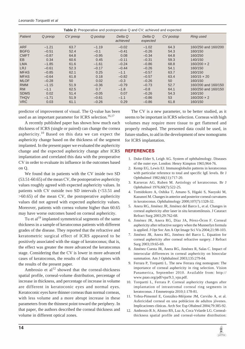
14
Leonardo Torquetti et al
predictor of improvement of visual. The Q-value has beenused as an important parameter for ICRS selection.16,17
A recently published paper has shown how much eachthickness of ICRS (single or paired) can change the corneaasphericity.10 Based on this data we can expect theasphericity change based on the thickness of the segmentimplanted. In the present paper we evaluated the asphericitychange and the expected asphericity change after ICRSimplantation and correlated this data with the preoperativeCV in order to evaluate its influence in the outcomes basedon Q.
We found that in patients with the CV inside two SD(53.51-60.65) of the mean CV, the postoperative asphericityvalues roughly agreed with expected asphericity values. Inpatients with CV outside two SD intervals (<53.51 and>60.65) of the mean CV, the postoperative asphericityvalues did not agreed with expected asphericity values.Moreover, patients with cornea volume higher than 60.65may have worse outcomes based on corneal asphericity.
Tu et al19 implanted symmetrical segments of the samethickness in a sample of keratoconus patients with differentgrades of the disease. They reported that the refractive andkeratometric surgical effect of ICRS appeared to bepositively associated with the stage of keratoconus; that is,the effect was greater the more advanced the keratoconusstage. Considering that the CV is lower in more advancedcases of keratoconus, the results of that study agrees withthe results of the present paper.
Ambrosio et al12 showed that the corneal-thicknessspatial profile, corneal-volume distribution, percentage ofincrease in thickness, and percentage of increase in volumeare different in keratoconic eyes and normal eyes.Keratoconic eyes have thinner corneas than normal corneas,with less volume and a more abrupt increase in theseparameters from the thinnest point toward the periphery. Inthat paper, the authors described the corneal thickness andvolume in different optical zones.
The CV is a new parameter, to be better studied, as itseems to be important in ICRS selection. Corneas with highvolumes may require more tissue to get flattened andproperly reshaped. The presented data could be used, infuture studies, to aid in the development of new nomogramsfor ICRS implantation.
REFERENCES
1. Duke-Elder S, Leigh AG. System of ophthalmology. Diseasesof the outer eye. London: Henry Kimpton 1965;964-76.
2. Kemp EG, Lewis EJ. Immunoglobulin patterns in keratoconuswith particular reference to total and specific IgE levels. Br JOphthalmol 1982;66(11):717-20.
3. Karseras AG, Ruben M. Aetiology of keratoconus. Br JOphthalmol 1976;60(7):522-25.
4. Tomidokoro A, Oshika T, Amano S, Higaki S, Naoyuki M,Kazunori M. Changes in anterior and posterior corneal curvaturesin keratoconus. Ophthalmology 2000;107(7):1328-32.
5. Anera RG, Jiménez JR, Jiménez del Barco L, et al. Changes incorneal asphericity after laser in situ keratomileusis. J CataractRefract Surg 2003;29:762-68.
6. Jiménez JR, Anera RG, Díaz JA, Pérez-Ócon F. Cornealasphericity after refractive surgery when the Munnerlyn formulais applied. J Opt Soc Am A Opt Image Sci Vis 2004;21:98-103.
7. Jiménez JR, Anera RG, Jiménez del Barco L. Equation forcorneal asphericity after corneal refractive surgery. J RefractSurg 2003;19:65-69.
8. Jiménez Cuesta JR, Anera RG, Jiménez R, Salas C. Impact ofinterocular differences in corneal asphericity on binocularsummation. Am J Ophthalmol 2003;135:279-84.
9. Ferrara P, Torquetti L. The new Ferrara ring nomogram: Theimportance of corneal asphericity in ring selection. VisionPanamerica, September 2010. Available from: http://www.paao.org/pdf/vpa/9.3_vpa.pdf.
10. Torquetti L, Ferrara P. Corneal asphericity changes afterimplantation of intrastromal corneal ring segments inkeratoconus. J Emmetropia 2010;1:178-81.
11. Yebra-Pimentel E, González-Méijome JM, Cerviño A, et al.Asfericidad corneal en una poblácion de adultos jóvenes.Implicaciones clínicas. Arch Soc Esp Oftalmol 2004;79:385-92.
12. Ambrosio R Jr, Alonso RS, Luz A, Coca Velarde LG. Corneal-thickness spatial profile and corneal-volume distribution:
Table 2: Preoperative and postoperative Q and CV; achieved and expected
Patient Q preop CV preop Q postop Delta Q Delta Q CV postop Ring usedachieved expected
ARF –1.21 63.7 –1.19 –0.02 –1.02 64.3 160/250 and 160/200BGFG –0.51 52.4 –0.1 –0.41 –0.26 54.3 160/150CMFT –0.87 64.8 –0.95 0.08 –0.34 64.8 160/250EB 0.34 60.6 0.45 –0.11 –0.31 59.3 140/150LMA –1.85 61.6 –1.61 –0.24 –0.86 68.8 160/200 × 2LRJ –0.61 52.3 –0.17 –0.44 –0.26 54.1 160/150MFAS –0.85 62.1 0.25 –1.1 –0.57 63.7 160/150MFAS –0.64 61.8 0.18 –0.82 –0.57 63.4 160/15 × 20MLOF –0.28 50 0.02 –0.3 –0.26 50 160/150RMM –1.15 51.9 –0.36 –0.79 –0.73 52.7 160/200 and 160/150RM –1.1 62.5 0.7 –1.8 –0.8 64.1 160/250 and 160/150SDMS 0.02 51.4 –0.05 0.07 –0.26 54.3 160/150THQV –1.71 51.9 –0.61 –1.1 –0.86 53 160/200 × 2VRC 0.03 61.1 –0.26 0.29 –0.86 61.8 160/150

International Journal of Keratoconus and Ectatic Corneal Diseases, January-April 2013;2(1):12-15 15
IJKECD
The Influence of Corneal Volume in Surgical Planning: Insights for a New Parameter for the Ferrara Ring Nomogram
tomographic indices to detect keratoconus. J Cataract RefractSurg 2006;32:1851-59.
13. Pinero D, Alio J, Aleson A, Vergara ME, Miranda M. Cornealvolume, pachymetry, and correlation of anterior and posteriorcorneal shape in subclinical and different stages of clinicalkeratoconus. J Cataract Refract Surg 2010;36:814-25.
14. Emre S, Doganay S, Yologlu S. Evaluation of anterior segmentparameters in keratoconic eyes measured with Pentacam system.J Cataract Refract Surg 2007;33:1708-12.
15. The Pentacam. The measurement principle. Available from:http://www.pentacam.com/sites/messprinzip.php. Assessed: 2-Jan 2013.
16. Cervino A, Gonzalez-Meijome JM, Ferrer-Blasco T, Garcia-Resua C, Montes-Mico R, Parafita M. Determination of cornealvolume from anterior topography and topographic pachymetry:application to healthy and keratoconic eyes. Ophthalmic PhysiolOpt 2009;29(6):652-60.
17. Torquetti L, Ferrara G, Ferrara P. Correlation of anterior segmentparameters in keratoconus. Int J Kerat Ect Cor Dis. 2012;1(2)87-91.
18. Mannion LS, Tromans C, O’Donnell C. Reduction in cornealvolume with severity of keratoconus. Curr Eye Res2011;36(6):522-27.
19. Tu KL, Sebastian RT, Owen M, Batterbury M, Kaye SB.Quantification of the surgically induced refractive effect of
intrastromal corneal ring segments in keratoconus withstandardized incision site and segment size. J Cataract RefractSurg 2011 Oct;37(10):1865-70.
ABOUT THE AUTHORS
Leonardo Torquetti
Lecturer, Department of Cornea, Paulo Ferrara Eye Clinic, MG, Brazil
Correspondence Address: Contorno Ave. 4747, Suite 615Lifecenter, Funcionários, Belo Horizonte, MG-30110-031, BrazilPhone: 553132860836, e-mail: [email protected]@gmail.com
Guilherme Ferrara
Lecturer, Department of Cornea, Paulo Ferrara Eye Clinic, MG, Brazil
Wagner Gomes Dias
Lecturer, Department of Cornea, Hilton Rocha Foundation, MG, Brazil
Paulo Ferrara
Professor, Department of Cornea, Paulo Ferrara Eye Clinic, MG, Brazil

122
Pablo F Rodrigues et al
ABSTRACTPurpose: The purpose of this study is to evaluate the topo-graphic, topometric and visual changes after implantation of Ferrara intrastromal corneal ring segments (ICRS) in grades I and II keratoconus patients.
Materials and methods: The chart records of 50 consecutively operated keratoconus patients were reviewed. The patients were operated on by the same surgeon, with the manual technique. All patients were preoperatively and postoperatively evaluated with the Pentacam (OCULUS Optikgeräte, Wetzlar, Germany). The studied parameters were: uncorrected distance visual acuity (UDVA), corrected distance visual acuity (CDVA), keratometry, corneal asphericity and corneal volume.
Results: Fifty eyes of 42 patients (26 males and 16 females) were analyzed. The mean preoperative UDVA was 0.91 ranging from LogMAR 0.10 to 1.30. The mean postoperative CDVA was 0.19 ranging from 0.00 to 0.54 LogMAR. The postoperative CDVA was equal or better than 0.18 in 37 cases (74%). The mean K1 decreased from 45.80D (±2.52) preoperatively to 44.27D (± 2.10) postoperatively and the mean K2 value, from 49.06D (± 2.09) to 46.22D (±1.89). The mean asphericity increased from –0.71 preoperatively to –0.29 postoperatively. The averagepreoperative corneal volume was 56.89 ± 3.11 mm3 while the average postoperative corneal volume was 57.64 ± 3.05 mm3.
Conclusion: The study supports the early indication of implantation of Ferrara ICRS in mild to moderate keratoconus cases in order to achieve good visual, keratometric and ashpericity outcomes.
Keywords: Mild keratoconus, Intrastromal corneal ring segment, Corneal volume, Asphericity.
How to cite this article: Rodrigues PF, Ferrara G, Ferrara P, Dias WG, Torquetti L. Intrastromal Corneal Ring Segments Implantation in Patients with Mild Keratoconus. Int J Kerat Ect Cor Dis 2014;3(3):122-126.
Source of support: Nil
Conflict of interest: None
INTRODUCTION
Keratoconus is a noninflammatory corneal condition in which there is thinning and protrusion of the cornea. It is commonly bilateral, involving two-third of the cornea, with its central apex just below the visual axis. The condi-tion, along with pellucid marginal degeneration, kerato-globus and posterior keratoconus represents pheno- typical variations of the same pathogenic mechanism. Asides from a significant variability in prevalence, it has an estimated incidence of 50 to 230 cases per 100,000.1 Initially, it is usually managed with the correction of ametropia through the use of glasses. In cases which glasses do not provide a satisfactory corrected visual acuity, rigid gas permeable contact lenses are used. They, in turn, create a new anterior refractive surface; however, they do not stop the progression of the disease. The use of intracorneal implants for high myopia correction was initially described at the beginning of the 60 seconds.1,2 Its application is an important tool for keratoconus patients who are intolerant to contact lenses. The Ferrara ring implant (1986) was first used in the correction of myopia of up to 15 diopters. Its applicability and reproducibility allowed for its use in keratoconus patients, irregular astigmatism and after corneal transplants.3-6
There is an important debate regarding the best moment in which intrastromal corneal ring segments (ICRS) should be implanted. Levinger et al7 showed significant reduction in myopia with improvement in regular astigmatism in initial and moderate keratoconus; Fahd et al8 also demonstrated good results in moderate and advanced keratoconus. Kahn et al demonstrated significant corneal flattening and improved patient contact lens tolerance after ICRS implants in advanced keratoconus. At this moment, there is no consensus in the current literature about the best moment to implant the ICRS in keratoconus patients.
The purpose of this study is to evaluate the topo-graphic, topometric and visual changes after implan-tation of Ferrara (ICRS) in grades I and II keratoconus patients.
Intrastromal Corneal Ring Segments Implantation in Patients with Mild Keratoconus1Pablo F Rodrigues, 2Guilherme Ferrara, 3Paulo Ferrara, 4Wagner G Dias, 5Leonardo Torquetti
IJKECD
CASE SERIES10.5005/jp-journals-10025-1091
1,2,5Cornea Subspecialty, 3Director, 4Professor1Department of Ophthalmology and Visual Sciences, Federal University of São Paulo, SP, Brazil2,3Department of Ophthalmology, Dr Paulo Ferrara Eye Clinic Belo Horizonte, MG, Brazil4Sector of Cornea and Cataract Hilton Rocha Institute, Belo Horizonte, MG, Brazil5Department of Ophthalmology, Center of Excellence in Ophthalmology, Pará de Minas, MG, Brazil
Corresponding Author: Pablo F Rodrigues, Department of Ophthalmology and Visual Sciences, 81 Benedito Lapin St Suit 101, Zip Code 04532-040, SP, Brazil, e-mail: [email protected]

International Journal of Keratoconus and Ectatic Corneal Diseases, September-December 2014;3(3):122-126 123
IJKECD
Intrastromal Corneal Ring Segments Implantation in Patients with Mild Keratoconus
MATERIALS AND METHODS
The chart records of 50 consecutively operated kerato-conus patients were reviewed. The patients were operated on by the same surgeon (Paulo Ferrara) from February to November 2011. This study was approved by the Ethics Committee of the Instituto Suel Abujamra (Plataforma Brasil) [Suel Abujamra Institute (Brazil Platform)], and follows the tenets of the Declaration of Helsinki. All patients signed an inform consent allowing the procedure to be done.
Inclusion criteria utilized: Topographic diagnosis of grades I and II keratoconus (Amslwer-Krumeich), absence of any other previous ocular disease or surgery, absence of corneal opacity, pachymetry greater than 450 micra at the incision site and minimal age of 16 years.
All patients were preoperatively and postoperatively evaluated with the Pentacam (OCULUS Optikgeräte, Wetzlar, Germany) for evaluation of anterior segment parameters.
All patients were operated using the manual surgical technique as previously described.9
INTRASTROMAL RING (ICRS) IMPLANTATION METHOD
Surgical Planning
The selection of ICRS was done according to the Ferrara asphericity nomogram.9 The most commonly accepted value of normal corneal asphericity is about –0.23 ± 0.08.10 In patients with unilateral keratoconus, the asphericity of the fellow eye should be used as the postoperative target. Thus, it will be calculated from the algebraic expression of the preoperative asphericity –0.23. This will be the target postoperative asphericity.
Surgical Technique
All surgeries were performed according to the standard manual technique.
A reference point was marked in the center of cornea, while asking to the patient to look to a red light attached
to surgical microscope (while turned off). The incision was made at the steepest meridian of the anterior cornea surface, using a calibrated diamond knife was set at approximately 80% of the mean corneal thickness deter-mined by Pentacam. Corneal pockets were then created using the spreader hook. One semicircular dissector was placed sequentially into the lamellar pocket to be steadily advanced by a rotational movement (counterclockwise and clockwise dissectors). After completion of the tunne-lization the ring was inserted inside the tunnels.
Postoperative evaluations were performed on the first day, 1 week, 1 and 3 months postoperatively.
DATA ANALYSIS
Evaluation of preoperative collected data and evaluation on the 1st and 3rd months of cornea analysis. The studied parameters were: corneal asphericity, volume, keratometry and visual acuity. The paired student t-test was used for statistical analysis.
RESULTS
Fifty eyes of 42 patients (26 males and 16 females) were analyzed. Of all of the surgeries performed 25 were on right eyes and 25 on left eyes. The results present pre-operative and last follow-up data. The age of patients ranged from 13 to 63 years (the mean age was 30.40 years old ±9.15). The mean preoperative corrected distance visual acuity (CDVA) was 0.91 (LogMAR) ranging from 0.10 to 1.30. The mean postoperative CDVA was 0.19 rang-ing from 0.00 to 0.54. The postoperative CDVA was equal or better than 0.18 in 37 cases (74%).
An observation between preoperative and postopera-tive asphericity and CDVA was described (Table 1). Forty-eight percent of the eyes present asphericity between –0.5 and –0.9 and achieve a mean CDVA post 0.19 LogMAR.
Table 1: Correlation between the preoperative Q and preoperative and postoperative CDVA (mean)
Q (30º) PreCDVA Pre Eyes
CDVA Post
Q (30º) Post
Q up to –0.3 0.84 4 0.28 0.09–0.3 ≤ Q < –0.5 0.81 8 0.15 –0.17–0.5 ≤ Q < –0.7 0.80 13 0.22 –0.25–0.7 ≤ Q < –0.9 0.79 11 0.16 –0.34–0.9 ≤ Q < –1.1 1.07 9 0.19 –0.35Less than or equal to –1.1
1.18 5 0.23 –0.77
Total 0.91 50 0.19 –0.29Graph 1: Correlation between corrected distance visual acuity
and pre- and postoperative asphericity

124
Pablo F Rodrigues et al
There is great difficulty in measuring medical visual acuity (LogMAR chart) in patients with Q (30º) equal to or less than 0.7 (Graph 1). The average CDVA in these patients was 1.0 still with a mean CDVA of 0.20. A CDVA better or equal to 0.18 occurred in 74% of all patients.
The mean K1 decreased from 45.80D (±2.52) preopera-tively to 44.27D (± 2.10) postoperatively and the mean K2 value, from 49.06D (± 2.09) to 46.22D (±1.89) (p < 0.05). The mean asphericity increased from –0.71 preoperatively to –0.29 postoperatively (p < 0.05) (Table 2). Currently, the most commonly accepted value in a young adult popula-tion is approximately –0.23 ± 0.08 measured at a 4.5 mm optical zone.10
Corneal volume (CV) was recently identified as an additional screening factor for keratoconus.11-13 Signi-ficant differences in CV have been reported between nor-mal and mode-rate keratoconic eyes (Pentacam system: 60.83 ± 3.27 mm3 controls vs 57.98 ± 2.65 mm3 moderate keratoconus).11
The average preoperative corneal volume was 56.89 ± 3.11 mm3 while the average postoperative corneal volume was 57.64 ± 3.05 mm.3 For better analysis of this additional screening, we divided the eyes into three groups: group 1—patients with corneal volume lower than normal and group 2—patients with normal corneal volumes. The study included 19 cases, 38% of total cases, already showed a volume lower than normal before the procedures. Twenty-five eyes were in the normal corneal volume range before (ICRS implantation). The group 3 (comprised of 6 eyes) shows eyes that had an upper limit of normality before the ICRS.
We analyzed the volumetric behavior of the cornea after surgeries and obtained 22 eyes with normal corneal volume (vs 25 eyes before surgeries). This total included 21 eyes, which maintained of normal range corneal volume
after the surgeries and originally belonged to group 2 (with normal range corneal volume), and one eye from group 1 (corneal volume lower than normal). Regarding the group 1, 11 cases increased the final volume being nine eyes were introduced in the normal corneal volume with normal asphericity in eight eyes of them (Table 3).
The group 1 showed a presurgical average kerato-metry of 46.45 vs of 42.33D after ICRS implantation with an average final asphericity of –0.29 (Qval preoperative –0.32). The group 2 reach an average keratometry 45.84D (vs a preoperative 47.89D) but the initial asphericity already were –0.76 and presented the final asphericity –0.32. The group 3 showed a presurgical average kerato-metry of 46.22D vs an average keratometry of 44.53D after ICRS implantation with an average final asphericitiy of –0.21 (vs –0.67 preoperative).
The mean volume of the ICRS implants was 1.31 mm3 ± 0.39 µm, with a variation between 0.75 to 1.71 mm3. The volumetric data of each ICRS segment were supplied by the company AJL (Miñano, Álava-Spain) manufacturer of the FerraraRings® orthoses (Table 3).
In the group 1 (19 eyes), 11 eyes presented a final volume larger than initial plus the ICRS implants being nine reached the normal range of volume, and eight presented final volume lower than initial plus the ICRS.
On the other hand, the group 2 (25 eyes) presented seven eyes a final volume bigger than initial plus the ICRS implants with asphericity of –0.34 but CDVA 0.15 vs 18 final volume lower than initial plus the ICRS with asphericity of –0.29 but CDVA 0.23.
In the group 3 (6 eyes), two eyes presented a final volume bigger than initial plus the ICRS implants with asphericity of –0.08 but VA 0.09 vs four eyes wich final volume lower than initial plus the ICRS implants with asphericity of –0.27 but VA 0.18.
An interesting relationship between volume and keratometry mean was observed from a 48D. In 71% of patients that underwent surgery who presented a Km less or equal that 48D had a reduction in the final corneal volume (Table 4).
Table 4: Volumetric comparison after discounting ICRS volume
KeratometryVolumetric comparison
Reduction (%) Increase (%) Km ≥ 48D 52 48 Km < 48D 71 29 Total 60 40
Table 2: K variation, Q variation and CV from preoperative to postoperative
K1 (D) K2(D) Km (D) Q (30º) CV p-valuePreop 45.80 ± 2.52 49.06 ± 2.09 47.10 ± 2.18 –0.71 ± 0.32 56.89 < 0.05Postop 44.27 ± 2.10 46.22 ± 1.89 45.18 ± 1.88 –0.29 ± 0.29 57.64 ± 3.05 < 0.05
Table 3: Volume of each segment from thickness and its respective arc length
Volume Thickness Arc angle (º)0.75 0.15 1400.94 0.20 1401.14 0.25 1400.85 0.15 1601.08 0.20 1601.30 0.25 1601.13 0.15 2101.42 0.20 2101.71 0.25 210

International Journal of Keratoconus and Ectatic Corneal Diseases, September-December 2014;3(3):122-126 125
IJKECD
Intrastromal Corneal Ring Segments Implantation in Patients with Mild Keratoconus
DISCUSSION
Aydin et al demonstrated the low visual quality in kerato-conus patients.14 This disease is progressive and vision may be seriously impaired, causing an important nega-tive impact on patients’ quality of life (QoL).15 A study conducted by the CLEK concluded that QoL is seriously affected in keratoconus patients and continues to decline over time, but an earlier study by our group showed that ICRS implantation improves visual QoL (V-QoL).16 The correlation between with quality of life (V-QoL) following ICRS implantation in these patients was established.17
The results demonstrated the low visual acuity pre-sent in patients suffering from keratoconus already in the initial grades (0.90 with a variability of ±0.74). The patients evaluated in the study presented a significant improvement in visual acuity (from 1.30 to 0.18 Log- MAR), and significant corneal flattening, with the mean keratometry decreasing from 47.14 to 45.18D. We believe that all of this is corroborated by the reduction in opti-cal aberration indices, and consequently improvement in visual quality.18
Some studies agree that the human cornea Q (asphe-ricity) values range from –0.01 to –0.80.19 However, in our studies, we observed that if we adopted such values as a reference we would have 56% of the patients within ‘normality’ with a mean visual acuity of 0.79 LogMAR before ICRS, i.e. completely outside of normality. The asphericity showed an important and direct relationship with the visual acuity. The Figure 1 demonstrates the progressive worsening of visual acuity preoperative and asphericity. This characteristic is more easily measured up to approximately 20/125. Beyond this point, we beli-eve the quantification of visual acuity (LogMAR chart) is relatively compromised. After ICRS implantation, 28% of eyes turn to the normal range of asphericity, and among these eyes, 71.42% had a final visual acuity better than or equal 20/30.
Asphericity before and after surgery is an important parameter to measure, as it allowed us to assert the significant change and variability, in the initial cases of keratoconus as well as their improvement after the surgi-cal procedure. This data supports the predictability and effectiveness of the nomogram based on asphericity also in mild keratoconus cases.
The correlation between keratometry and asphericity is evident with a strong correlation,20 however, no such corresponding correlation was shown with the corneal volume.
We measured the cornea volume after the procedure in all cases and, 58% of cases, the corneal volume turned to normal values and, in these cases, we obtained a final
asphericity –0.28 with a mean visual acuity 0.15 being better or equal to 0.10 LogMAR in 41%.
For the volumetric evaluation, we considered the volume of each ICRS in the cornea and therefore calculate the difference between the final (Vf) and initial volume (Vi) plus the ICRS volume (Vic). The result we call ‘volume error’ (Ver). If the result was positive after the surgery, the final volume is higher than initial. On the other hand, if ‘the volume error’ was negative, it means that the cornea was lower than initial.
We observed a final volume lower than the initial volume in 38% of all patients. Therefore, in 60% of cases we obtained a corneal remodeling whose final volume was lower than the initial volume, being the preoperative Km was 47.42 and 45.58 after the surgery and average final asphericity of –0.31. That suggests that the ICRS promote indirectly a kind of reorganization by collagen lamellas of corneal stroma contributing for better values of kerato-metry, asphericity and visual acuity, which raises the possibility of corneal shrinkage after the procedure. Such a presumption is corroborated based on other studies that demonstrated the formation of collagen and other extra-cellular matrix components21 after ICRS implantation.
Such characteristics lead us to consider the possible role of the ICRS in relation to the reorganization of col-lagen fibers allowing these ectasic corneas to resemble their primary form and, in this way, improve their visual performance.
The Ferrara ICRS demonstrates its effectiveness in the treatment of initial and moderate keratoconus. This ability can be verified through the application of the pro-posed nomogram based on an analysis and observation of asphericity as a remodeling parameter and corres-ponding corneal visual function.
CONCLUSION
The study supports the early indication of Ferrara ICRS in mild to moderate keratoconus cases in order to achieve good visual, keratometric and asphericity outcomes.
REFERENCES 1. Rabinowitz YS, Nesburn AB, McDonnell PJ. Videokeratography
of the fellow eye in unilateral keratoconus. Ophthalmology 1993; 100(2):181-186.
2. Barraquer JI. Modification of refraction by means of intra-corneal inclusions. Int Ophthalmol Clin 1966;6(1):53-78.
3. Holland DR, Maeda N, Hannush SB, Riveroll LH, Green MT, Klyce SD, Wilson SE. Unilateral keratoconus. Incidence and quantitative topographic analysis. Ophthalmology 1997;104(9): 1409-1413.
4. Lee LR, Hirst LW, Readshaw G. Clinical detection of uni- lateral keratoconus. Australian and New Zealand J Ophthalmol 1995;23(2):129-133.

126
Pablo F Rodrigues et al
5. Copeman PW. Eczema and keratoconus. Br Med J 1965;2(5468):977-979.
6. Nose W, Neves RA, Burris TE, Schanzlin DJ, Belfort R Jr.Intrastromal corneal ring: 12-month sighted myopic eyes. JRefrac Surg 1996;12(1):20-28.
7. Levinger S, Pokroy R. Keratoconus managed with intacs:one-year results. Archives of Ophthalmology 2005;123(10):1308-1314.
8. Fahd DC, Jabbur NS, Awwad ST. Intrastromal corneal ringsegment SK for moderate to severe keratoconus: a case series. J Refrac Surg 2012;28(10):701-705.
9. Torquetti L, Berbel RF, Ferrara P. Long-term follow-up ofintrastromal corneal ring segments in keratoconus. J CataractRefrac Surg 2009;35(10):1768-1773.
10. Yebra-Pimentel E, Gonzalez-Jeijome JM, Cervino A, GiraldezMJ, Gonzalez-Perez J, Parafita MA. Corneal asphericity ina young adult population. Clinical implications. Archi-vos de la Sociedad Espanola de Oftalmologia 2004;79(8):385-392.
11. Ambrosio R Jr, Alonso RS, Luz A, Coca Velarde LG. Corneal-thickness spatial profile and corneal-volume distribution:tomographic indices to detect keratoconus. J Cataract RefracSurg 2006;32(11):1851-1859.
12. Pinero DP, Alio JL, Aleson A, Escaf Vergara M, Miranda M.Corneal volume, pachymetry, and correlation of anterior andposterior corneal shape in subclinical and different stagesof clinical keratoconus. J Cataract Refrac Surg 2010;36(5):814-825.
13. Emre S, Doganay S, Yologlu S. Evaluation of anterior segmentparameters in keratoconic eyes measured with the Pentacam system. J Cataract Refrac Surg 2007;33(10):1708-1712.
14. Kurna AS, Altun A, Gencaga T, Akkaya S, Sengor T. Visionrelated quality of life in patients with keratoconus. JOphthalmol 2014;2014:694542.
15. Kymes SM, Walline JJ, Zadnik K, Sterling J, Gordon MO.Collaborative longitudinal evaluation of keratoconus studyG: changes in the quality-of-life of people with keratoconus.Am J Ophthalmol 2008;145(4):611-617.
16. de Freitas Santos Paranhos J, Avila MP, Paranhos A Jr, Schor P. Evaluation of the impact of intracorneal ring segments implan- tation on the quality of life of patients with keratoconususing the National Eye Institute Refractive Error Quality oflife (NEI-RQL) instrument. Br J Ophthalmol 2010;94(1):101-105.
17. Sahebjada S, Fenwick EK, Xie J, Snibson GR, Daniell MD,Baird PN. Impact of keratoconus in the better eye and theworse eye on vision-related quality of life. InvestigativeOphthalmology and Visual Science 2014;55(1):412-416.
18. Ferrara G, Torquetti L, Ferrara P, Merayo-Lloves J. Intrastro-mal corneal ring segments: visual outcomes from a large caseseries. Clin Experimental Ophthalmol 2012;40(5):433-439.
19. Davis WR, Raasch TW, Mitchell GL, Mutti DO, Zadnik K.Corneal asphericity and apical curvature in children: across-sectional and longitudinal evaluation. InvestigativeOphthalmol Visual Sci 2005;46(6):1899-1906.
20. Torquetti L. Corneal asphericity changes after implantationof intrastromal ring segments in keratoconus. J Emmtropia2010;1(4):178-181.
21. Maguen E, Rabinowitz YS, Regev L, Saghizadeh M, Sasaki T, Ljubimov AV. Alterations of extracellular matrix componentsand proteinases in human corneal buttons with INTACS forpost-laser in situ keratomileusis keratectasia and kerato-conus. Cornea 2008;27(5):565-573.

114
Leonardo Torquetti et al
ABSTRACTThe Ferrara intrastromal corneal ring segments (ICRS) are designed to treat ectatic corneal disorders, especially kerato-conus. They have been used to reshape keratoconic corneas to improve uncorrected visual acuity, best-corrected visual acuity, contact lens tolerance and to delay or prevent the need for keratoplasty. Intrastromal corneal ring segments have several distinct and important advantages. The Ferrara ICRS have been used largely in several countries for the treatment of primary and secondary ectatic corneal disorders. This article reviews the latest data published and the clinical experience/findings on the treatment of keratoconus by the Ferrara ICRS implantation.Keywords: Ferrara, Intrastromal corneal ring segments, Keratoconus.How to cite this article: Torquetti L, Sandes J, Ferrara G, Ferrara P. Ferrara Intrastromal Corneal Ring Segments. Int J Kerat Ect Cor Dis 2016;5(3):114-127.Source of support: NilConflict of interest: Dr. Paulo Ferrara and Guilherme Ferrara have proprietary interest in the Ferrara ICRS. Dr. Leonardo Torquetti does not have financial interest in any device cited in this review.
INTRODUCTION
The implantation of intrastromal corneal ring segments (ICRS) is a minimally invasive surgical option for reshap-ing the cornea in keratoconus and other secondary ectasias. Intrastromal corneal ring segments have been used to correct ectatic corneal diseases in order to reduce corneal steepening and irregular astigmatism and improve the visual acuity.1-6 Besides, the ICRS are a surgi-cal alternative to at least delay, if not eliminate, the need for lamellar keratoplasty or penetrating keratoplasty (PKP).7
In 1986, Ferrara started implanting modified poly-methylmethacrylate (PMMA) ICRS in rabbit corneas, and in 1994, he developed a better technique of corneal tunnel construction for implanting the ICRS.8
The Ferrara ICRS are made of PMMA Perspex camphorquinone (CQ) acrylic segments. They vary in
thickness and are available in 0.15, 0.20, 0.25, and 0.30 mm. The segment cross-section is triangular, and the base for every thickness and diameter is 0.60 mm. The segments have 90, 120, 140, 160, or 210° of arc.
Many studies have demonstrated the efficacy of ICRS to treat many corneal conditions as keratoconus,1-7 post-LASIK corneal ectasia,9,10 postradial keratotomy ectasia,11 astigmatism,12 and myopia.13-15 The changes in corneal structure induced by additive technologies can be roughly predicted by Barraquer’s thickness law, which states that when material is added to the periphery of the cornea or an equal amount of material is removed from the central area, a flattening effect is achieved. The cor-rective result varies in direct proportion to the thickness of the implant and in inverse proportion to its diameter. The thicker and smaller is the diameter of the device, the higher is the corrective result.16
Preliminary investigations have demonstrated that ICRS are effective in the treatment of astigmatism and myopia with astigmatism, with the preservation of cor-rected distance visual acuity (CDVA) and stable results over time.2,17-20 The objective of the addictive technology is to reinforce the cornea, decrease the corneal irregular-ity, and provide improvement of visual acuity in affected patients.
The research about the Ferrara ICRS began in 1985. In 1986, Dr. Paulo Ferrara (P.F) realized that to keep the implant in place, a large hole was needed in the center of the lens which resulted in an annular prostheses. Since then, several annular shapes and diameters were tried. From these researches, it was concluded that the best design would be the one, i.e., used nowadays, made of PMMA, with a total diameter of 5.0 mm, arch length ranging from 90 to 210°, and thickness ranging from 150 to 300 μm.
The ICRS were implanted in rabbit eyes, through a free hand dissection technique, at 50% depth of measured central corneal thickness. The eyes were examined for 12 months, and the animals were sacrificed for histo-pathological exams. The histopathological results revealed excellent tolerance of the cornea to the orthesis since there was only slight inflammatory reaction sur-rounding the implant and no evidence of extrusion.
The techniques, traditionally, used for the implant of corneal prostheses, free hand dissection, and keratectomy with a microkeratome showed some negative points, such as interface deposits, delay in refractive stability,
Ferrara Intrastromal Corneal Ring Segments1Leonardo Torquetti, 2Jordana Sandes, 3Guilherme Ferrara, 4Paulo Ferrara
IJKECD
REVIEW ARTICLE10.5005/jp-journals-10025-1132
1,4Head, 2,3Assistant Surgeon1Department of Ophthalmology, Center for Excellence in Ophthalmology, Pará de Minas, Brazil2Department of Ophthalmology, Center of Reference in Ophthalmology, Goiânia, Brazil3,4Department of Ophthalmology, Paulo Ferrara Eye Clinic Belo Horizonte, BrazilCorresponding Author: Leonardo Torquetti, Head, Department of Ophthalmology, Center for Excellence in Ophthalmology, Pará de Minas, Brazil, e-mail: [email protected]
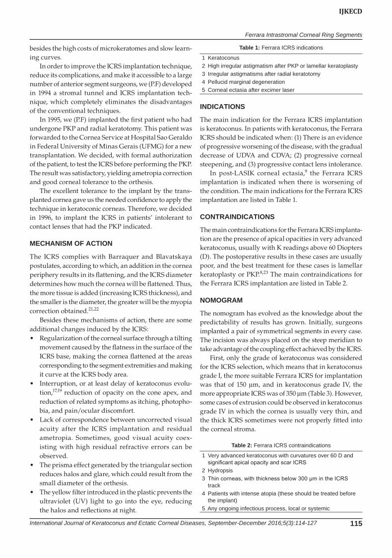
International Journal of Keratoconus and Ectatic Corneal Diseases, September-December 2016;5(3):114-127 115
IJKECD
Ferrara Intrastromal Corneal Ring Segments
besides the high costs of microkeratomes and slow learn-ing curves.
In order to improve the ICRS implantation technique, reduce its complications, and make it accessible to a large number of anterior segment surgeons, we (P.F) developed in 1994 a stromal tunnel and ICRS implantation tech-nique, which completely eliminates the disadvantages of the conventional techniques.
In 1995, we (P.F) implanted the first patient who had undergone PKP and radial keratotomy. This patient was forwarded to the Cornea Service at Hospital Sao Geraldo in Federal University of Minas Gerais (UFMG) for a new transplantation. We decided, with formal authorization of the patient, to test the ICRS before performing the PKP. The result was satisfactory, yielding ametropia correction and good corneal tolerance to the orthesis.
The excellent tolerance to the implant by the trans-planted cornea gave us the needed confidence to apply the technique in keratoconic corneas. Therefore, we decided in 1996, to implant the ICRS in patients’ intolerant to contact lenses that had the PKP indicated.
MECHANISM OF ACTION
The ICRS complies with Barraquer and Blavatskaya postulates, according to which, an addition in the cornea periphery results in its flattening, and the ICRS diameter determines how much the cornea will be flattened. Thus, the more tissue is added (increasing ICRS thickness), and the smaller is the diameter, the greater will be the myopia correction obtained.21,22
Besides these mechanisms of action, there are some additional changes induced by the ICRS:• Regularizationofthecornealsurfacethroughatilting
movement caused by the flatness in the surface of the ICRS base, making the cornea flattened at the areas corresponding to the segment extremities and making it curve at the ICRS body area.
• Interruption,oratleastdelayofkeratoconusevolu-tion,17,19 reduction of opacity on the cone apex, and reduction of related symptoms as itching, photopho-bia, and pain/ocular discomfort.
• Lackofcorrespondencebetweenuncorrectedvisualacuity after the ICRS implantation and residual ametropia. Sometimes, good visual acuity coex-isting with high residual refractive errors can be observed.
• Theprismaeffectgeneratedbythetriangularsectionreduces halos and glare, which could result from the small diameter of the orthesis.
• Theyellowfilterintroducedintheplasticpreventsthe ultraviolet (UV) light to go into the eye, reducing the halos and reflections at night.
INDICATIONS
The main indication for the Ferrara ICRS implantation is keratoconus. In patients with keratoconus, the Ferrara ICRS should be indicated when: (1) There is an evidence of progressive worsening of the disease, with the gradual decrease of UDVA and CDVA; (2) progressive corneal steepening, and (3) progressive contact lens intolerance.
In post-LASIK corneal ectasia,9 the Ferrara ICRS implantation is indicated when there is worsening of the condition. The main indications for the Ferrara ICRS implantation are listed in Table 1.
CONTRAINDICATIONS
The main contraindications for the Ferrara ICRS implanta-tion are the presence of apical opacities in very advanced keratoconus, usually with K readings above 60 Diopters (D). The postoperative results in these cases are usually poor, and the best treatment for these cases is lamellar keratoplasty or PKP.8,23 The main contraindications for the Ferrara ICRS implantation are listed in Table 2.
NOMOGRAM
The nomogram has evolved as the knowledge about the predictability of results has grown. Initially, surgeons implanted a pair of symmetrical segments in every case. The incision was always placed on the steep meridian to take advantage of the coupling effect achieved by the ICRS.
First, only the grade of keratoconus was considered for the ICRS selection, which means that in keratoconus grade I, the more suitable Ferrara ICRS for implantation was that of 150 μm, and in keratoconus grade IV, the more appropriate ICRS was of 350 μm (Table 3). However, some cases of extrusion could be observed in keratoconus grade IV in which the cornea is usually very thin, and the thick ICRS sometimes were not properly fitted into the corneal stroma.
Table 1: Ferrara ICRS indications
1 Keratoconus2 High irregular astigmatism after PKP or lamellar keratoplasty3 Irregular astigmatisms after radial keratotomy4 Pellucid marginal degeneration5 Corneal ectasia after excimer laser
Table 2: Ferrara ICRS contraindications
1 Very advanced keratoconus with curvatures over 60 D and significant apical opacity and scar ICRS
2 Hydropsis3 Thin corneas, with thickness below 300 μm in the ICRS
track4 Patients with intense atopia (these should be treated before
the implant)5 Any ongoing infectious process, local or systemic

116
Leonardo Torquetti et al
Table 3: Ferrara ICRS first-generation nomogram
Diameter 5.00 mm Thickness (mm) Diopters to be correctedFruste 0.150 –2.00 to –4.00Cone I 0.200 –4.25 to –6.00Cone II 0.250 –6.25 to –8.00Cone III 0.300 –8.25 to –10.00Cone IV 0.350 –10.25 to –12.00
Table 4: Distribution of the area of corneal ectasia
MapDistribution of ectasia Description0/100% All the ectatic area is located at one
side of the cornea
25/75% 75% of the ectatic area is located at one side of the cornea
33/66% 66% of the ectatic area is located at one side of the cornea
50/50% The ectatic area is symmetrically distributed on the cornea
The second generation of the nomogram considered the refraction for ICRS selection, besides the distribution of the ectatic area on the cornea. Therefore, as the spheri-cal equivalent increased, the selected ICRS thickness also increased. However, in many keratoconus cases, the myopia and astigmatism could not be caused by the ectasia itself but by an increase in the axial length of the eye (axial myopia). In these cases, hypercorrection by implanting a thick ICRS was observed in a keratoconus in which a thinner segment should be indicated.
In the third generation of the Ferrara ICRS nomogram, the ICRS selection depends on the type of keratoconus, its location in the cornea (Table 4), topographic astigmatism (Tables 5 to 7), and pachymetry.17,24
Table 5: Segment thickness choice in symmetric bow-tie keratoconus
Topographic astigmatism (D) Segment thickness<1.00 150/1501.25–2.00 200/2002.25–3.00 250/250>3.25 300/300
Table 6: Asymmetrical segment thickness choice in sag cones with 0/100 and 25/75% of asymmetry index (Graph 1)
Topographic astigmatism (D) Segment thickness<1.00 none/1501.25–2.00 none/2002.25–3.00 none/2503.25–4.00 none/3004.25–5.00 150/2506.25–6.00 200/300
Table 7: Asymmetrical segment thickness choice in sag cones with 0/100 and 33/66% of asymmetry index (Graph 1)
Topographic astigmatism (D) Segment thickness<1.00 none/1501.25–2.00 150/2002.25–3.00 200/2503.25–4.00 250/300
For symmetric bow-tie patterns of keratoconus, two equal segments are selected. For the nipple type of kera-toconus, a single 210 μm segment is chosen based on the nomogram (Table 8). For peripheral cones, the most common type asymmetrical segments are selected. It is important to emphasize that the ICRS thickness cannot exceed 50% of the thickness of the cornea on the track of the ICRS.
Using this third generation of the nomogram, we usually found that in some patients, there was significant corneal flattening without considerable improvement of visual acuity. We realized that, in these cases, the cornea usually presented oblate (positive Q values) postopera-tively, what could explain the lack of significant improve-ment in these cases.25-27
This finding led us to retrospectively review the charts of 147 eyes operated in 2008, and we evaluated the asphericity changes induced by the implantation of each thickness of ICRS (or pair of ICRS). Surprisingly, we found a direct correlation between ICRS thickness and change of Q values; i.e., the thicker is the ICRS the more is the effect in the change of Q value.
One paper published by our group27 showed that, using the previous nomograms, the CDVA was 20/60 or better in 70% of patients. When using the Q-based nomogram, we found a CDVA of 20/40 or better in 70% of patients.
The asphericity should be the first parameter to be considered in the ICRS selection. However, all the other parameters are considered secondarily (topographic astigmatism and pachymetry).
We have described27 that for each ICRS segment, there is a correspondent Q value change (Graph 1). A target postoperative Q value as close as possible of –0.23 is the goal after the Ferrara ICRS implantation.28-30 Based on this nomogram, one could predict the Q value change after the implantation of a specific ICRS (or pair of ICRS)
Table 8: Segment thickness choice in Nipple cones (210 μm ICRS)
Spherical equivalent (D) Segment thicknessUp to 2.00 1502.25–4.00 2004.25–6.00 250>6.25 300

International Journal of Keratoconus and Ectatic Corneal Diseases, September-December 2016;5(3):114-127 117
IJKECD
Ferrara Intrastromal Corneal Ring Segments
thickness; e.g., a single segment of 200 μm changes the asphericity as 0.31 (Graph 1); therefore, this segment would be the most appropriate in patients with a pre-operative Q value of –0.54, to achieve a postoperative Q value close to –0.23 (theoretical normal value).28-30
The pachymetry at the incision site (steep axis of the cornea) must be determined. The incision depth must be 80% of the corneal thickness at the incision site. The pachymetry should be measured in all ICRS track to avoid superficial ICRS, which could lead to future extrusion.
The actual guidelines for ICRS selection are described in Table 9.
SURGICAL TECHNIQUE
Manual Technique
The surgery is performed under topical anesthesia after miosis is achieved with 2% pilocarpine. An eyelid speculum is used to expose the eye, and 2.5% of povidone-iodine eye drops is instilled in the cornea and conjunctival cul-de-sac. The visual axis is marked by pressing the Sinskey hook on the central corneal epi-thelium while asking the patient to fixate on the corneal light reflex of the microscope light. Using a marker tinted with gentian-violet ink, a 5.0-mm optical zone and incision site are aligned to the desired axis in which the incision will be made. This site can be the steepest topographic axis of the cornea (in case of implantation of two segments) or 90° (in case of implantation of only one segment – one of the tips of the ICRS must be located on the steepest axis).17
The depth of a 1.0-mm square diamond blade is set at 80% of corneal thickness at the incision site, and this blade is used to make the incision. Using a “stromal spreader,” a pocket is formed in each side of the incision. Two (clockwise and counterclockwise) 270° semicircular dissecting spatu-las are consecutively inserted through the incision and gently pushed with some, quick, rotary “back and forth” tunneling movements. Following channel creation, the ICRS are inserted using a modified McPherson forceps. The ICRS are properly positioned with the aid of a Sinskey hook.
Femtosecond Laser Technique
The femtosecond laser (IntraLase Corp.) has recently been introduced in clinical practice whose surgical effect through photodisruption can be used as an alternative to the traditional mechanical techniques. Several recent articles31-36 have reported its efficacy and safety for tunnel creation and intrastromal ICRS implantation. The femtosecond laser can easily and quickly create a predetermined depth and channel size.
There is a controversy over channel size nomograms with the technique. Some authors conclude that more effect can be achieved by making the stromal channels narrower than the ICRS size, leading to faster visual results.37
The use of the femtosecond laser in corneal tunnel creation made the procedure faster, easier (especially for inexperienced surgeons), and more comfortable for the patient. However, the main advantage of IntraLase-assisted channel creation over the mechanical technique seems to be the precise depth of implantation. The only disadvantage of this technique is the cost of the equip-ment, which is still relatively high.
The technique: Tunnel depth is set at 80% of the thinnest corneal thickness on the tunnel location in the femtosecond laser. Special attention must be given in centralizing the disposable suction ICRS to mark the
Graph 1: Q (asphericity) variation according to the ICRS thickness (160-arc ICRS)
Table 9: Actual Ferrara ICRS nomogram (Q-based): step-by-step
1 Define the keratoconus type: sag, bow-tie, or nipple2 Corneal asphericity (Q)3 Pachymetry at incision site and ICRS track

118
Leonardo Torquetti et al
central point to minimize decentration. The channel’s inner diameter is set to 4.4 mm, the outer diameter is 5.6 mm, the entry cut thickness is 1 m (at the steepest topographic axis), the ICRS energy used for channel creation is 1.30j, and the entry cut energy is 1.30j. Channel creation timing with the femtosecond laser is 15 seconds. The intracorneal ICRS are implanted immediately after the channel creation before the disappearance of the bubbles, which reveals the exact tunnel location. The segments are placed in the final position with a Sinskey hook through a dialing hole at both ends of the segment.
The postoperative regimen, for both techniques, con-sists of moxifloxacin 0.5% (Vigamox®, Alcon, USA) and dexamethasone 0.1% (Maxidex®, Alcon, USA) eye drops four times per day for 2 weeks. The patients were instructed to avoid rubbing the eye and to use preservative-free artificial tears frequently – polyethylene glycol 400 0.4% (Oftane®, Alcon, USA).
Manual × Femtosecond Laser Technique
Initially, the tunnel for ICRS implantation was created manually with a mechanical spreader in every case. The complications of ICRS implantation with mechanical devices include epithelial defects, perforation, asym-metric segment placement, and extension of the incision toward the central visual axis or the limbus.38-41
More recently, surgeons began using a femtosecond laser to create the tunnel. It has been proposed that this method results in precise tunnel and keratotomy depth, width, and location as well as a uniform 360° channel; causes minimum haze and edema; and minimizes sur-gical complications. The femtosecond laser acts through photodisruption and can be programmed to create tunnels for segment placement at predictable corneal depths. Studies show that tunnel creation with the femto-second laser is easier, more precise, and more predictable than with the traditional mechanical spreaders.36
Some studies have compared the visual and refractive outcomes and the complications of mechanical tunnel creation and femtosecond-assisted tunnel creation for ICRS implantation to manage keratoconus.31,33,42,43
The results of these clinical trials suggest that visual and refractive results of femtosecond-assisted tunnel creation are comparable to those of mechanical tunnel creation.31 However, the femtosecond method is faster, easier, and more comfortable for both patient and surgeon. Another potential advantage of the femtosecond method is the precise depth of implantation, especially in thinner corneas.
LONG-TERM FOLLOW-UP AFTER FERRARA ICRS IMPLANTATION IN KERATOCONUS
We retrospectively reviewed patient records of 94 eyes of 76 patients, which were consecutively operated
(Ferrara ICRS implantation).17 There were 33 females and 61 males. The average age of the patients was 28.1 years. All procedures were performed by the same surgeon (P.F) between June 1996 and September 2007. Patients included in the study presented clear cornea and a minimal corneal thickness of 300 μm at the ICRS track. Patients were intolerant to contact lens and/or showed progression of ectasia.
Fifty-eight subjects underwent a single eye treatment, whereas 18 subjects had both eye treated. Seventy-three eyes had a 2-year follow-up, 66 eyes had a 3-year follow-up, 48 eyes had a 4-year follow-up, and 34 eyes had a 5-year follow-up. All patients completed at least a 2-year follow-up. No intraoperative complications occurred. All patients returned for ocular examination on day one, 1 week and a month after the surgery, and then 3, 6, and 12 months. Thereafter, the following eye examinations occurred yearly.
Preoperative and postoperative UDVA, CDVA, and keratometry data were collected from all patients. The mean UDVA (decimal) at the preoperative period was 0.12, and the mean CDVA (decimal) was 0.41. At the first month, the mean UDVA improved to 0.25, and the mean CDVA improved to 0.56. At 2-year follow-up, the mean UDVA improved from 0.12 preoperatively to 0.29. At 3-year follow-up, the mean UDVA improved to 0.34, at 4-year follow-up, the mean UDVA improved to 0.42, and at 5-year follow-up, the mean UDVA decreased to 0.31 postoperatively. The mean CDVA, at the first month, improved to 0.56. At 2-year follow-up, the mean CDVA improved from 0.41 preoperatively to 0.68. At the third-year follow-up, the mean CDVA decreased to 0.63; at the fourth-year follow-up, the mean CDVA improved to 0.65; and at the fifth-year follow-up, the mean CDVA decreased to 0.59 postoperatively.
The mean keratometry decreased significantly from the preoperative to the last postoperative follow-up. Preoperative mean keratometry was 50.36, which decreased to 47.29 at the first-month postoperative follow-up. The mean keratometry follow-up along the second to fifth years was 45.96, 45.83, 46.44, and 46.24 respectively.
As shown in previous studies, the intrastromal ICRS flatten the cornea and keep this effect for a long period of time. There is no significant resteepening of the cornea over time.
A study published by Pesando et al44 found similar results in a 5-year follow-up. A total of 93.84% (122 patients) of the eyes gained lines of UDVA, and only 1.53% (2 eyes) lost them. A total of 97.69% (127 patients) of the treated eyes gained lines of CDVA, and no eyes lost them. The values of K1 and K2 were considerably reduced over 5 years. The preoperative value of K average of 49.27 D became 4.68 D postoperatively. Both UDVA and the CDVA

International Journal of Keratoconus and Ectatic Corneal Diseases, September-December 2016;5(3):114-127 119
IJKECD
Ferrara Intrastromal Corneal Ring Segments
showed an increase. The UDVA changed from 0.14 lines preoperatively to 0.32 lines postoperatively, while the best-corrected visual acuity (BCVA) changing from 0.40 to 0.59. The spherical equivalent changed from –8.34 D before the operation to –2.83 D after the operation.
In 2014, we published a paper19 with the longest follow-up ever described, after ICRS implantation. The mean UDVA (logMAR) improved from 1.01 ± 0.28 to 0.71° ± °0.38 at 5 years (p = 0.000) and 0.67° ± °0.25 at 10 years (p = 0.735). The mean CDVA (logMAR) improved from 0.45° ± °0.45 to 0.24° ± °0.19 at 5 years (p = 0.004) and 0.29° ± °0.09 at 10 years (p = 0.292). The mean maximum K value decreased from 54.99° ± °6.33 to 50.58° ± °5.11 D at 5 years (p = 0.000) and 50.65° ± °5.17 D at 10 years (p = 0.854). The mean minimum K value decreased from 48.85° ± °5.70 to 46.90° ± °5.08 D at 5 years (p = 0.000) and 47.12° ± °4.22 D at 10 years (p = 0.945). The central corneal thickness decreased from 457.42° ± °58.21 to 421.34° ± °74.12 μm at 5 years (p = 0.039) and 434.32° ± °77.65 μm at 10 years (p = 0.427).
These studies showed that the Ferrara ICRS could be a valuable tool to provide topographic and visual stabil-ity, delay the progression of keratoconus, and postpone a corneal grafting surgery to more physiological position.4,18
THE 140 FERRARA ICRS
There are different models of Ferrara ICRS with varying sizes and arc thickness. These segments induce an arch shortening effect in the lamella, leading to central flat-tening of the cornea. There are three main corneal ring arc diameters, 140, 160, and 210°. The shorter the segment, the greater the astigmatic correction, the lesser the asphe-ricity change.27 A new Ferrara intrastromal ring model has a short arc length of 140° (140-ICRS) and has been recently used. The main advantage of this 140 arc length is its effect in astigmatism reduction. That is why, they have as a main indication, pellucid marginal degenera-tion,45 which leads to high astigmatism, besides corneal deformity. It has also been used in cases of astigmatic cones. These are central cones with high astigmatism and high keratometry.
We retrospectively reviewed the chart records of 65 consecutive patients implanted with Ferrara ICRS, which has a 140° arch length (unpublished study). Patients included in the study presented with clear corneas and minimum corneal thickness of 300 μm at the ring track. Patients were contact lens intolerant and/or showed pro-gression of ectasia. The average follow-up was 16 months. The average age was 33.38 (±13.25), 54 females (80.6%) and 13 males (19.4%).
The UDVA improved from 0.22 (decimal) preopera-tively (±0.15) to 0.42 postoperatively (±0.42) (p < 0.001). The CDVA improved from 0.38 (±0.20) to 0.59 (±0.21) (p < 0.001). The mean minimum keratometry (K1) did
not change significantly; it reduced from 45.49 (±6.38) to 45.14 (±5.10) (p = 0.354). The mean K2 decreased from 54.11 (±8.40) to 49.54 (±5.11) (p < 0.001). The average keratometry reduced from 49.87 (±7.018) to 47.34 (±4.90) (p < 0.001). The average asphericity changed from –0.60 (±0.86) to –0.23 (±0.67) (p < 0.001). The refractive astigmatism decreased from –4.95 (±1.61) to –2.55 D (±1.31) (p < 0.001). The mean preoperative astigmatism topography decreased from –8.00 (±3.45) to –4.53 (±2.52) (p < 0.001).
In selected cases of keratoconus with high astigma-tism, the short arch segments (140 arch) seem to be more effective, in order to get a significant astigmatic reduction.
THE 210 FERRARA ICRS
The 210° of arc Ferrara intrastromal ICRS have three major advantages over the conventional ICRS (160°): (1) Minimal astigmatic induction; (2) corneal flattening, and (3) implantation of a single segment. These ICRS are especially useful for the nipple type of keratoconus. The 210-ICRS are an efficient method for keratoconus correc-tion, significantly decreasing the keratometric values and spherical equivalent and improving UDVA and CDVA.
We retrospectively reviewed patient records of 80 eyes of 76 patients, which were consecutively oper-ated, in which the 210-ICRS were implanted.24 Statistical analysis included preoperative and postoperative UDVA, BCVA, spherical equivalent, and keratometry.
The mean follow-up time was 6.65 months. The mean UDVA increased from 20/350 to 20/136 (p° = °0.001). The mean CDVA increased from 20/125 to 20/55 (p° = °0.0001). The mean preoperative spherical equivalent decreased from –5.22 D, preoperative, to –2.26 D (p° = °0.050), postoperative.
Corneal tomography (Pentacam®) showed corneal flat-tening in all eyes. The mean K1 decreased from 51.49 D to 47.40 D (p° = °0.00014), and the mean K2 decreased from 54.33 D to 49.14 D (p° = °0.00022). The mean keratometric astigmatism decreased from 3.65 D (preoperative) to 2.69 D (postoperative) (p° = °0.0001).
IMPLANTATION OF FERRARA ICRS IN POSTREFRACTIVE SURGERY CORNEAL ECTASIA
We evaluated 25 eyes of 20 patients with corneal ectasia (13 males [15 eyes] and 7 females [10 eyes]) who under-went Ferrara intracorneal ICRS implantation.46
The mean follow-up time was 39.8° ± °21.1 months (Table 1). All patients had implanted only one segment of ICRS. A 160° arc ICRS (160-ICRS) was implanted in 18 eyes, and the 210° arc ICRS (210-ICRS) was implanted in 7 eyes.
The mean UDVA increased from 20/185 to 20/66 (p = 0.005). The mean CDVA increased from 20/125 to 20/40 (p = 0.008) (Graph 1). The mean asphericity values

120
Leonardo Torquetti et al
decreased from –0.95, preoperatively, to –0.23 (p = 0.006), postoperatively.
The mean pachymetry at the apex of the cornea increased from (mean) 457.7 ± 48.7 μm (361–542) to 466.2 ± 49.8 μm (381–559) (p = 0.025), and the pachymetry at the thinnest point of the cornea increased from 436.3 ± 46.2 μm (348–533) to 453.9 ± 49.3 μm (370–548) (p = 0.000). A signifi-cant reduction in keratometric values was found at the last follow-up examination; mean preoperative keratometry was decreased from 45.41 ± 5.63 D (37.3–55.5) and changed significantly to 42.88 ± 4.44 D (31.2–54.1) (p = 0.000).
Our postoperative results show a significant improve-ment in UDVA and CDVA. Moreover, there was signifi-cant increase in corneal thickness. This can be explained by a theoretical cornea collagen remodeling induced by the implantation of ICRS.
We found a significant increase in asphericity values after the implantation of ICRS in this study. Interest-ingly, the mean postoperative value was –0.23, which is considered the “normal” value for the general popula-tion.25,28-30,47 This value means that the normal physiologic asphericity of the cornea shows a significant individual variation ranging from mild oblate to moderate prolate. In an unpublished study, where we evaluated the corneal asphericity changes induced by the ICRS in keratoconus, we found that the Ferrara intrastromal ICRS implanta-tion significantly increased the mean corneal asphericity from –0.85 to –0.32. It is well known that most corneas after ablation laser procedures tend to become oblate, and when ectasia develops these corneas usually become prolate. However, the excess of prolatism usually found in keratoconus (primary) is usually of a much larger amount that that found in postrefractive surgery ectasia. The probable reason is that the Q value after Ferrara ICRS becomes much closer to “normal” values than when the ICRS is used for keratoconus. As asphericity is one of the markers of visual quality, turning it “normal” can be a predictor of improvement of visual quality.
The keratometry values reduced significantly in all eyes. It can be realized that the mean preoperative values are usually lower than ones found in keratoconus (primary). This can be explained somewhat by the corneal flattening induced by the refractive procedure, usually in an optic zone of greater extent than the location of the ectasia.
Most of the implanted ICRS were 160-ICRS, the “con-ventional” ICRS. The remainder of eyes received the 210-ICRS. The latter is usually reserved for central cones of nipple type.24 Some ectasia assume the same topographi-cal pattern of nipple cones, in which we usually use the 210-ICRS with excellent results. These ICRS are reserved for cases with low astigmatism, in which we desire to flatten the cornea with minimal astigmatic induction.
The potential advantages of ICRS implantation over keratoplasty in eyes with post-LASIK ectasia are
many.23,48 First, it avoids further laser treatment, eliminat-ing central corneal wound healing. This leaves the optical center of the cornea untouched, enhancing the refractive outcome. Second, the technique is reversible in cases of an unsatisfactory refractive or clinical outcome, and minimal postoperative care is required. Third, adjustment can be performed using thinner or thicker ICRS. In cases of unexpected corneal shape changes, one segment can be removed or exchanged.49 Fourth, it avoids the complica-tions of intraocular surgery.
These data are confirmed by several studies.9,50-52 Some long-term studies (ICRS in ectasia after LASIK) showed that ICRS yielded improvements in visual acuity, refractive status, and keratometric values without any progression in cases with post-LASIK corneal ectasia.53
ENDOTHELIUM EVALUATION AFTER FERRARA ICRS IMPLANTATION
We retrospectively reviewed patient records of 102 eyes of 81 patients, which were followed for a period of at least 1 year (mean follow-up: 45.7 months, standard deviation: 16.4 months; range: 13–71 months).54 All patients had the diagnosis of keratoconus, post-LASIK ectasia, or pellucid degeneration. Statistical analysis included preoperative and postoperative keratometry and endothelial characteristics (cell count, average cell size, and coefficient of variation).
All patients completed at least 1 year of follow-up (13–71 months). Mean age was 30.5 ± 8 years. The mean cell count decreased from (mean ± standard deviation [SD]) 2714 ± 372 to 2562 ± 406 cells/mm2 (p < 0.001). The calculated exponential cell loss rate over the mean inter-val of follow-up (4 years) was 1.4% per year. The average cell size increased from (mean ± SD) 375 ± 56 to –399 ± 61 μ2 (p < 0.001). The coefficient of variation increased from (mean ± SD) 0.22 ± 0.075 to 0.26 ± 0.010 (p = 0.001).
The mean maximum cell size increased from (mean ± SD) 529 ± 116 to 639 ± 225 μ2 (p < 0.001). The mean minimum cell size varied from (mean ± SD) 225 ± 36 to 226 ± 54 μ2 (p = 0.936).
There was significant corneal flattening as shown by keratometry changes. The mean K decreased from 47.70 ± 2.29 (43.70–53.80) to 44.86 ± 2.02 (41.20–51.20) (p = 0.0001).
In our study, we found a 1.4% loss of endothelial cells per year. Considering that most of the studied patients were young, the rate of endothelial cell loss was slightly higher than in normal eyes (1.1%).55,56 Moreover, there is no study in the current literature that shows the profile of the “normal” endothelial loss in keratoconus corneas, which could be higher than in normal corneas. The only report in the literature regarding the endothelium profile of keratoconus is nonprospective and studied only 12 eyes.57

International Journal of Keratoconus and Ectatic Corneal Diseases, September-December 2016;5(3):114-127 121
IJKECD
Ferrara Intrastromal Corneal Ring Segments
Endothelial cell loss after PKP is known to be an ongoing process even years after surgery. It is well known that the cell loss is higher than in the early time course after surgery and decreases 3 to 5 years after surgery. There is a great variation of rates of cell loss after PKP, ranging from 4.230 to 9.4% per year, at the long-term follow-up.58-60 Even after deep anterior lamellar kerato-plasty (DALK), which is a surgical technique that spares the receptor endothelium, cell loss has been reported. In one study, a decrease in average preoperative endothelial cell count of approximately 200 cells/mm2 was observed during the first 12 months of surgery.61,62
The only study that assessed the endothelial after intrastromal ICRS (Intacs, Addition Technology Inc.) implantation reported that after 24 months of surgery, all corneal regions had a slight decrease in cell density.63 In all eyes, mean central and peripheral endothelial cell counts remained above 2,495 cells/mm2. Our results are similar, we obtained a higher average postoperative cell count (2,562 cells/mm2), and we had a longer follow-up (4 years).
Wollensak et al,64 in a collagen cross-linking study in keratoconus, showed that the corneal transparency and the endothelial cell density (p = 0.45) remained unchanged. The follow-up was 23 months, and the sample was only 23 eyes. The same author, in an experimental study in rabbits,23 showed that riboflavin–ultraviolet A (UVA) treatment should be safe as long as the dose is less than the endothelial cytotoxic dose of 0.65 J/cm2. In human corneas, the endothelial cytotoxic UVA dose is reached in corneas thinner than 400 μ, which is not uncommon in keratoconus patients. Moreover, the data obtained from normal corneas of rabbit cannot be extra-polated to human keratoconic corneas, which can have a different metabolism and response to cross-linking. The study has a limitation of measure the endothelial toxic-ity only at 4 and 24 hours after treatment. The long-term endothelial cytotoxicity was not evaluated by the study.
Our study suggests that some endothelial changes occur after Ferrara ICRS implantation. However, these changes are minimal and nonclinically significant, since the endothelial cell loss rate is not much higher than that normally expected for normal corneas. In contrast, the long-term endothelial cell loss after other therapies for keratoconus is much higher (as in PKP, or even DALK, in which the receptor endothelium is spared) or unknown (as in cross-linking).
CONTACT LENS FITTING AFTER FERRARA ICRS IMPLANTATION
Contact lens fitting in keratoconus patients can be consid-erably facilitated after Ferrara ICRS implantation. Once there is corneal surface regularization with reduction of
the excess of prolatism, the majority of patients can be well fitted with contact lens after the surgery.
The contact lens trial must be done after 3 months of surgery, which is the period required for keratometry and refraction stabilization. It is very common that patients that usually were intolerant to rigid gas-permeable contact lens in the preoperative period become toler-ant after the surgery. It is frequently possible to fit soft contact lenses in these patients. Moreover, there is a very good stability of the contact lenses after the surgery, with “losses of lenses” caused by instability (a common complaint before the surgery) not occurring anymore.
There are a few studies about contact lens fitting after ICRS implantation. One study evaluated the fitting of a lathed soft toric contact lens (STCL) after the implant of ICRS to treat keratoconus. It was found that STCL fitting was successful in 75, 66.66, and 0% of the ICRS implanted eyes with stages I–III keratoconus respectively. Spectacle-CDVA was 1.5 lines better, and mean corneal power was 3.62 D lower in the successful STCL group. Piggy-back (PB) refitting achieved a PB-CDVA ≤ 0.2 logMAR in all cases. A similar difference in the CDVA change achieved by contact lenses vs spectacles was observed in the suc-cessful STCL and PB refitted groups. They concluded that STCL fitting is a feasible option in a large proportion of patients implanted with ICRS. When these lenses are unsatisfactory, a PB system is a good alternative.65
Scleral contact lenses (ScCl) are used for improving vision in patients with high or irregular astigmatism, such as keratoconus, pellucid marginal degeneration, ker-atoglobus, and post-keratoplasty astigmatism (Fig. 1).66 Scleral lenses are lenses with bearing only on the sclera with diameter of the lens being 15 mm and above. Mini-scleral lenses have diameters between 15 and 18 mm, and true scleral lenses have 18 mm diameter (more than 6 mm bearing on sclera). Mini-scleral lenses have less corneal clearance as compared with true sclerals. They were
Fig. 1: Scleral contact lens in a patient implanted with Ferrara ICRS

122
Leonardo Torquetti et al
used for a long time and then abandoned. Recently, after significant improvement in its manufacturing process, it has been used with great success in keratoconus patients with or without previous ICRS implantation.
High cost and the special training required in assess-ing the fitting and then modifying the design is quite challenging. Still, with the advances in technology, the field of ScCl has led to its resurgence as specialized lenses. Despite these challenges, improvement in visual acuity with scleral lenses is significant and worth its fitting.66
SURGICAL CORRECTION OF RESIDUAL AMETROPIA AFTER FERRARA ICRS IMPLANTATION
Keratoconus patients frequently have high myopia. Although the corneal shape in these eyes may improve after ICRS implantation, most patients require contact lenses or spectacles to correct the residual refractive error. Some studies67-69 found that implantation of a phakic intraocular lens (pIOL) was a safe, effective, and predict-able way to correct myopia associated with keratoconus (Figs 2A and B).
The success of the sequential procedure (ICRS fol-lowed by pIOL) requires knowledge of when the refraction is stable after ICRS insertion and whether the progression of keratoconus is halted because keratoconus progression leading to a refractive change can be a problem after pIOL implantation. A previous study18 evaluated the long-term results and stability of ICRS implantation for keratoconus correction. The authors found that CDVA stability was achieved, with no significant differences in refraction from 6 to 36 months after ICRS implantation; there was also an improvement in corneal topography. However, there was a significant increase in K values over time. It has been a consensus that pIOL should be implanted at least 6 months after ICRS implantation.
FERRARA ICRS IMPLANTATION FOR CORRECTION OF IRREGULAR ASTIGMATISM AFTER KERATOPLASTY
After keratoplasty, postoperative astigmatism is a common condition in clinical practice. Some studies32,70 have investigated the use of ICRS as an alternative surgical option for the treatment of astigmatism in patients who underwent keratoplasty for keratoconus (Figs 3A and B).
One of these studies70 found that the mean CDVA (LogMAR) improved from 0.45 ± 0.17 (0.18–1.00) to 0.30 ± 0.17 (0.00–1.00). The mean preoperative standard error was –6.34 ± 3.40 D (0.37 to –16.50 D) and –2.66 ± 2.52 D (0.87 to –10.50 D) postoperatively. The mean spherical refractive error reduced from –7.10 ± 3.07 D (2.15–16.68 D) preoperatively to –3.46 ± 2.05 D (0.88–10.79 D) postopera-tively. No patient lost visual acuity. The mean corneal topographic astigmatism decreased from 3.37 ± 1.51 D preoperatively to 1.69 ± 1.04 D postoperatively. The mean maximum K value decreased from 48.09 ± 2.56 to 44.17 ± 2.67 D, and the mean minimum K value decreased from 44.90 ± 2.54 to 42.46 ± 2.63 D. All changes were sta-tistically significant (p < 0.0001).
There are several potential advantages of ICRS implan-tation over other surgical techniques in eyes with high astigmatism after PKP. First, ICRS implantation avoids any excimer laser treatment, eliminating central corneal wound healing, which could be unsatisfactory in post-PKP corneas. This leaves the optical center of the cornea untouched, enhancing refractive outcomes. Second, the technique is reversible in cases of an unsatisfactory refractive or clini-cal outcome. Third, adjustment can be performed using thinner or thicker rings. In cases of unexpected corneal shape changes, one segment can be removed or exchanged. Fourth, it avoids the complications of intraocular surgery.
The results of these studies suggested that ICRS seem to be a promising treatment for astigmatism after kerato-plasty, especially in those with thin and irregular corneas.
Figs 2A and B: Phakic intraocular lens in patients implanted with Ferrara ICRS
A B

International Journal of Keratoconus and Ectatic Corneal Diseases, September-December 2016;5(3):114-127 123
IJKECD
Ferrara Intrastromal Corneal Ring Segments
ABERROMETRY AFTER FERRARA ICRS IMPLANTATION
The wavefront corneal changes induced by the implanta-tion of the Ferrara ICRS have been evaluated by several studies.33,70 The decrease of vision in patients with kerato-conus is caused not only by sphero-cylindrical refractive errors (low-order aberrations), but also to a significant extent by high-order ones. This is clearly explained by the fact that spectacles in most cases are not able to provide full correction. The predominant defect is in the coma aberration, specifically its vertical component.
The studies describe a statistically significant reduc-tion of all evaluated aberrations, not only the reduction of wavefront aberrations, but also the prediction of pos-sible surgical-induced aberrations. These data (reduction and induction of aberrations) could be considered in a nomogram to assist in the ring (or pair of rings) selection for the best visual quality results.
FERRARA ICRS × KERATOPLASTY
Barbara and Barbara71 have published the only paper that compares the clinical outcomes between ICRS implantation and keratoplasty in keratoconus patients. They found better UDVA and CDVA in the ICRS group of patients; the PKP group has more myopia and astigma-tism but lower keratometry readings; all these differences were not statistically significant. intrastromal corneal ring segments implantation has been shown to be a less invasive procedure with less postoperative complications than PKP.
COMPLICATIONS
The incidence of complications after the learning curve is very low.72 Postoperative complications can be related to (1) The surgical technique, (2) the nomogram, and (3) the ICRS itself. The complications related to the surgi-cal technique are as follows: Extrusion (due to a shallow tunnel), infection, bad centration of the segment (wrong placement of the ICRS), migration, and misplacement or asymmetry of the segments.
The complications related to the nomogram are linked to the corneal biomechanics and can be (1) Over-correction and (2) undercorrection. Although the predict-ability of postoperative results is high, in some cases, overcorrection and undercorrection can occur due to viscoelastic and biomechanic profile of the different keratoconic corneas.
The complications related to the ICRS itself are as follows: (1) Halos and glare, (2) periannular deposits, and (3) neovascularization. Halos are reported by 10% of patients and can be related to the pupil size. This symptom tends to fade or at least diminish over time. In very symptomatic cases, we usually prescribe pilocarpine or brimonidine tartarate at night, to constrict the pupil and alleviate the undesired reflexes. The periannular opacities are small white debris lying along the ICRS internal face (Fig. 4). They do not tend to grow and do not harm visual performance, being only antiesthetical when submitted to biomicroscopic examination. Neovasculari-zation of the stromal tunnel is rare and usually occurs in atopic patients (Fig. 5). Subconjunctival bevacizumab has shown to be an effective option to treat neovasculariza-tion of the tunnel.73,74
Coskunseven et al38 reported the complication rate after implantation of ICRS assisted by the femtosecond laser in 850 keratoconus eyes. They found an incidence of 2.7% of incomplete channel formation and 0.6% of endothelial
Fig. 4: White deposits in a patient implanted with Ferrara ICRS
Figs 3A and B: Ferrara ICRS implantation in a patient with corneal graft
A
B

124
Leonardo Torquetti et al
perforation. About postoperative complications, there were 1.3% cases of segment migration, 0.2% of corneal melting, and 0.1% case of mild infection. The overall complication rate was 5.7% (49 cases out of 850 eyes).
Kwitko and Severo41 reported ICRS decentration in 3.9% of cases, segment extrusion in 19.6%, and bacterial keratitis in 1.9%. As the author mentioned in his paper, the surgeon’s learning curve and different healing pro-cesses in keratoconic corneas can cause the majority of complications related to the surgical technique. Once the surgical procedure is mastered, the complication rate related to the surgery itself is very low. The surgical steps must be followed carefully (the stromal tunnel must be constructed with the adjustable diamond knife set at 80% of local corneal thickness to reduce the chance of a shallow tunnel and subsequent ICRS extrusion) to avoid surgery-related complications.
As a general rule, it must be assumed that the thickest segment of a pair of segments cannot exceed half thick-ness of the cornea in its bed. If this happens, a pair of segments that fit this condition has to be chosen even if the achieved correction is smaller than the desired one.
The majority of the complications can be managed by ICRS exchange, reposition, addition, or removal. One study49 evaluated the visual, refractive, and topographic changes occurring after reoperation in keratoconic eyes. In this study, the incidence of patients requiring follow-up surgery due to the overcorrection or undercorrection was 3.4%. For these patients, there was improvement of UDVA, CDVA, keratometry, and pachymetry. However, asphe-ricity and spherical equivalent did not improve in these patients undergoing subsequent surgery, perhaps due to the scarring of corneal tissue and/or stroma secondary to the first procedure. The mean follow-up time after the reoperation was 30.5 ± 9.7 months. Uncorrected distance visual acuity improved from 20/300 to 20/80 (p = 0.005); CDVA improved from 20/160 to 20/50 (p = 0.0002),
the mean keratometry reduced from 49.33 ± 4.19 D to 46.16 ± 3.90 D (p = 0.0001), and the mean pachymetry at the thinnest point increased from 450 ± 42.9 to 469 ± 40.8 μm (p = 0.0001).
Good outcomes can be obtained even after removal, addition, reposition, or exchange of ICRS. Ferrara ICRS implantation has been shown to be a reversible and read-justable surgical procedure for keratoconus treatment.
COMMENTS
Preliminary investigations have demonstrated that intra-corneal ICRS are effective in the treatment of astigmatism and myopia with astigmatism,9 with preservation of CDVA and stable results over time.10 The objective of the addictive technology is to reinforce the cornea, decrease the corneal irregularity, and provide an improvement of the visual acuity in affected patients.
It is important to note an important reduction of keratometric values after the Ferrara ICRS implantation, with corneal regularization and return to its physiological values when the intervention is made early in the course of the disease.75,76 However, in a late intervention, with values of keratometry superior to 56 D, a reduction of the K is also shown, high keratometry values remain when compared with a normal corneal keratometry.77
Ferrara’s ICRS technique has the objective of reshap-ing the abnormal cornea, flattening the periphery, and decreasing the corneal astigmatism. With the objective to avoid, or at least postpone2,17-19,39 the keratoplasty, the technique is within the options of visual rehabilitation of patients with keratoconus.
Observing the clinical outcomes of our patients, we could realize that the visual rehabilitation curve and refractive stabilization occurs on average after 3 months of surgery. The visual rehabilitation process follows a certain pattern. In general, vision improvement is quick and on the day following surgery, the patients usually report subjective and objective improvement of the visual acuity. However, it usually reverts within the first weeks, and at the end of the first month, the patient reports his/her vision was better immediately after surgery. The same fluctuation is detected in relation to refraction and keratometry. From the first month on, the vision starts to improve and refractive and keratometric fluctuation decreases. From the third month on, it stabilizes. Then, it is possible to correct the residual ametropia, if necessary, by means of eyeglasses, rigid or soft contact lenses, or even implanting plOL for high myopia correction.
We could notice that patients having central cones show a longer rehabilitation time,24 which means that the central flattening is slower, while patients with decentral-ized cones have faster rehabilitation.39 We believe that this is due to the dislocation or the corneal apex toward
Fig. 5: Neovascularization of the tunnel in a patient implanted with Ferrara ICRS

International Journal of Keratoconus and Ectatic Corneal Diseases, September-December 2016;5(3):114-127 125
IJKECD
Ferrara Intrastromal Corneal Ring Segments
its physiological position in front of the pupil. In some cases, we could observe that after ICRS implantation, there was an increase in myopia and in the keratometric readings, caused by this same phenomenon.
Symptoms, such as photophobia, visual discomfort, eye strain, and itching reduce or disappear after surgery. Most of the keratoconus patients are allergic; therefore, we recommend strongly that they should not rub their eyes, which could displace the segments, and stimulate the disease progression. In addition, rubbing could theoreti-cally change the regularity of the corneal surface leading to visual acuity loss. In some cases, it is necessary to use eye shields at night to prevent the patient from rubbing the eyes compulsively and unwarily.
The satisfaction level is high. We could observe that the fear of becoming blind in those patients, along with the fear-some possibility of a transplant in case of a con tinuously evolving condition, is very common. The possibility of postponing those eventualities generates great relief in the patients. Our cases show that, besides correcting the corneal deformity, the cone evolution is interrupted. Along with this interruption, we could also observe a decrease in corneal opacity and the other symptoms aforementioned.
The surgery is simple and well reproducible, although it is not an easy procedure. As in any other procedure, it must be well executed to attain a consistent result.
The incidence of complications is very low, around 3 to 5%, compatible with the levels required for refractive procedures. It should be emphasized that the corneal ICRS implant is essentially an orthopedic technique designed to enable the correction of a structural deform-ity. As an advantage, it provides simultaneous refractive correction, although not well predictable, as with other refractive procedures.
Whenever it was necessary to perform keratoplasty, the ICRS not only helped the procedure, but also provided a better centralization during trephination.
We could also notice that, after surgery, there was a decrease in the corneal sensitivity, resulting in greater comfort in contact lens fitting, which was not possible before the operation.
The incidence of complications is greater in more advanced stages, because the cornea is thinner, and the pressure generated inside the stroma after the ICRS implantation can cause the displacement of segments toward the incisions, eventually extruding the segments.
CONCLUSION
From the results obtained, we can state that this thera-peutic approach has the following benefits:• Lowmorbidity,becauseitpreservesthecornealstruc-
ture and has a low rate of complications,38,39 allowing 95% of the operated patients to quickly reintegrate themselves to their everyday activities.
• Reversibility,becauseitenablesthecorneatorevertto the preoperative dimensions when the segments are removed.49
• Readjustability through segment replacement. Insome cases, it was possible to correct hypercorrection removing just one of the segments.49
• Lackofrejection–theacrylicwhichtheICRSismadeof is inert and biocompatible.
• Patients’highsatisfactionrate.• Asanorthopedictechnique,itcorrectscornealdeform-
ity and restores the physiologic curvature. After the surgery it is possible to correct the residual ametropia with conventional optical correction or contact lenses.65
• Stabilizationordelayofconeprogression.19
• Lack of a minimum age for surgery, thus makingit possible to reduce the waitlists for transplants in eye banks (30% of the transplants in eye banks are attributed to keratoconus).
• Possibilityofassociationwithotherprocedureslikecontact lens fitting and intraocular lenses.
• Nointerferencewhatsoeverwithcornealtransplant.
REFERENCES
1. Colin J, Velou S. Implantation of Intacs and a refractive intraocular lens to correct keratoconus. J Cataract Refract Surg 2003 Apr;29(4):832-834.
2. Kymionis GD, Siganos CS, Tsiklis NS, Anastasakis A, Yoo SH, Pallikaris AI, Astyrakakis N, Pallikaris IG. Long-term follow-up of Intacs in keratoconus. Am J Ophthalmol 2007 Feb;143(2):236-244.
3. Colin J, Cochener B, Savary G, Malet F. Correcting kerato-conus with intracorneal rings. J Cataract Refract Surg 2000 Aug;26(8):1117-1122.
4. Colin J, Cochener B, Savary G, Malet F, Holmes-Higgin D. INTACS inserts for treating keratoconus: one-year results. Ophthalmology 2001 Aug;108(8):1409-1414.
5. Siganos CS, Kymionis GD, Kartakis N, Theodorakis MA, Astyrakakis N, Pallikaris IG. Management of keratoconus with Intacs. Am J Ophthalmol 2003 Jan;135(1):64-70.
6. Siganos D, Ferrara P, Chatzinikolas K, Bessis N, Papastergiou G. Ferrara intrastromal corneal rings for the correction of kera-toconus. J Cataract Refract Surg 2002 Nov;28(11):1947-1951.
7. Beniz LA, Queiroz GH, Queiroz CF, Lopes WL, Moraes LF, Beniz J. Intrastromal corneal ring segments delay corneal grafting in patients with keratoconus. Arq Bras Oftalmol 2016 Feb;79(1):30-32.
8. Miranda D, Ferrara P. Ferrara intrastromal corneal ring seg-ments for severe keratoconus. J Refract Surg 2003 Nov-Dec; 19(6):645-653.
9. Brenner LF, Alió JL, Vega-Estrada A, Baviera J, Beltrán J, Cobo-Soriano R. Indications for intrastromal corneal ring segments in ectasia after laser in situ keratomileusis. J Cataract Refract Surg 2012 Dec;38(12):2117-2124.
10. Suiter BG, Twa MD, Ruckhofer J, Schanzlin DJ. A comparison of visual acuity, predictability, and visual function outcomes after intracorneal ring segments and laser in situ keratomileusis. Trans Am Ophthalmol Soc 2000;98:51-55; discussion 55-57.

126
Leonardo Torquetti et al
11. Baïkoff G, Maia N, Poulhalec D, Fontaine A, Giusiano B. Diurnal variations in keratometry and refraction with intra-corneal ring segments. J Cataract Refract Surg 1999 Aug;25(8): 1056-1061.
12. Ruckhofer J, Stoiber J, Twa MD, Grabner G. Correction of astigmatism with short arc-length intrastromal corneal ring segments: preliminary results. Ophthalmology 2003 Mar;110(3):516-524.
13. Ruckhofer J, Stoiber J, Alzner E, Grabner G. One year results of European multicenter study of intrastromal corneal ring seg-ments. Part 2: complications, visual symptoms, and patient satisfaction. J Cataract Refract Surg 2001 Feb;27(2):287-296.
14. Nosé W, Neves RA, Burris TE, Schanzlin DJ, Belfort Júnior R. Intrastromal corneal ring: 12-month sighted myopic eyes. J Refract Surg 1996 Jan-Feb;12(1):20-28.
15. Burris TE, Ayer CT, Evensen DA, Davenport JM. Effects of intrastromal corneal ring size and thickness on corneal flat-tening in human eyes. Refract Surg 1991 Jan-Feb;7(1):46-50.
16. Zare MA, Hashemi H, Salari MR. Intracorneal ring segment implantation for the management of keratoconus: safety and efficacy. J Cataract Refract Surg 2007 Nov;33(11):1886-1891.
17. Torquetti L, Berbel RF, Ferrara P. Long-term follow-up of intrastromal corneal ring segments in keratoconus. J Cataract Refract Surg 2009 Oct;35(10):1768-1773.
18. Alió JL, Shabayek MH, Artola A. Intracorneal ring segments for keratoconus correction: long-term follow-up. J Cataract Refract Surg 2006 Jun;32(6):978-985.
19. Torquetti L, Ferrara G, Almeida F, Cunha L, Araujo LP, Machado A, Marcelo Lyra J, Merayo-Lloves J, Ferrara P. Intrastromal corneal ring segments implantation in patients with keratoconus: 10-year follow-up. J Refract Surg 2014 Jan;30(1):22-26.
20. Barbara A, Barbara R. Long-term follow-up of Ferrara rings segments for the treatment of keratoconus. Int J Keratoconus Ectatic Corneal Dis 2013 Jan-Apr;2(1):34-39.
21. Holmes-Higgin DK, Burris TE. Corneal surface topography and associated visual performance with INTACS for myopia: phase III clinical trial results. The INTACS Study Group. Ophthalmology 2000 Nov;107(11):2061-2071.
22. Rabinowitz YS. Keratoconus. Surv Ophthalmol 1998 Jan-Feb; 42(4):297-319.
23. Rodríguez LA, Guillén PB, Benavides MA, Garcia L, Porras D, Daqui-Garay RM. Penetrating keratoplasty versus intrastromal corneal ring segments to correct bilateral corneal ectasia: pre-liminary study. J Cataract Refract Surg 2007 Mar;33(3):488-496.
24. Ferrara P, Torquetti L. Clinical outcomes after implantation of a new intrastromal corneal ring with a 210-degree arc length. J Cataract Refract Surg 2009 Sep;35(9):1604-1608.
25. Bottos KM, Leite MT, Aventura-Isidro M, Bernabe-Ko J, Wongpitoonpiya N, Ong-Camara NH, Purcell TL, Schanzlin DJ. Corneal asphericity and spherical aberration after refractive surgery. J Cataract Refract Surg 2011 Jun;37(6):1109-1115.
26. Koller T, Iseli HP, Hafezi F, Mrochen M, Seiler T. Q-factor customized ablation profile for the correction of myopic astigmatism. J Cataract Refract Surg 2006 Apr;32(4):584-589.
27. Torquetti L, Ferrara P. Corneal asphericity changes after implantation of intrastromal corneal ring segments in kera-toconus. J Emmetropia 2010;1:178-181.
28. Yebra-Pimentel E, Gonzalez-Jeijome JM, Cervino A, Giraldez MJ, Gonzalez-Perez J, Parafita MA. Corneal asphericity in a young adult population. Clinical implications. Arch Soc Esp Oftalmol 2004 Aug;79(8):385-392.
29. Calossi A. Corneal asphericity and spherical aberration. J Refract Surg 2007 May;23(5):505-514.
30. Sheridan M, Douthwaite WA. Corneal asphericity and refrac-tive error. Ophthalmic Physiol Opt 1989 Jul;9(3):235-238.
31. Kubaloglu A, Sari ES, Cinar Y, Cingu K, Koytak A, Coşkun E, Ozertürk Y. Comparison of mechanical and femtosecond laser tunnel creation for intrastromal corneal ring segment implantation in keratoconus: prospective randomized clini-cal trial. J Cataract Refract Surg 2010 Sep;36(9):1556-1561.
32. Lisa C, García-Fernández M, Madrid-Costa D, Torquetti L, Merayo-Lloves J, Alfonso JF. Femtosecond laser-assisted intrastromal corneal ring segment implantation for high astig-matism correction after penetrating keratoplasty. J Cataract Refract Surg 2013 Nov;39(11):1660-1667.
33. Piñero DP, Alio JL, El Kady B, Coskunseven E, Morbelli H, Uceda-Montanes A, Maldonado MJ, Cuevas D, Pascual I. Refractive and aberrometric outcomes of intracorneal ring segments for keratoconus: mechanical versus femtosecond-assisted procedures. Ophthalmology 2009 Sep;116(9):1675-1687.
34. Coskunseven E, Kymionis GD, Bouzoukis DI, Aslan E, Pal-likaris I. Single intrastromal corneal ring segment implantation using the femtosecond laser after radial keratotomy in a kera-toconic patient. J Cataract Refract Surg 2009 Jan;35(1):197-199.
35. Yeung SN, Ku JYF, Lichtinger A, Low SAW, Kim P, Rootman DS. Efficacy of single or paired intrastromal corneal ring segment implantation combined with collagen crosslinking in kerato-conus. J Cataract Refract Surg 2013 Aug;39(8):1146-1151.
36. Pinero DP, Alio JL, El Kady B, Pascual I. Corneal aberro-metric and refractive performance of 2 intrastromal corneal ring segment models in early and moderate ectatic disease. J Cataract Refract Surg 2010 Jan;36(1):102-109.
37. Shabayek MH, Alió JL. Intrastromal corneal ring segment implantation by femtosecond laser for keratoconus correc-tion. Ophthalmology 2007 Sep;114(9):1643-1652.
38. Coskunseven E, Kymionis GD, Tsiklis NS, Atun S, Arslan E, Siganos CS, Jankov M, Pallikaris IG. Complications of intras-tromal corneal ring segment implantation using a femto-second laser for channel creation: a survey of 850 eyes with keratoconus. Acta Ophthalmol 2011 Feb;89(1):54-57.
39. Ferrara G, Torquetti L, Ferrara P, Merayo-Lloves J. Intrastro-mal corneal ring segments: visual outcomes from a large case series. Clin Exp Ophthalmol 2012 Jul;40(5):433-439.
40. Coskunseven E, Kymionis GD, Talu H, Aslan E, Diakonis VF, Bouzoukis DI, Pallikaris I. Intrastromal corneal ring seg-ment implantation with the femtosecond laser in a post-keratoplasty patient with recurrent keratoconus. J Cataract Refract Surg 2007 Oct;33(10):1808-1810.
41. Kwitko S, Severo NS. Ferrara intracorneal ring segments for keratoconus. J Cataract Refract Surg 2004 Apr;30(4):812-820.
42. Rabinowitz YS, Li X, Ignacio TS, Maguen E. INTACS inserts using the femtosecond laser compared to the mechanical spreader in the treatment of keratoconus. J Refract Surg 2006 Oct;22(8):764-771.
43. Kouassi F-X, Buestel C, Raman B, Melinte D, Touboul D, Gallois A, Colin J. Comparison of the depth predictability of intra corneal ring segment implantation by mechanical versus femtosecond laser-assisted techniques using optical coherence tomography (OCT Visante®). J Fr Ophtalmol 2012 Feb;35(2):94-99.
44. Pesando PM, Ghiringhello MP, Di Meglio G, Romeo S. Treat-ment of keratoconus with Ferrara ICRS and consideration of the efficacy of the Ferrara nomogram in a 5-year follow-up. Eur J Ophthalmol 2010 Sep-Oct;20(5):865-873.

International Journal of Keratoconus and Ectatic Corneal Diseases, September-December 2016;5(3):114-127 127
IJKECD
Ferrara Intrastromal Corneal Ring Segments
45. Rodriguez-Prats J, Galal A, Garcia-Lledo M, De La Hoz F,Alió JL. Intracorneal rings for the correction of pellucidmarginal degeneration. J Cataract Refract Surg 2003 Jul;29(7):1421-1424.
46. Torquetti L, Ferrara P. Intrastromal corneal ring segmentimplantation for ectasia after refractive surgery. J CataractRefract Surg 2010 Jun;36(6):986-990.
47. Gonzalez-Meijome JM, Villa-Collar C, Montés-Micó R,Gomes A. Asphericity of the anterior human cornea with differ-ent corneal diameters. J Cataract Refract Surg 2007 Mar;33(3):465-473.
48. Ozertürk Y, Sari ES, Kubaloglu A, Koytak A, Piñero D, Akyol S. Comparison of deep anterior lamellar keratoplasty andintrastromal corneal ring segment implantation in advanced keratoconus. J Cataract Refract Surg 2012 Feb;38(2):324-332.
49. Torquetti L, Ferrara G, Almeida F, Cunha L, Ferrara P, Merayo-Lloves J. Clinical outcomes after intrastromal corneal ringsegments reoperation in keratoconus patients. Int J Ophthal-mol 2013 Dec;6(6):796-800.
50. Stival LR, Nassaralla BR, Figueiredo MN, Bicalho F,Nassaralla JJ Jr. Intrastromal corneal ring segment implanta-tion for ectasia after refractive surgery. Arq Bras Oftalmol2015 Jul-Aug;78(4):212-215.
51. Tunc Z, Helvacioglu F, Sencan S. Evaluation of intrastromalcorneal ring segments for treatment of post-LASIK ectasiapatients with a mechanical implantation technique. IndianJ Ophthalmol 2011 Nov-Dec;59(6):437-443.
52. Piñero DP, Alio JL, Uceda-Montanes A, El Kady B, Pascual I. Intracorneal ring segment implantation in corneas with post-laser in situ keratomileusis keratectasia. Ophthalmology 2009 Sep;116(9):1665-1674.
53. Yildirim A, Cakir H, Kara N, Uslu H. Long-term outcomesof intrastromal corneal ring segment implantation for post-LASIK ectasia. Cont Lens Anterior Eye 2014 Dec;37(6):469-472.
54. Ferrara P, Torquetti L. Corneal endothelial profile afterFerrara Ring implantation. J Emmetropia 2010;1:29-32.
55. Patel SV, McLaren JW, Bachman LA, Bourne WM. Com-parison of flex-center, center, and corner methods of corneal endothelial cell analysis. Cornea 2010 Sep;29(9):1042-1047.
56. Higa A, Sakai H, Sawaguchi S, Iwase A, Tomidokoro A,Amano S, Araie M. Corneal endothelial cell density andassociated factors in a population-based study in Japan: theKumejima study. Am J Ophthalmol 2010 May;149(5):794-799.
57. Laing RA, Sandstrom MM, Berrospi AR, Leibowitz HM. The human corneal endothelium in keratoconus: a specular micro-scopic study. Arch Ophthalmol 1979 Oct;97(10):1867-1869.
58. Regis-Pacheco LF, Binder PS. What happens to the cornealtransplant endothelium after penetrating keratoplasty? Cornea 2014 Jun;33(6):587-596.
59. Langenbucher A, Seitz B, Nguyen NX, Naumann GO. Cornealendothelial cell loss after nonmechanical penetrating kerato-plasty depends on diagnosis: a regression analysis. GraefesArch Clin Exp Ophthalmol 2002 May;240(5):387-392.
60. Seitz B, Langenbucher A, Nguyen NX, Kus MM, Küchle M,Naumann GO. Graft endothelium and thickness after pene-trating keratoplasty, comparing mechanical and excimer laser trephination: a prospective randomised study. Graefes Arch Clin Exp Ophthalmol 2001 Jan;239(1):12-17.
61. Sarnicola V, Toro P, Sarnicola C, Sarnicola E, Ruggiero A.Long-term graft survival in deep anterior lamellar kerato-plasty. Cornea 2012 Jun;31(6):621-626.
62. Reinhart WJ, Musch DC, Jacobs DS, Lee WB, Kaufman SC,Shtein RM. Deep anterior lamellar keratoplasty as an alter-native to penetrating keratoplasty a report by the American Academy of Ophthalmology. Ophthalmology 2011 Jan;118(1):209-218.
63. Azar RG, Holdbrook MJ, Lemp M, Edelhauser HF. Two-year corneal endothelial cell assessment following INTACSimplantation. J Refract Surg 2001 Sep-Oct;17(5):542-548.
64. Wollensak G, Spoerl E, Wilsch M, Seiler T. Endothelial celldamage after riboflavin-ultraviolet – a treatment in the rabbit. J Cataract Refract Surg 2003 Sep;29(9):1786-1790.
65. Carballo-Alvarez J, Puell MC, Cuiña R, Diaz-Valle D,Vazquez JM, Benitez-Del-Castillo JM. Soft contact lens fitting after intrastromal corneal ring segment implantation to treat keratoconus. Cont Lens Anterior Eye 2014 Oct;37(5):377-381.
66. Rathi VM, Mandathara PS, Taneja M, Dumpati S, Sangwan VS. Scleral lens for keratoconus: technology update. Clin Oph-thalmol 2015 Oct 28;9:2013-2018.
67. Alfonso JF, Lisa C, Fernández-Vega L, Madrid-Costa D, Poo-López A, Montés-Micó R. Intrastromal corneal ring segments and posterior chamber phakic intraocular lens implantation for keratoconus correction. J Cataract Refract Surg 2011Apr;37(4):706-713.
68. Coskunseven E, Onder M, Kymionis GD, Diakonis VF, Arslan E, Tsiklis N, Bouzoukis DI, Pallikaris I. Combined Intacs andposterior chamber toric implantable Collamer lens implan-tation for keratoconic patients with extreme myopia. Am JOphthalmol 2007 Sep;144(3):387-389.
69. El-Raggal TM, Abdel Fattah AA. Sequential Intacs and Verisyse phakic intraocular lens for refractive improvement in kerato-conic eyes. J Cataract Refract Surg 2007 Jun;33(6):966-970.
70. Coscarelli S, Ferrara G, Alfonso JF, Ferrara P, Merayo-Lloves J, Araújo LP, Machado AP, Lyra JM, Torquetti L. Intrastromalcorneal ring segment implantation to correct astigmatismafter penetrating keratoplasty. J Cataract Refract Surg 2012Jun;38(6):1006-1013.
71. Barbara R, Barbara A. Intrastromal corneal ring segment with and without collagen corneal crosslinking vs penetratingkeratoplasty for the treatment of keratoconus – case series.Int J Kerat Ect Cor Dis 2014 May-Aug;3(2):88-94.
72. Barbara A, Barbara R. Intacs intracorneal ring segmentscomplications in patients suffering from keratoconus. Int JKerat Ect Cor Dis 2013 Sep-Dec;2(3):121-128.
73. Bahar I, Kaiserman I, McAllum P, Rootman D, Slomovic A.Subconjunctival bevacizumab injection for corneal neo-vascularization in recurrent pterygium. Curr Eye Res 2008Jan;33(1):23-28.
74. Bahar I, Kaiserman I, McAllum P, Rootman D, Slomovic A.Subconjunctival bevacizumab injection for corneal neovas-cularization. Cornea 2008 Feb;27(2):142-147.
75. Rodrigues P, Ferrara G, Ferrara P, Dias W, Torquetti L.Intrastromal corneal ring segments implantation in patients with mild keratoconus. Int J Kerat Ect Cor Dis 2014;3(3):122-126.
76. Torquetti L, Ferrara G, Ferrara P. Predictors of clinical out-comes after intrastromal corneal ring segments implantation. Int J Kerat Ect Cor Dis 2012;1(1):26-30.
77. Khan MI, Injarie A, Muhtaseb M. Intrastromal cornealring segments for advanced keratoconus and cases withhigh keratometric asymmetry. J Cataract Refract Surg 2012Jan;38(1):129-136.

[FerraraG.ICRS Keratoconus in Children] 1 . Int J Kerat Ect Cor Dis Submitted April 5 2017
INTRASTROMAL CORNEAL RING SEGMENTS IN CHILDREN WITH
KERATOCONUS
Ferrara G1, Merayo-Lloves J2, Ferrara P1. Torquetti L1,3.
Int J Kerat Ect Cor Dis Submitted April 5 2017.
1. Departament of Ophthalmolgoy, Paulo Ferrara Eye Clinic, Minas
Gerais, Brazil 2. Instituto Universitario Fernández-Vega. FIO Universidad
de Oviedo. Spain. 3. Departament of Ophthalmolgoy, Center for
Excellence in Ophthalmology, Para de Minas, Brazil
ABSTRACT
Purpose: To evaluate the long-term follow-up of Ferrara intrastromal corneal
ring segments (ICRS) (Ferrara Ophthalmics, Belo Horizonte, Brazil)
implantation for the management of keratoconus in children.
Setting: Dr. Paulo Ferrara Eye Clinic, Belo Horizonte, MG, Brazil
Methods: Fifty-eight eyes of 37 children with keratoconus were included. One
or two ring segments were inserted in the cornea, embracing the keratoconus
area. Statistical analysis included preoperative and postoperative uncorrected
distance visual acuity (UDVA), corrected distance visual acuity (CDVA),
asphericity, pachymetry and keratometry.
Results: Ferrara ICRS implantation significantly improved the mean UDVA and
CDVA. Corneal tomography (Pentacam®) showed corneal flattening in all eyes

implanted with the Ferrara ring. The mean K decreased, and the corneal
asphericity and pachymetry increased in all cases.
Conclusion: The Ferrara ICRS improved all parameters after two years of
implantation in children with keratoconus. There was significant corneal
flattening after ring implantation with improvement of the UDVA and the CDVA.
All studied parameters remained stable over time.
Key-words: Ferrara ring, keratoconus, children, intrastromal corneal ring segments
INTRODUCTION
Keratoconus is a corneal ectatic disease characterized by non-
inflammatory progressive thinning of unknown cause in which the cornea
assumes a conical shape.
Intrastromal corneal ring segments (ICRS) have been used to correct
ectatic corneal diseases in order to reduce the corneal steepening, reduce the
irregular astigmatism and improve the visual acuity. 1-7 Besides, the segments
may be a surgical alternative to at least delay, if not eliminate, the need of
lamellar or penetrating keratoplasty.
The Ferrara ICRS are made of PMMA Perspex CQ acrylic. They vary in
thickness, and are available in 0.15, 0.20, 0.25, 0.30 and 0.35 mm. The
[FerraraG.ICRS Keratoconus in Children] 3 . Int J Kerat Ect Cor Dis Submitted April 5 2017
segment cross-section is triangular, and the base for every thickness and
diameter is 0.60 mm. The segments have 90, 120, 160 or 210 degrees of arc.
Many studies have demonstrated the efficacy of intrastromal rings to
treat many corneal conditions as keratoconus,1-7 post–LASIK corneal ectasia,8
post-radial keratotomy ectasia,9 astigmatism10 and myopia.11-14 The changes in
corneal structure induced by additive technologies can be roughly predicted by
the Barraquer´s thickness law; that is when material is added to the periphery of
the cornea or an equal amount of material is removed from the central area, a
flattening effect is achieved. The corrective result varies in direct proportion to
the thickness of the implant an in inverse proportion to its diameter. The thicker
and smaller the diameter of the device, the higher the corrective result.15
In order to investigate the long-term visual acuity and mechanical stability
after Ferrara ICRS implantation in children with keratoconus, we conducted the
current retrospective study.
METHODS
We retrospectively reviewed patient records of 58 eyes of 37 children
with keratoconus, which were followed for a period of at least 6 months. The
main indication for ICRS implantation was contact lens intolerance and/or
progression of the ectasia. The progression of the disease was defined by:

worsening of uncorrected distance visual acuity (UDVA) and corrected distance
visual acuity (CDVA) (loss of at least 1 line of VA), progressive intolerance to
contact lens wear and progressive corneal steepening documented by corneal
topography. Patients were excluded if any of the following criteria applied after
preoperative examination: advanced keratoconus with significant apical opacity
and scarring, hydropsis, thin corneas, with thickness below 300 µm in the ring
track, intense atopia and any ongoing infectious process, local or systemic.
Patients were separated into two groups (initial and advanced) according
to Amsler-Krumeich classification*.
Statistical analysis included preoperative and postoperative UDVA,
CDVA, keratometry, pachymetry at the thinnest point of the cornea and corneal
asphericity. The anterior segment parameters were obtained from Pentacam
(Oculus Pentacam®, Germany). Statistical analysis was carried out using the
MINITAB software (version .3.3.1). Student´s t test for paired data was used to
compare preoperative and postoperative data.
All surgeries were performed by the same surgeon (PF) using the
manual technique for ICRS implantation, as previously described.1-5 The
segments were implanted according to a previously described Ferrara
Nomogram16.
After surgery Ketorolac drops were used every 15 minutes for 3 hours,
and a combination of 0.1% dexamethasone and 0.3% moxifloxacin or
ciprofloxacin drops was used every 4 hours for 7 days, as well as hypromelose
(Alcon) every 6 hours for 30 days.

[FerraraG.ICRS Keratoconus in Children] 5 . Int J Kerat Ect Cor Dis Submitted April 5 2017
RESULTS
Fifty-eight eyes of 37 patients were studied. Thirteen eyes remained
untreated and 3 eyes underwent lamellar keratoplasty due to advanced
keratoconus. The mean age of patients was 13 ± 2.1 years old (range 8 to 16
years). All patients completed at least 6 months of follow-up (average 20
months, range 6 to 81). No peroperative or postoperative complications
occurred.
Preoperative and postoperative UDVA, CDVA, asphericity, pachymetry
and keratometry data were collected from all patients. The mean UDVA at the
preoperative period was 0.41 LogMAR, and the mean CDVA was 0.36
LogMAR. At the first month, the mean UDVA improved to 0,29 LogMAR and the
mean CDVA improved to 0.20 LogMAR. At the first year follow-up, the mean
UDVA was 0.30 LogMAR postoperatively. The mean CDVA, at the first year
follow-up, the mean CDVA improved to 0.15 LogMAR, at the second year
follow-up, the mean UDVA increased slightly 0.25 LogMAR, and the mean
CDVA decreased slightly, to 0.16 LogMAR postoperatively (Table 1).
Corneal topography showed corneal flattening in all eye. The mean
Kminimum and the mean Kmaximum decreased in all groups and there was an
increase of corneal asphericity and pachymetry. One patient needed
crosslinking and one patient needed a lamellar keratoplasty due to progressive
steepening despite ICRS implantation.
Minimum Keratometry
Evaluating the results obtained between preoperative and the first month
postoperatively, the minimum keratometry reduced, on average, 4 units (range:
3 to 5), with 95% confidence.
Between the first month and the second year of follow up, there was no
change in minimum keratometry values (p-value = 0.412). (Graphic 1)
Maximum Keratometry
From the preoperative to the evaluation in the first month, the maximum
keratometry decreased, on average, 6 units (range: 4.7 to 7,3), with 95%
confidence.
From the first month and the second year of follow up, the maximum
keratometry increased (p-value =0,002). At each year evaluated, the maximum
keratometry increased, on average, 0.7 units (95%CI: 0.2 to 0.5) (Graphic 2)
Asphericity
From the preoperative to the first month postoperative, there was an
increase in asphericity, which was, on average, 0,61 unit (p-values < 0.001).

[FerraraG.ICRS Keratoconus in Children] 7 . Int J Kerat Ect Cor Dis Submitted April 5 2017
From the first month and the second year of follow up, the asphericity
values remained stable over time (p-value = 0,275) (Graphic 3).
Corneal Thickness Variation
From the preoperative evaluation to the first month postoperative, there
was corneal thickening of, on average, 8.5 units (p-values = 0.05).
From the first month to the second year of follow up, the corneal
thickness remained stable over time (p-value = 0,112) (Graphic 4).

DISCUSSION
Preliminary investigations have demonstrated that intracorneal rings are
effective in the treatment of astigmatism and myopia with astigmatism15, with
preservation of CDVA and stable results over time16. The objective of the
addictive technology is to reinforce the cornea, decrease the corneal irregularity
and to improve the visual acuity in affected patients.
This is the first study to show the long-term follow-up of children with
keratoconus in which the Ferrara ICRS was implanted for at least 6 months.
This study is in agreement with some other studies: Miranda et al. obtained on
their study a significant reduction in the mean central corneal curvature
postoperatively. CDVA and UDVA improved in 87.1% and 80.6% of the eyes,
respectively. Siganos et al 4 showed an increase of the mean UDVA from 0.07
+ 0.08 preoperatively to 0.20 + 0.13 and 0.30 + 0.21 after 1 and 6 months,
respectively, and the mean CDVA improved from 0.37 + 0.25 preoperatively to
0.50 + 0.43 and 0.60 + 0.17 after 1 and 6 months, respectively. Kwitko and
Severo18 reported that, after implantation of Ferrara ring in keratoconus eye, the
CDVA improved in 86.4% of eyes, was unchanged in 1.9% and worsened in
11.7%. The UDVA improved in 86.4% of eyes, was unchanged in 7.8%, and
worsened in 5.8%. The mean corneal curvature was reduced from 48.76 ± 3.97
to 43.17 ± 4.79.
The minimum and maximum keratometry decreased from the pre-
surgical evaluation to the first month, while the values of asphericity and
pachymetry increased during this same period. Between the first month of

[FerraraG.ICRS Keratoconus in Children] 9 . Int J Kerat Ect Cor Dis Submitted April 5 2017
follow-up to the second year, we observed that minimum keratometry did not
change over time, and maximum keratometry had a slight increase over time.
The value of asphericity does not change over time; the pachymetry did not
change over time. Although there was a slight increase in maximum
keratometry over time (0.7 diopters per year, on average), this increase was not
clinically significant.
Based on our personal (unpublished) data, about 5% of patients go to
penetrating or lamellar keratoplasty due to progressive corneal scarring, despite
proper ICRS implantation. It is important to emphasize that these patients
usually had ring implantation in very advanced phases of the disease and does
not mean necessarily keratoconus evolution but rather an unsatisfactory visual
outcome. In our study, two patients (5,4%) underwent keratoplasty, and one
patient (2.7%) underwent corneal crosslinking due to keratoconus progression.
Alió et al.20 conducted a retrospective study to evaluate the long-term (up
to 48 months) results after implantation of Intacs in patients with keratoconus.
They found that the mean CDVA increase significantly (p<0.01) from 0.46
(20/50) preoperatively to 0.66 (20/30) 6 months after implantation. Also the
mean average K-value decreased significantly (p<0.01) by 3.13 D. The
comparison of results 6 months and 36 months after implantation showed
refraction and topography stability.
Kimionis et al.21 studied 17 eyes of patients with keratoconus that had
Intacs implantation for corneal flattening. They found that the pre-Intacs UDVA
was 20/50 or worse in all eyes, whereas, at the last follow-up examination 59%

had UDVA of 20/50 or better. Most of eyes (59%) experienced a gain of one up
to 8 lines of visual acuity.
Previous studies showed that the intrastromal ring flattens the cornea
and keeps this effect for a long period of time. There is no significant re-
steepening of the cornea over time, in adults. We found that the preoperative
keratometry values were higher in our study when compared with similar
studies.20,21,22 Moreover, the preoperative UDVA and CDVA were worse in our
study. This can be explained by the behaviour of keratoconus in some children,
which can rapidly evolve, and when the surgery is indicated the disease is more
advanced.
There was improvement of all parameters in our study. The pattern of
reduction of keratometry parameters was similar to other studies. Despite of
that, at 5 years of follow-up we found corneal re-steepening on maximum
keratometry, what was not found in the other studies we compared our results.
Regarding the visual acuity, there was improvement in UDVA and CDVA, but
not as much as in adults.
The present study showed that the Ferrara ring, despite the small sample
of patients, could be a valuable tool to provide topographic and visual
improvement in children with keratoconus. Corneal re-steepening can occur
after ICRS implantation. It plays an important role in delaying the progression of
keratoconus and postpones a corneal grafting surgery. Further studies with
larger samples and longer follow-up periods must be warranted to confirm the
presented results.

[FerraraG.ICRS Keratoconus in Children] 11 . Int J Kerat Ect Cor Dis Submitted April 5 2017

1. Colin J, Velou S. Implantation of Intacs and a refractive intraocular lens to correct
keratoconus. J Cataract Refract Surg 2003;29:832–834
2. Asbell PA, Ucakhan O. Long-term follow-up of Intacs from a single center. J Cataract Refract
Surg 2001; 27:1456–1468
3. Colin J, Cochener B, Savary G, et al. Correcting keratoconus with intracorneal rings. J
Cataract Refract Surg. 2000;26:1117–1122.
4. Siganos D, Ferrara P, Chatzinikolas K, et al. Ferrara intrastromal corneal rings for the
correction of keratoconus. J Cataract Refract Surg 2002; 28:1947-1951.
5. Colin J, Cochener B, SavaryG, et al. INTACS inserts for treating keratoconus; one-year
results. Ophthalmology 2001; 108:1409–1414
6. Siganos CS, Kymionis GD, Kartakis N, et al. Management of keratoconus with Intacs. Am J
Ophthalmol 2003; 135:64–70.
7. Assil KK, Barrett AM, Fouraker BD, Schanzlin DJ. One-year results of the intrastromal
corneal ring in nonfunctional human eyes; the Intrastromal Corneal Ring Study Group. Arch
Ophthalmol 1995; 113:159–167
8. Siganos CS, Kymionis GD, Astyrakakis N, et al. Management of corneal ectasia after laser in
situ keratomileusis with INTACS. J Refract Surg. 2002;18:43–46.
9. Silva FBD, Alves EAF, Cunha PFA. Utilização do Anel de Ferrara na estabilização e correção
da ectasia corneana pós PRK. Arq Bras Oftalmol. 2000;63:215–218.
10. Ruckhofer J, Stoiber J, Twa MD, et al. Correction of astigmatism with short arc-length
intrastromal corneal ring segments: preliminary results. Ophthalmology. 2003;110:516–524.

[FerraraG.ICRS Keratoconus in Children] 13 . Int J Kerat Ect Cor Dis Submitted April 5 2017
11. Nose W, Neves RA, Burris TE, et al. Intrastromal corneal ring: 12-month sighted myopic
eyes. J Refract Surg 1996; 12:20–28
12. Schanzlin DJ, Asbell PA, Burris TE, Durrie DS. The intrastromal ring segments; phase II
results for the correction of myopia. Ophthalmology 1997; 104:1067–1078
13. Holmes-Higgin DK, Burris TE, Lapidus JA, Greenlick MR. Risk factors for self-reported
visual symptoms with Intacs inserts for myopia. Ophthalmology 2002; 109:46–56
14. Asbell PA, Ucakhan O, Abbott RL, et al. Intrastromal corneal ring segments: reversibility of
refractive effect. J Refract Surg 2001; 17:25–31
15. Barraquer JI. Modification of refraction by means of intracorneal inclusions. Int Ophthalmol
Clin, 1966; 6(1)53-78.
16. Ferrara de A Cunha P. Técnica cirúrgica para correção de miopia; Anel corneano intra-
estromal. Rev Bras Oftalmol 1995;54:577-588.
17- Bock RH, Maumenee AE. Corneal fluid metabolism: experiments and observations. Arch
Ophthalmol 1953; 50(3):282-5.
18 - Miranda D, Sartori M, Francesconi C, Allemann N, Ferrara P, Campos M.Ferrara
intrastromal corneal ring segments for severe keratoconus. J Refract Surg. 2003 Nov-
Dec;19(6):645-53.
19. Kwitko S, Severo N. Ferrara intracorneal ring segments for keratoconus. J Cataract Refract
Surg 2004; 30:812-820.
20. Alió, JI, Shabayek MH, Artola A. Intracorneal ring segments for keratoconus correction:
long-term follow-up. J Cataract Refract Surg 2006; 32:978-985.
21. Kymionis GD, Siganos CS, Tsiklis NS, Anastasakis A, Yoo SH, Pallikaris AI, Astyrakakis N,
Pallikaris I. Long-term follow-up of Intacs in Keratoconus. Am J Ophthalmol 2007; 143:236-244.

22. Colin J, Malet F. Intacs for the correction of keratoconus: two-year follow-up. J Cataract
Refract Surg 2007; 33:69-74.
Pre and Post operative data
Postoperative
Preoperative 1st month p 2nd year p
UDVA 0.41 0.29 0.004 0.25 0.262 CDVA 0.36 0.2
0.16 0.983
Table 1: UDVA and CDVA variation
Tempo
Cera
tom
etria
mín
ima
2 an
os1 a
no
6 me
ses
1 mê
sPr
é
70
65
60
55
50
45
40
35
Graphic 1: Variation of minimum keratometry over time

[FerraraG.ICRS Keratoconus in Children] 15 . Int J Kerat Ect Cor Dis Submitted April 5 2017
Tempo
Cera
tom
etria
máx
ima
2 an
os1 a
no
6 mes
es
1 mêsPr
é
75
70
65
60
55
50
45
40
Graphic 2: Variation of maximum keratometry over time
Tempo
Valo
r da
asf
erici
dade
2 an
os1 a
no
6 mes
es
1 mêsPr
é
1
0
-1
-2
-3
-4
-5
Graphic 3: Variation of asphericity over time

Tempo
Varia
ção
da e
spes
sura
2 an
os1 a
no
6 mes
es
1 mêsPr
é
550
500
450
400
350
300
Graphic 4: Variation of pachymetry over time