
TYPEI / TYPE2 / TYPE3 / TYPE4 ALFA use TYPE2B 1 TEL.. 075 ...

JACC Vol. 5, No.3March 1985:609-16
Angiographic Morphology and the Pathogenesis of UnstableAngina Pectoris
JOHN A. AMBROSE, MD, FACC, STEPHEN L. WINTERS, MD, AUDREY STERN, MD,
ANGIE ENG, BS, LOUIS E. TEICHHOLZ, MD, FACC, RICHARD GORLIN, MD, FACC,
VALENTIN FUSTER, MD, FACC
New York, New York
609
In 110patients with either stable or unstable angina, themorphology of coronary artery lesionswas qualitativelyassessedat angiography. Each obstruction reducing theluminal diameter of the vessel by 50% or greater wascategorized into one of the foUowing morphologic groups:concentric (symmetric narrowing); type I eccentric(asymmetric narrowing with smoothborders and a broadneck); type II eccentric (asymmetric with a narrow neckor irregular borders, or both); and multiple irregularcoronary narrowings in series. For the entire group,type II eccentric lesionswere significantly more frequentin the 63 patients with unstable angina (p < 0.001),whereas concentric and type I eccentric lesionswereseenmore frequently in the 47 patients with stable angina(p < 0.05).
In unstable angina pectoris, patients often present with acuteonset of chest pain or rapid progression of symptoms. Insome cases, these presentations may indicate a decrease incoronary blood flow rather than an increase in myocardialoxygen demand as the primary mechanism for chest pain.Even though the clinical manifestations and pathophysiology of ischemia in unstable angina may differ from thoseof stable angina (1), quantitative coronary anatomic variables are similar. Angiographic comparisons of the coronaryanatomy in patients with unstable angina have shown nosignificant differences from patients with stable angina interms of the number of diseased vessels and the degree ofobstruction (2,3).
Although differentiation on the basis of quantitative variables has not proven helpful, the qualitative appearance ofcoronary lesions in unstable angina is unknown. The qual-
From the Department of Medicine, Division of Cardiology, MountSinai Medical Center, New York, New York. Manuscript received September 9, 1984; revised manuscript received October 19, 1984, acceptedOctober 31, 1984.
Address for reprints: John A. Ambrose, MD, Division of Cardiology,Mount Sinai Medical Center, One Gustave L. Levy Place, New York,New York 10029.
© 1985 by the American College of Cardiology
Type II eccentric lesions were also present in 29 of41 arteries in patients with unstable angina comparedwith 4 of 25 arteries in those with stable angina (p <0.0001) in whom an "angina-producing" artery couldbe identified. Therefore, type II eccentric lesions arefrequent in patients with unstable angina and probablyrepresent ruptured atherosclerotic plaques or partiallyocclusive thrombi, or both. A temporary decrease incoronary perfusion secondary to these plaques with orwithout superimposed transient platelet thrombi or altered vasomotor tone may be responsible for chest painin some of these patients with unstable angina.
(J Am Coli CardioI1985;5:609-16)
itative appearance of coronary lesions on postmortem angiography as demonstrated by Levin and Fallon (4) hasprovided important information concerning the pathologicsignificance of various coronary lesions. Qualitative analysisof coronary morphology at cardiac catheterization may provide a foundation for the angiographic stratification of patients with coronary artery disease on the basis of clinicalpresentation.
MethodsPatient selection. Between June and September 1983,
the history and coronary angiograms of all patients referredto cardiac catheterization for evaluation of angina pectorisor for angioplasty were prospectively reviewed. The historywas obtained before catheterization by either one of twocardiologists. The coronary angiograms were reviewed bythree experienced angiographers unaware of the historicaldata. The criteria for exclusion of patients with coronaryartery disease or angina pectoris from the study includedthe following: normal coronary arteries or nonobstructivecoronary artery disease «50% obstruction), concomitant
0735-1097/85/$3.30

610 AMBROSE ET AL.ANGIOGRAPHIC MORPHOLOGY IN ANGINA
JACC Vol. 5, No.3March 1985:609-16
Figure 1. Representative left coronary angiogram (LCA) and schematic diagram of a concentric coronary stenosis. There is a severe stenosis of the proximal left anterior descendingarteryas seenin the rightanterioroblique(RAO)projection. The thick arrow indicates the concentriclesion;the thinarrow indicates the distalleft anterior descending artery.
valvular or nonischemic cardiomyopathy, acute infarctionwith entry into a streptokinase reperfusion trial or atypicalor unclear history.
Patient classification. Patients were classified as havingstable angina pectoris if they manifested one of the following: 1) typical exertional angina (class I or II) relieved byrest or nitroglycerin with stable symptoms; 2) slowly progressive exertional angina (increasing by less than two Canadian Heart classifications) without pain at rest; or 3) severestable angina (class III to IV), including chronic pain at rest(>6 months' duration) and no recent change in symptoms.
Patients were classified as having unstable angina pectoris if they manifested one of the following: new onset ofchest pain at a low work load (class III) or at rest (class IV)
of less than 6 months' duration, crescendo angina definedas an abrupt increase in angina (2': two Canadian Heartclassifications) within 6 months of catheterization or in apatient with previous stable angina or recent « 2 weeks)well documented subendocardial or transmural infarctionand recurrent anginal pain at rest in the hospital withoutserum enzyme evidence of additional infarction.
Study patients. Of the 143 patients who met the criteriafor admission into the study, 110 were entered. This included 47 patients with stable angina and 63 patients withunstable angina; no patient had variant angina. All patientswere categorized into anginal subsets without knowledge ofthe coronary anatomy. The most common reason for elimination of the other 33 patients was the inability to clearly
Figure 2. Representative tight coronary angiograms(RCA) and schematic diagrams of two different rightcoronary arteries with type I eccentric lesions. Thearrows indicate the type I eccentric lesion.
JACC Vol. 5, No.3March 1985:609-16
AMBROSE ET AL.ANGIOGRAPHIC MORPHOLOGY IN ANGINA
611
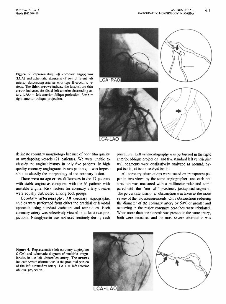
Figure 3. Representative left coronary angiograms(LCA) and schematic diagrams of two different leftanterior descending arteries with type II eccentric lesions. The thick arrows indicate the lesions; the thinarrow indicates the distal left anterior descending artery. LAO = left anterior oblique projection, RAO =right anterior oblique projection.
delineate coronary morphology because of poor film qualityor overlapping vessels (21 patients). We were unable toclassify the anginal history in only five patients. In highquality coronary angiograms in two patients, it was impossible to classify the morphology of the coronary lesion.
There were no age or sex differences in the 47 patientswith stable angina as compared with the 63 patients withunstable angina. Risk factors for coronary artery diseasewere equally distributed among both groups.
Coronary arteriography. All coronary angiographicstudies were performed from either the brachial or femoralapproach using standard catheters and techniques. Eachcoronary artery was selectively viewed in at least two projections. Nitroglycerin was not used routinely during each
procedure. Left ventriculography was performed in the rightanterior oblique projection, and five standard left ventricularwall segments were qualitatively analyzed as normal, hypokinetic, akinetic or dyskinetic.
All coronary obstructions were traced on transparent paper in two views by the same angiographer, and each obstruction was measured with a millimeter ruler and compared with the "normal" proximal, juxtaposed segment.The percent stenosis of an obstruction was taken as the moresevere of the two measurements. Only obstructions reducingthe diameter of the coronary artery by 50% or greater andoccurring in the major coronary branches were tabulated.When more than one stenosis was present in the same artery,both were measured and the most severe obstruction was
Figure 4. Representative left coronary angiogram(LCA) and schematic diagram of multiple irregularities in the left circumflex artery. The arrowsindicate severe obstructions in the proximal portionof the left circumflex artery. LAO = left anterioroblique projection.

612 AMBROSE ET AL.ANGIOGRAPHIC MORPHOLOGY IN ANGINA
Table 1. Angiographic Characteristics of 1I0 Patients With Stable or Unstable Angina Pectoris
lACC Vol. 5, No.3March 1985:609-16
No. of vessels with > 50% obstructionDistribution of coronary artery disease
One vesselTwo vesselThree vessel
Distribution of coronary arteries with "" 50% stenosisLeft mainLeft anterior descendingLeft circumflexRight
No. of totally obstructed vesselsNo. of vessels with > 50% and
< I00% obstructionMean % of stenosis (± SO) in vessels with
"" 50% and < I00% obstructionPresence of collateral vesselsCalcified obstructions
*p < 0.05 by Student's t test. SO = standard deviation.
StableAnginaPectoris
(n = 47)
91
12 (25%)22 (47%)13 (28%)
5 (6%)36 (40%)25 (27%)25 (27%)36 (40%)*
55 (60%)79.7 ± 13.7
21 (23%)6 (7%)
UnstableAnginaPectoris
(n = 63)
121
22 (35%)25 (40%)16 (25%)
4 (3%)
45 (37%)29 (24%)43 (36%)29 (24%)
92 (70%)79.8 ± 11.8
18 (28%)16 (25%)
*p < 0.05, tp < 0.01, tp < 0.001 by Student's t test.
Table 2. Angiographic Morphology of Coronary ArteryObstructions ~ 50% and < 100% in All 110 Patients
without knowledge of the first reading. Ninety-seven percentof all stenoses, including all type II eccentric lesions, wereread in an identical fashion. In the other 3%, the secondreading was utilized for analysis of data.
"Angina-producing" coronary artery. In all patients,an attempt was made to localize the coronary artery with50% or more and less than 100% obstruction that was responsible for the anginal syndrome and characterize its morphology. This artery was classified as the "angina-producing" artery. Determinationof this presumed angina-producingcoronary artery was possible in 66 of the 110study patients.
This artery was characterized in the following manner.In the 32 patients with significant one vessel disease, thisvessel was designated as the "angina-producing" artery.An angina-producing artery was also identifiable in 34 (latients with multivessel disease by one of the following criteria: reversible ST-T changes on electrocardiograms obtained during anginal episodes at rest localized the anginaproducing artery in 13 patients, while reversible thallium
utilized. Calcification of coronary obstructions as well asthe presence of any coronary collateral vessels filling thevessel distal to the obstruction were recorded. The samecinefilm viewer (Tagamo) was used for viewing all coronaryangiograms.
Coronary morphology. Coronary lesions were morphologically classified as follows by a consensus of the samethree angiographers on the basis of qualitative analysis ofeach lesion in at least two projections:
1) Concentric stenosis: symmetric narrowing of a coronaryartery. The borders of this lesion were smooth or onlyslightly irregular (Fig. 1).
2) Eccentric stenosis: asymmetric narrowing of a coronaryartery. Two subgroups of eccentric lesions werecategorized:a. Type 1 eccentric lesion: any asymmetric stenosis with
smooth borders and a broad neck (Fig. 2).b. Type 11 eccentric lesion: an asymmetric stenosis usu
ally in the form of a convex intraluminal obstructionwith a narrow base or neck due to one or more overhanging edges or borders that were very irregularor scalloped (Fig. 3).
3) Multiple irregularities: three or more serial and severe(2: 70%) closely spaced obstructions in a coronary artery. This classification also included coronary arterieswith severe diffuse irregularities or arteries in which thesegment of a coronary artery between two severe obstructions also exhibited significant diffuse luminal irregularities (Fig. 4).
Reproducibility for classifying a stenosis in its morphologic subset was determined by repeat evaluation of 54 films
No. of vesselsConcentricType I eccentricType II eccentricMultiple irregularities
Stable AnginaPectoris (n = 47)
5526 (47%)t19 (35%)*4 (7%)6 (11%)*
Unstable AnginaPectoris (n = 63)
9224 (26%)16 (17%)50 (54%):1:
2 (2%)

l ACC Vol. 5. No.3March 1985:609- 16
AMBROSE ET AL.ANGIOGRAPHIC MORPHOLOGY IN ANGINA
Table 3. Angiographic Characteristics of Coronary Obstructions ;::= 50% and < 100% in" Angina-Producing" Arteries
613
Distribution of coronary artery diseaseOne vesselMultivessel
Mean % stenosis (± SD)Presence of collate rals to
"angina-producing" arteriesCalc ified obst ructio ns
SD = standard deviation .
Stable Angina Pectoris(n = 25)
9 (36%)
16 (64%)
85.0 ± 9.9
2 (8%)7 ( 16%)
Unstable Angina Pectoris(n = 41)
20 (49%)21 (5 1%)
84.0 ± 10 .2
4 (10%)5 (12%)
Table 4. Angiographic Morphology of ;::= 50% and < 100%Coronary Artery Obstructions in " Angina-Producing" Arteries
perfus ion defects on exercise testing localized this artery in4 additional patients . In patients with a significant obstruction in a left anterior descending artery without obstructionin a large circumflex branch , isolated anterior precordialST-T changes localized the left anterior descending arteryas the angina-producing vessel. In patients with a significantobstruct ion in a dominant right coronary artery without obstruction in a large circumfle x branch or in a left anteriordescend ing artery that "wrapped" completely around theapex, isolated inferior ST-T changes localized the right coronary artery as the angina-producing vessel.
Angiographic analysis localized this artery ill the remaining J7 patients . Eight of these patient s had a priorhistory of myocard ial infarction and one or more totallyoccluded arterie s subserving akinet ic myocardium. The remaining vessel with a significant obstruction was designatedas the angina-producing artery if it supplied normal myocardium. In the remaining nine patients, all coronary obstructions of a given patient had the same morphology. Theartery with the most severe obstruction was arbitrarily chosen as the angina-producing artery. In no patient with twosignificant obstructions in a single vessel was this vesselincluded as an angina-producing artery if the morphologicclassifications differed .
Statistical analysis. All analyses between groups wereperformed using the two tailed Student 's t test or chi-squareanalysis. Significance was defined as a probability (p) valueof less than 0. 05.
*p < 0.005. tp < 0.001 by Studen t' s I test.
ConcentricType I eccentric
Type " eccentricMultipl e irregularities
Stable AnginaPectoris (n = 25)
12 (48%)*8 (32%)4 (16%)
1(4%)
6 (15%)6 (15%)
29 (7I%)to
ResultsCoronary arteriographic findings. The findings at
coronary arteriography in all 110 patient s with stable andunstable angina are listed in Table I . There were 91 vesselswith significant obstruction in the stable angina group compared with 121 significantly obstructed vessels in the unstable angina group (1.9 coronary obstructions/patient inboth groups). There were more totall y occluded vessels inthe stable than in the unstable angina group (p < 0.05 );however , the incidence of one , two and three vessel diseaseand the distribution of coronary obstructions were similar.In addition, the percent stenosis for all nontotall y occludedarteries in the stable and unstable angina groups was notsignificantly different.
Coronary morphology. Morphologic findings at coronary arterio graphy in vessels with less than 100% obstruction are listed in Table 2. Two by four chi-square analy sisrevealed a significant difference in the coronary morphologybetween the groups with stable and unstable angina (p <O.00 I). Concentric and type I eccentric lesions were morefrequent in patients with stable angina, whereas in the groupwith unstable angina type II eccentric lesions were presentin 50 (54%) of 92 vessels (p < 0.00l) .
"Angina-producing" coronary artery. Presumed angina-producing arteries were identifiable in 25 patient s withstable angina and 4 1 patients with unstable angina . Thedistributi on of coronary obstructions and their morphologyin these two groups are listed in Tables 3 and 4 , respectively.The percent stenosis of the angina-producing artery wassimilar in both groups. The left anterior descending arterywas the most common angina-producing vessel in both groups.
Analysis of coronary morphology revealed that significant differences were present between the two groups (twoby four chi-square analysis, p < 0 .00 1). Concentric lesionswere more prevalent in the group with stable angin a (p <O.005) . Type II eccentr ic lesions were present in 29 (7 1%)of 4 1 arteries in the unstable angina group and in only 4(16%) of 25 arteries in the stable angina group (p < 0.00 1).Of the 12 patients in the unstable angina group without typeII eccentric lesions in the angin a-producing artery, only 2
614 AMBROSE ET AL.ANGIOGRAPHIC MORPHOLOGY IN ANGINA
lACC Vol. 5. No.3March 1985:609-16
TYPE II LESIONS IN TH E "ANGINA- PRODUCING" ARTERY
100r----------------- ----,
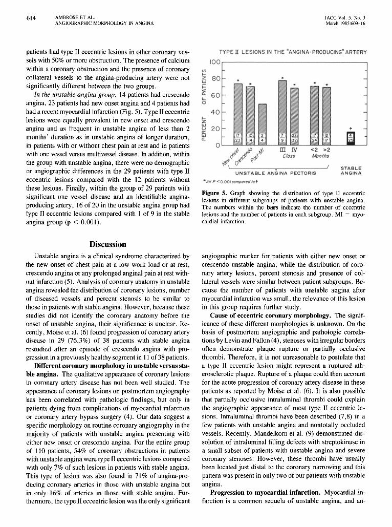
Figure 5. Graph showing the distribution of type II eccentriclesions in different subgroups of patients with unstable angina.The numbers within the bars indicate the number of eccentriclesions and the number of patients in each subgroup. MI = myocardial infarction.
.. All P <0.001comport!d fa t
STABLEANG INA
<2 >2Months
ill NClass
vU N S T A BL E ANG INA P EC TOR IS
4 0IZW
~ 20wa.
patients had type II eccentric lesions in other coronary vessels with 50% or more obstruction. The presence of calciumwithin a coronary obstruction and the presence of coronarycollateral vessels to the angina-producing artery were notsignificantly different between the two groups.
In the unstable angina group, 14 patients had crescendoangina, 23 patients had new onset angina and 4 patients hadhad a recent myocardial infarction (Fig. 5). Type II eccentriclesions were equally prevalent in new onset and crescendoangina and as frequent in unstable angina of less than 2months' duration as in unstable angina of longer duration,in patients with or without chest pain at rest and in patientswith one vessel versus multivessel disease. In addition, withinthe group with unstable angina, there were no demographicor angiographic differences in the 29 patients with type IIeccentric lesions compared with the 12 patients withoutthese lesions. Finally, within the group of 29 patients withsignificant one vessel disease and an identifiable anginaproducing artery, 16 of 20 in the unstable angina group hadtype II eccentric lesions compared with 1 of 9 in the stableangina group (p < 0.001).
Discussion
Unstable angina is a clinical syndrome characterized bythe new onset of chest pain at a low work load or at rest,crescendo angina or any prolonged anginal pain at rest without infarction (5). Analysis of coronary anatomy in unstableangina revealed the distribution of coronary lesions, numberof diseased vessels and percent stenosis to be similar tothose in patients with stable angina. However, because thesestudies did not identify the coronary anatomy before theonset of unstable angina, their significance is unclear. Recently, Moise et al. (6) found progression of coronary arterydisease in 29 (76.3%) of 38 patients with stable anginarestudied after an episode of crescendo angina with progression in a previously healthy segment in II of 38 patients.
Different coronary morphology in unstable versus stable angina. The qualitative appearance of coronary lesionsin coronary artery disease has not been well studied. Theappearance of coronary lesions on postmortem angiographyhas been correlated with pathologic findings, but only inpatients dying from complications of myocardial infarctionor coronary artery bypass surgery (4). Our data suggest aspecific morphology on routine coronary angiography in themajority of patients with unstable angina presenting witheither new onset or crescendo angina. For the entire groupof 110 patients, 54% of coronary obstructions in patientswith unstable angina were type II eccentric lesions comparedwith only 7% of such lesions in patients with stable angina.This type of lesion was also found in 71% of angina-producing coronary arteries in those with unstable angina butin only 16% of arteries in those with stable angina. Furthermore, the type II eccentric lesion was the only significant
angiographic marker for patients with either new onset orcrescendo unstable angina, while the distribution of coronary artery lesions, percent stenosis and presence of collateral vessels were similar between patient subgroups. Because the number of patients with unstable angina aftermyocardial infarction was small, the relevance of this lesionin this group requires further study.
Cause of eccentric coronary morphology. The significance of these different morphologies is unknown. On thebasis of postmortem angiographic and pathologic correlations by Levin and Fallon (4), stenoses with irregular bordersoften demonstrate plaque rupture or partially occlusivethrombi. Therefore, it is not unreasonable to postulate thata type II eccentric lesion might represent a ruptured atherosclerotic plaque. Rupture of a plaque could then accountfor the acute progression of coronary artery disease in thesepatients as reported by Moise et al. (6). It is also possiblethat partially occlusive intraluminal thrombi could explainthe angiographic appearance of most type II eccentric lesions. Intraluminal thrombi have been described (7,8) in afew patients with unstable angina and nontotally occludedvessels. Recently, Mandelkorn et al. (9) demonstrated dissolution of intraluminal filling defects with streptokinase ina small subset of patients with unstable angina and severecoronary stenoses. However, these thrombi have usuallybeen located just distal to the coronary narrowing and thispattern was present in only two of our patients with unstableangina.
Progression to myocardial infarction. Myocardial infarction is a common sequela of unstable angina, and an-

lACC Vol. 5, No. 3March 1985:609-16
AMBROSEET AL.ANGIOGRAPHIC MORPHOLOGY IN ANGINA
6 15
giographic progression from severe coronary obstruction tototal occlusion has been documented (10) , Although themechanisms involved in this progression are unknown ,pathologic studies (11,12) in patients dying from acute infarction have revealed a high incidence of thrombus overlying a disrupted atherosclerotic plaque . Our study suggestsa possible mechani sm for this progression in that the exposedintimal surfaces of these irregular stenoses may be foci forplatelet aggregation with subsequent thrombu s formation(13,14) or possible vasospasm secondary to release ofthromboxane A2 , or both (15 ,16). Furthermore , as the percent stenosis and presence of collateral vessels were similaramong patients with stable or unstable angina, transientplatelet aggregates or vasospasm on an exposed intimal surface, or both. might be respon sible for episodes of ischemicpain at rest in the absence of myocardial infarction . Conceivably, a decrease in perfusion in arter ies with these eccentri c lesions could be due solely to a decrease in thecoronary distending pressure (passive vasomotion) withoutimplicating vasospasm or platelet thrombi (17).
Limitations of the study. Patients. In this study . patients with unstable angina were limited primarily to thosewith new onset or crescendo angina. This included mostpatients with pain at rest , but excluded a small group ( I Ipatients) with a long history of severe angina and chronicpain at rest (> 6 month s' duration) that was included in thestable group . In this latter group , coronary angiographyrevealed multivessel disease in 8 of II patients and therewas only I patien t with a type II eccentric lesion. Therefore .the morphologic findings in new onset or crescendo anginaare not appl icable to all patient s with ischemic pain at rest.
Angiographic analysis and coronary morphology . Ouranalysis of coronary morphology was purely descriptive ,All previou s angiographic analyses in unstable angina havenot commented on the appearance of coronary lesions. Asnoted previously , Levin and Fallon (4) performed postmortem angiography in patients dying from compli cation sof myocardi al infarction or during coronary artery bypasssurgery. They noted that coronary lesions with irregularborders or intraluminal lucencies that were termed " complicated lesions" exhibited plaque rupture or partially occlusive thromb i on pathologic sectioning, Their definitionof complicated lesions would probably have included ourclassification of type II eccentric and multiple irregularities.However. since patient selection and angiographic techniques were not comparable , these differences in morphologic description cannot be resolved at the present time.
Although we suspect that these type II eccentric lesionsrepresent ruptured plaques or thrombi . the significance ofthe other morphologic subtypes is unclear. Although coronary lesions classified as multiple irregularities may havecontained plaque rupture or thrombi. no correlation to newonset or crescendo angina was found. In addit ion. the lengthof the stenosis, the minimal cross-sectional area or the ef-
fects of serial stenoses were not considered in this study.Therefore, further study in this area appears warranted .
" Angina-producing" artery . The concept of an " angina-producing" artery was developed to localize the coronary obstruction responsible for angina pectoris in patientswith single and multive ssel disease. Since no patient hadvariant angina. chest pain was assumed to originate onlyfrom vessels with significant obstructions. In the majorityof patients (49 of 66). either a single obstruction or ischemi carea localized by noninvasive tests was present , whereas inthe other 17. indirect methods were utilized . Therefore. theangina-producing artery was probabl y correctly identifiedin all patients.
Clinical implications. Unstable angina is intermediatein its presentation between stable angina and myocardialinfarction . Although the risk of a subsequent coronary eventis higher in unstable ang ina than in stable angina. the greatest period of risk appears to be soon after the change insymptoms (18). Identification of this patient group at riskfor a subsequent coron ary event would be extremely important in plann ing appropriate therapy.
We have shown that the type II eccentric lesion is acommon angiographic finding in the majorit y of patientswith either new onset or crescendo unstable angina. Wesuspect that this lesion is caused by a ruptured plaque or apartially occlusive thrombu s, or both , which acutel y compromises the coronary circulation and plays an importantrole as the source of the unstable ischem ia. Unfortunately.the natural history of type II eccentric lesions in this studyis unknown since most patients with unstable angina had anintervention after angiography . As progression to completecoronary occlusion and myocardial infarction is thought tobe related to thrombu s formation. the " thrombogenicity"of various morphologic lesions should be a topic of futurestudy. Analysis of coron ary sinus blood for the presence ofvarious vasoactive substances may ident ify the group athighest risk for a subsequent coronary event. It may beprudent . however. in the absence of these data to administeranticoagulants to all patient s with new onset or crescendounstable angina using either antiplatelet or heparin-like agentsbecause most of these patients will have type II lesions.Two recent studies (19,20) that showed a decreased incidence of subsequent coronary event s in patients with unstable angina treated with either low dose aspirin or heparinwould support this suggestion.
ReferencesI. Maseri A, Chierchia S, L'Abb ate A. Pathogenetic mechanisms un
derlying the clinical events associated with atherosclerotic heart disease. Circulation 1980;62(suppl V):V-3- 13.
2. Alison HW, Russel RO Jr, Mantle lA , Kouchoukos NT, Moraski RE,Rackley CEo Coronary anatomy and arteriography in patients withunstable angina pectoris. Am J Cardiol 1978;41:204-9.
3. Fuster V, Frye RL, Connolly DC, Danielson MA. Elveback LR.

616 AMBROSE ET AL.ANGIOGRAPHIC MORPHOLOGY IN ANGINA
lACC Vol. 5, No.3March 1985:609-16
Kurland LT. Arteriographic patterns early in the onset of the coronarysyndromes. Br Heart 1 1975;37:1250-5.
4. Levin DC, Fallon JT. Significance of the angiographic morphologyof localized coronary stenosis. Histopathologic correlations. Circulation 1982;66:316-20.
5. Cohn PF, Braunwald E. Chronic coronary artery disease. In: Braunwald E, ed. Heart Disease, vol. I. Philadelphia: WB Saunders,1980:1480-512.
6. Moise A, Theroux P, Taeymans Y, et al. Unstable angina and progression of coronary atherosclerosis. N Engl 1 Med 1983;309:685-9.
7. Vetrovec GW, Cowley Ml, Overton H, Richardson OW. Intracoronary thrombus in syndromes of unstable myocardial ischemia. AmHeart 11981;102:1202-8.
8. Holmes DR Jr, Hartzler GO, Smith HC, Fuster V. Coronary arterythrombosis in patients with unstable angina. Br Heart 1 1981;45:411-6.
9. Mandelkorn IB, Wolf MN, Singh S, et al. Intracoronary thrombus innontransmural myocardial infarction and in unstable angina pectoris.Am 1 Cardiol 1983;52:1-6.
10. Neill WA, Wharton TP Jr, Fluri-Lundeen 1, Cohen IS. Acute coronaryinsufficiency. Coronary occlusion after intermittent ischemic attacks.N Engl 1 Med 1980;302:1157-62.
11. Chapman I. Morphogenesis of occluding coronary artery thrombosis.Arch Pathol 1965;80:256-61.
12. Chandler AB, Chapman I, Erhardt LR, et al. Coronary thrombosis in
myocardial infarction. Report of a workshop on the role of coronarythrombosis in the pathogenesis of acute myocardial infarction. Am 1Cardiol 1974;34:823-33.
13. Erhardt LR, Lundman T, Mellstedt H. Incorporation of '25I-labelledfibrinogen into coronary arterial thrombi in acute myocardial infarctionin man. Lancet 1973;1:387-90.
14. Fuster V, Chesebro JH. Current concepts of thrombogenesis. Role ofplatelets. Mayo Clin Proc 1981;56:102-12.
15. Hamberg M, Svensson 1, Samuelsson B. Thromboxanes: a new groupof biologically active compounds derived from prostaglandin endoperoxides. Proc Natl Acad Sci USA 1975;72:2994-8.
16. Hirsh PO, Hillis LD, Campbell WB, Firth BG, Willerson JT. Releaseof prostaglandins and thromboxane into the coronary circulation inpatients with ischemic heart disease. N Engl 1 Med 1981;304:685-91.
17. Schwartz IS, Carlyle PF, Cohen IN. Effect of coronary arterial pressure on coronary stenosis resistance. Circulation 1980;61:70-6.
18. Duncan B, Fulton M, Morrison SL, et al. Prognosis of new andworsening angina pectoris. Br Med 1 1976;1:981-5.
19. Telford AM, Wilson C. Trial of heparin versus atenolol in preventionof myocardial infarction in intermediate coronary syndrome. Lancet1981;1:1225-8.
20. Lewis HD Jr. Protective effects of aspirin against acute myocardialinfarction and death in men with unstable angina: results of a VeteransAdministration Cooperative Study. N Engl 1 Med 1983;309:396-403.


