Angiogenesis: Potentials for Pharmacologic … Potentials for Pharmacologic Intervention in the...
32
Angiogenesis: Potentials for Pharmacologic Intervention in the Treatment of Cancer, Cardiovascular Diseases, and Chronic Inflammation ARJAN W. GRIFFIOEN 1 AND GRIETJE MOLEMA Tumor Angiogenesis Laboratory (A.W.G.), Department of Internal Medicine, University Hospital Maastricht, Maastricht; Groningen University Institute for Drug Exploration (G.M.), Department of Pathology and Laboratory Medicine, Tumor Immunology Laboratory, and Department of Pharmacokinetics and Drug Delivery, Groningen, The Netherlands This paper is available online at http://www.pharmrev.org Abstract ................................................................................ 238 I. General aspects of angiogenesis ........................................................... 238 A. Introduction ......................................................................... 238 B. Function of endothelial cells in normal physiology ....................................... 239 C. Molecular control of angiogenesis ...................................................... 239 1. Initiation of the angiogenic response ................................................ 239 2. Endothelial cell migration and proliferation .......................................... 240 3. Maturation of the neovasculature ................................................... 241 4. Other mechanisms implicated in angiogenesis control ................................. 242 II. Angiogenesis stimulation ................................................................. 243 A. Target diseases for angiogenesis stimulation ............................................ 243 B. Proangiogenic compounds ............................................................. 243 1. Vascular endothelial growth factor .................................................. 243 2. Fibroblast growth factors ........................................................... 245 3. Angiopoietin-1 .................................................................... 245 C. Effects of angiogenesis stimulation in preclinical studies ................................. 245 D. First clinical studies on angiogenesis stimulation ........................................ 246 III. Angiogenesis inhibition .................................................................. 246 A. Angiogenesis and cancer .............................................................. 246 B. In vitro and in vivo models to study angiogenesis ........................................ 247 C. Ways to interfere with angiogenesis .................................................... 248 1. Intervention with endothelial cell growth ............................................ 248 2. Intervention with endothelial cell adhesion and migration ............................. 249 3. Intervention with metalloproteinases ................................................ 249 D. Preclinical use of angiogenesis inhibitors in cancer ...................................... 250 E. Clinical trials with inhibitors of angiogenesis for cancer treatment ........................ 250 F. Novel approaches to interfere with tumor blood flow ..................................... 253 1. Targeted strategies to induce tumor blood coagulation ................................ 253 2. Targeted strategies to kill tumor endothelial cells .................................... 254 3. The quest for new targets on tumor endothelium ..................................... 255 IV. The interplay between angiogenesis and cells of the immune system.......................... 256 A. Angiogenesis regulates leukocyte recruitment ........................................... 256 B. The role of angiogenesis in chronic inflammation ........................................ 257 C. Inhibition of angiogenesis in chronic inflammation....................................... 259 D. Clinical trials with inhibitors of angiogenesis for noncancerous diseases ................... 260 V. Back to the drawing board ............................................................... 260 A. Angiogenesis stimulation.............................................................. 260 B. Antiangiogenic strategies in cancer therapy ............................................. 260 C. Antiangiogenic strategies in chronic inflammation ....................................... 262 1 Address for correspondence: Dr. A.W. Griffioen, Tumor Angiogenesis Laboratory, Dept. of Internal Medicine, University Hospital Maastricht, P.O. Box 5800, 6202 AZ Maastricht, The Netherlands. E-mail: [email protected] 0031-6997/00/5202-0237$03.00/0 PHARMACOLOGICAL REVIEWS Vol. 52, No. 2 Copyright © 2000 by The American Society for Pharmacology and Experimental Therapeutics Printed in U.S.A. PR 52:237–268, 2000 /3000/826808 237
Transcript of Angiogenesis: Potentials for Pharmacologic … Potentials for Pharmacologic Intervention in the...

Angiogenesis: Potentials for PharmacologicIntervention in the Treatment of Cancer,
Cardiovascular Diseases, and Chronic InflammationARJAN W. GRIFFIOEN1 AND GRIETJE MOLEMA
Tumor Angiogenesis Laboratory (A.W.G.), Department of Internal Medicine, University Hospital Maastricht, Maastricht; GroningenUniversity Institute for Drug Exploration (G.M.), Department of Pathology and Laboratory Medicine, Tumor Immunology Laboratory, and
Department of Pharmacokinetics and Drug Delivery, Groningen, The Netherlands
This paper is available online at http://www.pharmrev.org
Abstract . . . . . . . . . . . . . . . . . . . . . . . . . . . . . . . . . . . . . . . . . . . . . . . . . . . . . . . . . . . . . . . . . . . . . . . . . . . . . . . . 238I. General aspects of angiogenesis . . . . . . . . . . . . . . . . . . . . . . . . . . . . . . . . . . . . . . . . . . . . . . . . . . . . . . . . . . . 238
A. Introduction . . . . . . . . . . . . . . . . . . . . . . . . . . . . . . . . . . . . . . . . . . . . . . . . . . . . . . . . . . . . . . . . . . . . . . . . . 238B. Function of endothelial cells in normal physiology . . . . . . . . . . . . . . . . . . . . . . . . . . . . . . . . . . . . . . . 239C. Molecular control of angiogenesis . . . . . . . . . . . . . . . . . . . . . . . . . . . . . . . . . . . . . . . . . . . . . . . . . . . . . . 239
1. Initiation of the angiogenic response . . . . . . . . . . . . . . . . . . . . . . . . . . . . . . . . . . . . . . . . . . . . . . . . 2392. Endothelial cell migration and proliferation . . . . . . . . . . . . . . . . . . . . . . . . . . . . . . . . . . . . . . . . . . 2403. Maturation of the neovasculature . . . . . . . . . . . . . . . . . . . . . . . . . . . . . . . . . . . . . . . . . . . . . . . . . . . 2414. Other mechanisms implicated in angiogenesis control . . . . . . . . . . . . . . . . . . . . . . . . . . . . . . . . . 242
II. Angiogenesis stimulation . . . . . . . . . . . . . . . . . . . . . . . . . . . . . . . . . . . . . . . . . . . . . . . . . . . . . . . . . . . . . . . . . 243A. Target diseases for angiogenesis stimulation . . . . . . . . . . . . . . . . . . . . . . . . . . . . . . . . . . . . . . . . . . . . 243B. Proangiogenic compounds . . . . . . . . . . . . . . . . . . . . . . . . . . . . . . . . . . . . . . . . . . . . . . . . . . . . . . . . . . . . . 243
1. Vascular endothelial growth factor . . . . . . . . . . . . . . . . . . . . . . . . . . . . . . . . . . . . . . . . . . . . . . . . . . 2432. Fibroblast growth factors. . . . . . . . . . . . . . . . . . . . . . . . . . . . . . . . . . . . . . . . . . . . . . . . . . . . . . . . . . . 2453. Angiopoietin-1 . . . . . . . . . . . . . . . . . . . . . . . . . . . . . . . . . . . . . . . . . . . . . . . . . . . . . . . . . . . . . . . . . . . . 245
C. Effects of angiogenesis stimulation in preclinical studies . . . . . . . . . . . . . . . . . . . . . . . . . . . . . . . . . 245D. First clinical studies on angiogenesis stimulation . . . . . . . . . . . . . . . . . . . . . . . . . . . . . . . . . . . . . . . . 246
III. Angiogenesis inhibition . . . . . . . . . . . . . . . . . . . . . . . . . . . . . . . . . . . . . . . . . . . . . . . . . . . . . . . . . . . . . . . . . . 246A. Angiogenesis and cancer . . . . . . . . . . . . . . . . . . . . . . . . . . . . . . . . . . . . . . . . . . . . . . . . . . . . . . . . . . . . . . 246B. In vitro and in vivo models to study angiogenesis. . . . . . . . . . . . . . . . . . . . . . . . . . . . . . . . . . . . . . . . 247C. Ways to interfere with angiogenesis . . . . . . . . . . . . . . . . . . . . . . . . . . . . . . . . . . . . . . . . . . . . . . . . . . . . 248
1. Intervention with endothelial cell growth . . . . . . . . . . . . . . . . . . . . . . . . . . . . . . . . . . . . . . . . . . . . 2482. Intervention with endothelial cell adhesion and migration . . . . . . . . . . . . . . . . . . . . . . . . . . . . . 2493. Intervention with metalloproteinases . . . . . . . . . . . . . . . . . . . . . . . . . . . . . . . . . . . . . . . . . . . . . . . . 249
D. Preclinical use of angiogenesis inhibitors in cancer . . . . . . . . . . . . . . . . . . . . . . . . . . . . . . . . . . . . . . 250E. Clinical trials with inhibitors of angiogenesis for cancer treatment . . . . . . . . . . . . . . . . . . . . . . . . 250F. Novel approaches to interfere with tumor blood flow . . . . . . . . . . . . . . . . . . . . . . . . . . . . . . . . . . . . . 253
1. Targeted strategies to induce tumor blood coagulation . . . . . . . . . . . . . . . . . . . . . . . . . . . . . . . . 2532. Targeted strategies to kill tumor endothelial cells . . . . . . . . . . . . . . . . . . . . . . . . . . . . . . . . . . . . 2543. The quest for new targets on tumor endothelium . . . . . . . . . . . . . . . . . . . . . . . . . . . . . . . . . . . . . 255
IV. The interplay between angiogenesis and cells of the immune system. . . . . . . . . . . . . . . . . . . . . . . . . . 256A. Angiogenesis regulates leukocyte recruitment . . . . . . . . . . . . . . . . . . . . . . . . . . . . . . . . . . . . . . . . . . . 256B. The role of angiogenesis in chronic inflammation . . . . . . . . . . . . . . . . . . . . . . . . . . . . . . . . . . . . . . . . 257C. Inhibition of angiogenesis in chronic inflammation. . . . . . . . . . . . . . . . . . . . . . . . . . . . . . . . . . . . . . . 259D. Clinical trials with inhibitors of angiogenesis for noncancerous diseases . . . . . . . . . . . . . . . . . . . 260
V. Back to the drawing board . . . . . . . . . . . . . . . . . . . . . . . . . . . . . . . . . . . . . . . . . . . . . . . . . . . . . . . . . . . . . . . 260A. Angiogenesis stimulation. . . . . . . . . . . . . . . . . . . . . . . . . . . . . . . . . . . . . . . . . . . . . . . . . . . . . . . . . . . . . . 260B. Antiangiogenic strategies in cancer therapy . . . . . . . . . . . . . . . . . . . . . . . . . . . . . . . . . . . . . . . . . . . . . 260C. Antiangiogenic strategies in chronic inflammation . . . . . . . . . . . . . . . . . . . . . . . . . . . . . . . . . . . . . . . 262
1 Address for correspondence: Dr. A.W. Griffioen, Tumor Angiogenesis Laboratory, Dept. of Internal Medicine, University HospitalMaastricht, P.O. Box 5800, 6202 AZ Maastricht, The Netherlands. E-mail: [email protected]
0031-6997/00/5202-0237$03.00/0PHARMACOLOGICAL REVIEWS Vol. 52, No. 2Copyright © 2000 by The American Society for Pharmacology and Experimental Therapeutics Printed in U.S.A.PR 52:237–268, 2000 /3000/826808
237

VI. Concluding remarks . . . . . . . . . . . . . . . . . . . . . . . . . . . . . . . . . . . . . . . . . . . . . . . . . . . . . . . . . . . . . . . . . . . . . 263Acknowledgments . . . . . . . . . . . . . . . . . . . . . . . . . . . . . . . . . . . . . . . . . . . . . . . . . . . . . . . . . . . . . . . . . . . . . . . 263References . . . . . . . . . . . . . . . . . . . . . . . . . . . . . . . . . . . . . . . . . . . . . . . . . . . . . . . . . . . . . . . . . . . . . . . . . . . . . . 263
Abstract——Angiogenesis, or the formation of newblood vessels out of pre-existing capillaries, is a se-quence of events that is fundamental to many physio-logic and pathologic processes such as cancer, isch-emic diseases, and chronic inflammation. With theidentification of several proangiogenic moleculessuch as the vascular endothelial cell growth factor, thefibroblast growth factors (like in FGFs), and the an-giopoietins, and the recent description of specific in-hibitors of angiogenesis such as platelet factor-4, an-giostatin, endostatin, and vasostatin, it is recognizedthat therapeutic interference with vasculature forma-tion offers a tool for clinical applications in various
pathologies. Whereas inhibition of angiogenesis canprevent diseases with excessive vessel growth such ascancer, diabetes retinopathy, and arthritis, stimula-tion of angiogenesis would be beneficial in the treat-ment of diseases such as coronary artery disease andcritical limb ischemia in diabetes. In this review wehighlight the current knowledge on angiogenesis reg-ulation and report on the recent findings in angiogen-esis research and clinical studies. We also discuss thepotentials, limitations, and challenges within this fieldof research, in light of the development of new thera-peutic strategies for diseases in which angiogenesisplays an important role.
I. General Aspects of Angiogenesis
A. Introduction
The formation of new blood vessels out of pre-existingcapillaries, or angiogenesis, is a sequence of events thatis of key importance in a broad array of physiologic andpathologic processes. Normal tissue growth, such as inembryonic development, wound healing, and the men-strual cycle, is characterized by dependence on new ves-sel formation for the supply of oxygen and nutrients aswell as removal of waste products. Also, a large numberof different and nonrelated diseases is associated withformation of new vasculature. Among these pathologiesare diseases, such as tissue damage after reperfusion ofischemic tissue or cardiac failure, where angiogenesis islow and should be enhanced to improve disease condi-tions (Carmeliet et al., 1999; Ferrara and Alitalo, 1999).In several diseases, excessive angiogenesis is part of thepathology. These diseases include cancer (both solid andhematologic tumors), cardiovascular diseases (athero-sclerosis), chronic inflammation (rheumatoid arthritis,Crohn’s disease), diabetes (diabetic retinopathy), psori-asis, endometriosis, and adiposity. These diseases may
benefit from therapeutic inhibition of angiogenesis(Folkman, 1995; Hanahan and Folkman, 1996).
The initial recognition of angiogenesis being a thera-peutically interesting process began in the area of oncol-ogy in the early 1970s, when Drs. Folkman andDenekamp put forward the idea that tumors are highlyvascularized and thereby vulnerable at the level of theirblood supply. In those early years, it was already hy-pothesized that the process of angiogenesis might be atarget for therapy. It was only after the discovery of thefirst compounds with specific angiostatic effects in theearly 1990s (Ingber et al., 1990; O’Reilly et al., 1994)that the research field of angiogenesis rapidly expandedand provided an increasing body of evidence that inhi-bition of angiogenesis could attenuate tumor growth.More recently, novel angiogenesis inhibitors have showngreat potential in the treatment of cancer in preclinicalstudies. Several of those compounds are currently beingtested in clinical trials (Molema and Griffioen, 1998).With increasing insight into the role of angiogenesis inother diseases as well, modulation of vascular outgrowthis now also regarded as a therapeutic target in thesediseases.
To date, antiangiogenesis therapy is considered,worldwide, a promising approach, supposedly leading tothe desperately needed breakthrough in cancer therapyand other proangiogenic diseases. Nevertheless, manyquestions remain unanswered and many concepts un-verified at present. For example, it has to be establishedwhether the exciting effects seen in preclinical investi-gations using xenogeneic and syngeneic tumor trans-plant models and transgenic systems also prevail in thehuman situation. Furthermore, it has been shown thatthe angiogenesis inhibitors angiostatin and endostatin(O’Reilly et al., 1994, 1997) do not elicit drug-inducedresistance on prolonged treatment in tumor-bearing an-imals, although being highly effective in tumor growth
2 Abbreviations: BM, basement membrane; Ang-1, angiopoietin-1;BsAb, bispecific antibody; CAI, carboxyamidotriazole; ECM, extra-cellular matrix; HIF, hypoxia-inducible factor; HUVEC, human um-bilical vein endothelial cells; FGF, fibroblast growth factor; FGF-R,FGF receptor; ICAM-1, intercellular adhesion molecule-1; IFN, in-terferon; IL, interleukin; MAPK, mitogen-activated protein kinase;MEK, MAPK kinase; MMP, matrix metalloproteinase; NFAT, nu-clear factor of activated T cells; NO, nitric oxide; PDGF, platelet-derived growth factor; t-PA/u-PA, tissue type/urokinase plasminogenactivator; SPARC, secreted protein acidic and rich in cysteine; TGF,transforming growth factor; TNF, tumor necrosis factor; tTF, trun-cated tissue factor; VCAM-1, vascular cell adhesion molecule-1;VEGF, vascular endothelial growth factor; VEGF-R, VEGF receptor;phVEGF165, plasmid-encoding human VEGF165 isoform; vWF, vonWillebrand factor/factor VIII-related antigen.
238 GRIFFIOEN AND MOLEMA

reduction. This observation is of extreme importance,because it opens possibilities for long-term treatment orthe development of treatment modalities for the preven-tion of disease in high-risk populations prone to developtumors. It remains to be seen whether this scenario canbe extended to other angiogenesis inhibitors as well as toother proangiogenic diseases of interest. Another impor-tant issue is the concept of cancer treatment with angio-genesis inhibitors as a single-compound strategy. Is thisa feasible treatment strategy or should antiangiogenictherapy be used in combination with other treatmentmodalities such as immuno- or chemotherapy? Also, al-though antiangiogenesis therapy is considered to havelow toxicity, there is as yet little information on thesafety of therapeutic angiostatic strategies; there is littleor no information to what extent inhibition of angiogen-esis as tumor treatment will affect normal physiologicalprocesses in embryonic development or in wound heal-ing and what the long-term side effects will be.
Although current interest in angiogenesis comesmainly from oncology researchers, also nononcologicalresearch fields have now recognized that modulation ofangiogenesis may provide a tool for clinical interven-tions. This demonstrates that angiogenesis is a multi-disciplinary theme from a pharmacologic target point ofview. In addition, many disciplines of biomedical originare contributing to basic angiogenesis research, becausethe processes involved are so complex. In this review,the molecular players of vessel growth, methodology ofangiogenesis research, and preclinical and clinical use ofangiogenesis as a target for therapy will be discussed.
B. Function of Endothelial Cells in Normal Physiology
The blood vessels in the body have long been consid-ered to merely function as a transport compartment ofthe blood. Nowadays, it is appreciated that the vascula-ture is one of the main organs in the body, extendingmore than 900 m2 and playing a major role in maintain-ing the body’s integrity in various ways.
Blood vessels consist of endothelial cells that are indirect contact with the blood, and subendothelially lo-cated pericytes, smooth muscle cells, fibroblasts, base-ment membrane (BM),1 and extracellular matrix (ECM).Depending on the location in the body, the organ micro-environment, the cellular constituents, BM, and ECM ofthe vasculature differ in phenotype, composition, andfunction (Rajotte et al., 1998).
The endothelial cells form a monolayer in every singleblood vessel in the circulation and are actively involvedin several regulatory processes in the body (Fig. 1). Be-sides being metabolically active and selectively perme-able for small solutes and peptides/proteins, they regu-late blood coagulation. When their integrity is maintained,endothelial cells exert anticoagulative properties via thesynthesis of thrombomodulin, tissue factor (TF) path-way inhibitor and tissue-type plasminogen activator (t-PA). On activation or damage, endothelial cells quickly
release proteins like multimeric von Willebrand factor(vWF), which promotes platelet adhesion and aggrega-tion, and plasminogen activator inhibitor-1, a member ofthe serpin family. In addition, TF expression by endo-thelium leads to initiation of the extrinsic blood coagu-lation pathway (Verstraete, 1995). Another importantfeature of endothelial cells is their ability to direct cellsof the immune system to specific sites in the body. Con-stitutively expressed or cytokine-inducible cellular ad-hesion molecules [e.g., E-selectin and intercellular adhe-sion molecule-1 (ICAM-1)] and soluble factors such aschemoattractants, cytokines, and chemokines act in con-cert to recruit the immune cells to lymphoid organs orinflammatory sites (Carlos and Harlan, 1994). Last, en-dothelial cells are actively involved in vascular remod-eling during, for example, ovulation, wound healing,tumor growth, and diabetic retinopathy. Although com-plex in regulation and sometimes difficult to function-ally analyze in vitro, as well as during disease progres-sion, data have become available that link (parts of)these endothelial cell functions to various steps in theangiogenic cascade.
C. Molecular Control of Angiogenesis
In vasculogenesis during embryonic development,new endothelial cells differentiate from stem cells. Incontrast, in angiogenesis new blood vessels mainlyemerge from pre-existing ones (Risau, 1997). In adultlife, physiologic stimuli during wound healing and thereproductive cycle in women lead to angiogenesis,whereas vasculogenesis is absent. Pathologic conditionssuch as tumor growth, rheumatoid arthritis, and dia-betic retinopathy are characterized by abundant angio-genesis. The active vascular remodeling phase in tu-mors, e.g., is reflected by the fact that tumor endothelialcells proliferate 20 to 2000 times faster than normaltissue endothelium in the adult (Denekamp, 1984). Inthe last decade, several molecular players have beenidentified that significantly contribute to the molecularprocesses leading to new blood vessel formation. In thefollowing sections, recent advances in this area of re-search are discussed.
1. Initiation of the Angiogenic Response. Angiogen-esis is rapidly initiated in response to hypoxic or isch-emic conditions. Vascular relaxation, for example, medi-ated by nitric oxide (NO) is a prerequisite for endothelialcells to enter the angiogenic cascade. Likely, morpho-logic changes of the endothelial cells lead to a decreasein confluency status to make them susceptible to mito-gens (Folkman, 1997). In all types of angiogenesis, ei-ther under physiologic or pathologic conditions, endothe-lial cell activation is the first process to take place.Cytokines from various sources are released in responseto hypoxia or ischemia. It is suggested that vascularendothelial growth factor (VEGF) is a major player inangiogenesis initiation based on its ability to induce
ANGIOGENESIS AND DISEASE 239

vasodilation via endothelial NO production and its en-dothelial cell permeability increasing effect (Ziche et al.,1997). This allows plasma proteins to enter the tissue toform a fibrin-rich provisional network (Dvorak, 1986).The observation that VEGF production is under controlof hypoxia inducible factor (HIF) strengthens the sug-gestion of an early involvement of VEGF in the angio-genic response. Moreover, VEGF receptor (VEGFR) ex-pression is up-regulated under hypoxic or ischemicconditions as well (Forsythe et al., 1996).
VEGF is abundantly produced by hypoxic tumor cells,macrophages and other cells of the immune system(Brown et al., 1997). Besides affecting vasodilation andvascular permeability, VEGF can induce the expressionof proteases and receptors important in cellular invasionand tissue remodeling and is able to prevent endothelialcell apoptosis (Ferrara and Keyt, 1997; Gupta et al.,1999). That angiogenesis is not completely dependent onVEGF production was recently shown by Hansen-Algen-staedt et al. (1999); the consequences of which will bediscussed in more detail in Section V. For a more de-tailed overview on the role of VEGF in the regulation ofangiogenesis, the reader is referred to a recently pub-lished review by Ferrara (1999).
After proper activation of the endothelial cells, endo-thelial penetration into new areas of the body isachieved by degradation of the BM by matrix metallo-proteinases (MMPs). These extracellular endopepti-dases are secreted as zymogens that become activated inthe ECM compartment and subsequently selectively de-grade components of the ECM (Stetler Stevenson, 1999).They are produced by a variety of cells, including epi-thelial cells, fibroblasts, inflammatory cells, and endo-thelial cells. MMP activity and, hence, angiogenesis iscounteracted by the family of tissue inhibitors of metal-loproteinase (TIMPs) (Gomez et al., 1997; Valente et al.,1998).
2. Endothelial Cell Migration and Proliferation.Plasminogen activators u-PA and t-PA convert the ubiq-uitous plasma protein plasminogen to plasmin. Plasminhas a broad trypsin-like specificity and degrades, e.g.,fibronectin, laminin, and the protein core of proteogly-cans. In addition, plasmin activates certain metallopro-teinases. Plasmin is believed to be the most importantprotease for the mobilization of fibroblast growth fac-tor-2 (FGF-2 or basic FGF) from the ECM pool.
FGF members are directly acting proangiogenic mol-ecules. FGF-2 consists of, in two modifications, an 18-
FIG. 1. Endothelial cells exert several important functions in the body. A, the endothelium forms a semipermeable barrier for the transport ofblood-borne peptides, proteins, and other soluble molecules to underlying tissue; B, via the regulated expression of pro- and anticoagulative activities,endothelium actively participates in the hemostatic balance in the body; C, under the influence of proinflammatory cytokines, endothelial cellsup-regulate a variety of cellular adhesion molecules to tether and activate leukocytes and facilitate leukocyte adhesion and transmigration from theblood into the tissue; D, during wound healing and tumor growth, among others, angiogenesis takes place. In this process, an active role exists forendothelial cells. EC, endothelial cells; IgSF, Ig superfamily; PAI-1, plasminogen activator inhibitor; PBMC, peripheral blood mononuclear cells; sLex,sialyl Lewis X; TFPI, Tissue Factor pathway inhibitor; TM, thrombomodulin.
240 GRIFFIOEN AND MOLEMA

kDa low-molecular weight form and a 22- to 24-kDahigh-molecular weight form. During angiogenesis, low-molecular weight FGF-2 binding to endothelium inducesFGF receptor (FGF-R) down-regulation, increased mo-tility, proliferation and proteinase activity, and modu-lates integrin levels. High-molecular weight FGF-2 mayact on endothelial cell proliferation after nuclear trans-location in the endothelial cells (Gleizes et al., 1995;Klein et al., 1997). Recently, it was shown that a se-creted FGF-2-binding protein could bind FGF-2 that isnormally inactive due to strong adherence to heparansulfate proteoglycans in the ECM. The displaced FGF-2molecules were thus released to mediate biological func-tion. Of note is the observation in this and other studiesthat angiogenesis seems exquisitely sensitive to smallchanges in factors such as VEGF and FGF-2 that drivethe angiogenic process. This may have important ther-apeutic implications in treating angiogenesis-driven dis-orders (Czubayko et al., 1997). Besides its effect on an-giogenesis initiation, VEGF also affects endothelial cellproliferation. This effect can be (partly) attributed to NOand cGMP-mediated activation of the mitogen-activatedprotein kinase (MAPK) family (see Section II.B. for amore detailed description on VEGF and FGF-2-medi-ated signal transduction and cell activation pathways).
Integrins are transmembrane proteins composed of ana and b subunit in over 20 different heterodimeric com-binations. They bind to ECM proteins or cell surfaceligands through short peptide sequences. Combinationsof different integrins on cell surfaces allow cells to rec-ognize and respond to a variety of different ECM pro-teins (Varner, 1997). They are able to transduce signalsfrom within the cells to the outside as well as from theoutside into the cell (Aplin et al., 1998). Integrin-medi-ated cell adhesion impacts two key aspects of growthregulation. First, it can influence the activity of thebasal cell cycle machinery consisting of cyclin-dependentkinase complexes. Second, integrins play a pivotal rolein anchorage-dependent cell death or anoikis (Frischand Ruoslahti, 1997; Howe et al., 1998). Integrin avb3mediates cellular adhesion to vitronectin, fibrinogen,laminin, collagen, vWF, or osteopontin through theirexposed tripeptide Arg-Gly-Asp (RGD) moiety (Cheresh,1993). avb3 is minimally expressed on normal restingendothelium, but significantly up-regulated on activatedendothelium and is believed to play a critical role inangiogenesis. Both peptide and antibody inhibitors ofavb3 induced endothelial cell apoptosis, suggesting arole for this integrin in endothelial cell survival duringangiogenesis (Brooks et al., 1994a). Another av integrinassociated with angiogenesis is avb5. Whereas FGF-2 ortumor necrosis factor-a (TNF-a) induced avb3-depen-dent angiogenesis in vivo, VEGF or transforming growthfactor-b (TGF-b) initiated an angiogenesis pathwaymerely dependent on avb5 (Friedlander et al., 1995).
Components of the ECM also contribute to the regu-lation of endothelial cell morphology and function.
Thrombospondin, for example, inhibits endothelial cellproliferation when added in soluble form. When endo-thelial cells on the other hand are plated on matrixbound thrombospondin, they become more permissivefor proliferative signals. Furthermore, through bindingto and activation of TGF-b and affecting protease activ-ity, thrombospondin may be able to influence cellgrowth, migration, and differentation as well (DiPietro,1997). In patients with invasive bladder cancer, low-thrombospondin expression in the tumor was associatedwith increased recurrence rates, decreased overall sur-vival, and high-microvessel density counts. These dataare suggestive of an antiangiogenic role for this ECMconstituent under physiologic conditions (Grossfeld etal., 1997). Laminin is another ECM protein with func-tions in endothelial cell attachment, growth promotion,protease secretion, and interactions with other ECMcomponents. Laminin can bind to cell surface-bindingproteins including integrins, which leads to integrin sig-naling (Grant and Kleinman, 1997). SPARC (secretedprotein acidic and rich in cysteine), also known as BM40or osteonectin, is a protein of which the expression iselevated under stress conditions such as endotoxin stim-ulation, heat shock, and sparse cell density. SPARCoverexpression has been observed in tumors such ashuman esophageal carcinoma and cutaneous malignantmelanoma (Porte et al., 1998; Massi et al., 1999). Fur-thermore, transient expression of SPARC during endo-thelial cell injury and cellular activation indicated a rolein tissue repair, remodeling and angiogenesis (Jendras-chak and Sage, 1996).
3. Maturation of the Neovasculature. Endothelialcell interaction with ECM and mesenchymal cells is aprerequisite to form a stable vasculature. Therefore,after endothelial cell proliferation and maturation, andthe formation of endothelial tube structures, surround-ing vessel layers composed of mural cells (pericytes insmall vessels and smooth muscle cells in large vessels),need to be recruited. Endothelial cells may accomplishthis via the synthesis and secretion of platelet-derivedgrowth factor (PDGF), a mitogen and chemoattractantfor a variety of mesenchymal cells. Subsequent differen-tiation of the mural precursor cells into pericytes andsmooth muscle cells is believed to be a cell-cell contact-dependent process. On endothelial cell-mural cell con-tact, a latent form of TGF-b, produced by both endothe-lium and mural cells, is activated in a plasmin-mediatedprocess. Activated TGF-b can induce changes in myofi-broblasts and pericytes, which may contribute to theformation of a quiescent vessel, ECM production, andmaintenance of growth control. The coinciding invest-ment of growing capillaries by pericytes with the depo-sition of BM and cessation of vessel growth duringwound healing also indicates vessel growth regulationby pericytes (Hirschi and D’Amore, 1997). FGF-1 is alsoimplicated in endothelial cell differentiation leading tovascular tube formation. Besides inducing plasminogen
ANGIOGENESIS AND DISEASE 241

activator and endothelial cell proliferation and migra-tion, FGF-1 receptor signaling resulted in endothelialtube formation in collagen (Kanda et al., 1996).
Angiopoietins and receptor tyrosine kinase Tie1 andTie2 play critical roles in the later stages of angiogenesisas well. They are required for communication of endo-thelial cells with the surrounding mesenchyme to estab-lish stable cellular and biochemical interactions (Mai-sonpierre et al., 1997). Tie1 function is related toendothelial cell differentiation and the establishment ofblood vessel integrity. Tie2, on the other hand, is partic-ularly important for vascular network formation (Du-mont et al., 1994; Puri et al., 1995; Sato et al., 1995).Tie2 expression is restricted to the endothelial cells.Surprisingly, Tie2 was present on quiescent as well asangiogenic endothelium in the adult rat. Moreover, thereceptor tyrosine kinase was constitutively phosphory-lated in both types of vasculature. These data suggestthat Tie2 has a dual function involving both angiogene-sis and vascular maintenance (Wong et al., 1997). An-giopoietin-1 (Ang-1) and Angiopoietin-2 (Ang-2) areTie2-specific ligands that activate or antagonize Tie2signaling in endothelium, respectively. In postnatal neo-vascularization, Ang-1 is likely to promote vascular net-work maturation (see Section II.B. for a more detaileddescription of Ang-1 as a proangiogenic protein for ther-apeutic purposes). In contrast, Ang-2 rendered endothe-lium sensitive to angiogenic factors via induction ofsmooth muscle cell/pericyte loss and hence destabilizedthe neovasculature (Maisonpierre et al., 1997; Asaharaet al., 1998). The observation that Ang-2 was able tophosphorylate Tie2 when expressed by fibroblasts, indi-cates that in endothelial cells other regulatory mecha-nism(s) prevail leading to antagonistic activity. WhereasAng-1 is widely expressed, Ang-2 is only found at sites ofvascular remodeling. Here it may block vessel stabiliza-tion, maturation, or survival signals from Tie2 (Maison-pierre et al., 1997; Korpelainen and Alitalo, 1998). Inhuman glioblastomas, a cell-specific up-regulation ofTie2, Ang-1, and Ang-2 during tumor progression wasdemonstrated in a pattern compatible with a role intumor-induced angiogenesis (Stratmann et al., 1998).Using homology based cloning, two new members of theangiopoietins, Ang-3 (mouse-specific) and Ang-4 (hu-man-specific) were identified. They are distributed dif-ferently in the respective species, where Ang-3 acts asan antagonist and Ang-4 as an agonist of receptor ty-rosine kinase signaling. Their respective roles in vascu-lar maintenance have not been established yet (Valen-zuela et al., 1999).
4. Other Mechanisms Implicated in Angiogenesis Control.Although the roles of several factors during angiogenesishave been discussed here separately, it is important tonote that the activity of an angiogenesis-regulating cy-tokine depends on the presence and concentration ofother factors or cytokines in the environment of theresponding endothelium (Pepper et al., 1998). For exam-
ple, exogenous factors such as hormones can affect con-ditions leading to angiogenesis (Schiffenbauer et al.,1997). Isoforms of VEGF that bind to ECM-associatedheparan sulfate proteoglycans can release ECM-storedFGF-2 in a bioactive form (Jonca et al., 1997), and an-giopoietins potentiate the effects of VEGF (Asahara etal., 1998).
Although their relative role in angiogenesis is not yetfully elucidated, it is now well appreciated that cells ofthe immune system such as monocytes/macrophages,lymphocytes, and mast cells can affect pro- and antian-giogenic balances (Sunderkotter et al., 1996; Blair et al.,1997). T lymphocytes were able to activate endothelialexpression of various metalloproteinases via CD40/CD40-ligand interactions. As a consequence, increasedtube formation in a three-dimensional gel was observed(Mach et al., 1999). Based on the effect of cells of theimmune system on angiogenic parameters and the overtneovascularization in chronic inflammatory diseases,antiangiogenic strategies were put forward as treatmentmodalities for these diseases as well (see Section IV).
Recently, Keshet and coworkers identified the impor-tance of the presence of periendothelial cells in the mi-croenvironment as a control mechanism of angiogenesis.Loss of VEGF by androgen ablation therapy led to selec-tive apoptosis of endothelial cells in vessels devoid ofperiendothelial cells. Based on this observation, it is nowhypothesized that VEGF is required to maintain cellanchorage to a provisional ECM until periendothelialcells facilitate a more permanent mode of adhesion (Ben-jamin et al., 1999).
Besides the already mentioned proangiogenic factors,VEGF, FGF-1, and FGF-2, many others have now beenidentified in various settings of physiologic and patho-logic angiogenesis. Among them are TGF-a and TGF-b,granulocyte macrophage-colony-stimulating factor, epi-dermal growth factor, interleukin-1 (IL-1), scatter fac-tor, platelet-activating factor, IL-8, and substance P(Bouck et al., 1996; Yoshida et al., 1997). Their effectscan be either directly or indirectly on the endotheliumvia activation of surrounding cells to produce other fac-tors with proangiogenic activity or modulation of recep-tors/receptor activities (Yoshida et al., 1997; Giraudo etal., 1998).
Hypoxia is an important environmental factor thatleads to neovascularization. In the case of tumor growth,however, cancer-causing genetic changes, possibly inconjunction with environmental influences, are able toinduce angiogenesis as well (Rak et al., 1995; Okada etal., 1998). Many oncogenes, among which c-myb, sis, andsrc, were shown to stimulate the expression of a widevariety of molecules that induce angiogenesis. Further-more, mutant ras oncogenes strongly up-regulated theproangiogenic factors TGF-a, TGF-b, and VEGF. Acti-vated oncogenes can also indirectly contribute to theangiogenic phenotype by affecting the production andactivation of BM and ECM-degrading enzymes (Bouck et
242 GRIFFIOEN AND MOLEMA

al., 1996; Okada et al., 1998). Tumor suppressor geneshave now also been identified to play a role in angiogenicactivities of cells. Inactivation of p53, for example, down-regulated the antiangiogenic ECM component throm-bospondin (Dameron et al., 1994; Grossfeld et al., 1997).In nonsmall cell lung cancer, loss of p53 was associatedwith a high-vascular maturation index (Kakolyris et al.,1999). Besides the involvement of tumor cell-associatedchanges in p53, this tumor suppressor gene also plays arole in endothelial cell-mediated control of angiogenesis.Adenovirus-mediated overexpression of endothelial p53inhibited human umbilical vein endothelial cells(HUVEC) proliferation and capillary network formationin vitro (Riccioni et al., 1998). In endothelial cells exist-ing in atherosclerotic and normal human aorta, varia-tions in p53 expression levels could be detected. More-over, the multinucleated variant endothelial cellsexpressed a mutant p53 type, which may be indicativefor loss of endothelial cell growth control (Satoh et al.,1998). As with tumor cells, it is most likely that in vivoa combination of mutations in various tumor suppressorgenes and oncogenes leads to a proliferative proangio-genic character of the endothelial cells.
Most of the experimental data on angiogenesis controlpublished so far deal with angiogenesis in cancer. Theadvances made in this area are of prime importance forunderstanding molecular players involved in the regu-latory pathways. It should be realized, however, that theprocess of angiogenesis may be differentially regulatedin the various disease settings. Therefore, care should betaken in extrapolating data on, e.g., regulatory path-ways and their activators and inhibitors from these tu-mor growth-related experiments to other diseases.
II. Angiogenesis Stimulation
Much attention has been payed to therapeutic strate-gies that are able to stop the angiogenic cascade intumor growth (see Section III) and more recently, inchronic inflammatory situations such as rheumatoid ar-thritis (see Section IV). There are, however, various dis-eases affecting millions of people every year that wouldbenefit from the induction of angiogenesis, so-calledtherapeutic angiogenesis (Takeshita et al., 1994). Al-though the number of studies reported in this area ofresearch are not nearly as high as the number of studieson antiangiogenic therapies, the approach appeared tobe quite successful in a preclinical setting as well as inthe recently performed first clinical trials.
A. Target Diseases for Angiogenesis Stimulation
The disease conditions that may benefit from thera-peutic angiogenesis encompass ischemic diseases suchas ischemic coronary artery disease, critical limb isch-emia with various etiologies, and decubitus. In thesediseases, functional blood flow is partially lost in anorgan or limb. For coronary artery disease, the leading
cause of morbidity and mortality in Western countries,the therapeutic options (reducing the risk factors, resto-ration of the blood flow by angioplasty, or coronary by-pass grafting), are insufficient. In critical limb ischemia,estimated to develop in 500 to 1000 individuals permillion per year, the anatomic extent and the distribu-tion of the arterial occlusions render the patients unsuit-able for operative or percutaneous revascularization. Atpresent, no pharmacologic treatment could favorably af-fect the ischemia. Often, loss of the limb by amputationis the recommended treatment for these patients(Baumgartner et al., 1998). A specific form of vascularocclusive disease that leads to critical limb ischemia, isthromboangiitis obliterans, or Buerger’s disease. Thedisease afflicts arteries of young smokers and is charac-terized by the onset of distal extremity ischemic symp-toms, leading to ulceration and gangrene (Isner et al.,1998). Gastroduodenal ulcers, also caused by local insuf-ficient perfusion, have been subject of angiogenesis stim-ulation therapies (Wolfe et al., 1995). It has recentlybeen suggested that for congestive heart failure, possi-bly a result of myocardial ischemia, stimulation of an-giogenesis may also become a therapeutic option (Car-meliet et al., 1999; Isner and Losordo, 1999).
The treatment of arterial occlusions by balloon angio-plasty is frequently associated with delinquent re-endo-thelialization and smooth muscle cell proliferation. Onetherapeutic option to reduce subsequent intimal thick-ening is the induction of apoptosis in infiltrating im-mune cells (Sata et al., 1998). Therapeutic angiogenesisto facilitate endothelial cell regeneration in this specificpathology has been proposed as well (Callow et al., 1994;Asahara et al., 1996).
In the case of organ transplantation, surgical proce-dures dictate loss of vessel integrity and function of thetransplanted organ (Taub et al., 1998). Transplantationof encapsulated pancreatic islets as a treatment modal-ity for type I diabetes, for example, may be more suc-cessful when prevascularized solid supports are used orsolid supports are pretreated with proangiogenic factors(de Vos et al., 1997).
B. Proangiogenic Compounds
Ischemic diseases from different etiologies may im-prove when treated with agents that induce neovascu-larization. Although a vast number of proangiogenic fac-tors are available (see Section I.C), to date mostly VEGFand FGF-2 have been explored for this purpose. Morerecently, the proangiogenic protein angiopoietin-1 (Ang-1), ligand for the Tie2 receptor on endothelium, has beenapplied in therapeutic angiogenesis strategies as well.
1. Vascular Endothelial Growth Factor. Angiogen-esis is driven by numerous mediators produced by nu-merous cells under a variety of conditions. These medi-ators are either soluble, ECM or membrane boundgrowth factors, or components of the ECM itself. Of thesoluble factors, one of the best studied and the most
ANGIOGENESIS AND DISEASE 243
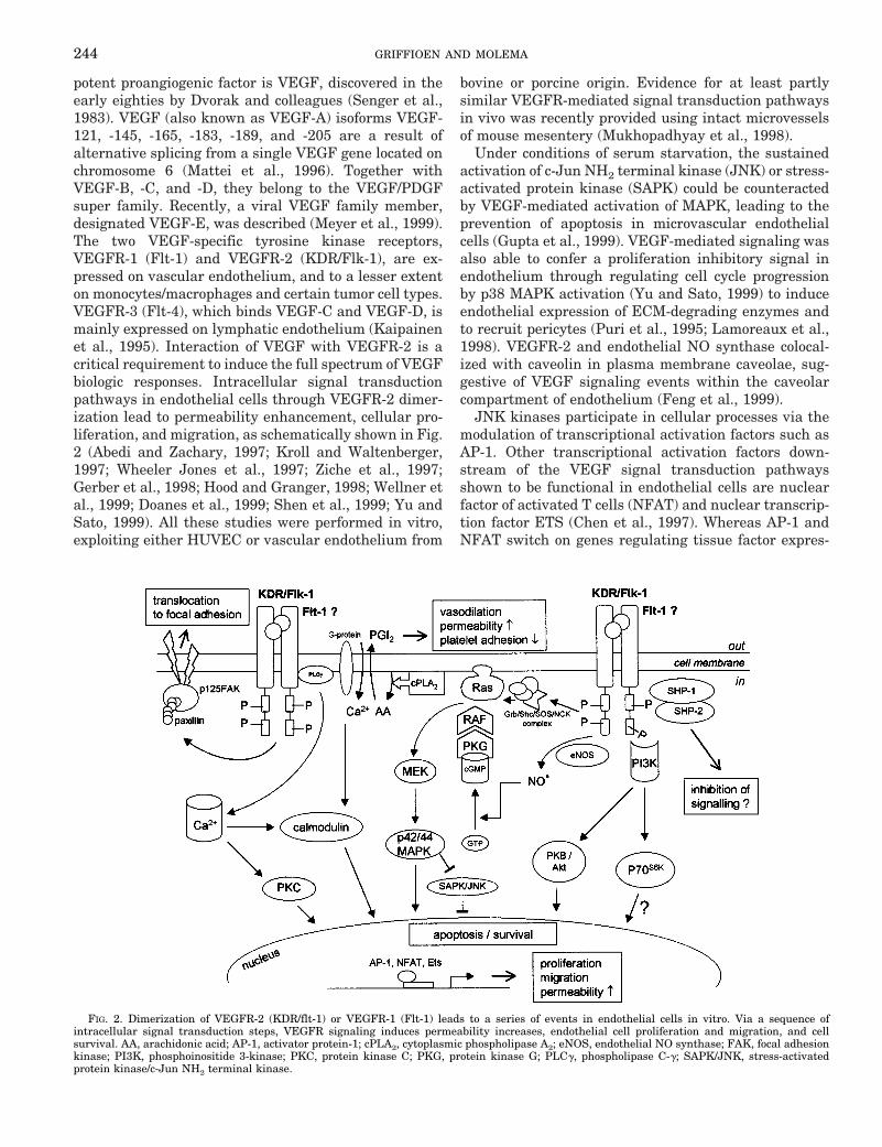
potent proangiogenic factor is VEGF, discovered in theearly eighties by Dvorak and colleagues (Senger et al.,1983). VEGF (also known as VEGF-A) isoforms VEGF-121, -145, -165, -183, -189, and -205 are a result ofalternative splicing from a single VEGF gene located onchromosome 6 (Mattei et al., 1996). Together withVEGF-B, -C, and -D, they belong to the VEGF/PDGFsuper family. Recently, a viral VEGF family member,designated VEGF-E, was described (Meyer et al., 1999).The two VEGF-specific tyrosine kinase receptors,VEGFR-1 (Flt-1) and VEGFR-2 (KDR/Flk-1), are ex-pressed on vascular endothelium, and to a lesser extenton monocytes/macrophages and certain tumor cell types.VEGFR-3 (Flt-4), which binds VEGF-C and VEGF-D, ismainly expressed on lymphatic endothelium (Kaipainenet al., 1995). Interaction of VEGF with VEGFR-2 is acritical requirement to induce the full spectrum of VEGFbiologic responses. Intracellular signal transductionpathways in endothelial cells through VEGFR-2 dimer-ization lead to permeability enhancement, cellular pro-liferation, and migration, as schematically shown in Fig.2 (Abedi and Zachary, 1997; Kroll and Waltenberger,1997; Wheeler Jones et al., 1997; Ziche et al., 1997;Gerber et al., 1998; Hood and Granger, 1998; Wellner etal., 1999; Doanes et al., 1999; Shen et al., 1999; Yu andSato, 1999). All these studies were performed in vitro,exploiting either HUVEC or vascular endothelium from
bovine or porcine origin. Evidence for at least partlysimilar VEGFR-mediated signal transduction pathwaysin vivo was recently provided using intact microvesselsof mouse mesentery (Mukhopadhyay et al., 1998).
Under conditions of serum starvation, the sustainedactivation of c-Jun NH2 terminal kinase (JNK) or stress-activated protein kinase (SAPK) could be counteractedby VEGF-mediated activation of MAPK, leading to theprevention of apoptosis in microvascular endothelialcells (Gupta et al., 1999). VEGF-mediated signaling wasalso able to confer a proliferation inhibitory signal inendothelium through regulating cell cycle progressionby p38 MAPK activation (Yu and Sato, 1999) to induceendothelial expression of ECM-degrading enzymes andto recruit pericytes (Puri et al., 1995; Lamoreaux et al.,1998). VEGFR-2 and endothelial NO synthase colocal-ized with caveolin in plasma membrane caveolae, sug-gestive of VEGF signaling events within the caveolarcompartment of endothelium (Feng et al., 1999).
JNK kinases participate in cellular processes via themodulation of transcriptional activation factors such asAP-1. Other transcriptional activation factors down-stream of the VEGF signal transduction pathwaysshown to be functional in endothelial cells are nuclearfactor of activated T cells (NFAT) and nuclear transcrip-tion factor ETS (Chen et al., 1997). Whereas AP-1 andNFAT switch on genes regulating tissue factor expres-
FIG. 2. Dimerization of VEGFR-2 (KDR/flt-1) or VEGFR-1 (Flt-1) leads to a series of events in endothelial cells in vitro. Via a sequence ofintracellular signal transduction steps, VEGFR signaling induces permeability increases, endothelial cell proliferation and migration, and cellsurvival. AA, arachidonic acid; AP-1, activator protein-1; cPLA2, cytoplasmic phospholipase A2; eNOS, endothelial NO synthase; FAK, focal adhesionkinase; PI3K, phosphoinositide 3-kinase; PKC, protein kinase C; PKG, protein kinase G; PLCg, phospholipase C-g; SAPK/JNK, stress-activatedprotein kinase/c-Jun NH2 terminal kinase.
244 GRIFFIOEN AND MOLEMA

sion upon VEGF activation, ETS regulates the expres-sion of u-PA, MMPs, and integrin 3 (Iwasaka et al.,1996; Oda et al., 1999).
The pleiotropic effects of VEGF in vitro on endothelialcell growth, proliferation and migration, among others,have now been extensively documented by many groups.However, plasma leakage and subsequent fibrin forma-tion is one of the hallmarks of angiogenesis initiation insolid tumor growth and initiation of wound healing. Thefibrin provides a functional matrix for endothelial cellsto become activated, to proliferate, and to migrate(Dvorak, 1986). Therefore, it may well be that in vivo thepermeability inducing capacity of VEGF is its most im-portant function in regulating physiologic and patho-logic angiogenesis.
2. Fibroblast Growth Factors. Members of the FGFfamily are also potent inducers of angiogenesis. Cellularresponses mediated by FGFs include cell migration, pro-liferation, and differentiation (Kanda et al., 1997). TheFGF family consists of nine structurally related polypep-tides, of which FGF-1 (acidic FGF) and FGF-2 (basicFGF) are the most extensively studied. Both FGF-1 andFGF-2 are devoid of a signal sequence for secretion.Export from cells without compromising cell integrity orrequiring cell death possibly follows a nonclassical, syn-aptotagmin-1-dependent exocytotic pathway (LaValleeet al., 1998).
The cellular effects of FGFs are mediated via specificbinding to high-affinity tyrosine kinase receptors (Kleinet al., 1997). In addition, low-affinity FGF receptorsexist, that consist of polysaccharide components of hepa-ran sulfate proteoglycans on cell surfaces and in ECM.Binding to the latter receptors has been proposed as amechanism to stabilize and protect FGF from inactiva-tion. Heparan sulfate on cell surfaces, on the other hand,plays a more active role in displacing ECM-bound FGF-2and subsequent presentation to the high-affinity signaltransducing receptors (Miao et al., 1996).
Receptor dimerization by FGF is facilitated by hepa-rin. It results in protein kinase activity and receptorautophosphorylation. As with VEGFR signaling, thisautophosphorylation enables adaptor proteins such asGrb2, Shc, and Nck to bind and subsequently activatethe Ras/Raf-MAPK pathway of endothelial cell prolifer-ation activation (Klein et al., 1997). p42 MAPK activa-tion was also implicated in endothelial cell motilityregulatory responses to FGF. p42 MAPK driven phos-phorylation of cytoplasmic phospholipase-A2 enabled ar-achidonic acid release upon FGF-2 activation of bovineaortic cells (Sa et al., 1995). Besides activating MAPK-mediated cell proliferation, FGF-2 induced murine brainendothelial cell proliferation via a serine/threonine ki-nase that phosphorylates ribosomal protein S6 (p70S6K).This proliferation activation route was restricted to en-dothelial cells cultured on fibronectin. When allowed todifferentiate to form tube-like structures in collagengels, p70S6K was not activated (Kanda et al., 1997).
In addition to initiating receptor signaling, FGF-1 canbe endocytosed and transported to the cell nucleus. Thistransport affects the cell cycle in the late G1 stage,promoting transition to the S stage (Imamura et al.,1994). Entrance of FGF-2 into the nucleus correlatedwith phosphorylation of nucleolin and subsequent in-creases of rDNA transcription, likely to be mediated bythe protein kinase CKII (Bouche et al., 1994). FGF-2uptake by endothelial cell was furthermore shown to bea route of growth factor degradation and can thereforeact to regulate FGF-2 activity (Gleizes et al., 1995).
3. Angiopoietin-1. Ang-1 is an endogenously secretedglycoprotein of approximately 75 kDa. Its receptor, Tie2,is generally restricted to the endothelium and of impor-tance in angiogenesis during development, tumorgrowth, and wound healing (Sato et al., 1995; Lin et al.,1997; Wong et al., 1997; Stratmann et al., 1998). In vitro,Ang-1 stimulated tyrosine phosphorylation of Tie2 inendothelial cells, inhibited serum starvation-inducedendothelial apoptosis, induced sprouting angiogenesis,and stabilized HUVEC network organization (Koblizeket al., 1998; Korpelainen and Alitalo, 1998; Kwak et al.,1999). When combined with other angiogenic factorssuch as VEGF or growth factor supplements containingFGF-1, the survival of both endothelial cells and vascu-lar networks increased even more (Kwak et al., 1999;Papapetropoulos et al., 1999). Although being chemotac-tic for endothelial cells and Tie2-transfected fibroblasts,no mitogenic responses of endothelial cells to Ang-1could be observed (Koblizek et al., 1998; Witzenbichleret al., 1998).
C. Effects of Angiogenesis Stimulation in PreclinicalStudies
In various animal models, the effects of either plasmidDNA encoding angiogenic factors or their respective pro-tein have been studied. In pigs, gradual narrowing of thecoronary artery leading to complete blood vessel occlu-sion was induced by use of an ameroid constructor. Inthis model, the continuous perivascular administrationof VEGF via an osmotic pump, or FGF-2 containingheparin-alginate microspheres, led to improved restingand stress-induced collateral blood flow values. Al-though the number of (vWF positive) blood vessels innonischemic heart tissue was unchanged, the vesseldensity significantly increased in the ischemic areas(Lopez and Simons, 1996).
In a mouse model of hindlimb ischemia, created byfemoral artery ligation, the question of whether diabetesleads to impaired neovascularization was addressed, be-cause diabetes is a major risk factor for artery diseases.It was shown, that nonobese diabetic mice exerted alower angiogenic response in ischemic tissue comparedwith normal mice. This impaired response could be re-duced by intramuscular (i.m.) gene therapy with recom-binant adenovirus expressing murine VEGF cDNA (Ri-vard et al., 1999). Local administration of a plasmid
ANGIOGENESIS AND DISEASE 245

encoding the 165-amino acid isoform of human VEGF165
(phVEGF165) during balloon cathetherization of the fem-oral arteries of rabbits resulted in an increased rate ofre-endothelialization. This effect was also observed inthe contralateral femoral artery that underwent simul-taneous balloon injury but was not transfected. As aresult of the increased re-endothelialization, the intimalthickening was diminished in both limbs, thromboticocclusions were less frequent, and recovery of the endo-thelial cell-dependent vasomotor reactivity was acceler-ated (Asahara et al., 1996).
For reconstructive surgery and organ transplantationprocedures, hypoxia or ischemia of the organs will neg-atively influence organ viability and function. The localadministration of VEGF cDNA or FGF-2 protein intoischemic experimental skin flaps in rats and rabbits,respectively, significantly enhanced survival time of iso-lated skin flaps after 1 week (Hickey et al., 1998; Taub etal., 1998). In the case of a solid support system exploitedfor the grafting of, e.g., pancreatic islets as a means ofbioartificial organ development, incorporation of FGF-1led to ingrowth of blood vessels 4 weeks after implanta-tion under the liver. Engrafting of pancreatic islets intothese FGF-1 prevascularized solid support systems re-sulted in a better survival of the graft compared withislets engrafted without a solid support, although isletfunction was somewhat less than in normal rats (de Voset al., 1997). This study indicates that therapeutic an-giogenesis may also have a potential in organ transplan-tation and bioartificial organ development. It should berealized, however, that in the case of allotransplanta-tion, immune cell activation will occur. Increased neo-vascularization may facilitate immune cell infiltrationby virtue of the fact that more blood vessels allow betteraccess to the graft. On the other hand, endotheliumunder the influence of proangiogenic factors may exhibitimpaired leukocyte recruitment functions (see SectionIV.A).
After i.m. administration of a plasmid encoding thehuman Ang-1 gene in a rabbit ischemic hindlimb model,human Ang-1 mRNA could be detected 3 to 14 days aftergene transfer. No mRNA was found in sites distant fromthe ischemic hindlimb. Both the angiographic score andthe capillary density were increased in the hindlimb 40days after Ang-1 encoding plasmid administration (Shyuet al., 1998).
The increase in re-endothelialization of balloon in-jured vessels in the femoral artery that was not treatedwith the phVEGF165 cDNA (Asahara et al., 1996), posesthe question whether angiogenic therapy for a specificpurpose may affect other sites in the body as well. Forexample, the question comes to mind whether therapeu-tic angiogenesis in a patient with myocardial ischemia isable to induce angiogenesis in an otherwise dormant,undetected, tumor nodule. Until now, however, labora-tory studies did not demonstrate that stimulation of
angiogenesis alone was sufficient for malignant growth(Isner et al., 1996).
D. First Clinical Studies on Angiogenesis Stimulation
So far, results are available from several pilot studieson the clinical application of therapeutic angiogenesis.In all three studies described, naked plasmid DNA en-coding human phVEGF165 under the cytomegaloviruspromoter/enhancer was administered. In the case ofischemic limbs in patients with critical limb ischemia orthromboangiitis obliterans, the DNA was i.m. injected inthe ischemic limb (Baumgartner et al., 1998; Isner et al.,1998). The DNA was administered directly in the myo-cardium of patients suffering from myocardial ischemia.In patients suffering from myocardial ischemia, theDNA was administered directly in the myocardium(Losordo et al., 1998).
Using contrast angiography, newly formed collateralblood vessels could be visualized in critical limb isch-emia and thromboangiitis obliterans patients treatedwith phVEGF165. Ischemic ulcers markedly improved orhealed, resulting in successful limb salvage in severalpatients. Documented adverse effects were transient an-kle or calf edema in some limbs. Patients suffering frommyocardial ischemia had significant reduction in anginaand reduced ischemia after phVEGF165 treatment.
VEGF was also administered as a protein to patientswith angina. Although the 120-day follow-up showedpromising results, the 60-day follow-up showed no dif-ference in exercise time or improvement of angina com-pared with placebo. An unexpected improvement in theplacebo group may be the reason for this result. Further-more, a clinical study on the applicability of FGF-2 in asimilar patient group has started. Results are expectedto be presented early 2000 (anonymous, 1999a).
It was concluded from these preliminary studies thattherapeutic angiogenesis was able to induce neovascu-larization, and if instituted at the proper time, it mayimprove ischemic disease conditions in humans. Thefinding that endothelial progenitors can be isolated fromhuman peripheral blood opens another possibility toaugment collateral vessel growth to ischemic tissue(Asahara et al., 1997). The homing potential of theseprogenitors to foci of angiogenesis may be exploited fortheir application as autologous vectors for gene therapywith, e.g., cDNA encoding VEGF after angiogenesis in-duction with nontargeted plasmid DNA.
III. Angiogenesis Inhibition
A. Angiogenesis and Cancer
Virchow was among the first to demonstrate the highvascularization in tumors in his publication DieKrankhaften Geschwulste published in 1863. He sug-gested that this was associated with the disorganizednature of tumor cells. The origin of the observed bloodvessels was uncertain by then, it either developed from
246 GRIFFIOEN AND MOLEMA

the transformed tumor cells or, alternatively, from nor-mal cells that had been derived from the neighboringbenign tissues. In a later period it was suggested thatthe trigger for enhanced blood vessel growth in tumorsemanated from the invading malignant cells. It wasproposed that the ability to attract new vasculature fromthe host was a characteristic feature of tumor cells.Recently, two paradigms were added to the vascularprocesses thought to prevail in tumor-induced neovas-cularization. During vessel co-option, tumors will ini-tially exploit the host vasculature for survival, whichcoincides with host vasculature regression. Ongoing tu-mor cell growth will subsequently lead to initiation ofangiogenesis (Holash et al., 1999). Furthermore, circu-lating endothelial progenitor cells can form an addi-tional source for postnatal vasculogenesis in tumorgrowth (Asahara et al., 1999).
Since then many researchers have studied angiogen-esis in a variety of test systems. Only after it was rec-ognized that new vessels at the tumor site were abso-lutely required for solid tumor expansion beyond the sizeof approximately 1 to 2 mm in diameter, it was sug-gested that the process of angiogenesis might be a targetfor therapy. Recent studies have demonstrated that lym-phoproliferative diseases, such as leukemia and lym-phoma, are dependent on angiogenesis as well. Elevatedexpression of FGF and VEGF has been observed in acutemyeloid leukemia, acute lymphoblastoid leukemia, andlymphomas (Fiedler et al., 1997; Foss et al., 1997;PerezAtayde et al., 1997). These studies indicate, there-fore, that angiogenesis might also be a therapeuticaltarget for hematologic tumors.
The first molecule identified as an angiogenic factorwas described in 1984 (Shing et al., 1984) after which alarge number of angiogenic factors followed. These fac-tors can be produced by the tumor cells themselves, cellspresent in the tumor stroma such as fibroblasts, smoothmuscle cells, or by infiltrating immune cells. To compli-cate matters, these cells are all able to produce angio-genesis inhibitors as well. More recent attention hasbeen paid to the isolation and characterization of theseangiogenesis inhibitors because they may have potentialas therapeutic agents. Most of them have been studiedfor their applicability in cancer therapy but may also besuitable for the therapy of chronic inflammatory condi-tions (see Section IV.C).
B. In Vitro and in Vivo Models to Study Angiogenesis
Angiogenesis can be qualitatively and quantitativelymeasured in a large variety of in vitro and in vivo modelsystems. As discussed in Section I.C, the angiogeniccascade can be dissected in different sequential steps sothat can each be studied separately in vitro. Researchhas mainly focused on proliferation and migration ofendothelial cells. For this research, different endothelialcell sources can be applied. For human research mostlaboratories make use of HUVECs. This is the best
available source of human endothelium, but the majordrawback of these cells is their macrovascular origin,which makes them less suitable for studies on angiogen-esis, a microvascular process. Although more laborious,human microvascular endothelial cells can be isolatedfrom other organs such as foreskin or adipose tissue. Forall primary isolates, the number of in vitro passages(4–5, and in the presence of growth factors, approxi-mately 10) is, however, limited, which poses a majorproblem for their application. The required regular iso-lations furthermore introduce significant donor varia-tion. To circumvent these drawbacks, one can use im-mortalized endothelial cell lines, such as HMEC-1 (Adeset al., 1992), EA.hy926 (Edgell et al., 1983), or ECL4n(Griffioen et al., 1996b). ECV304, a spontaneous immor-talized endothelial cell line, has been applied for in vitroangiogenesis studies, although recently it has becomeapparent that this cell line contains a nonendothelialbackground as well. Endothelial cells from other speciesare also available, e.g., bovine capillary endothelial cellsor cell lines from mouse and rat origin. It should be keptin mind that the effects of angiogenesis-inducing or -in-hibiting factors can be different in the different species.
Assays to study proliferation of endothelial cells arebased on cell counting or radiolabeled thymidine incor-poration, or on colorimetric systems for measurement ofmitochondrial activity [cell counting kit-8 (CCK-8) ordimethylthiazol diphenyl tetrazolium bromide (MTT)].Alternatively, proliferation of endothelial cells can beanalyzed by DNA profiling or determination of cell cycle-dependent expression of molecules such as proliferatingcell nuclear antigen or Ki-67. Also, detection of cell deathis a commonly used approach to average cell growth;e.g., apoptosis induction can be studied by detection ofsubdiploid cells or analysis of DNA degradation profiles,cell morphology or nick-end labeling by terminal dUTPnick-end labeling analysis.
For analysis of migration of endothelial cells, Boydenchambers are primarily used. An easier method is thewound assay. This assay system is based on wounding ofa confluent monolayer of endothelial cells and measure-ment of the wound width in time.
Although the advantage of these in vitro assays isclearly the control over the few parameters present, theangiogenic cascade consists of multiple steps. This as amore extended process, can be studied in vitro, too. Mostof these assays studying more complex processes of an-giogenesis are based on tube formation of long-termcultured endothelial cells in a 3-dimensional seminatu-ral matrix microenvironment. The most commonly usedassay system to measure tube formation is the growthfactor-induced sprouting of capillary-like structuresfrom a confluent monolayer of endothelial cells grown ona thick gel. These gels can either be composed of aseminatural matrix with or without growth factors (e.g.,Matrigel), or be based on collagen (Barendsz-Janson etal., 1998) or fibrin (Koolwijk et al., 1996). The demon-
ANGIOGENESIS AND DISEASE 247

stration of lumina in these endothelial cell sprouts isregarded as a criterion for vessel growth, in contrast tojust migration of endothelial cells in the matrix or justrearrangement of endothelial cells on the gel. An elegantmethod to measure capillary formation has been de-scribed where endothelial cells grown on gelatin-coatedcytodex-3 microcarrier beads were cultured in a fibringel (Nehls and Drenckhahn, 1995; Trochon et al., 1998).Quantification of sprouting can subsequently be per-formed by either measurement of maximal sproutingdistance or by computer-based determination of totalvessel length. Assays based on the sprouting of capillar-ies out of fresh tissue embedded in matrix gels moreclosely reflect the in vivo situation. This has been de-scribed for rat aortic rings (Nicosia and Ottinetti, 1990;Malinda et al., 1999) and human placenta tissue (Brownet al., 1996). This procedure is not applicable for alltissues because it has been described that, e.g., tumorbiopsies often produce too much proteases digesting thematrix and thereby prevent endothelial cell sprouting(Barendsz-Janson et al., 1998). Figure 3 shows someexamples of in vitro angiogenesis assays. A recentlypublished novel way of measurement of angiogenesis invitro, which is even more close to the in vivo situation, isthe use of embryoid bodies (Wartenberg et al., 1998). Invitro cultured mouse blastocyst cells (Evans and Kauf-man, 1981) recapitulate several steps of murine embry-ogenesis, including vasculogenesis and angiogenesis (Ri-sau et al., 1988). There is a complete blood vesseldevelopment in these embryoid bodies (Vittet et al.,1996) making this system suitable for the study of an-giogenesis modulators.
Besides the advantages that in vitro angiogenesis as-says clearly have, the major drawback of all these assaysis that they require the endothelial cells to be removed
from their natural microenvironment, which may altertheir physiologic properties. To study angiogenesis invivo, the most frequently used assay systems are thechicken chorioallantoic membrane assay (Nguyen et al.,1994), the corneal pocket (Conrad et al., 1994), transpar-ent chamber preparations such as the dorsal skin-foldchamber (Algire, 1943; Lichtenbeld et al., 1998), thecheek pouch window (Shubik et al., 1976), and the poly-mer matrix implants (Mahadevan et al., 1989; Plunkettand Hailey, 1990). However, in vivo assays also haveseveral disadvantages: the pharmacokinetic propertiesof the compounds tested, necessary for proper interpre-tation of results, are often not known and the host willrespond nonspecifically to the implantation. In this re-view, these assays will not be discussed in more detailbecause recently an elegant review on this issue ap-peared, discussing the pro’s and con’s of in vivo quanti-tative angiogenesis assays (Jain et al., 1997).
C. Ways to Interfere with Angiogenesis
A broad spectrum of strategies for modulation of an-giogenesis has been described. As discussed in SectionI.C, angiogenesis mainly depends on proper activation,proliferation, adhesion, migration, and maturation ofendothelial cells. Most approaches to modulate angio-genesis are therefore focused on these endothelial func-tions during blood vessel formation.
1. Intervention with Endothelial Cell Growth. Themost successful approach to modulate angiogenesis, todate, is the use of agents that specifically inhibit thegrowth of the endothelial cells. One of the first compoundsidentified to exhibit inhibitory effects on cell growth withspecificity for endothelial cells was O-chloroacetylcar-bamoyl fumagillol or AGM-1470/TNP-470, an analog of thefungus-derived antibiotic fumagillin (Ingber et al., 1990;
FIG. 3. In vitro angiogenesis assays. Tube formation of human umbilical vein endothelial cells on the seminatural matrix, Matrigel, 2 h (panel A)and 16 h (panel B) after seeding. Growth factor induced (20 ng/ml bFGF) sprouting of bovine microvascular endothelial cells grown as a monolayeron a collagen-based gel (panel C) can be inhibited by the angiogenesis inhibitor platelet factor-4 (panel D). Similar regulation is shown in panels E andF in a tube formation/migration assay of bovine endothelial cells grown on cytodex-3 beads and embedded in a fibrin gel. Panel G represents the rataortic ring assay under control conditions and after exposure for 5 days with 200 mg/ml endothelial cell growth supplement (H). Panels A, B,G, andH were kindly provided by Dr. H. Kleinman (Bethesda, MD).
248 GRIFFIOEN AND MOLEMA

Kusaka et al., 1991). The mechanism of action of thiscompound was found to be prevention of endothelial cellsto enter G1 phase of the cell cycle, resulting in a decrease inproliferation (reviewed in Castronovo and Belotti, 1996). Inlater years, several endogenous molecules with angiostaticactivity were described. Among these molecules are throm-bospondin-1 (Rastinejad et al., 1989; Good et al., 1990;Grossfeld et al., 1997), platelet factor-4 (Gupta et al., 1995;Kolber et al., 1995), and interferon-inducible protein-10(Luster et al., 1995). Two other members of this class ofendogenously produced antiangiogenic proteins are an-giostatin (O’Reilly et al., 1994) and endostatin (O’Reilly etal., 1997). Angiostatin is an internal fragment of plasmin-ogen with multiple antiangiogenic activities in vitro and invivo. Endostatin is a proteolytic fragment of collagen XVIIIthat affects endothelial cell survival via the induction of animbalance between the antiapoptotic proteins Bcl-2 andBcl-XL and the proapoptotic protein Bax (Dhanabal et al.,1999). Both induced tumor regression, not only growthinhibition, in tumor-bearing mice, an effect that was mostpronounced with endostatin and demonstrates the poten-tial of these proteins. Direct inhibition of endothelial cellgrowth was also obtained with two other recently de-scribed endogenously produced angiostatic proteins,namely vasostatin (Pike et al., 1998) and restin (Ramchan-dran et al., 1999). Detailed mechanisms of action have notbeen described yet for these angiogenesis inhibitors.
A separate method for modulation of angiogenesis isthe interference with angiogenic factors such as VEGFor FGF and their receptors. VEGF is also a major rulerduring the development of tumors. Angiogenesis andsubsequent tumor growth can be inhibited by blockingthese factors (Kim et al., 1993). This can be performed bytreatment with humanized blocking antibodies to thesefactors, antibodies to their receptors, with soluble recep-tors functioning as antagonists, dominant negativegrowth factor variants, or VEGF antisense constructs.Functional interference with growth factor signaling canalso be performed by specific inhibitors of growth factorreceptor signaling, as has been described for SU5416, aspecific inhibitor of VEGFR-2 phosphorylation (Fong etal., 1999).
Recently a new nonendothelial cell-specific inhibitorof angiogenesis was described. Carboxyamidotriazole(CAI) is an inhibitor of tumor cell motility; the mecha-nism of action is the inhibition of transmembrane cal-cium influx. The inhibition of calcium influx preventsthe activation of the focal adhesion kinase and RhoApathways. CAI inhibited invasion by its ability to de-crease the production of MMPs, blocked migration ofcells, and caused cytostasis in tumor cells and endothe-lial cells. By interference in the biochemical pathwaysinvolved in endothelial spreading on extracellular ma-trix, the integrity of the vascular tube as well as stabi-lization of newly formed vessels were affected. Localadministration of CAI inhibited capillary expansion inthe chick chorioallantoic membrane assay. In vivo stud-
ies confirmed the antiangiogenic and anticancer effect ofCAI for, e.g., ovarian cancer (Kohn and Liotta, 1995;Kohn et al., 1995).
2. Intervention with Endothelial Cell Adhesion andMigration. Because the process of angiogenesis alsodepends on endothelial cell adhesion events to, and mi-gration of cells through, the extracellular matrix, effortis put in the search for modulators of these interactions.The first identified member of this group of compoundsis the endogenously produced cytokine interferon. Anti-endothelial activity was recognized by the observationthat interferon could inhibit the migration of capillaryendothelial cells (Brouty and Zetter, 1980). Subse-quently, both interferons a and b were shown to have invivo antiangiogenic activity (Sidky and Borden, 1987).Although interferons are probably not sufficiently activefor treatment of all tumors, benign tumors predomi-nantly comprised of endothelial cells are particularlysensitive to treatment with interferon (Ezekowitz et al.,1992). When it was found that activated endothelial cellsup-regulate receptors for extracellular matrix compo-nents (Re et al., 1994; Frisch et al., 1996; Griffioen et al.,1997), interaction of endothelial cells with the matrixwas chosen as a target for inhibition of angiogenesis.This proved to be a relevant approach by the demonstra-tion that avb3 integrin molecules, the biological functionof which is binding of vitronectin and other RGD-con-taining matrix components, are overexpressed in angio-genically stimulated blood vessels. Ligation of these re-ceptors with an antibody called LM609 interferes withendothelial cell growth leading to inhibition of subse-quent tumor growth (Brooks et al., 1994a). In addition,the exposure of endothelial cells to anti-avb3 antibodiesresulted in the induction of apoptosis in these cells vialoss of cell anchorage to the extracellular matrix (Brookset al., 1994b). This is most likely the mechanism bywhich proliferation of endothelial cells and angiogenesisin vivo is blocked by avb3-directed antibodies.
3. Intervention with Metalloproteinases. Anothermechanism of angiogenesis inhibition, related to theinhibition of endothelial cell adhesion and migration, isthe use of specific inhibitors of proteinases that dissolvethe connective tissue, thereby facilitating endothelialcell migration and subsequent vessel formation. Matrixmetalloproteinases are a homologous family of enzymesthat are involved in tissue remodeling and morphogen-esis. Collectively, these enzymes are capable of degrad-ing all components of the extracellular matrix (Rasmus-sen and McCann, 1997). Increased activity of theseenzymes has been observed in tumor formation, andtherefore inhibitors of MMPs represent an attractiveapproach to treat cancer. MMP inhibitors can be dividedin synthetic protease inhibitors and naturally occurringMMP inhibitors, the tissue inhibitors of metalloprotein-ase. Belonging to the former group, batimastat, mari-mastat, and prinomastat/AG3340 are potent broad-spec-trum inhibitors of the major MMPs and can prevent or
ANGIOGENESIS AND DISEASE 249

reduce spread and growth of several different malignanttumors in numerous animal models (Brown and Gia-vazzi, 1995; Shalinsky et al., 1999). Cell adhesion andproteolytic mechanisms are functionally associated, asrecently demonstrated by the observation that the col-lagenase MMP-2 can bind to integrin avb3 on angiogenicblood vessels. Most interestingly, it was found that anaturally occurring MMP-2 breakdown product, calledPEX, can inhibit cell-associated collagenolytic activity.It is suggested that this breakdown product is an impor-tant regulator of protease activity during angiogenesisand vasculogenesis. A recombinant form of PEX wasuseful in blocking angiogenesis and tumor growth invivo, providing a novel therapeutic approach for angio-genesis inhibition at this level (Brooks et al., 1998).
D. Preclinical Use of Angiogenesis Inhibitors in Cancer
The pivotal role of angiogenesis in tumor progressionand metastasis has urged researchers to test newly de-veloped angiogenesis inhibitors in a broad variety ofanimal tumor growth models. Studies with one of thefirst angiogenesis inhibitors, AGM-1470/TNP-470, wereperformed in the early 1990s. Although in vitro thesensitivity for TNP-470 was not completely restricted toendothelial cells, doses of the compound had to be 10 to100 times higher for comparable inhibition of tumor celllines. Treatment of tumor-bearing mice resulted in asignificantly increased survival time of 260% over un-treated control animals. Daily treatment was not neces-sary; optimal treatment regimens were s.c. or i.v. admin-istration once every three days. Oral administration hadweaker effects on tumor growth, likely a result of lowerbioavailability. The fact that sensitivity of tumor cells invitro and effect on tumor growth in vivo did not correlateis seen as evidence for antitumor effects through thetumor vasculature (Ingber et al., 1990; Yamaoka et al.,1993). Angiogenesis inhibitors have also been expectedto be efficient metastasis inhibitors based on the conceptthat tumors require new vasculature for spreading andoutgrowth at a secondary site. TNP-470 reduced boththe number and size of metastases of the B16BL6 mel-anoma and the M5076 reticulum cell sarcoma cell line inmice by 80 to 90% (Yamaoka et al., 1993). Also in rats,inhibition of tumor growth and metastasis was observed(Futami et al., 1996).
The next breakthrough in the search for novel antian-giogenic compounds occurred when the hypothesis thata primary tumor, whereas capable of stimulating angio-genesis for its own blood supply, can produce angiogen-esis inhibitors that suppress the outgrowth of distantmetastases, was proven to hold true. This hypothesiscame from the observation that the removal of primarytumors could lead to the accelerated growth of metasta-ses (Sugarbaker et al., 1977). To test this hypothesis, theLewis lung carcinoma mouse model was used in whichthe primary tumor completely suppresses the growth ofits metastases. From the urine of these mice a cleavage
fragment of plasminogen called angiostatin was puri-fied, which completely replaced the inhibitory activity ofthe primary tumor (O’Reilly et al., 1994). Treatment oftumor-bearing mice with angiostatin almost completelyprevented metastasis formation in the lung. Using asimilar strategy, endostatin was discovered (O’Reilly etal., 1997). Treatment of mice carrying different synge-neic malignant tumors with endostatin led to a rapidregression of tumors. As with angiostatin, there was nosign of toxicity, and continued endostatin therapy main-tained the tumors in a state of dormancy. Discontinua-tion of treatment led to renewed growth at the primarysite, which would eventually lead to death of the ani-mals. However, when therapy was restarted tumors re-gressed again for nearly 100%. Subsequent intermittentcycles of treatment were able to maintain the tumors ina state of dormancy and showed no sign of drug-inducedresistance (Boehm et al., 1997). When angiostatin andendostatin therapy were combined for 25 days, both atrelatively high doses (20 mg/kg/day), tumors regressedcompletely yielding tumor-free survival of up to 11months after start of therapy.
The nature of this dormancy is still obscure sincethese tumors have the capacity of renewed outgrowthwhen transplanted to the other flank or to another ani-mal. This makes the involvement of the immune system,i.e., the development of a specific antitumor immuneresponse in these mice, unlikely. It has been suggestedthat the sequential accumulation of endostatin duringthese cycles of growth and regression can locally lead toa high concentration, and hence keep the tumor dormant(Black and Agner, 1998). The data on endostatin suggestthat the endothelial cell compartment is directly in-volved in the dormancy of the tumor cells, demonstrat-ing the powerful control exerted by the vascular endo-thelial cell population over the tumor cell population.
The discovery of overexpression of avb3 integrin intumor vessels and the dependence of angiogenesis onthis matrix receptor (Brooks et al., 1994a) introducedthe first strategy for angiogenesis inhibition by target-ing specific tumor endothelial cell determinants. Anti-body and specific RGD motif containing peptide ligandsboth inhibited human tumor growth in animal models(Brooks et al., 1994b; Friedlander et al., 1995). Theprevention of the formation of a suitable matrix to mi-grate through by the inhibition of MMPs is related tothis inhibition of cell adhesion to suppress angiogenesis.Treatment of mice carrying the highly metastatic tumorB16BL6 with batimastat resulted in reduction of lungcolony number. It was found that not cell arrest in thelungs but actually extravasation of the cells was inhib-ited (Chirivi et al., 1994). Batimastat also inhibited pri-mary tumor cell outgrowth. A 58% inhibition was ob-served when treatment was started from the day oftumor cell inoculation. In numerous other models, suchas ovarian cancer and an orthotopic colon cancer model,similar results were obtained (Davies et al., 1993; Wang
250 GRIFFIOEN AND MOLEMA

et al., 1994). Several other low-molecular weight syn-thetic MMP inhibitors is currently under development.One of them is prinomastat/AG3340, which has one ofthe lowest Ki values (Ki is the concentration that inhibitsenzyme activity by 50%) for gelatinase A (MMP-2) andgelatinase B (MMP-9). In a chemoresistant human non-small cell lung carcinoma mouse model, AG3340 waswell tolerated at 400 mg/kg/day (administered twicedaily, 7 days a week) and exhibited significant inhibitionof angiogenesis (up to 77%) and tumor growth (up to65%). A suboptimal dose of AG3340 in combination withcarboplatin or paclitaxel resulted in greater inhibitionthan was observed with either agent alone. In colon andprostatic tumors, angiogenesis was inhibited by approx-imately 50% (Shalinsky et al., 1998).
The potential of the use of inhibitors of specific signaltransduction pathways is nicely exemplified by the useof CAI. This inhibitor of calcium mobilization, arachi-donic acid release, and generation of inositol phos-phates, inhibits growth of tumor cell lines. In the sameway that it inhibits tumor cells, endothelial cells areaffected, resulting in inhibition of angiogenesis. Oraladministration of CAI to mice carrying human xeno-grafts resulted in a .80% inhibition of tumor outgrowth(Kohn et al., 1992). CAI was not toxic as determined bymacroscopic and histological evaluation. These studiesare highly interesting because one might expect thatpharmacologic interference with intracellular signalingpathways, which are so fundamental to cellular functionin both pathologic and normal tissues, will lead to mas-sive toxicity. Because this is not the case, the conclusionseems justified that tumors display an enhanced depen-dence and sensitivity to these second messenger path-ways or are exposed to higher drug concentrations byincreased uptake or accumulation at the tumor site.
E. Clinical Trials with Inhibitors of Angiogenesis forCancer Treatment
Currently some 30 antiangiogenesis compounds arebeing tested in human clinical anticancer trials. Most ofthem are in phase I or II testing, to determine optimaldosage, nature and severeness of side effects, safety andpreliminary effectivity. Only a few compounds, as dis-cussed in this chapter, are further down the clinical roadand currently being tested in phase III trials. Usuallythe angiogenesis targeted studies are designed in a waythat patients receive either standard (chemo- or im-muno-) therapy and a placebo, or standard therapy com-bined with a new antiangiogenic drug. The NationalCancer Institute, which is involved in some of theseclinical trials, has classified these drugs in differentcategories. Without trying to be complete, a few exam-ples of clinical studies with these compounds will bediscussed here.
i. Drugs that inhibit endothelial cell proliferation di-rectly. As a member of the “first generation” of angio-genesis inhibitors, TNP-470 was used in the first formal
clinical trial of antiangiogenic therapy. In cooperationwith the National Cancer Institute, currently three clin-ical studies are operative: 1) a phase I study of TNP-470in patients with recurrent or refractory pediatric solidtumors, lymphomas, and acute leukemias; 2) a phase IIstudy for advanced or recurrent squamous cell carci-noma of the cervix; and 3) a phase III randomized studyof TNP-470 versus synchronous radiotherapy and che-motherapy for locally advanced nonresectable nonmeta-static pancreatic cancer. Recently, one phase II multi-institutional clinical study in metastatic renal cellcarcinoma patients was reported. In the first 20 patientswith an adequate follow-up, there was one partial re-sponse and one minor (40% shrinkage) response. Five ofthese 20 patients remained progression-free for 16weeks or more. The mild toxicity reported and the highincidence of apparent prolonged progression-free sur-vival in this heavily pretreated cohort is encouraging(Stadler et al., 1998).
ii. Drugs that block matrix breakdown. Among theclinical trials with angiogenesis inhibitors, the metallo-proteinase inhibitors have probably been the most ex-tensively studied in clinical trials to date. The first trialin patients with a MMP inhibitor, batimastat, started in1991 in patients with ovarian carcinoma. Because thedrug showed low bioavailability, other administrationroutes were used. A phase I study using an i.p. formu-lation yielded high and sustained plasma concentrationsof the drug, low toxicity, and early signs of efficacy. Arandomized controlled trial of marimastat in 400 pa-tients with advanced pancreatic cancer has now beencompleted. The study was designed to compare the effectof orally and twice daily administered marimastat withthe conventional treatment with gemcitabine. The studydid not meet its primary endpoint, designated as a 16%or greater reduction in mortality versus gemcitabinetreatment, i.e., there was no significant difference be-tween the survival curves for gemcitabine and the high-est marimastat dose. Safety data revealed no differencebetween the treatment groups other than the expectedmusculoskeletal events (stiffness and pain) caused bythe MMP inhibitor that have been reported in moststudies. Although this study has not shown any benefitof treatment with marimastat over the conventionaltreatment, there is evidence that marimastat is as effec-tive as the “drug of first choice” treatment. This holdspromise for the use of marimastat in future combinationregimens, studies which are currently being enrolled.Oral administration of prinomastat/AG3340 was testedin a single dose phase I study in healthy volunteers.Favorable absoption, pharmacokinetics, and toxicityprofiles were observed. In a phase I safety and tolerabil-ity study of AG3340 administered orally and twice dailyin patients with advanced cancer, including lung, pros-tate, kidney, and colorectal cancers as well as sarcomaand melanoma, disease was stabilized in more than 25%of 47 evaluable patients who were treated for periods of
ANGIOGENESIS AND DISEASE 251

16 to 40 weeks. Three patients (one each with nonsmallcell lung cancer, renal carcinoma, and melanoma) werefound to have minor reductions in tumor volume.AG3340 was found to be generally well tolerated.
A phase III clinical trial of AG3340 has been initiatedin combination with chemotherapy in patients with ad-vanced nonsmall cell lung cancer. This trial is designedto evaluate the safety and efficacy of AG3340 as part offirst-line therapy in combination with chemotherapy. Ina study being conducted at sites in North America, Eu-rope, and Australia, patients with advanced nonsmallcell lung cancer will be randomized to receive eitherAG3340 (tablet form) in combination with gemcitabineand cisplatin or placebo in combination with gemcitab-ine and cisplatin. The primary objective of this study isto compare time of overall survival between patientsreceiving AG3340 or placebo in combination with gem-citabine and cisplatin.
iii. Drugs that inhibit endothelial-specific integrin/survival signaling. The results of a completed phase Iclinical trial with Vitaxin, the humanized avb3-directedLM609 monoclonal antibody have been presented(Gutheil et al., 1998). This trial of 14 patients revealedno significant toxicities, and in addition, showed thattwo-thirds of the patients experienced an objective clin-ical response to the Vitaxin treatment. Most recently, aphase I/II study was initiated to determine the optimaldosage, whereas a phase II clinical trial to determineefficacy is planned to start early 2000.
iv. Drugs with nonendothelial cell-specific mecha-nism(s) of action. Phase I clinical trials with CAI, theinhibitor of transmembrane calcium influx, have beencompleted, demonstrating that oral treatment with asingle dose daily was relatively well tolerated (Kohn etal., 1997). Cytostasis in the form of disease stabilizationwas observed in almost half the solid tumor patientstreated. Several refractory ovarian cancer patients werefound to have disease stabilization lasting up to 10months. This led to the initiation of a phase II clinicaltrial for women with relapsed epithelial ovarian cancerand subjected to no more than three prior therapeuticregimens. This single agent trial is aimed at an outcomeof 6 or more months of disease stabilization.
v. Drugs that block activators of angiogenesis. Angio-genesis seems exquisitely sensitive to small changes infactors such as VEGF and FGF-2 that drive the angio-genic process. This has important therapeutic implica-tions in treating angiogenesis-driven disorders (Rak andKerbel, 1997). Next to anti-VEGF antibodies used toneutralize the cytokine in serum/extracellular fluids,another approach is the blockade of VEGF receptor sig-naling. SU5416 is a specific inhibitor of the signalingpathway of the VEGFR-2 found on endothelial cells.This compound entered phase I clinical testing in the fallof 1997 and has shown good safety profiles in approxi-mately 100 patients with solid tumors. The phase IIIclinical study in colorectal and lung carcinoma patients
will compare the efficacy of standard chemotherapy reg-imens with the efficacy when combined with SU5416.SU6668 is a broad-spectrum inhibitor of angiogenesisand tumor growth, acting by interference with VEGF,FGF, and PDGF receptors. This drug is currently enter-ing phase I studies in both i.v. and oral formulations.
A listing of angiogenesis inhibitors that are currentlyin clinical trials, can be found on an internet website ofthe National Cancer Institute (http://cancertrials.nci.ni-h.gov). Moreover, most of the data available so far canonly be retrieved from the internet. Inhibition of angio-genesis is a new strategy of cancer therapy, in which theconcept of treatment is fundamentally different fromconventional therapies. In standard chemotherapy thedesired endpoint is shrinkage of the tumor and completeremission, whereas in antiangiogenic therapy the end-point aimed at is achievement of stable disease andprolonged progression-free periods leading to a rela-tively long survival. This underscores the novelty ofantiangiogenesis therapy because until now, to ourknowledge, there has never been a drug for oncologicaluse that has been approved by the FDA on the basis ofstabilization of the disease. As a consequence, the sur-rogate endpoints, such as stabilization or decrease ofcirculating tumor markers [e.g., prostate specific anti-gen (PSA) in prostate cancer, carcino-embryonic antigen(CEA) in gastrointestinal cancer, and CA15 in breastcancer], do not necessarily need to be the same. Animportant issue in antiangiogenesis treatment that con-cerns most clinicians, is the question whether there aresurrogate endpoints for angiostatic treatment. The mostcommonly used parameter for ongoing angiogenesis isthe circulating (plasma) level of VEGF. However, this isan indirect method and in some instances, e.g., duringtreatment with VEGF-blocking antibody, this parame-ter cannot be used. More useful surrogate endpointshave not been described yet, although circulating levelsof E-selectin and vascular cell adhesion molecule-1(VCAM-1) have been put forward for this purpose (Fer-rara, 1995; Koch et al., 1995). Considering the fact thattheir counter ligands are abundantly present in the se-rum, it is however likely that these soluble adhesionmolecules are rapidly cleared from the circulation, andthereby mask ongoing angiogenesis.
Although stabilization of tumor size in antiangiogen-esis therapy may be an endpoint in the clinic, in preclin-ical animal studies it is obvious that the most promisingangiogenesis inhibitors do not stabilize the tumor. In-stead, they induce partial or sometimes even completeregression. This may partly be explained by the rapidturnover of cells in animal tumors, even in a stabilizedtumor, in which case inhibition of angiogenesis wouldlead to a disturbed balance between cell growth and celldeath leading to an overall enhanced cell death.
Animal experiments also may provide a clue towhether benefit from antiangiogenesis therapy shouldbe expected in patients with large tumor burdens. Some
252 GRIFFIOEN AND MOLEMA

unpublished studies indicate that the largest benefitfrom angiogenesis inhibition is expected when the ther-apies are started as early as possible, with the largesteffects seen on starting the treatment even before theangiogenic switch occurred. Because this does not meetthe situation in the clinic, where cancer is normallydiagnosed at a much later stage, combination therapiesare most likely to give the best results.
F. Novel Approaches to Interfere with TumorBlood Flow
Angiogenesis inhibitors are compounds that specifi-cally interfere with regulatory processes of the varioussteps of angiogenesis, as described above. Tumor vascu-lature targeting aims at specifically delivering pharma-cologic agents to the site of the blood vessels of a tumor.The pharmacologically active moieties can exert a vari-ety of activities, but in general all strategies aim to blocktumor blood flow and hence tumor growth. Moreover, asa consequence, site-specific immunologic responses maybe brought about and potentiate anti-tumor effects. Be-sides the choice of the effector modality (e.g., coagulationfactors or toxins), the choice of the target epitope is ofgreat importance for feasibility and effectiveness ofthese approaches, as will be discussed below.
1. Targeted Strategies to Induce Tumor Blood Coagu-lation. At present, two studies on targeting blood coag-ulation-inducing activity to tumor endothelium as atherapy for cancer have been reported. In the first study,an animal model with artificially induced target
epitopes on the tumor vasculature was exploited. In thismodel, transfected tumor cells s.c. inoculated locally pro-duced high amounts of interferon (IFN), leading to tu-mor endothelium-specific expression of major histocom-patibility complex (MHC) class II. Bispecific antibodies(BsAb) against MHC class II and a truncated form of theactivator of the extrinsic coagulation pathway, tTF,were subsequently developed. Intravenous administra-tion of a mixture consisting of BsAb and tTF (BsAbztTF),the so called “coaguligand” formulation, to mice withclinically relevant tumor burden resulted in dramatictumor reduction without concurrent toxicity in otherorgans. Site-specific blood coagulation in the tumorblood vessels caused an almost instantaneous and per-sisting blockade of tumor blood flow. Treatment of micebearing s.c. tumors twice with BsAbztTF coaguligandlead to 38% complete tumor regressions and 24% partialresponses (Huang et al., 1997). The attractiveness of thecoaguligand approach is the use of a truncated form ofTF that is completely devoid of coagulation-inducingactivity as long as it is kept from complexation with thelipophilic factor X at cell membranes. On cross-linking ofthe hydrophilic tTF with the target cell membranes bythe BsAb, tTF becomes complexed with factor X in thepresence of factor VII/VIIa, leading to the induction ofblood coagulation (Fig. 4). It is thought that a thresholdin the number of tTF cross-linked to factor X-containingcell membranes exists, above which the coagulation cas-cade is initiated. This allows the use of target epitopes
FIG. 4. Schematic representation of drug-targeting approaches aimed at selectively blocking tumor blood flow. A, coaguligand approach: bydelivering the coagulation factor truncated tissue factor to the tumor endothelium, blood coagulation is initiated and the tumor blood flow blocked; B,intracellular delivery of doxorubicin by RGD peptide, leading to endothelial cell death via doxorubicin-related cytotoxic effects such as DNAtopoisomerase inhibition and the generation of free radicals; C, immunotoxins or “angiotoxins” deliver deglycosylated ricin A chain or diphtheria toxinin the target cells, leading to endothelial cell killing. fVIIa, factor VIIa; fX(a), factor X(a).
ANGIOGENESIS AND DISEASE 253

that are highly but not exclusively expressed on tumorendothelium.
Using a similar approach of tumor infarction, mousesolid Hodgkin’s tumors spontaneously expressing endo-thelial VCAM-1 were significantly retarded in out-growth (Ran et al., 1998). The antitumor effect was notas dramatic as that seen in the MHC class II model.Possibly, the number of tTF molecules delivered at thesite of the tumor endothelium was not sufficient to cre-ate a rapid and more or less generalized blood coagula-tion throughout the tumor vasculature. Only if the bloodin the majority of vessels becomes coagulated by thecoaguligand, the number of tumor cells that will bedeprived of nutrients will be sufficient enough to resultin strong antitumor effects. Furthermore, anticoagula-tive activities may be strong enough to counteract theeffects when the kinetics of coagulation induction by thecoaguligand are insufficient to perturb local pro- andanticoagulative activities.
The coagulation induction potency of coaguligand for-mulations are mainly determined by the following fac-tors: i) the number of target epitopes on the tumor en-dothelium that allow BsAb-mediated interactionbetween tTF and factor X on the target cell membrane;ii) locally present counteracting anti-coagulative activ-ity; and iii) kinetics of cross-linking of the BsAb and thetarget epitopes in relation to the kinetics of coagulationinduction capacity. No detailed information is availableon species differences in coagulation induction activityof the tTF coaguligand formulation, although we areaware of differences in pro- and anticoagulative proper-ties between species. Furthermore, the number of MHCclass II and VCAM-1 molecules expressed on the tumorvasculature of the animal models discussed, were high,as were the antibody affinities. This enabled a signifi-cant number of tTF to be rapidly cross-linked to thetarget cell membrane. For clinically relevant targetepitopes and targeting devices, it needs to be establishedwhether these parameters are as important with respectto therapy efficacy.
The research in the area of coaguligand therapy forcancer is still in its infancy. Important for its future is todefine appropriate target epitopes on human tumor en-dothelium (Section III.F.3), qualitatively as well asquantitatively. Furthermore, the issue of the occurrenceof distant embolism following local thrombus formationin the tumor vasculature, needs to be addressed in theappropriate settings. By obtaining more insight in theseand other above-discussed processes in the comingyears, we will be able to better understand the criticalplayers in this type of therapy, and hence the value of itspotential for future clinical application.
2. Targeted Strategies to Kill Tumor Endothelial Cells. Inthe development of drug-targeting approaches for therapy ofcancer, toxins, chemotherapeutics, and ionizing radiationhave been extensively studied as effector molecules (Meijerand Molema, 1995). The majority of approaches aim at tar-
geting the effector molecules to the tumor cells, thereby in-creasing therapeutic efficacy and decreasing toxicity else-where in the body. Treatment of solid tumors, however, hasnot been successful, which is believed to be due to poor pen-etration of the drug-targeting conjugates in the solid tumormass (Jain, 1996; Molema et al., 1997). In this respect, tumorendothelial cells are now considered a better target candidatefor cancer therapy, because they are easily accessible forblood-borne therapeutics. Furthermore, hundreds of tumorcells rely on the functionality of only one blood vessel formedby relatively few endothelial cells. The number of target cellsto be destroyed is therefore significantly less than when tu-mor cells themselves are the target for killing.
The first study on a strategy to directly kill tumorendothelium reported on the targeting of deglycosylatedricin A chain toxin to MHC class II on tumor endothe-lium in the IFN mouse model (Burrows and Thorpe,1993). Treatment of tumor-bearing mice with the immu-notoxin caused complete occlusion of the tumor vascula-ture and strong regression of large solid tumors. An-other tumor endothelial cell-targeted immunotoxinrecently reported consisted of deglycosylated ricin Achain conjugated with antibody against endoglin, aTGF-binding protein specifically expressed on prolifer-ating cells (Matsuno et al., 1999). In addition, “angiotox-ins” consisting of diphtheria toxin domains fused orchemically conjugated to VEGF isoforms, VEGF-121and -165, have been investigated for this purpose. Theseapproaches were potent in inhibiting angiogenesis andtumor outgrowth in in vitro and in vivo angiogenesismodels (Ramakrishnan et al., 1996; Olson et al., 1997;Arora et al., 1999).
Targeted radioimmunotherapy of pulmonary micro-metastases was feasible in mice with an antibody di-rected against thrombomodulin, expressed selectivelyand in large amounts on the luminal surfaces of capil-laries and small blood vessels in the lungs. The short-lived (t1/2 5 45 min) a-particle emitter 213Bi conjugatedto the antibody was delivered to healthy lung and tumorcapillaries, resulting in significant tumor growth reduc-tion and extended life span of animals treated at lowdoses. At higher doses, tumors almost completely re-gressed, but animals died of lung fibrosis induced as aresult of concurrent damage to healthy tissue (Kennel etal., 1999).
By covalently conjugating the chemotherapeuticagent doxorubicin to a peptide containing an RGD motif,Arap et al. (1998) constructed drug-targeting conjugatesspecific for integrins avb3 and avb5 on endothelial cell oftumor vasculature. Treatment of human breast carcino-ma-bearing mice with the conjugate at a doxorubicinequivalent 10 to 40 times lower than that of free doxo-rubicin, caused vascular damage in the tumors and astrong antitumor effect, whereas liver and heart toxicitywas less compared with free doxorubicin (Arap et al.,1998). Whether this effect was caused by the selectivedelivery of the chemotherapeutic drug to the tumor en-
254 GRIFFIOEN AND MOLEMA

dothelial cells and/or tumor cells, direct caspase-3 acti-vation (Buckley et al., 1999), or a combination has notbeen studied yet. More recently, the applicability of thisRGD peptide-mediated tumor endothelium targetingwas extended to dipeptides consisting of the RGD-tar-geting peptide and an apoptosis-inducing peptide. Thedipeptides were selectively toxic to angiogenic endothe-lium and showed anticancer activity in mice (Ellerby etal., 1999).
Some major steps forward have been made in thedevelopment of novel drug-targeting approaches aimedat selectively killing tumor endothelial cells. The exten-sive “from the bench to the bed” experience with tumorcell targeted immunotoxins (Frankel et al., 1996) will beused for the further development of these tumor endo-thelial cell-targeted strategies.
3. The Quest for New Targets on Tumor Endotheli-um. The angiogenic phenotype of the tumor vascula-ture provides the most prominent markers for differen-tiation between tumor and normal endothelial cells. Oneof the first studies on the development of moleculesspecifically recognizing tumor endothelium was pub-lished by Hagemeier et al. (1986). They developed anantibody recognizing a 30.5-kDa antigen present at thetip of budding capillaries in proliferating tissue (placen-ta, umbilical vein, intestine) and in acute inflammatoryreactions and tumors. This study was followed by sev-eral others, all describing molecular markers more orless selectively expressed on endothelial cells in tumortissue (Table 1).
Most of the targets identified in the 1980s were eithernot evaluated for drug-targeting strategies or failed tobe selective for tumor endothelium once tested in vivo.However, in recent years, several targets evolved thathave now shown their potential for future developmentfor clinical application. These targets include avb3 inte-grin and VEGFR. Not only was the avb3 targeted doxo-rubicin a potent inhibitor of tumor endothelial cell pro-
liferation in mice (Arap et al., 1998), avb3 was also asuitable target for the detection of tumor angiogenesisby magnetic resonance imaging in rabbits (Sipkins et al.,1998). The conjugation of diptheria toxin to VEGF re-sulted in an angiotoxin with endothelial cell killing ac-tivity in vivo (see Section III.F.2). One may considermodifying these small peptide conjugates, because clear-ance from the systemic circulation can be rapid andunwanted. Modifications can be brought about by in-creasing molecular weight, for example, an approachalso followed in the development of antibody fragmentswith unfavorable pharmacokinetics. A longer circulationtime of the conjugates allows longer exposure of thetarget endothelium to the conjugates, although un-wanted toxicity elsewhere may simultaneously increase.
Induction of target epitopes by irradiation of the tu-mor or cytokine administration may be considered anoption. Using X-ray radiation, P-selectin was rapidlytranslocated to the vascular lumen of tumors in rats andmice (Hallahan et al., 1998). It has been known for quitesome time, that TNF, combined with IFN, exerts selec-tive cytotoxic effect on tumor macro- and microvascula-ture. In vitro studies demonstrated a decrease in avb3
integrin expression on exposure to these cytokines(Ruegg et al., 1998). One can hypothesize that this com-bination of cytokines is likely to affect the differentialexpression of other molecules as well. Up-regulated mol-ecules may then be identified as potential targets. Es-pecially in the context of multiple metastases in anorgan, which can be subjected to isolated organ perfu-sion (e.g., limb, liver, kidney), cytokine-induced targetepitopes may be worth considering.
CM101 is a polysaccharide exotoxin of group B strep-tococcus. In tumor-bearing mice, i.v. injection of CM101led to a rapid binding of the compound to tumor neovas-culature. As a result, complement was activated andleukocytes started to infiltrate, followed by the produc-tion of proinflammatory cytokines (Yan et al., 1998).Besides having a quite potent effect on tumor growthitself, CM101 could be exploited as a carrier to deliverpharmacologically active compounds at the site of thetumor vasculature. By incorporating two entities with adifferent mechanism of action, an approach called “dualtargeting” (i.e., the carrier itself and the attached drugboth exert an effect), synergistic effects can occur (Meijeret al., 1996). Similarly endostatin and TNF may be con-sidered carrier molecules for dual targeting purposes,because tumor endothelium is more prone to endostatin-and TNF-induced cytotoxicity. The recent demonstra-tion that the putative binding sites for endostatin arepresent on endothelial cells in normal tissue as well(Chang et al., 1999) challenges the use of endostatin forthis purpose. As a result of its high toxicity, applicationof TNF for such an approach seems only feasible inisolated organ perfusion systems (Eggermont et al.,1996).
TABLE 1Epitopes on tumor/angiogenic vascular endothelium which differentiate
between normal and disease vasculature may be suitable for drugtargeting or diagnostic purposes. Targets presented by molecules specific
for tumor-associated basement membrane, extracellular matrix, ornonendothelial cell components have not been included.
Target Epitope Reference
30.5-kDa. antigen Hagemeier et al., 1986CD34 Schlingemann et al., 1990VEGF/VEGF receptor complex Ramakrishnan et al., 1996VEGF receptor Dvorak et al., 1991Endosialin Rettig et al., 1992E-selectin Nguyen et al., 1993av integrins Brooks et al., 1994aEndoglin Burrows et al., 1995Tie2 Sato et al., 1995TNFa receptor Eggermont et al., 1996CD44 Griffioen et al., 1997Angiostatin receptor Moser et al., 1999Endostatin receptor not identified at presentCM101-binding protein not identified at presentMMP-2/MMP-9 Koivunen et al., 1999
ANGIOGENESIS AND DISEASE 255

The observation in the MHC class II directed coagu-ligand studies that tumor endothelium of those animalsnot responding to treatment were devoid of the targetepitope is important in light of intra- and intertumoralheterogeneity in human. In human tumors, the vascu-lature exists in different stages of activation and angio-genesis. In some areas the endothelium is quiescent,whereas at the same time in other areas neovessel mat-uration takes place. Therefore, targeting effector mole-cules to one target epitope only will not be successful inblocking the majority of the tumor blood flow. Conju-gates targeted to a combination of epitopes should beapplied for effective solid tumor debulking. In this re-spect, one can think of targeting to VEGFR in the earlyangiogenic cascade, avb3 integrin expressed in laterstages and Tie2 during the maturation phase.
IV. The Interplay between Angiogenesis andCells of the Immune System
A. Angiogenesis Regulates Leukocyte Recruitment
It is well established that the immune system plays animportant role in the regulation of angiogenesis. Multi-ple studies indicate that leukocytes can induce vascularproliferation (Sidky and Auerbach, 1975; Polverini et al.,1977; Camussi et al., 1997), and specific leukocyte-de-rived cytokines have been identified to induce angiogen-esis (Koch et al., 1992; Hashimoto et al., 1994; Richard-son et al., 1994; Vanhee et al., 1994; Freeman et al.,1995). Next to the regulation of angiogenesis by leuko-cytes, angiogenic processes can have a major impact oncells of the immune system and on the development ofan immune response as well. Normal endothelial cellscontribute to the recruitment of immune cells to the siteof inflammation by the expression of adhesion molecules(depicted in Fig. 1). Different families of adhesion mol-ecules play a role in this process. The most importantfamilies identified at present are 1) the Ig superfamily ofIg related molecules, such as ICAM-1, VCAM-1, andCD31; 2) the selectins, molecules that initiate the adhe-sion cascade by mediating leukocyte rolling through rec-ognition of carbohydrate epitopes; and 3) a group con-sisting of among others, CD34 an L-selectin bindingglycoprotein that is expressed on hematopoietic progen-itor cells and on the luminal side of vascular endothelialcells (Kuzu et al., 1992) and CD44, the lymphocyte hom-ing receptor that is expressed on activated endothelialcells (Griffioen et al., 1997). Expression of endothelialadhesion molecules is controlled by cytokines such asTNF, IL-1, and IFN. These cytokines facilitate leukocyteadhesion to endothelial cells and extravasation into tis-sues by inducing an enhanced expression of ICAM-1,VCAM-1, and E-selectin among others (Carlos and Har-lan, 1994).
In angiogenically stimulated tissues angiogenesis me-diates the formation of new blood vessels (Hanahan andFolkman, 1996). The exposure of endothelial cells to
angiogenic factors down-regulates adhesion molecule ex-pression (Kitayama et al., 1994; Griffioen et al., 1996b,1999b). Also, the induction of adhesion molecule expres-sion by the proinflammatory cytokines TNF, IL-1, orIFN, is severely hampered (Fig. 5), a phenomenon calledendothelial cell anergy (Griffioen et al., 1996a). Theseobservations are in line with and provided a mechanisticbackground for earlier observations of reduced leuko-cyte-vessel wall interactions in tumors (Wu et al., 1994;Dellian et al., 1995; Fukumura et al., 1995; Borgstrom etal., 1997). In one study (Melder et al., 1996), it was foundthat during angiogenesis FGF-2 and VEGF have oppo-site functions with regard to natural killer cell adhesionto endothelium. Whereas FGF-2 inhibited adhesion bothin vivo and in vitro, the effect of VEGF, which was onlystudied in vitro, consisted of stimulation of adhesion.The above results may partly explain the findings thatsome adhesion molecules that are absent on restingendothelial cells become overexpressed on tumor endo-thelial cells (Kraling et al., 1996).
A link between leukocyte-endothelium adhesion andangiogenesis seems to be present as suggested by theobservation that endothelial markers originally identi-fied to play a role in leukocyte recruitment appear to beinvolved in neovascularization as well (Ferrara, 1995).E-selectin has a function in the formation of capillaries(Nguyen et al., 1993), whereas platelet endothelial celladhesion molecule-1 or CD31 has been reported to playa role in tube formation of endothelial cells (Berger etal., 1993; Fox et al., 1995; Delisser et al., 1997). Theseobservations were extended for VCAM-1 being ex-pressed in medullo- and neuroblastoma, lung and renalcell carcinoma (Kuzu et al., 1993; Patey et al., 1996). Thedescription of soluble VCAM-1 and E-selectin as activa-tors and attractors of endothelial cells (Koch et al.,1995), added to this leukocyte endothelial cell adhesion-angiogenesis link.
At the molecular level, expression of endothelial celladhesion molecules required to facilitate leukocyte re-cruitment is differentially regulated from the expressionof adhesion molecules in angiogenesis. Although inflam-matory signals increase expression, angiogenic stimulioften down-regulate adhesion molecules involved in leu-kocyte-endothelial cell interactions. The net adhesionmolecule receptor make-up on the endothelium is hencedetermined by both signals.
Recently, additional studies addressing the role ofVEGF and FGF in leukocyte vessel wall interactionsshowed that in general a strong induction of angiogen-esis inhibits leukocyte adhesion (Booth et al., 1999).These studies are especially important in the light oftumor biology because these phenomena contribute tothe escape of tumors from immune surveillance. It hasbeen suggested that this “aberrant” responsiveness inadhesion molecule expression is not specific for tumorsbut has its origin in embryonic development where
256 GRIFFIOEN AND MOLEMA

growing tissue should not be infiltrated by excessivenumbers of leukocytes.
Inhibition of angiogenesis may counteract the down-regulation of adhesion molecules and endothelial cellanergy in tumors, and thereby restore the inflammatoryresponse of endothelial cells and enhance the infiltrationof leukocytes (Griffioen et al., 1999a) (Fig. 5). Thiswould, next to the direct inhibition of tumor growth by
prevention of blood vessel formation, be beneficial in thetreatment of cancer and could be enhanced even furtherby combination of the therapy with, e.g., bispecific anti-bodies (Kroesen et al., 1998).
B. The Role of Angiogenesis in Chronic Inflammation
The relation between angiogenesis and leukocyte in-filtration in cardiovascular diseases and chronic inflam-
FIG. 5. Angiogenesis down-regulates endothelial cell adhesion molecules, an effect that can be overcome by angiogenesis inhibitors. Tumorendothelial cells (C, D), identified with CD31 antibodies display a suppressed ICAM-1 expression as compared with endothelial cells obtained fromnormal tissue (A, B). Up-regulation of ICAM-1 with TNF-a (B) is less efficient in tumor endothelial cells (D). Video images of PMN perfusionexperiments with control (E), FGF-2 (bFGF) treated (10 ng/ml; G), TNF-a treated (4 ng/ml; F), and TNF-a plus FGF-2 treated HUVEC (H) demonstratethe suppression of rolling and adhesion by the proangiogenic factor FGF-2. Rolling of PMN is visualized by showing the track of rolling during a 2.5-speriod. PMN without a track represents the firmly adhered population. The angiogenesis inhibitor platelet factor-4 down-regulates endothelial cellproliferation (I) and overrules ICAM-1 downregulation (J).
ANGIOGENESIS AND DISEASE 257

mation has attracted a lot of attention in the last years.In these diseases, the leukocyte involvement, e.g., mac-rophage infiltration in atherosclerotic plaques and theimmune response against cartilage in rheumatoid ar-thritis, is expected to mediate the pathology.
In the first acute phase of inflammation, functionalchanges in the vasculature such as dilatation, increasein permeability, and endothelial activation occur. In thesecond subacute phase, capillaries and venules remodelwith extensive endothelial mitotic activity (Majno,1998). Upon chronic stimulation, both increases in cap-illary density and vascular dilatation can be observed,although these responses can differ significantly be-tween strains of mice and possibly between species(Thurston et al., 1998). In many chronic inflammatorydiseases in human, neovascularization can be identifiedin the inflamed lesions. In rheumatoid arthritis, neovas-cularization, which is one of the earliest histopatholog-ical findings, is thought to be required for pannus devel-opment. Rheumatoid synovial endothelium is constantlysubjected to remodeling. Besides nurturing the pannus,the blood vessels also play an active role in the inflam-mation by being a source of cytokines, chemokines, andproteases (Storgard et al., 1999). Synoviocytes in therheumatoid lesions exhibit characteristics of tumor cells,including somatic mutations in regulatory genes such asHa-ras and p53. Therefore, the rheumatoid synoviumcan be envisioned as a tumor-like mass that invades anddestroys its local environment and is enriched in angio-genesis-promoting cytokines, such as FGF-2, VEGF, andIL-8, and soluble adhesion molecules VCAM-1 and E-selectin (Koch, 1998; Firestein, 1999). Also in psoriasis,expansion of the dermal microvasculature is a promi-nent feature (Creamer et al., 1997). Capillary leakinessand vascular anomalies develop in psoriatic skin afterthe occurrence of epidermal alterations. TGF, overex-pressed in psoriatic epidermis, induced VEGF produc-tion by keratinocytes in vitro. Furthermore, extensiveVEGF production in psoriatic skin lesions coincided withVEGFR-2 overexpression in lesional endothelium (Det-mar et al., 1994). Interleukin-8 may play a key role inpsoriasis because it is expressed in the stratum granu-losum, attracts polymorphonuclear cells, and stimulatesangiogenesis and keratinocyte mitogenesis (Kon-stantinova et al., 1996).
As is demonstrated for psoriasis, obesity is also depen-dent on angiogenesis (Crandall et al., 1997). Rats thatare exposed to cold perform adaptive hyperplasia of ad-ipose tissue. This coincides with abundant overexpres-sion of VEGF in this tissue. This reaction was regulatedby b-adrenergic stimulation of the adipose tissue andwas mediated by VEGF120, the short heparin bindingsite lacking isoform of VEGF (Asano et al., 1997).
Interestingly, the endothelial adhesion molecule E-selectin, which was identified many years ago as a primemediator of leukocyte tethering from the blood stream,was more recently identified as a marker of angiogenic
endothelium (Luscinskas et al., 1989; Nguyen et al.,1993). It may well be that specific isoforms of E-selectinprevail during the angiogenic process, although atpresent, the mechanism(s) leading to this and the func-tional consequences of such molecular modulations havenot been extensively investigated (Verkarre et al., 1999).
In vitro, activated T cells were able to induce endothe-lial expression of MMPs and vascular tube formation ina three-dimensional gel assay by CD40-CD40 ligandinteractions. In sites of chronic inflammation such asatherosclerotic plaques, vascular expression of CD40and its ligand has been reported. These data suggestthat ligation of CD40 on endothelium can mediate sev-eral aspects of vascular remodeling and neovasculariza-tion during atherogenesis and other chronic inflamma-tory diseases (Mach et al., 1999). In addition, VEGF wasshown to be moderately to strongly expressed in athero-sclerotic human arteries. Smooth muscle cells as well asmacrophage-derived foam cells and ECM near macro-phages contained extensive VEGF levels. In regions richin macrophages, the prominent T cell infiltrate was amajor source of VEGF (Couffinhal et al., 1997; Inoue etal., 1998; Chen et al., 1999a). The more the atheroscle-rotic lesions advanced, the more often the lesions con-tained intimal blood vessels. VEGFR-1 and -2 were bothdistinctly upregulated in macrophages and endothelialcells in the lesions (Inoue et al., 1998).
The increased serum levels of VEGF that correlatedwith disease activity in patients with inflammatorybowel diseases, Crohn’s disease and ulcerative colitis,indicate a role for this cytokine in promoting inflamma-tion in these chronic inflammatory diseases as well. Themechanism of action may be through increasing thevascular permeability and/or wound healing via itsproangiogenic effects (Bousvaros et al., 1999).
In endometriosis, excessive endometrial angiogenesisis proposed as a mechanism in the pathogenesis of thisdisease. The endometrium of women with endometriosishas an increased capacity to proliferate, implant, andgrow in the peritoneal cavity. There is enhanced endo-thelial cell proliferation in the endometrium of womenwith this disease, and these vessels express the celladhesion molecule avb3 integrin. This demonstratesthat endometriosis is a disease with a proangiogeniccharacter, which suggests that novel new medical treat-ments for endometriosis can be aimed at the inhibitionof angiogenesis (Rogers and Gargett, 1999).
Intraocular neovascularization causes a functionaldisorder of the eye and contributes to loss of vision. It isassociated with diseases such as diabetic retinopathy,retinal vein occlusion, and age-related macular degen-eration. In diabetic retinopathy and other retinal disor-ders, vitreous levels of soluble E-selectin and ICAM-1were significantly elevated, indicating a strong inflam-matory component in these diseases (Esser et al., 1995;Limb et al., 1999). Furthermore, several types and lay-ers of retinal cells can produce VEGF, the major medi-
258 GRIFFIOEN AND MOLEMA

ator of intraocular neovascularization and permeability(Brown et al., 1997). Proangiogenic factors such as pla-centa growth factor and TGF-b1 may also be involved indeposition of other proangiogenic proteins in the bloodvessel walls leading to neovascularization (Spirin et al.,1999). Recently, a hierarchical relationship between in-sulin-like growth factor-1 and VEGFR signaling in ret-inopathy was identified. This can (partially) explain theobservation in diabetic patients who are treated withinsulin that a rise in insulin-like growth factor-1 levelsis followed by VEGF-induced retinopathy (Smith et al.,1999).
In various tumor types, inflammatory cells arepresent in the stromal areas. These cells may account forthe production of cytokines that attract additional in-flammatory cells but can have a proangiogenic activitythemselves as well (Chen et al., 1999b; Goede et al.,1999). In wound healing, inflammation is followed byangiogenesis before resolution of the wound occurs(Witte and Barbul, 1997). Similarly, in foreign body re-actions against biomaterial implants, neovasculariza-tion and giant cell formation take place in concert. Re-cently, a macrophage-derived peptide, PR39, was shownto inhibit proteasome-mediated degradation of HIF-1.This resulted in accelerated vasculature formation inmice (Li et al., 2000). All these observations indicatethat in a variety of conditions, angiogenesis and inflam-mation exist at the same time. The differences in theexpression of molecules such as CD40 and in the com-position of cellular infiltrates between, e.g., rheumatoidarthritis lesions and solid tumors are highly suggestiveof pathology-related variations in the angiogenic andinflammatory processes. Possibly, mechanisms drivingthe angiogenic cascade are differentially regulated de-pending on the disease pathology. Future studies willprovide clues on whether regulatory fine tuning of an-giogenic processes under the various pathologic condi-tions can be exploited therapeutically. Such an approachseems justified when undesired side effects in physio-logic angiogenesis occur during therapy directed againstpathologic angiogenesis.
C. Inhibition of Angiogenesis in Chronic Inflammation
In theory, several advantages of antiangiogenic ther-apy for chronic inflammatory diseases can be envi-sioned, similar to those of tumor growth-directed anti-angiogenic strategies. First of all, suppression of bloodvessel growth leads to diminished nutrient supply to themetabolically active cells present in inflamed tissue.Second, by preventing blood vessel formation, the entryroute of inflammatory cells into the tissue becomesblocked. A third potential advantage of inhibiting endo-thelial cell activation, proliferation and vascular remod-eling in chronically inflamed lesions is the inhibition ofthe production of endothelial cell-derived soluble factorssuch as MMPs and cytokines.
One of the first studies on the pharmacologic effects ofangiogenesis inhibition in chronic inflammatory diseasewas reported by Peacock et al. (1992). They showed thatthe antiangiogenic fumagillin analog AGM-1470 (TNP-470) could prevent and even reverse established arthri-tis in rats. In combination with taxol, a microtubuleinhibitor interfering with cell mitosis, migration, chemo-taxis, and intracellular transport functions, an evengreater reduction of arthritis was observed (Oliver et al.,1994).
Recent research on atherosclerosis investigated therole of angiogenesis (Carmeliet and Collen, 1998). Inapolipoprotein E-deficient mice, neovascularization sim-ilar to that observed in human atherosclerotic lesionsoccurs. Treatment with either endostatin or AGM-1470for 16 weeks inhibited plaque growth by 85 and 70%,respectively, whereas intimal smooth muscle cell con-tent did not change during the treatment period (Moul-ton et al., 1999). This study clearly demonstrated thatneovascularization is involved in the promotion ofplaque formation and that antiangiogenic therapy inthis disease may be an effective strategy.
Thalidomide is a drug that has now been tried inhumans for various diseases, among which are rheuma-toid arthritis and gastrointestinal ulcerations. It is be-lieved to act primarily as an inhibitor of TNF expressionby destabilization of TNF mRNA. It may, however, alsoimpair angiogenesis, possibly by down-regulating endo-thelial integrin expression (Marriott et al., 1999; Sandsand Podolsky, 1999). In an arthritis rat model, on theother hand, inhibited collagen induced arthritis bymechanisms other than TNF or VEGF down-regulation(Oliver et al., 1998). Also, thrombospondin-1 has beenstudied in arthritis models. Surprisingly, this angiogen-esis inhibitor augmented the severity of the disease(Koch et al., 1998). These findings may be explained byinterference in the numerous angiogenesis-independentfunctions of thrombospondin-1. Alternatively, it may re-flect disadvantageous features of antiangiogenic treat-ment options for arthritic disease.
The observation that synovial blood vessels from rheu-matoid arthritis patients have an increased expressionof integrin avb3 prompted Cheresh and collaborators(Storgard et al., 1999) to study the effects of inhibition ofthis integrin on arthritic disease in rabbits. Intra-artic-ular administration of a cyclic peptide antagonist of avb3induced vascular apoptosis and inhibition of synovialangiogenesis. This was paralleled by a reduction of jointswelling, synovial infiltrate, and pannus formation inboth early and well established arthritis. Moreover, theantagonist was able to protect against cartilage ero-sions.
Antiangiogenic strategies have enormous potential forclinical application in the treatment of arthritis andother chronic inflammatory diseases accompanied byovert neovascularization (Firestein, 1999). Further-more, application of antiangiogenic strategies will allow
ANGIOGENESIS AND DISEASE 259

us to study in more detail the functional role of angio-genesis during chronic inflammation. Initiation of theangiogenic cascade during inflammation is most likelycaused by activated and proliferating cells that are incontinuous demand of nutrients and oxygen. It may wellbe that during a prolonged inflammatory response, cellsinvolved in the angiogenic cascade start to function au-tonomously. Continuous production of cytokines in-volved in angiogenesis as well as leukocyte recruitmentand activation may be the consequence. These issuesneed to be studied to better understand the role of an-giogenesis in the pathology of chronic inflammation andto design tailor made drugs to interfere at specific levelsof regulation of the disease.
D. Clinical Trials with Inhibitors of Angiogenesis forNoncancerous Diseases
At present, data on the effects of drugs in patientssuffering from chronic inflammatory diseases with re-spect to their antiangiogenic activity are limited. Onestudy on interference with macular degeneration wasannounced to be initiated soon. The neovascular form ofage-related macular degeneration is the leading cause ofblindness and vision impairment in people aged 60 yearsand older. A phase II clinical trial will assess the safetyof the MMP inhibitor AG3340 in approximately 100patients aged 50 years and older affected by this diseaseand determine the optimal dose and regimen to use insubsequent phase III trials.
In rheumatoid arthritis patients treated with TNF-a-blocking antibodies, a decrease in synovial vascularitywas observed. This effect is likely to be mediated byVEGF, because treatment with anti-TNF-a antibodiessignificantly decreased serum VEGF levels. Studieswith human rheumatoid arthritis synovial membranecultures furthermore demonstrated that a combinedneutralization of TNF-a and IL-1 elicited a more pro-nounced reduction of VEGF production than separateinhibition of either cytokine. Whether IL-1 also partici-pates in in vivo effects exerted by anti-TNF-a antibodiesis not clear yet (Paleolog et al., 1998).
Thalidomide was shown to be efficacious in some pa-tients with refractory Crohn’s disease. In an open labeltrial, 9 of 22 patients with either luminal disease orfistulas achieved clinical remissions (Ehrenpreis et al.,1999). Although at present the mechanism throughwhich thalidomide induced these remissions remains tobe elucidated, it is believed to act via a combination ofdirect inhibition of TNF production and angiogenesis.
V. Back to the Drawing Board
An enormous research effort in the last decade hasresulted in important advances in our understanding ofthe role of angiogenesis in health and disease. Still,several issues will have to be addressed in the comingyears before we can fully appreciate the options and
limitations of modulation of angiogenesis as a therapeu-tic tool.
A. Angiogenesis Stimulation
The first results of the angiogenesis stimulation trialsin patients with ischemic disease are encouraging. How-ever, the company that developed VEGF protein for thispurpose recently announced that they would not pursuefurther clinical trials with this protein. This decisionfollowed disappointing results with a phase II trial in178 patients. The 60-day results of this trial showed nodifference in clinical improvement compared with pla-cebo, and although the 120-day follow-up was morepromising, the trial was halted. Because the protein wasvery effective in animal models, it has now taken backinto the laboratory to improve its effectiveness An enor-mous research effort in the last decade has resulted inimportant advances in our understanding of the role ofangiogenesis in health and disease. Still, several issueswill have to be addressed in the coming years before wecan fully appreciate the options and limitations of mod-ulation of angiogenesis as a therapeutic tool. An impor-tant obstacle in this respect is the fact that the responsesof patients suffering from ischemia and those of healthyanimals with a relatively short-term ischemic insult arelikely to be incomparable (Ferrara and Alitalo, 1999).
As with angiogenesis inhibition, it may well be thatthe effects of angiogenesis induction also takes monthsto become evident. Although the clinical studies on thenaked plasmid VEGF administration in myocardialischemia patients challenges this, it is important to notethat these were not placebo-controlled studies. The de-velopment of a catheter that can inject the plasmidwithout the need for opening the chest now allows pla-cebo-controlled studies and proper evaluation of the ef-fects. If indeed prolonged levels of proangiogenic factorswill be required for treatment success, the effects ofprolonged treatment on tumor dormancy will be an issueto reconsider. Even though it is believed that the angio-genic factors alone do not affect a tumor’s angiogenicswitch, this belief has been based on relatively short-term studies in animals. Markers other than VEGF orFGF-2 plasma levels used to measure the occurrence oftumor-specific angiogenesis would be enormously usefulin this respect.
At present, all studies on angiogenesis induction madeuse of a single proangiogenic factor. Given the complex-ity of the angiogenic cascade, this may result in incom-pletely functioning or unstable endothelial channelswith defects in differentiation (Ferrara and Alitalo,1999).
B. Antiangiogenic Strategies in Cancer Therapy
One of the main messages during the angiogenesissymposia at the 1999 Annual Meeting of the AmericanAssociation of Cancer Research was that “we need to goback to the drawing board”. Although many antiangio-
260 GRIFFIOEN AND MOLEMA

genic therapies for treating cancer were highly active inanimal models, clinical results so far are disappointing.This may either be a result of the most promising anti-angiogenic compounds having not been tested in theclinic yet or that the read-out systems available formeasuring clinical efficacy of antitumor drugs are notsuitable for measuring antiangiogenic effects. For a bet-ter understanding of the pharmacologic effects of anti-angiogenic therapies in cancer patients, methods need tobe developed that are able to determine the response(s)of the body to the therapy. In the clinical trials recentlyperformed on angiogenesis stimulation with ph-VEGF165, markers of blood flow, imaging techniques,and simply increased limb viability nicely demonstratedclinical improvement. For antiangiogenic therapy fortumor growth or chronic inflammatory diseases, theread-out system is less clear. In addition to this, biolog-ical agents need a longer period of time to induce aresponse, in contrast to, e.g., cytotoxic drugs. An exam-ple of this has been described by Folkman and colleagues(Ezekowitz et al., 1992), who treated a child with hem-angioma with IFN-a for almost a year before diseaseimprovement was visible. Furthermore, biologicalagents are likely to exert their effects in a concentration-dependent way. At higher concentrations, antagonisticeffects or loss of effects may occur, as has been describedfor TNF-a and granulocyte macrophage-colony-stimu-lating factor, for example. A complete inventory on cy-tokine levels in sera/plasma of patients before and aftertreatment with antiangiogenic drugs would be ideal tosee whether it is possible to define a set of (surrogate)markers for antiangiogenic effects. In several tumortypes, VEGF plasma levels, for example, can be of valuein predicting tumor vascularity and disease outcome,and hence serve as a parameter of treatment effective-ness. These levels are, however, not applicable to alltumor types and disease stages (Salven et al., 1998;Gadducci et al., 1999). It is likely, therefore, that tumortype-specific sets of markers need to be defined. In thisrespect, it would be of enormous help, but extremelydifficult to achieve, to define markers related to endo-thelial cell death and decreased endothelial cell prolif-eration rate, because these are the common denomina-tors of antiangiogenic activity.
Of great importance is the observation by Kennel etal. (1999) that a better antitumor response of targeted213Bi was observed in immunocompetent mice. Manyantitumor studies, either exploiting “conventional” che-motherapeutic drugs or drugs with a pronounced anti-angiogenic effect, are performed in immunodeficientmice. They accept human tumor grafts but lack addi-tional antitumor mechanisms brought about by cells ofthe immune system. It is very possible, that upon tumorendothelial cell inhibition/killing or blood coagulationinduction, an inflammatory response is initiated whenan intact immune system is available. In contrast to cellkilling via apoptosis, a mechanism aimed at eliminating
the dying cells without eliciting an inflammatory reac-tion, target cell killing via necrosis is expected to bemore effective in initiating such an inflammatory re-sponse. These effects are underestimated in immunode-ficient animal models. In cancer patients, defects in theimmune defense may exist, and additional immunologicresponses on antiangiogenic therapies may or may notbe present. To date, we are not capable of fully inter-preting the consequences of the lack of certain immuno-logic specificities on the effects of antiangiogenic thera-pies due to lack of knowledge. Another major drawbackof the use of xenograft mouse models is the xenogeneicinterplay between the mouse vessels and the humantumor. It is unknown to what extent mouse vessels in ahuman microenvironment are more fragile than vesselsin an entirely syngeneic mouse model.
An important issue, which has not been addressed tothe full extent, is the potential occurrence of side effectsduring antiangiogenic treatment. Recently, Ferrara andcolleagues (Gerber et al., 1999) demonstrated thatVEGF-driven angiogenesis is an important feature ofbone formation in the growth plate. This may have im-portant implications for antiangiogenic therapies in theelderly when the growth plate undergoes active remod-eling. From the clinical studies with MMP inhibitors, itwas observed that at generally well tolerated doses, inmost cases musculoskeletal- and joint-related events oc-curred approximately 4 weeks after the start of treat-ment. These events included joint stiffness and swelling,and, limited to a few patients, the mobility of certainjoints. The side effects typically begin in a dose- andtime-dependent manner and may be explained by unin-tentional inhibition of proteolytic processes of macro-phages that are involved in bone and cartilage remodel-ing in the joints. The side effects are reversible and canbe managed by treatment rests and subsequent dosereduction (Wilding et al., 1998). The recent withdrawalof two MMP inhibitors from clinical trials indicates theoccurrence of toxicities that were not obvious in animalmodels (Anonymous, 1999b).
Because angiogenesis is a process that occurs duringmany normal physiologic processes, great care should betaken to address the issue of side effects of the tumorvasculature-directed antiangiogenic therapy in patientsas well.
One of the advantages of antiangiogenic therapy isbelieved to be the lack of induction of resistance to ther-apy (Boehm et al., 1997). This idea is based on the factthat processes exerted by endothelial cells in angiogen-esis are not a result of endothelial cell genetic alter-ations in the oncogene/tumor suppressor gene activity.In other words, the absence of drug resistance to angio-genesis inhibitors is most likely explained by the factthat endothelial cells are genetically stable cells that areconsidered not to mutate into drug-resistant variants.Although this is a highly promising feature of this kindof treatment, absence of resistance strongly depends on
ANGIOGENESIS AND DISEASE 261

the type of angiostatic therapy applied. If the therapy isaimed interference at the level of tumor cell produced(growth) signals, it can be expected that the therapyeventually will run into drug-induced resistance due toadaptation or mutation of the tumor cells. For example,when the approach consists of intervention at VEGFsignaling, regardless of which approach (blocking anti-bodies, soluble receptors, or inhibition of signal trans-duction), the tumor will either switch to dependence onother growth factors, or selection of other growth factor-dependent cells will occur. The recent observation byJain and coworkers (Hansen-Algenstaedt et al., 1999),that upon long-term blocking of VEGF activity a secondwave of VEGF-independent angiogenesis occurred,shows the importance of this concept. Next to this phe-nomenon, the success of the use of angiogenesis inhibi-tors that are not specific for endothelial cells, such asMMP inhibitors and the calcium influx inhibitor CAI,may also very well be dependent on effects on the tumorcells. In that case, mutation of tumor cells will reducethe efficacy of the compound. Also, it may well be thatthe dual activity of these compounds dictates their effi-cacy and loss of one effector function reduces therapeuticoutcome. A therapeutic consequence of these consider-ations is that treatment of excessive angiogenesis withonly one angiogenesis inhibitor is not an option. More-over, one should realize that in animal models of eithertumor growth or inflammatory angiogenesis, the major-ity of the vasculature is in a proangiogenic state. Incontrast, in human tumors the percentage of proangio-genic vessels is variable, often quite low, and henceantiangiogenic therapy may only affect the minority ofvessels. Furthermore, fine-tuned strategies to targetspecific stages of the disease progression and hence an-giogenesis were recently proposed (Bergers et al., 1999).Such strategies require an enormous preclinical re-search effort on the most potent formulations, dosingregimens, and so on, and therefore, clinical applicationsof these optimized strategies are not expected to bestarted soon.
The heterogeneity in angiogenic stages in human tu-mor vasculature makes that antiangiogenic therapyalone is believed to be never sufficiently effective on itsown. Combining antiangiogenic therapy with conven-tional, tumor cell-directed therapies seem contradictory;blockade of the formation of new blood vessels will hamperthe availability of the chemotherapeutics in the tumortissue. Whereas antiangiogenic therapeutics act on ves-sels in hypoxic tumor sites, chemotherapeutics wouldbecome available in sites where the tumor vasculature isat rest. One can ask the question whether the tumorcells in such a site are nondividing cells, requiring fewnutrients and being unresponsive to chemotherapeuticsacting on cell cycle regulatory processes. On the otherhand, by lowering the tumor mass with antiangiogenicdrugs, intratumoral pressure may concurrently drop. Asa result, the availability of chemotherapeutics can in-
crease. Furthermore, radiotherapy had an additionalantitumor effect when combined with angiostatin in an-imals and hence may be considered for combinationtherapies as well (Mauceri et al., 1998). Still, one has tokeep in mind that these therapies have all been tested inanimal models with a homogeneous proangiogenic tu-mor vasculature make-up. Lack of proper knowledge onthe mechanistic backgrounds of this potentiating effectprohibits any prediction on usefulness in cancer patientsat this moment.
C. Antiangiogenic Strategies in Chronic Inflammation
Many of the above considerations on tumor-directedantiangiogenic therapies seem to be applicable tochronic inflammatory diseases. The codependence of an-giogenesis and chronic inflammation (Jackson et al.,1997) seems to justify the development of inhibitors ofchronic inflammation for specific tumor types and inhib-itors of angiogenesis for the treatment of chronic inflam-mation. Still, the data obtained so far do not allow adefinite conclusion about whether inflammation-in-duced angiogenesis and tumor growth-induced angio-genesis are analogous processes. For example, in animalmodels, thrombospondin-1 inhibited tumor-induced an-giogenesis but worsened the disease parameters in ad-juvant-induced arthritis (Koch et al., 1998). Therefore,care should be taken in just extrapolating the knowledgeon tumor angiogenesis to the situation of chronic inflam-mation. And similar to anticancer therapy, knowledgeon the stages of angiogenesis during chronic inflamma-tion in human and animal models is a prerequisite fordesigning optimal treatment strategies.
Maintenance of the vascular wall integrity is a gen-eral function of endothelial cells. If the integrity of en-dothelium is lost, the integrity of the whole body will benegatively affected. Therefore, regulatory processes inendothelial cells are such that they acquire a phenotypethat protects them from activation-induced damage,e.g., cytokine-induced apoptosis. With this function, theendothelium distinguishes itself from almost all othercell types in the body (Badrichani et al., 1999). Thischaracteristic is believed to play a pivotal role in endo-thelial cell function and possibly dysfunction especiallyunder proinflammatory conditions. This subject of pro-tective gene expression has up to now only been exten-sively addressed in transplantation-related research.One can easily envision that under chronic inflamma-tory conditions, both proangiogenic and proinflamma-tory factors affect these specific functions of the endo-thelium. Moreover, they may significantly contribute tothe outcome of therapeutic interventions aimed atproangiogenic processes in chronic inflammation. Ad-dressing these issues in full detail and comparing thestatus of the endothelium in patient lesions and lesionsin animal models with respect to these specific charac-teristics will provide insight based on which drugs canbe developed.
262 GRIFFIOEN AND MOLEMA

Taking into account that inhibitors of angiogenesismay be quite toxic to normal tissues, it is worthwhile toconsider the selective delivery of drugs to endothelialcells to block their proangiogenic behavior. But, in con-trast to endothelial cell killing as an effector modality intumor endothelial cell targeting, a more sophisticatedapproach will be required to obtain anti-inflammatoryeffects.
VI. Concluding Remarks
Angiogenesis is an important process during normalphysiology and pathologic conditions such as ischemicdiseases, chronic inflammatory diseases, and tumorgrowth. Important advances have been made in unrav-eling the regulatory pathways involved in the varioussteps that take place during angiogenesis. The cellularplayers, soluble factors, and environmental conditionsthat are able to affect one or more of the angiogenic stepshave been identified in studies referred to in this reviewand in many others. In the coming years, it will be agreat challenge to unravel the causal versus the conse-quential relation between the various disease patholo-gies and the spatiotemporal disbalance in pro- and an-tiangiogenic entities in human disease in light of thedevelopment of better therapies. In this respect, theadvent of novel molecular biological techniques such asthe isolation of single cells from patient biopsies andsubsequent profiling of gene expression with DNA arraytechnology will be a helpful tool.
Acknowledgments. This work was supported by research grantsfrom the University Hospital Maastricht (to A.W.G.), and The RoyalNetherlands Academy of Arts and Sciences KNAW (G.M.). We thankDrs. H.F.P. Hillen (Maastricht, The Netherlands), H. Kleinman (Be-thesda, MD), W. Leenders (Nijmegen, The Netherlands), D.W.J. vander Schaft (Maastricht) for valuable input.
REFERENCES
Abedi H and Zachary I (1997) Vascular endothelial growth factor stimulates tyrosinephosphorylation and recruitment to new focal adhesions of focal adhesion kinaseand paxillin in endothelial cells. J Biol Chem 272:15442–15451
Ades EW, Candal FJ, Swerlick RA, George VG, Summers S, Bosse DC and Lawley TJ(1992) HMEC-1: Establishment of an immortalized human microvascular endo-thelial cell line. J Invest Dermatol 99:683–690.
Algire GH (1943) An adaptation of the transparant chamber technique to the mouse.J Natl Cancer Inst 4:1–11.
Anonymous (1999a) Genentech takes VEGF back to lab. SCRIP 2493:24.Anonymous (1999b) Bayer drug casts shadow over MMP inhibitors in cancer. SCRIP
2477:20.Aplin AE, Howe A, Alahari SK and Juliano RL (1998) Signal transduction and signal
modulation by cell adhesion receptors: The role of integrins, cadherins, immuno-globulin-cell adhesion molecules, and selectins. Pharmacol Rev 50:197–263.
Arap W, Pasqualini R and Ruoslahti E (1998) Cancer treatment by targeted drugdelivery to tumor vasculature in a mouse model. Science (Wash DC) 279:377–380.
Arora N, Masood R, Zheng T, Cai J, Smith DL and Gill PS (1999) Vascular endo-thelial growth factor chimeric toxin is highly active against endothelial cells.Cancer Res 59:183–188.
Asahara T, Chen D, Takahashi T, Fujikawa K, Kearney M, Magner M, YancopoulosGD and Isner JM (1998) Tie2 receptor ligands, angiopoietin-1 and angiopoietin-2,modulate VEGF-induced postnatal neovascularization. Circ Res 83:233–240.
Asahara T, Chen D, Tsurumi Y, Kearney M, Rossow S, Passeri J, Symes JF and IsnerJM (1996) Accelerated restitution of endothelial integrity and endothelium-dependent function after PhVEGF165 gene transfer. Circulation 94:3291–3302.
Asahara T, Masuda H, Takahashi T, Kalka C, Pastore C, Silver M, Kearne M,Magner M and Isner JM (1999) Bone marrow origin of endothelial progenitor cellsresponsible for postnatal vasculogenesis in physiological and pathological neovas-cularization. Circ Res 85:221–228.
Asahara T, Murohara T, Sullivan A, Silver M, van der Zee R, Li T, Witzenbichler B,
Schatteman G and Isner JM (1997) Isolation of putative progenitor endothelialcells for angiogenesis. Science (Wash DC) 275:964–967.
Asano A, Morimatsu M, Nikami H, Yoshida T and Saito M (1997) Adrenergicactivation of vascular endothelial growth factor mRNA expression in rat brownadipose tissue: Implication in cold-induced angiogenesis. Biochem J 328:179–183.
Badrichani AZ, Stroka DM, Bilbao G, Curiel DT, Bach FH and Ferran C (1999) Bcl-2and Bcl-XL serve an anti-inflammatory function in endothelial cells throughinhibition of NF-kB. J Clin Invest 103:543–553.
Barendsz-Janson AF, Griffioen AW, Muller AD, Van Dam-Mieras MCE and HillenHFP (1998) In vitro tumor angiogenesis assays: Plasminogen lysine binding site 1(LBS-1) inhibits in vitro tumor induced angiogenesis. J Vasc Res 35:109–114.
Baumgartner I, Pieczek A, Manor O, Blair R, Kearney M, Walsh K and Isner JM(1998) Constitutive expression of PhVEGF165 after intramuscular gene transferpromotes collateral vessel development in patients with critical limb ischemia.Circulation 97:1114–1123.
Benjamin LE, Golijanin D, Itin A, Pode D and Keshet E (1999) Selective ablation ofimmature blood vessels in established human tumors follows vascular endothelialgrowth factor withdrawal. J Clin Invest 103:159–165.
Berger R, Albelda SM, Berd D, Ioffreda M, Whitaker D and Murphy GF (1993)Expression of platelet-endothelial cell adhesion molecule-1 (PECAM-1) duringmelanoma induced angiogenesis in vivo. J Cutan Pathol 20:399–406.
Bergers G, Javaherian K, Lo KM, Folkman J and Hanahan D (1999) Effects ofangiogenesis inhibitors on multistage carcinogenesis in mice. Science (Wash DC)284:808–812.
Black WR and Agner RC (1998) Tumour regression after endostatin therapy. Nature(Lond) 391:450.
Blair RJ, Meng H, Marchese MJ, Ren S, Schwartz LB, Tonnesen MG and Gruber BL(1997) Human mast cells stimulate vascular tube formation. Tryptase is a novel,potent angiogenic factor. J Clin Invest 99:2691–2700.
Boehm T, Folkman J, Browder T and OReilly M S (1997) Antiangiogenic therapy ofexperimental cancer does not induce acquired drug resistance. Nature (Lond)390:404–407.
Booth G, Scalia R, Lefer DJ and Lefer AM (1999) Vascular endothelium growthfactor (VEGF) inhibits leukocyte-endothelium interaction via nitric oxide (Ab-stract). FASEB J 13:A815.
Borgstrom P, Hughes GK, Hansell P, Wolitsky BA and Sriramarao P (1997) Leuko-cyte adhesion in angiogenic blood vessels. Role of E-selectin, P-selectin, and b2integrin in lymphotoxin-mediated leukocyte recruitment in tumor microvessels.J Clin Invest 99:2246–2253.
Bouche G, Baldin V, Belenguer P, Prats H and Amalric F (1994) Activation of rDNAtranscription by FGF-2: Key role of protein kinase CKII. Cell Mol Biol Res 40:547–554.
Bouck N, Stellmach V and Hsu SC (1996) How tumors become angiogenic. AdvCancer Res 69:135–174.
Bousvaros A, Leichtner A, Zurakowski D, Kwon J, Law T, Keough K and Fishman S(1999) Elevated serum vascular endothelial growth factor in children and youngadults with Crohn’s disease. Dig Dis Sci 44:424–430.
Brooks PC, Clark RA and Cheresh DA (1994a) Requirement of vascular integrinavb3 for angiogenesis. Science (Wash DC) 264:569–571.
Brooks PC, Montgomery AMP, Rosenfeld M, Reisfeld RA, Hu T, Klier G and ChereshDA (1994b) Integrin Alpha-v Beta-3 antagonists promote tumor regression byinducing apoptosis of angiogenic blood vessels. Cell 79:1157–1164.
Brooks PC, Silletti S, vonSchalscha TL, Friedlander M and Cheresh DA (1998)Disruption of angiogenesis by PEX, a noncatalytic metalloproteinase fragmentwith integrin binding activity. Cell 92:391–400.
Brouty BD and Zetter BR (1980) Inhibition of cell motility by interferon. Science(Wash DC) 208:516–518.
Brown KJ, Maynes SF, Bezos A, Maguire DJ, Ford MD and Parish CR (1996) A novelin vitro assay for human angiogenesis. Lab Invest 75:539–555.
Brown LF, Detmar M, Claffey K, Nagy JA, Feng D, Dvorak AM and Dvorak HF(1997) Vascular permeability factor/vascular endothelial growth factor: A multi-functional angiogenic cytokine. EXS 79:233–269.
Brown PD and Giavazzi R (1995) Matrix metalloproteinase inhibition: A review ofanti-tumour activity. Ann Oncol 6:967–974.
Buckley CD, Pilling D, Henriquez N V, Parsonage G, Threlfall K, Scheel Toellner D,Simmons DL, Akbar AN, Lord JM and Salmon M (1999) RGD peptides induceapoptosis by direct caspase-3 activation. Nature (Lond) 397:534–539.
Burrows FJ, Tazzari PL, Amlot P, Gazdar AF, Derbyshire EJ, King SW, Vitetta ESand Thorpe PE (1995) Endoglin is an endothelial cell proliferation marker that isselectively expressed in tumor vasculature. Clin Cancer Res 1:1623–1634.
Burrows FJ and Thorpe PE (1993) Eradication of large solid tumors in mice with animmunotoxin directed against tumor vasculature. Proc Natl Acad Sci U S A90:8996–9000.
Callow AD, Choi ET, Trachtenberg JD, Stevens SL, Connolly DT, Rodi C and RyanUS (1994) Vascular permeability factor accelerates endothelial regrowth followingballoon angioplasty. Growth Factors 10:223–228.
Camussi G, Montrucchio G, Lupia E, Soldi R, Comoglio PM and Bussolino F (1997)Angiogenesis induced in vivo by hepatocyte growth factor is mediated by platelet-activating factor synthesis from macrophages. J Immunol 158:1302–1309.
Carlos TM and Harlan JM (1994) Leukocyte-endothelial adhesion molecules. Blood84:2068–2101.
Carmeliet P and Collen D (1998) Vascular development and disorders: Molecularanalysis and pathogenic insights. Kidney Int 53:1519–1549.
Carmeliet P, Ng Y S, Nuyens D, Theilmeier G, Brusselmans K, Cornelissen I, EhlerE, Kakkar V V, Stalmans I, Mattot V, Perriard J C, Dewerchin M, Flameng W,Nagy A, Lupu F, Moons L, Collen D, D’Amore P A and Shima D T (1999) Impairedmyocardial angiogenesis and ischemic cardiomyopathy in mice lacking the vascu-lar endothelial growth factor isoforms VEGF164 and VEGF188. Nat Med 5:495–502.
Castronovo V and Belotti D (1996) TNP-470 (AGM-1470): Mechanisms of action andearly clinical development. Eur J Cancer 32A:2520–2527.
ANGIOGENESIS AND DISEASE 263

Chang Z, Choon A and Friedl A (1999) Endostatin binds to blood vessels in situindependent of heparan sulfate and does not compete for fibroblast growth factor-2binding. Am J Pathol 155:71–76.
Chen YX, Nakashima Y, Tanaka K, Shiraishi S, Nakagawa K and Sueishi K (1999a)Immunohistochemical expression of vascular endothelial growth factor/vascularpermeability factor in atherosclerotic intimas of human coronary arteries. Arte-rioscler Thromb Vasc Biol 19:131–139.
Chen Z, Fisher RJ, Riggs CW, Rhim JS and Lautenberger JA (1997) Inhibition ofvascular endothelial growth factor-induced endothelial cell migration by ETS1antisense oligonucleotides. Cancer Res 57:2013–2019.
Chen Z, Malhotra P S, Thomas G R, Ondrey F G, Duffey D C, Smith C W, EnamoradoI, Yeh N T, Kroog G S, Rudy S, McCullagh L, Mousa S, Quezado M, Herscher L Land Van Waes C (1999b) Expression of proinflammatory and proangiogenic cyto-kines in patients with head and neck cancer. Clin Cancer Res 5:1369–1379.
Cheresh DA (1993) Integrins: Structure, function and biological properties. Adv MolCell Biol 6:225–252.
Chirivi RG, Garofalo A, Crimmin MJ, Bawden LJ, Stoppacciaro A, Brown PD andGiavazzi R (1994) Inhibition of the metastatic spread and growth of B16-BL6murine melanoma by a synthetic matrix metalloproteinase inhibitor. Int J Cancer58:460–464.
Conrad TJ, Chandler DB, Corless JM and Klintworth GK (1994) In vivo measure-ment of corneal angiogenesis with video data acquisition and computerized imageanalysis. Lab Invest 70:426–434.
Couffinhal T, Kearney M, Witzenbichler B, Chen D, Murohara T, Losordo DW,Symes J and Isner JM (1997) Vascular endothelial growth factor/vascular perme-ability factor (VEGF/VPF) in normal and atherosclerotic human arteries. Am JPathol 150:1673–1685.
Crandall DL, Hausman GJ and Kral JG (1997) A review of the microcirculation ofadipose tissue: Anatomic, metabolic, and angiogenic perspectives. Microcirculation4:211–232.
Creamer D, Allen MH, Sousa A, Poston R and Barker JN (1997) Localization ofendothelial proliferation and microvascular expansion in active plaque psoriasis.Br J Dermatol 136:859–865.
Czubayko F, Liaudet Coopman ED, Aigner A, Tuveson AT, Berchem GJ and Well-stein A (1997) A secreted FGF-binding protein can serve as the angiogenic switchin human cancer. Nat Med 3:1137–1140.
Dameron KM, Volpert O V, Tainsky MA and Bouck N (1994) The P53 tumorsuppressor gene inhibits angiogenesis by stimulating the production of throm-bospondin. Cold Spring Harbor Symp Quant Biol 59:483–489.
Davies B, Brown PD, East N, Crimmin MJ and Balkwill FR (1993) A syntheticmatrix metalloproteinase inhibitor decreases tumor burden and prolongs survivalof mice bearing human ovarian carcinoma xenografts. Cancer Res 53:2087–2091.
de Vos P, Hillebrands JL, De Haan BJ, Strubbe JH and Van Schilfgaarde R (1997)Efficacy of a prevascularized expanded polytetrafluoroethylene solid support sys-tem as a transplantation site for pancreatic islets. Transplantation 63:824–830.
Delisser HM, Christofidou SM, Strieter RM, Burdick MD, Robinson CS, Wexler RS,Kerr JS, Garlanda C, Merwin JR, Madri JA and Albelda SM (1997) Involvementof endothelial PECAM-1/CD31 in angiogenesis. Am J Pathol 151:671–677.
Dellian M, Abels C, Kuhnle GE and Goetz AE (1995) Effects of photodynamictherapy on leucocyte-endothelium interaction: Differences between normal andtumour tissue. Br J Cancer 72:1125–1130.
Denekamp J (1984) Vascular endothelium as the vulnerable element in tumours.Acta Radiol Oncol 23:217–225.
Detmar M, Brown LF, Claffey KP, Yeo KT, Kocher O, Jackman RW, Berse B andDvorak HF (1994) Overexpression of vascular permeability factor/vascular endo-thelial growth factor and its receptors in psoriasis. J Exp Med 180:1141–1146.
Dhanabal M, Ramchandran R, Waterman MJ, Lu H, Knebelmann B, Segal M andSukhatme VP (1999) Endostatin induces endothelial cell apoptosis. J Biol Chem274:11721–11726.
DiPietro LA (1997) Thrombospondin as a regulator of angiogenesis. EXS 79:295–314.Doanes AM, Hegland DD, Sethi R, Kovesdi I, Bruder JT and Finkel T (1999) VEGF
stimulates MAPK through a pathway that is unique for receptor tyrosine kinases.Biochem Biophys Res Commun 255:545–548.
Dumont DJ, Gradwohl G, Fong GH, Puri MC, Gertsenstein M, Auerbach A andBreitman ML (1994) Dominant-negative and targeted null mutations in the en-dothelial receptor tyrosine kinase, Tek, reveal a critical role in vasculogenesis ofthe embryo. Genes Dev 8:1897–1909.
Dvorak HF (1986) Tumors: Wounds that do not heal. Similarities between tumorstroma generation and wound healing. N Engl J Med 315:1650–1659.
Dvorak HF, Sioussat TM, Brown LF, Berse B, Nagy JA, Sotrel A, Manseau EJ, VanDe Water L and Senger DR (1991) Distribution of vascular permeability factor(vascular endothelial growth factor) in tumors: Concentration in tumor bloodvessels. J Exp Med 174:1275–1278.
Edgell CJ, McDonald CC and Graham JB (1983) Permanent cell line expressinghuman factor VIII-related antigen established by hybridization. Proc Natl AcadSci U S A 80:3734–3737.
Eggermont AM, Schraffordt K H, Lienard D, Kroon B B, van Geel A N, Hoekstra HJ and Lejeune F J (1996) Isolated limb perfusion with high-dose tumor necrosisfactor-alpha in combination with interferon-gamma and melphalan for nonresect-able extremity soft tissue sarcomas: A multicenter trial. J Clin Oncol 14:2653–2665.
Ehrenpreis ED, Kane S V, Cohen LB, Cohen RD and Hanauer SB (1999) Thalido-mide therapy for patients with refractory Crohn’s disease: An open-label trial [SeeComments]. Gastroenterology 117:1271–1277.
Ellerby HM, Arap W, Ellerby LM, Kain R, Andrusiak R, Rio GD, Krajewski S,Lombardo CR, Rao R, Ruoslahti E, Bredesen DE and Pasqualini R (1999) Anti-cancer activity of targeted pro-apoptotic peptides. Nat Med 5:1032–1038.
Esser P, Bresgen M, Fischbach R, Heimann K and Wiedemann P (1995) Intercellularadhesion molecule-1 levels in plasma and vitreous from patients with vitreoretinaldisorders. Ger J Ophthalmol 4:269–274.
Evans MJ and Kaufman MH (1981) Establishment in culture of pluripotential cellsfrom mouse embryos. Nature (Lond) 292:154–156.
Ezekowitz RA, Mulliken JB and Folkman J (1992) Interferon Alfa-2a therapy forlife-threatening hemangiomas of infancy. N Engl J Med 326:1456–1463.
Feng Y, Venema VJ, Venema RC, Tsai N, Behzadian MA and Caldwell RB (1999)VEGF-induced permeability increase is mediated by caveolae. Invest OphthalmolVis Sci 40:157–167.
Ferrara N (1995) Leukocyte adhesion. Missing link in angiogenesis. Nature (Lond)376:467.
Ferrara N (1999) Role of vascular endothelial growth factor in the regulation ofangiogenesis. Kidney Int 56:794–814.
Ferrara N and Alitalo K (1999) Clinical Applications of angiogenic growth factorsand their inhibitors. Nat Med 5:1359–1364.
Ferrara N and Keyt B (1997) Vascular endothelial growth factor: Basic biology andclinical implications. EXS 79:209–232.
Fiedler W, Graeven U, Ergun S, Verago S, Kilic N, Stockschlader M and Hossfeld DK(1997) Vascular endothelial growth factor, a possible paracrine growth factor inhuman acute myeloid leukemia. Blood 89:1870–1875.
Firestein GS (1999) Starving the synovium: Angiogenesis and inflammation inrheumatoid arthritis. J Clin Invest 103:3–4.
Folkman J (1995) Angiogenesis in cancer, vascular, rheumatoid and other disease.Nat Med 1:27–31.
Folkman J (1997) Angiogenesis and angiogenesis inhibition: An overview. EXS79:1–8.
Fong TA, Shawver L K, Sun L, Tang C, AH, Powell T J, Kim Y H, Schreck R, WangX, Risau W, Ullrich A, Hirth K P and McMahon G (1999) SU5416 is a potent andselective inhibitor of the vascular endothelial growth factor receptor (Flk-1/KDR)that inhibits tyrosine kinase catalysis, tumor vascularization, and growth of mul-tiple tumor types. Cancer Res 59:99–106.
Forsythe JA, Jiang BH, Iyer N V, Agani F, Leung SW, Koos RD and Semenza GL(1996) Activation of Vascular Endothelial Growth Factor Gene Transcription byHypoxia-Inducible Factor 1. Mol Cell Biol 16:4604–4613.
Foss HD, Araujo I, Demel G, Klotzbach H, Hummel M and Stein H (1997) Expressionof Vascular Endothelial Growth Factor in Lymphomas and Castleman’s Disease.J Pathol 183:44–50.
Fox SB, Turner GD, Gatter KC and Harris AL (1995) The increased expression ofadhesion molecules ICAM-3, E- and P-selectins on breast cancer endothelium.J Pathol 177:369–376.
Frankel A-E, Fitzgerald D, Siegall C and Press O-W (1996) Advances in immuno-toxin biology and therapy: A summary of the Fourth International Symposium onImmunotoxins (Myrtle Beach, South Carolina, June 8–10, 1995). Cancer Res56:926–932.
Freeman MR, Schneck FX, Gagnon ML, Corless C, Soker S, Niknejad K, Peoples GEand Klagsbrun M (1995) Peripheral blood T lymphocytes and lymphocytes infil-trating human cancers express vascular endothelial growth factor: A potential rolefor T cells in angiogenesis. Cancer Res 55:4140–4145.
Friedlander M, Brooks PC, Shaffer RW, Kincaid CM, Varner JA and Cheresh DA(1995) Definition of two angiogenic pathways by distinct Alpha v integrins. Science(Wash DC) 270:1500–1502.
Frisch SM and Ruoslahti E (1997) Integrins and anoikis. Curr Opin Cell Biol9:701–706.
Frisch SM, Vuori K, Ruoslahti E and Chan Hui PY (1996) Control of adhesion-dependent cell survival by focal adhesion kinase. J Cell Biol 134:793–799.
Fukumura D, Salehi HA, Witwer B, Tuma RF, Melder RJ and Jain RK (1995) Tumornecrosis factor alpha-induced leukocyte adhesion in normal and tumor vessels:Effect of tumor type, transplantation site, and host strain. Cancer Res 55:4824–4829.
Futami H, Iseki H, Egawa S, Koyama K and Yamaguchi K (1996) Inhibition oflymphatic metastasis in a syngeneic rat fibrosarcoma model by an angiogenesisinhibitor, AGM-1470. Invasion Metastasis 16:73–82.
Gadducci A, Ferdeghini M, Fanucchi A, Annicchiarico C, Ciampi B, Prontera C andGenazzani AR (1999) Serum preoperative vascular endothelial growth factor(VEGF) in epithelial ovarian cancer: Relationship with prognostic variables andclinical outcome. Anticancer Res 19:1401–1405.
Gerber H-P, Vu TH, Ryan AM, Kowalski J, Werb Z and Ferrara N (1999) VEGFcouples hypertrophic cartilage remodeling, ossification and angiogenesis duringendochondral bone formation. Nat Med 5:623–628.
Gerber HP, McMurtrey A, Kowalski J, Yan M, Keyt BA, Dixit V and Ferrara N(1998) Vascular endothelial growth factor regulates endothelial cell survivalthrough the phosphatidylinositol 39-kinase/Akt signal transduction pathway. Re-quirement for Flk-1/KDR activation. J Biol Chem 273:30336–30343.
Giraudo E, Primo L, Audero E, Gerber H P, Koolwijk P, Soker S, Klagsbrun M,Ferrara N and Bussolino F (1998) Tumor necrosis factor-alpha regulates expres-sion of vascular endothelial growth factor receptor-2 and of its co-receptor neuro-pilin-1 in human vascular endothelial cells. J Biol Chem 273:22128–22135.
Gleizes PE, Noaillac Depeyre J, Amalric F and Gas N (1995) Basic fibroblast growthfactor (FGF-2) internalization through the heparan sulfate proteoglycans-mediated pathway: An ultrastructural approach. Eur J Cell Biol 66:47–59.
Goede V, Brogelli L, Ziche M and Augustin HG (1999) Induction of inflammatoryangiogenesis by monocyte chemoattractant protein-1. Int J Cancer 82:765–770.
Gomez DE, Alonso DF, Yoshiji H and Thorgeirsson UP (1997) Tissue inhibitors ofmetalloproteinases: Structure, regulation and biological functions. Eur J Cell Biol74:111–122.
Good DJ, Polverini PJ, Rastinejad F, Le Beau MM, Lemons RS, Frazier WA andBouck NP (1990) A tumor suppressor-dependent inhibitor of angiogenesis is im-munologically and functionally indistinguishable from a fragment of throm-bospondin. Proc Natl Acad Sci U S A 87:6624–6628.
Grant DS and Kleinman HK (1997) Regulation of capillary formation by laminin andother components of the extracellular matrix. EXS 79:317–333.
Griffioen AW, Coenen MJH, Damen CA, Hellwig SMM, Van Weering DHJ, Vooys W,
264 GRIFFIOEN AND MOLEMA

Blijham GH and Groenewegen G (1997) CD44 is an activation antigen on humanendothelial cells, involvement in tumor angiogenesis. Blood 90:1150–1159.
Griffioen AW, Damen CA, Blijham GH and Groenewegen G (1996a) Tumor angio-genesis is accompanied by a decreased inflammatory response of tumor associatedendothelium. Blood 88:667–673.
Griffioen AW, Damen CA, Martinotti S, Blijham GH and Groenewegen G (1996b)Endothelial ICAM-1 expression is suppressed in human malignancies, role ofangiogenic factors. Cancer Res 56:1111–1117.
Griffioen AW, Damen CA, Mayo K, Barendsz-Janson AF, Martinotti S, Blijham GHand Groenewegen G (1999a) Angiogenesis inhibitors overcome tumor inducedendothelial cell anergy. Int J Cancer 80:315–319.
Griffioen AW, Relou IAM, Gallardo Torres HI, Damen CA, De Graaf JC, Zwaginga JJand Groenewegen G (1999b) Tumor angiogenesis impairs leukocyte adhesion androlling under flow conditions. Angiogenesis 2:45–50.
Grossfeld GD, Ginsberg DA, Stein JP, Bochner BH, Esrig D, Groshen S, Dunn M,Nichols PW, Taylor CR, Skinner DG and Cote RJ (1997) Thrombospondin-1 ex-pression in bladder cancer: Association with P53 alterations, tumor angiogenesis,and tumor progression. J Natl Cancer Inst 89:219–227.
Gupta K, Kshirsagar S, Li W, Gui L, Ramakrishnan S, Gupta P, Law PY and HebbelRP (1999) VEGF prevents apoptosis of human microvascular endothelial cells viaopposing effects on MAPK/ERK and SAPK/JNK signaling. Exp Cell Res 247:495–504.
Gupta SK, Hassel T and Singh JP (1995) A potent inhibitor of endothelial cellproliferation is generated by proteolytic cleavage of the chemokine platelet factor4. Proc Natl Acad Sci U S A 92:7799–7803.
Gutheil JC, Campbell TN, Pierce PR, Watkins JD, Huse WD, Bodkin DJ, Hart J andCheresh DA (1998) Phase I study of vitaxin, an anti-angiogenic humanized mono-clonal antibody to vascular integrin alpha-v-beta-3 (Abstract). Proc Am Soc ClinOncol 39:832.
Hagemeier HH, Vollmer E, Goerdt S, Schulze Osthoff K and Sorg C (1986) Amonoclonal antibody reacting with endothelial cells of budding vessels in tumorsand inflammatory tissues, and non-reactive with normal adult tissues. Int JCancer 38:481–488.
Hallahan DE, Staba Hogan MJ, Virudachalam S and Kolchinsky A (1998) X-ray-induced P-selectin localization to the lumen of tumor blood vessels. Cancer Res58:5216–5220.
Hanahan D and Folkman J (1996) Patterns and emerging mechanisms of theangiogenic switch during tumorigenesis. Cell 86:353–364.
Hansen-Algenstaedt N, Fukumura D, Stoll B, Hicklin D and Jain RK (1999) Secondwave of angiogenesis during KDR/Flk-1 antibody therapy (Abstract). Proc AmAssoc Canc Res 40:620.
Hashimoto M, Shingu M, Ezaki I, Nobunaga M, Minamihara M, Kato K and SumiokiH (1994) Production of soluble ICAM-1 from human endothelial cells induced byIL-1 beta and TNF-alpha. Inflammation 18:163–173.
Hickey MJ, Wilson Y, Hurley J V and Morrison WA (1998) Mode of vascularizationof control and basic fibroblast growth factor-stimulated prefabricated skin flaps.Plast Reconstr Surg 101:1296–1304.
Hirschi KK and D’Amore PA (1997) Control of angiogenesis by the pericyte: Molec-ular mechanisms and significance. EXS 79:419–428.
Holash J, Maisonpierre PC, Compton D, Boland P, Alexander CR, Zagzag D, Yan-copoulos GD and Wiegand SJ (1999) Vessel cooption, regression, and growth intumors mediated by angiopoietins and VEGF. Science (Wash DC) 284:1994–1998.
Hood J and Granger HJ (1998) Protein kinase G mediates vascular endothelialgrowth factor-induced Raf-1 activation and proliferation in human endothelialcells. J Biol Chem 273:23504–23508.
Howe A, Aplin AE, Alahari SK and Juliano RL (1998) Integrin signaling and cellgrowth control. Curr Opin Cell Biol 10:220–231.
Huang X, Molema G, King S, Watkins L, Edgington TS and Thorpe PE (1997) Tumorinfarction in mice by antibody-directed targeting of tissue factor to tumor vascu-lature. Science (Wash DC) 275:547–550.
Imamura T, Oka S, Tanahashi T and Okita Y (1994) Cell cycle-dependent nuclearlocalization of exogenously added fibroblast growth factor-1 in BALB/c3T3 andhuman vascular endothelial cells. Exp Cell Res 215:363–372.
Ingber D, Fujita T, Kishimoto S, Sudo K, Kanamaru T, Brem H and Folkman J(1990) Synthetic analogues of fumagillin that inhibit angiogenesis and suppresstumour growth. Nature (Lond) 348:555–557.
Inoue M, Itoh H, Ueda M, Naruko T, Kojima A, Komatsu R, Doi K, Ogawa Y, TamuraN, Takaya K, Igaki T, Yamashita J, Chun T H, Masatsugu K, Becker A E andNakao K (1998) Vascular endothelial growth factor (VEGF) expression in humancoronary atherosclerotic lesions: Possible pathophysiological significance of VEGFin progression of atherosclerosis. Circulation 98:2108–2116.
Isner JM, Baumgartner I, Rauh G, Schainfeld R, Blair R, Manor O, Razvi S andSymes JF (1998) Treatment of thromboangiitis obliterans (Buerger’s disease) byintramuscular gene transfer of vascular endothelial growth factor: Preliminaryclinical results. J Vasc Surg 28:964–973.
Isner JM and Losordo DW (1999) Therapeutic angiogenesis for heart failure. NatMed 5:491–492.
Isner JM, Walsh K, Symes J, Pieczek A, Takeshita S, Lowry J, Rosenfield K, Weir L,Brogi E and Jurayj D (1996) Arterial gene transfer for therapeutic angiogenesis inpatients with peripheral artery disease. Hum Gene Ther 7:959–988.
Iwasaka C, Tanaka K, Abe M and Sato Y (1996) Ets-1 Regulates angiogenesis byinducing the expression of urokinase-type plasminogen activator and matrix met-alloproteinase-1 and the migration of vascular endothelial cells. J Cell Physiol169:522–531.
Jackson JR, Seed MP, Kircher CH, Willoughby DA and Winkler JD (1997) TheCodependence of angiogenesis and chronic inflammation. FASEB J 11:457–465.
Jain RK (1996) 1995 Whitaker Lecture: Delivery of molecules, particles and cells tosolid tumors. Ann Biomed Eng 24:457–473.
Jain RK, Schlenger K, Hockel M and Yuan F (1997) Quantitative angiogenesisassays: Progress and problems. Nat Med 3:1203–1208.
Jendraschak E and Sage EH (1996) Regulation of angiogenesis by SPARC andangiostatin: Implications for tumor cell biology. Semin Cancer Biol 7:139–146.
Jonca F, Ortega N, Gleizes PE, Bertrand N and Plouet J (1997) Cell release ofbioactive fibroblast growth factor 2 by exon 6-encoded sequence of vascular endo-thelial growth factor. J Biol Chem 272:24203–24209.
Kaipainen A, Korhonen J, Mustonen T, van Hinsbergh VW, Fang GH, Dumont D,Breitman M and Alitalo K (1995) Expression of the Fms-like tyrosine kinase 4 genebecomes restricted to lymphatic endothelium during development. Proc Natl AcadSci U S A 92:3566–3570.
Kakolyris S, Giatromanolaki A, Koukourakis M, Leigh IM, Georgoulias V, Kana-varos P, Sivridis E, Gatter KC and Harris AL (1999) Assessment of vascularmaturation in non-small cell lung cancer using a novel basement membranecomponent, LH39: Correlation with P53 and angiogenic factor expression. CancerRes 59:5602–5607.
Kanda S, Hodgkin MN, Woodfield RJ, Wakelam MJ, Thomas G and Claesson WelshL (1997) Phosphatidylinositol 39-kinase-independent P70 S6 kinase activation byfibroblast growth factor receptor-1 is important for proliferation but not differen-tiation of endothelial cells. J Biol Chem 272:23347–23353.
Kanda S, Landgren E, Ljungstrom M and Claesson Welsh L (1996) Fibroblastgrowth factor receptor 1-induced differentiation of endothelial cell line establishedfrom TsA58 large T transgenic mice. Cell Growth Differ 7:383–395.
Kennel SJ, Boll R, Stabin M, Schuller HM and Mirzadeh S (1999) Radioimmuno-therapy of micrometastases in lung with vascular targeted 213Bi. Br J Cancer80:175–184.
Kim KJ, Li B, Winer J, Armanini M, Gillett N, Phillips HS and Ferrara N (1993)Inhibition of vascular endothelial growth factor-induced angiogenesis suppressestumour growth in vivo. Nature (Lond) 362:841–844.
Kitayama J, Nagawa H, Yasuhara H, Tsuno N, Kimura W, Shibata Y and Muto T(1994) Suppressive effect of basic fibroblast growth factor on transendothelialemigration of CD4(1) T-lymphocyte. Cancer Res 54:4729–4733.
Klein S, Roghani M and Rifkin DB (1997) Fibroblast growth factors as angiogenesisfactors: New insights into their mechanism of action. EXS 79:159–192.
Koblizek TI, Weiss C, Yancopoulos GD, Deutsch U and Risau W (1998) Angiopoi-etin-1 induces sprouting angiogenesis in vitro. Curr Biol 8:529–532.
Koch AE (1998) Review: Angiogenesis: Implications for rheumatoid arthritis. Arthri-tis Rheum 41:951–962.
Koch AE, Halloran MM, Haskell CJ, Shah MR and Polverini PJ (1995) Angiogenesismediated by soluble forms of E-selectin and vascular cell adhesion molecule-1.Nature (Lond) 376:517–519.
Koch AE, Polverini PJ, Kunkel SL, Harlow LA, DiPietro LA, Elner VM, Elner SG andStrieter LM (1992) Interleukin-8 as a macrophage-derived mediator of angiogen-esis. Science (Wash DC) 258:1798–1801.
Koch AE, Szekanecz Z, Friedman J, Haines GK, Langman CB and Bouck NP (1998)Effects of thrombospondin-1 on disease course and angiogenesis in rat adjuvant-induced arthritis. Clin Immunol Immunopathol 86:199–208.
Kohn EC, Alessandro R, Spoonster J, Wersto RP and Liotta LA (1995) Angiogenesis:Role of calcium-mediated signal transduction. Proc Natl Acad Sci U S A 92:1307–1311.
Kohn EC, Figg WD, Sarosy GA, Bauer KS, Davis PA, Soltis MJ, Thompkins A, LiottaLA and Reed E (1997) Phase I trial of micronized formulation carboxyamidotria-zole in patients with refractory solid tumors: Pharmacokinetics, clinical outcome,and comparison of formulations. J Clin Oncol 15:1985–1993.
Kohn EC and Liotta LA (1995) Molecular insights into cancer invasion: Strategies forprevention and intervention. Cancer Res 55:1856–1862.
Kohn EC, Sandeen MA and Liotta LA (1992) In vivo efficacy of a novel inhibitor ofselected signal transduction pathways including calcium, arachidonate, and ino-sitol phosphates. Cancer Res 52:3208–3212.
Koivunen E, Arap W, Valtanen H, Rainisalo A, Medina O P, Heikkila P, Kantor C,Gahmberg C G, Salo T, Konttinen Y T, Sorsa T, Ruoslahti E and Pasqualini R(1999) Tumor targeting with a selective gelatinase inhibitor. Nat Biotechnol 17:768–774.
Kolber DL, Knisely TL and Maione TE (1995) Inhibition of development of murinemelanoma lung metastases by systemic administration of recombinant plateletfactor 4. J Natl Cancer Inst 87:304–309.
Konstantinova NV, Duong DM, Remenyik E, Hazarika P, Chuang A and Duvic M(1996) Interleukin-8 is induced in skin equivalents and is highest in those derivedfrom psoriatic fibroblasts. J Invest Dermatol 107:615–621.
Koolwijk P, van Erck M G, de Vree W J, Vermeer M A, Weich H A, Hanemaaijer Rand van Hinsbergh V W (1996) Cooperative effect of TNFa, BFGF, and VEGF onthe formation of tubular structures of human microvascular endothelial cells in afibrin matrix. Role of Urokinase Activity. J Cell Biol 132:1177–1188.
Korpelainen EI and Alitalo K (1998) Signaling angiogenesis and lymphangiogenesis.Curr Opin Cell Biol 10:159–164.
Kraling BM, Razon MJ, Boon LM, Zurakowski D, Seachord C, Darveau RP, MullikenJB, Corless CL and Bischoff J (1996) E-selectin is present in proliferating endo-thelial cells in human hemangiomas. Am J Pathol 148:1181–1191.
Kroesen BJ, Helfrich W, Molema G and de Leij L (1998) Bispecific antibodies for thetreatment of cancer in experimental animal models and man. Adv Drug Deliv Rev31:105–129.
Kroll J and Waltenberger J (1997) The vascular endothelial growth factor receptorKDR activates multiple signal transduction pathways in porcine aortic endothelialcells. J Biol Chem 272:32521–32527.
Kusaka M, Sudo K, Fujita T, Marui S, Itoh F, Ingber D and Folkman J (1991) Potentanti-angiogenic action of AGM-1470: Comparison to the fumagillin parent. Bio-chem Biophys Res Commun 174:1070–1076.
Kuzu I, Bicknell R, Fletcher CD and Gatter KC (1993) Expression of adhesionmolecules on the endothelium of normal tissue vessels and vascular tumors. LabInvest 69:322–328.
Kuzu I, Bicknell R, Harris AL, Jones M, Gatter KC and Mason DY (1992) Hetero-geneity of vascular endothelial cells with relevance to diagnosis of vascular tu-mours. J Clin Pathol 45:143–148.
ANGIOGENESIS AND DISEASE 265

Kwak HJ, So JN, Lee SJ, Kim I and Koh GY (1999) Angiopoietin-1 is an apoptosissurvival factor for endothelial cells. FEBS Lett 448:249–253.
Lamoreaux WJ, Fitzgerald ME, Reiner A, Hasty KA and Charles ST (1998) Vascularendothelial growth factor increases release of gelatinase A and decreases release oftissue inhibitor of metalloproteinases by microvascular endothelial cells in vitro.Microvasc Res 55:29–42.
LaVallee TM, Tarantini F, Gamble S, Carreira CM, Jackson A and Maciag T (1998)Synaptotagmin-1 is required for fibroblast growth factor-1 release. J Biol Chem273:22217–22223.
Li J, Post M, Volk R, Gao Y, Li M, Metais C, Sato K, Tsai J, Aird W, Rosenberg R D,Hampton T G, Sellke F, Carmeliet P and Simons M (2000) PR39, a peptideregulator of angiogenesis. Nat Med 6: 49–55.
Lichtenbeld H, Barendsz-Janson AF, Van Essen H, Struijker Boudier HAJ, GriffioenAW and Hillen HFP (1998) Angiogenic potential of malignant and non-malignanthuman breast tissues in an in vivo angiogenesis model. Int J Cancer 77:455–459.
Limb GA, Hickman-Casey J, Hollifield RD and Chignell AH (1999) Vascular adhe-sion molecules in vitreous from eyes with proliferative diabetic retinopathy. InvestOphthalmol Vis Sci 40:2453–2457.
Lin P, Polverini P, Dewhirst M, Shan S, Rao PS and Peters K (1997) Inhibition oftumor angiogenesis using a soluble receptor establishes a role for Tie2 in patho-logic vascular growth. J Clin Invest 100:2072–2078.
Lopez JJ and Simons M (1996) Local extravascular growth factor delivery in myo-cardial ischemia. Drug Delivery 3:143–147.
Losordo DW, Vale PR, Symes JF, Dunnington CH, Esakof DD, Maysky M, AshareAB, Lathi K and Isner JM (1998) Gene therapy for myocardial angiogenesis: Initialclinical results with direct myocardial injection of PhVEGF165 as sole therapy formyocardial ischemia. Circulation 98:2800–2804.
Luscinskas FW, Brock AF, Arnaout MA and Gimbrone MA Jr (1989) Endothelial-leukocyte adhesion molecule-1-dependent and leukocyte (CD11/CD18)-dependentmechanisms contribute to polymorphonuclear leukocyte adhesion to cytokine-activated human vascular endothelium. J Immunol 142:2257–2263.
Luster AD, Greenberg SM and Leder P (1995) The IP-10 chemokine binds to aspecific cell surface heparan sulfate site shared with platelet factor 4 and inhibitsendothelial cell proliferation. J Exp Med 182:219–231.
Mach F, Schonbeck U, Fabunmi RP, Murphy C, Atkinson E, Bonnefoy JY, Graber Pand Libby P (1999) T Lymphocytes induce endothelial cell matrix metalloprotein-ase expression by a CD40L-dependent mechanism—Implications for tubule for-mation. Am J Pathol 154:229–238.
Mahadevan V, Hart IR and Lewis GP (1989) Factors influencing blood supply inwound granuloma quantitated by a new in vivo technique. Cancer Res 49:415–419.
Maisonpierre PC, Suri C, Jones PF, Bartunkova S, Wiegand S, Radziejewski C,Compton D, McClain J, Aldrich TH, Papadopoulos N, Daly TJ, Davis S, Sato TNand Yancopoulos GD (1997) Angiopoietin-2, a natural antagonist for Tie2 thatdisrupts in vivo angiogenesis. Science (Wash DC) 277:55–60.
Majno G (1998) Chronic inflammation. Links with angiogenesis and wound healing.Am J Pathol 153:1035–1039.
Malinda KM, Nomizu M, Chung M, Delgado M, Kuratomi Y, Yamada Y, KleinmanHK and Ponce ML (1999) Identification of laminin a1 and b1 chain peptides activefor endothelial cell adhesion, tube formation, and aortic sprouting. FASEB J13:53–62.
Marriott JB, Muller G and Dalgleish AG (1999) Thalidomide as an emerging immu-notherapeutic agent. Immunol Today 20:538–540.
Massi D, Franchi A, Borgognoni L, Reali UM and Santucci M (1999) Osteonectinexpression correlates with clinical outcome in thin cutaneous malignant melano-mas. Hum Pathol 30:339–344.
Matsuno F, Haruta Y, Kondo M, Tsai H, Barcos M and Seon BK (1999) Induction oflasting complete regression of preformed distinct solid tumors by targeting thetumor vasculature using two new anti-endoglin monoclonal antibodies. Clin Can-cer Res 5:371–382.
Mattei MG, Borg JP, Rosnet O, Marme D and Birnbaum D (1996) Assignment ofvascular endothelial growth factor (VEGF) and placenta growth factor (PLGF)genes to human chromosome 6p12–P21 and 14q24–Q31 regions, respectively.Genomics 32:168–169.
Mauceri HJ, Hanna NN, Beckett MA, Gorski DH, Staba MJ, Stellato KA, Bigelow K,Heimann R, Gately S, Dhanabal M, Soff GA, Sukhatme VP, Kufe DW and Weich-selbaum RR (1998) Combined effects of angiostatin and ionizing radiation inantitumour therapy. Nature (Lond) 394:287–291.
Meijer DKF and Molema G (1995) Targeting of drugs to the liver. Semin Liver Dis15:202–256.
Meijer DKF, Molema G, Moolenaar F, De Zeeuw D and Swart PJ (1996) (Glyco)-protein drug carriers with an intrinsic therapeutic activity: The concept of dualtargeting. J Control Release 39:163–172.
Melder RJ, Koenig GC, Witwer BP, Safabakhsh N, Munn LL and Jain RK (1996)During angiogenesis, vascular endothelial growth factor and basic fibroblastgrowth factor regulate natural killer cell adhesion to tumor endothelium. Nat Med2:992–997.
Meyer M, Clauss M, Lepple Wienhues A, Waltenberger J, Augustin HG, Ziche M,Lanz C, Buttner M, Rziha HJ and Dehio C (1999) A novel vascular endothelialgrowth factor encoded by Orf virus, VEGF-E, mediates angiogenesis via signallingthrough VEGFR-2 (KDR) but not VEGFR-1 (Flt-1) receptor tyrosine Kinases.EMBO J 18:363–374.
Miao HQ, Ishai Michaeli R, Atzmon R, Peretz T and Vlodavsky I (1996) Sulfatemoieties in the subendothelial extracellular matrix are involved in basic fibroblastgrowth factor sequestration, dimerization, and stimulation of cell proliferation.J Biol Chem 271:4879–4886.
Molema G, de Leij LFMH and Meijer DKF (1997) Tumor vascular endothelium:Barrier or target in tumor directed drug delivery and immunotherapy. Pharm Res14:2–10.
Molema G and Griffioen AW (1998) Rocking the foundations of solid tumor growth byattacking the tumor’s blood supply. Immunol Today 19:392–394.
Moser TL, Stack MS, Asplin I, Enghild JJ, Hojrup P, Everitt L, Hubchak S, Schnaper
HW and Pizzo S V (1999) Angiostatin binds ATP synthase on the surface of humanendothelial cells. Proc Natl Acad Sci U S A 96:2811–2816.
Moulton KS, Heller E, Konerding MA, Flynn E, Palinski W and Folkman J (1999)Angiogenesis inhibitors endostatin or TNP-470 reduce intimal neovascularizationand plaque growth in apolipoprotein E-deficient mice. Circulation 99:1726–1732.
Mukhopadhyay D, Nagy JA, Manseau EJ and Dvorak HF (1998) Vascular perme-ability factor/vascular endothelial growth factor-mediated signaling in mouse mes-entery vascular endothelium. Cancer Res 58:1278–1284.
Nehls V and Drenckhahn D (1995) A novel, microcarrier-based in vitro assay forrapid and reliable quantification of three-dimensional cell migration and angio-genesis. Microvasc Res 50:311–322.
Nguyen M, Shing Y and Folkman J (1994) Quantitation of angiogenesis and anti-angiogenesis in the chick embryo chorioallantoic membrane. Microvasc Res 47:31–40.
Nguyen M, Strubel NA and Bischoff J (1993) A role for sialyl Lewis-X/A glycoconju-gates in capillary morphogenesis. Nature (Lond) 365:267–269.
Nicosia RF and Ottinetti A (1990) Modulation of microvascular growth and morpho-genesis by reconstituted basement membrane gel in three-dimensional cultures ofrat aorta: A comparative study of angiogenesis in matrigel, collagen, fibrin, andplasma clot. In Vitro Cell Dev Biol 26:119–128.
O’Reilly MS, Boehm T, Shing Y, Fukai N, Vasios G, Lane WS, Flynn E, Birkhead JR,Olsen BR and Folkman J (1997) Endostatin: An endogenous inhibitor of angio-genesis and tumor growth. Cell 88: 277–285.
O’Reilly MS, Holmgren L, Shing Y, Chen C, Rosenthal RA, Moses M, Lane WS, CaoY, Sage EH and Folkman J (1994) Angiostatin: A novel angiogenesis inhibitor thatmediates the suppression of metastases by a Lewis lung carcinoma. Cell 79:315–328.
Oda N, Abe M and Sato Y (1999) ETS-1 converts endothelial cells to the angiogenicphenotype by inducing the expression of matrix metalloproteinases and integrinb3. J Cell Physiol 178:121–132.
Okada F, Rak JW, Croix BS, Lieubeau B, Kaya M, Roncari L, Shirasawa S, SasazukiT and Kerbel RS (1998) Impact of oncogenes in tumor angiogenesis: Mutant K-Rasup-regulation of vascular endothelial growth factor/vascular permeability factor isnecessary, but not sufficient for tumorigenicity of human colorectal carcinomacells. Proc Natl Acad Sci U S A 95:3609–3614.
Oliver SJ, Banquerigo ML and Brahn E (1994) Supression of collagen-inducedarthritis using an angiogenesis inhibitor AGM-1470 and microtubule stabilizertaxol. Cell Immunol 157:291–299.
Oliver SJ, Cheng TP, Banquerigo ML and Brahn E (1998) The effect of thalidomideand 2 analogs on collagen induced arthritis. J Rheumatol 25:964–969.
Olson TA, Mohanraj D, Roy S and Ramakrishnan S (1997) Targeting the tumorvasculature: Inhibition of tumor growth by a vascular endothelial growth factor-toxin conjugate. Int J Cancer 73:865–870.
Paleolog EM, Young S, Stark AC, McCloskey R V, Feldmann M and Maini RN (1998)Modulation of angiogenic vascular endothelium growth factor by tumor necrosisfactor a and interleukin-1 in rheumatoid arthritis. Arthritis Rheum 41:1258–1265.
Papapetropoulos A, Garcia Cardena G, Dengler T J, Maisonpierre P C, YancopoulosG D and Sessa W C (1999) Direct actions of angiopoietin-1 on human endothelium:Evidence for network stabilization, cell survival, and interaction with other an-giogenic growth factors. Lab Invest 79:213–223.
Patey N, Vazeux R, Canioni D, Potter T, Gallatin WM and Brousse N (1996)Intercellular adhesion molecule-3 on endothelial cells. Expression in tumors butnot in inflammatory responses. Am J Pathol 148:465–472.
Peacock DJ, Banquerigo ML and Brahn E (1992) Angiogenesis inhibition suppressescollagen arthritis. J Exp Med 175:1135–1138.
Pepper MS, Mandriota SJ, Jeltsch M, Kumar V and Alitalo K (1998) Vascularendothelial growth factor (VEGF)-C synergizes with basic fibroblast growth factorand VEGF in the induction of angiogenesis in vitro and alters endothelial cellextracellular proteolytic activity. J Cell Physiol 177:439–452.
PerezAtayde AR, Sallan SE, Tedrow U, Connors S, Allred E and Folkman J (1997)Spectrum of tumor angiogenesis in the bone marrow of children with acute lym-phoblastic leukemia. Am J Pathol 150:815–821.
Pike SE, Yao L, Jones KD, Cherney B, Appella E, Sakaguchi K, Nakhasi H, TeruyaFJ, Wirth P, Gupta G and Tosato G (1998) Vasostatin, a calreticulin fragment,inhibits angiogenesis and suppresses tumor growth. J Exp Med 188:2349–2356.
Plunkett ML and Hailey JA (1990) An in vivo quantitative angiogenesis model usingtumor cells entrapped in alginate. Lab Invest 62:510–517.
Polverini PJ, Cotran PS, Gimbrone MA Jr and Unanue ER (1977) Activated macro-phages induce vascular proliferation. Nature (Lond) 269:804–806.
Porte H, Triboulet J P, Kotelevets L, Carrat F, Prevot S, Nordlinger B, DiGioia Y,Wurtz A, Comoglio P, Gespach C and Chastre E (1998) Overexpression of strome-lysin-3, BM-40/SPARC, and MET genes in human esophageal carcinoma: Impli-cations for prognosis. Clin Cancer Res 4:1375–1382.
Puri MC, Rossant J, Alitalo K, Bernstein A and Partanen J (1995) The receptortyrosine kinase TIE is required for integrity and survival of vascular endothelialcells. EMBO J 14:5884–5891.
Rajotte D, Arap W, Hagedorn M, Koivunen E, Pasqualini R and Ruoslahti E (1998)Molecular heterogeneity of the vascular endothelium revealed by in vivo phagedisplay. J Clin Invest 102:430–437.
Rak J and Kerbel RS (1997) BFGF and tumor angiogenesis—back in the limelight?Nat Med 3: 1083–1084.
Rak J, Filmus J, Finkenzeller G, Grugel S, Marme D and Kerbel RS (1995) Onco-genes as inducers of tumor angiogenesis. Cancer Metastasis Rev 14:263–277.
Ramakrishnan S, Olson TA, Bautch VL and Mohanraj D (1996) Vascular endothelialgrowth factor-toxin conjugate specifically inhibits KDR/Flk-1-positive endothelialcell proliferation in vitro and angiogenesis in vivo. Cancer Res 56:1324–1330.
Ramchandran R, Dhanabal M, Volk R, Waterman MJF, Segal M, Knebelmann B andSukhatme VP (1999) Antiangiogenic activity of restin, NC10 domain of humancollagen XV: Comparison to endostatin. Biochem Biophys Res Commun 255:735–739.
Ran S, Gao B, Duffy S, Watkins L, Rote N and Thorpe PE (1998) Infarction of solid
266 GRIFFIOEN AND MOLEMA

Hodgkin’s tumors in mice by antibody-directed targeting of tissue factor to tumorvasculature. Cancer Res 58:4646–4653.
Rasmussen HS and McCann (1997) Matrix metalloproteinase inhibition as a novelanticancer strategy: A review with special focus on batimastat and marimastat.Pharmacol Ther 75:69–75.
Rastinejad F, Polverini PJ and Bouck N (1989) Regulation of the activity of a newinhibitor of angiogenesis by a cancer suppressor gene. Cell 56:345–355.
Re F, Zanetti A, Sironi M, Polentarutti N, Lanfrancone L, Dejana E and Colotta F(1994) Inhibition of anchorage-dependent cell spreading triggers apoptosis incultured human endothelial cells. J Cell Biol 127:537–546.
Rettig WJ, Garin Chesa P, Healey JH, Su SL, Jaffe EA and Old LJ (1992) Identifi-cation of endosialin, a cell surface glycoprotein of vascular endothelial cells inhuman cancer. Proc Natl Acad Sci U S A 89:10832–10836.
Riccioni T, Cirielli C, Wang X, Passaniti A and Capogrossi MC (1998) Adenovirus-mediated wild-type P53 overexpression inhibits endothelial cell differentiation invitro and angiogenesis in vivo. Gene Ther 5:747–754.
Richardson M, Hadcock SJ, Dereske M and Cybulsky MI (1994) Increased expressionin vivo of VCAM-1 and E-selectin by the aortic endothelium of normolipemic andhyperlipemic diabetic rabbits. Arterioscler Thromb 14:760–769.
Risau W (1997) Mechanisms of angiogenesis. Nature (Lond) 386:671–674.Risau W, Sariola H, Zerwes HG, Sasse J, Ekblom P, Kemler R and Doetschman T
(1988) Vasculogenesis and angiogenesis in embryonic-stem-cell-derived embryoidbodies. Development 102:471–478.
Rivard A, Silver M, Chen D, Kearney M, Magner M, Annex B, Peters K and Isner JM(1999) Rescue of diabetes-related impairment of angiogenesis by intramusculargene therapy with adeno-VEGF. Am J Pathol 154:355–363.
Rogers PAW and Gargett CE (1999) Endometrial angiogenesis. Angiogenesis 2:287–294.
Ruegg C, Yilmaz A, Bieler G, Bamat J, Chaubert P and Lejeune FJ (1998) Evidencefor the involvement of endothelial cell integrin avb3 in the disruption of the tumorvasculature induced by TNF and IFN. Nat Med 4:408–414.
Sa G, Murugesan G, Jaye M, Ivashchenko Y and Fox PL (1995) Activation of cytosolicphospholipase A2 by basic fibroblast growth factor via a P42 mitogen-activatedprotein kinase-dependent phosphorylation pathway in endothelial cells. J BiolChem 270:2360–2366.
Salven P, Ruotsalainen T, Mattson K and Joensuu H (1998) High pre-treatmentserum level of vascular endothelial growth factor (VEGF) is associated with pooroutcome in small-cell lung cancer. Int J Cancer 79:144–146.
Sands BE and Podolsky DK (1999) New life in a sleeper: Thalidomide and Crohn’sdisease. Gastroenterology 117:1485–1488.
Sata M, Perlman H, Muruve DA, Silver M, Ikebe M, Libermann TA, Oettgen P andWalsh K (1998) Fas ligand gene transfer to the vessel wall inhibits neointimaformation and overrides the adenovirus-mediated T cell response. Proc Natl AcadSci U S A 95:1213–1217.
Sato TN, Tozawa Y, Deutsch U, Wolburg Buchholz K, Fujiwara Y, Gendron MaguireM, Gridley T, Wolburg H, Risau W and Qin Y (1995) Distinct roles of the receptortyrosine kinases Tie-1 and Tie-2 in blood vessel formation. Nature (Lond) 376:70–74.
Satoh T, Sasatomi E, Yamasaki F, Ishida H, Wu L and Tokunaga O (1998) Multinu-cleated variant endothelial cells (MVECs) of human aorta: Expression of tumorsuppressor gene P53 and relationship to atherosclerosis and aging. Endothelium6:123–132.
Schiffenbauer YS, Abramovitch R, Meir G, Nevo N, Holzinger M, Itin A, Keshet Eand Neeman M (1997) Loss of ovarian function promotes angiogenesis in humanovarian carcinoma. Proc Natl Acad Sci U S A 94:13203–13208.
Schlingemann RO, Rietveld F J, de Waal R M, Bradley N J, Skene A I, Davies A J,Greaves M F, Denekamp J and Ruiter D J (1990) Leukocyte antigen CD34 isexpressed by a subset of cultured endothelial cells and on endothelial abluminalmicroprocesses in the tumor stroma. Lab Invest 62:690–696.
Senger DR, Galli SJ, Dvorak AM, Perruzzi CA, Harvey VS and Dvorak HF (1983)Tumor cells secrete a vascular permeability factor that promotes accumulation ofascites fluid. Science (Wash DC) 219:983–985.
Shalinsky DR, Brekken J, Robinson S R, Varki N V, Daniels R, Bender S, Zook S,Kolis S, Khelemsaaya N, Gabriel L, Wood A W, Webber S and Appelt K (1998)Marked inhibition of the proliferation of human adenocarcinoma colon tumors invivo by orally-administrated AG3340, a novel matrix metalloproteinase (MMP)inhibitor (Abstract). Proc Am Assoc Cancer Res 39:302.
Shalinsky DR, Brekken J, Zou H, Bloom LA, McDermott CD, Zook S, Varki NM andAppelt K (1999) Marked antiangiogenic and antitumor efficacy of AG3340 inchemoresistant human non-small cell lung cancer tumors: Single agent and com-bination chemotherapy studies. Clin Cancer Res 5:1905–1917.
Shen BQ, Lee DY and Zioncheck TF (1999) Vascular endothelial growth factorgoverns endothelial nitric-oxide synthase expression via a KDR/Flk-1 receptor anda protein kinase C signaling pathway. J Biol Chem 274:33057–33063.
Shing Y, Folkman J, Sullivan R, Butterfield C, Murray J and Klagsbrun M (1984)Heparin affinity: Purification of a tumor-derived capillary endothelial cell growthfactor. Science (Wash DC) 223:1296–1299.
Shubik P, Feldman R, Garcia H and Warren BA (1976) Vascularization induced inthe cheek pouch of the Syrian hamster by tumor and nontumor substances. J NatlCancer Inst 57:769–774.
Shyu KG, Manor O, Magner M, Yancopoulos GD and Isner JM (1998) Direct intra-muscular injection of plasmid DNA encoding angiopoietin-1 but not angiopoietin-2augments revascularization in the rabbit ischemic hindlimb. Circulation 98:2081–2087.
Sidky YA and Auerbach R (1975) Lymphocyte-induced angiogenesis: A quantitativeand sensitive assay of the graft-vs.-host reaction. J Exp Med 141:1084–1100.
Sidky YA and Borden EC (1987) Inhibition of angiogenesis by interferons: Effects ontumor- and lymphocyte-induced vascular responses. Cancer Res 47:5155–5161.
Sipkins DA, Cheresh DA, Kazemi MR, Nevin LM, Bednarski MD and Li KCP (1998)Detection of tumor angiogenesis in vivo by avb3-targeted magnetic resonanceimaging. Nat Med 4:623–626.
Smith LE, Shen W, Perruzzi C, Soker S, Kinose F, Xu X, Robinson G, Driver S,Bischoff J, Zhang B, Schaeffer JM and Senger DR (1999) Regulation of vascularendothelial growth factor-dependent retinal neovascularization by insulin-likegrowth factor-1 receptor. Nat Med 5:1390–1395.
Spirin KS, Saghizadeh M, Lewin SL, Zardi L, Kenney MC and Ljubimov A V (1999)Basement membrane and growth factor gene expression in normal and diabetichuman retinas. Curr Eye Res 18:490–499.
Stadler WM, Shapiro C L, Sossman. J., Clark J, Vogelzang N J and Kuzel T (1998)A Multi-institutional study of the angiogenesis inhibitor TNP-470 in metastaticrenal cell carcinoma (RCC) (Abstract). Proc Am Soc Clin Oncol 17:1192.
Stetler Stevenson WG (1999) Matrix metalloproteinases in angiogenesis: A movingtarget for therapeutic intervention. J Clin Invest 103:1237–1241.
Storgard CM, Stupack DG, Jonczyk A, Goodman SL, Fox RI and Cheresh DA (1999)Decreased angiogenesis and arthritic disease in rabbits treated with an avb3antagonist. J Clin Invest 103:47–54.
Stratmann A, Risau W and Plate KH (1998) Cell type-specific expression of angio-poietin-1 and angiopoietin-2 suggests a role in glioblastoma angiogenesis. Am JPathol 153:1459–1466.
Sugarbaker EV, Thornthwaite J and Ketcham AS (1977) Inhibitory Effect of aPrimary Tumor on Metastasis, in Progress in Cancer Research and Therapy (DaySB and Meyers WPL eds) pp 227–240, Raven Press, New York.
Sunderkotter C, Steinbrink K, Henseleit U, Bosse R, Schwarz A, Vestweber D andSorg C (1996) Activated T cells induce expression of E-selectin in vitro and in anantigen-dependent manner in vivo. Eur J Immunol 26:1571–1579.
Takeshita S, Zheng LP, Brogi E, Kearney M, Pu LQ, Bunting S, Ferrara N, SymesJF and Isner JM (1994) Therapeutic angiogenesis. A single intraarterial bolus ofvascular endothelial growth factor augments revascularization in a rabbit isch-emic hind limb model. J Clin Invest 93:662–670.
Taub PJ, Marmur JD, Zhang WX, Senderoff D, Nhat PD, Phelps R, Urken ML, SilverL and Weinberg H (1998) Locally administered vascular endothelial growth factorcDNA increases survival of ischemic experimental skin flaps. Plast Reconstr Surg102:2033–2039.
Thurston G, Murphy TJ, Baluk P, Lindsey JR and McDonald DM (1998) Strain-dependent differences in angiogenesisin mice with chronic airway inflammation.Am J Pathol 153:1099–1112.
Trochon V, Li H, Vasse M, Frankenne F, Thomaidis A, Soria J, Lu H, Gardner C andSoria C (1998) Endothelial metalloprotease-disintegrin protein (ADAM) is impli-cated in angiogenesis in vitro. Angiogenesis 2:277–285.
Valente P, Fassina G, Melchiori A, Masiello L, Cilli M, Vacca A, Onisto M, Santi L,Stetler Stevenson WG and Albini A (1998) TIMP-2 over-expression reduces inva-sion and angiogenesis and protects B16F10 melanoma cells from apoptosis. Int JCancer 75:246–253.
Valenzuela DM, Griffiths J A, Rojas J, Aldrich T H, Jones P F, Zhou H, McClain J,Copeland N G, Gilbert D J, Jenkins N A, Huang T, Papadopoulos N, MaisonpierreP C, Davis S and Yancopoulos G D (1999) Angiopoietins 3 and 4: Diverging genecounterparts in mice and humans. Proc Natl Acad Sci U S A 96:1904–1909.
Vanhee D, Delneste Y, Lassalle P, Gosset P, Joseph M and Tonnel AB (1994)Modulation of endothelial cell adhesion molecule expression in a situation ofchronic inflammatory stimulation. Cell Immunol 155:446–456.
Varner JA (1997) The role of vascular cell integrins avb3 and avb5 in angiogenesis.EXS 79:361–390.
Verkarre V, Patey Mariaud de Serre N, Vazeux R, Teillac Hamel D, ChretienMarquet B, Le Bihan C, Leborgne M, Fraitag S and Brousse N (1999) ICAM-3 andE-selectin endothelial cell expression differentiate two phases of angiogenesis ininfantile hemangiomas. J Cutan Pathol 26:17–24.
Verstraete M (1995) Endothelial Cell-Mediated Coagulation, Anticoagulation andFibrinolysis, in The Endothelial Cell in Health and Disease (Vane JR, Born GVRand Welzel D eds) pp 147–164, Schattauer, Stuttgart, New York.
Vittet D, Prandini MH, Berthier R, Schweitzer A, Martin SH, Uzan G and Dejana E(1996) Embryonic stem cells differentiate in vitro to endothelial cells throughsuccessive maturation steps. Blood 88:3424–3431.
Wang X, Fu X, Brown PD, Crimmin MJ and Hoffman RM (1994) Matrix metallo-proteinase inhibitor BB-94 (batimastat) inhibits human colon tumor growth andspread in a patient-like orthotopic model in nude mice. Cancer Res 54:4726–4728.
Wartenberg M, Gunther J, Hescheler J and Sauer H (1998) The embryoid body as anovel in vitro assay system for antiangiogenic agents. Lab Invest 78:1301–1314.
Wellner M, Maasch C, Kupprion C, Lindschau C, Luft FC and Haller H (1999) Theproliferative effect of vascular endothelial growth factor requires protein kinaseC-a and protein kinase C-z. Arterioscler Thromb Vasc Biol 19:178–185.
Wheeler Jones C, Abu Ghazaleh R, Cospedal R, Houliston RA, Martin J and ZacharyI (1997) Vascular endothelial growth factor stimulates prostacyclin production andactivation of cytosolic phospholipase A2 in endothelial cells via P42/P44 mitogen-activated protein kinase. FEBS Lett 420:28–32.
Wilding G, Small E, Ripple G, Keller M, Yuen G and Collier M (1998) Phase I studyof AG3340, a matrix metalloprotease inhibitor, in combination with mitoxantrone/prednisone in patients having advanced prostate cancer (Abstract). Ann Oncol9:280.
Witte MB and Barbul A (1997) General principles of wound healing. Surg Clin NorthAm 77:509–528.
Witzenbichler B, Maisonpierre PC, Jones P, Yancopoulos GD and Isner JM (1998)Chemotactic properties of angiopoietin-1 and -2, ligands for the endothelial-specific receptor tyrosine kinase Tie2. J Biol Chem 273:18514–18521.
Wolfe MM, Bynum TE, Parsons WG, Malone KM, Szabo S and Folkman J (1995)Safety and efficacy of an angiogenic peptide, basic fibroblast growth factor (BFGF),in the treatment of gastroduodenal ulcers: A preliminary report (Abstract). Gas-troenterology 106:A212.
Wong AL, Haroon ZA, Werner S, Dewhirst MW, Greenberg CS and Peters KG (1997)Tie2 expression and phosphorylation in angiogenic and quiescent adult tissues.Circ Res 81:567–574.
Wu NZ, Ross BA, Gulledge C, Klitzman B, Dodge R and Dewhirst MW (1994)
ANGIOGENESIS AND DISEASE 267

Differences in leucocyte-endothelium interactions between normal and adenocar-cinoma bearing tissues in response to radiation. Br J Cancer 69:883–889.
Yamaoka M, Yamamoto T, Masaki T, Ikeyama S, Sudo K and Fujita T (1993)Inhibition of tumor growth and metastasis of rodent tumors by the angiogenesisinhibitor O-(chloroacetyl-carbamoyl)fumagillol (TNP-470, AGM-1470). Cancer Res53:4262–4267.
Yan H-P, Carter CE, Wang E, Page DL, Washington K, Wamil BD, Yakes FM,Thurman GB and Hellerqvist CG (1998) Functional studies on the anti-pathoangiogenic properties of CM101. Angiogenesis 2:219–233.
Yoshida S, Ono M, Shono T, Izumi H, Ishibashi T, Suzuki H and Kuwano M (1997)
Involvement of interleukin-8, vascular endothelial growth factor, and basic fibro-blast growth factor in tumor necrosis factor a-dependent angiogenesis. Mol CellBiol 17:4015–4023.
Yu Y and Sato JD (1999) MAP kinases, phosphatidylinositol 3-kinase, and P70 S6kinase mediate the mitogenic response of human endothelial cells to vascularendothelial growth factor. J Cell Physiol 178:235–246.
Ziche M, Morbidelli L, Choudhuri R, Zhang HT, Donnini S, Granger HJ and BicknellR (1997) Nitric oxide synthase lies downstream from vascular endothelial growthfactor-induced but not basic fibroblast growth factor-induced angiogenesis. J ClinInvest 99:2625–2634.
268 GRIFFIOEN AND MOLEMA