Angiogenesis and Metastasis - Buffalo, NY · •Angioprevention- Targeting angiogenesis advocated...
93
Angiogenesis and Metastasis 10/31/2017 Tariq Bhat (Ph.D.)
Transcript of Angiogenesis and Metastasis - Buffalo, NY · •Angioprevention- Targeting angiogenesis advocated...

Angiogenesis and Metastasis
10/31/2017
Tariq Bhat
(Ph.D.)

Definition of Angiogenesis: “Formation of new blood vessels/capillaries from the pre-existing vessels/capillaries”
• Involves - sprouting
- splitting
- Remodeling (of the existing vessels)
• Why is it important??? - supply of oxygen and nutrients to tissues/organs
- removal of waste products from tissues/organs
- fuels tumor growth, progression and metastasis

• Vasculogenesis:
- Blood vessel formation by endothelial cells (ECs) that differentiate from stem cells”
- Seen during embryonic development (primary vasculature)
• Angiogenesis:
- New capillaries or vessels from the pre-existing vessels
- Seen during the embryonic development and in adult life
- Physiologic stimuli (wound/injury Or reproductive cycle in women)
• Arteriogenesis
- Growth of large arteries from pre-existing small vessels/capillaries
• Lymphangiogenesis
- Formation of the lymphatic vasculature
Types of new vessel/ capillary formation


Tumor angiogenesis

Vasculogenesis:
Vessels formation by ECs differentiating from angioblasts in the yolk sac of embryo - differentiation and proliferation of ECs in a non-vascularized tissue - leads to formation of a primitive tubular network - undergoes angiogenic remodeling to stabilize vascular system

Hemangioblast Angioblast Endothelial cell (EC)
Postnatal vasculogenesis

Structure of vessels and capillaries
Small artery: Monocellular layer of endothelial cells

4 major steps by endothelial cells during angiogenesis
1. Breaking through of the basal lamina that envelops
existing blood vessels
2. Migration toward a source signal (chemotactic signal)
3. Proliferation (cell division)
4. Formation of tubes
Angiogenesis is a multi-step process:

Key Stage Markers
Stage One: Endothelial cell activation in response to angiogenic factors.
Basic Fibroblast Growth Factor (bFGF): a potent stimulatory factor for endothelial cell migration and proliferation. Vascular Endothelial Growth Factor (VEGF): initiates cell proliferation and migration.
Stage Two: Degradation of the capillary wall by extracellular proteinases.
Matrix Metalloproteinases (MMPs): MMP1 (a collagenase) and MMP2 are expressed during angiogenesis and act to degrade extracellular matrix components.
Stage Three: Formation of a branch point in the vessel wall.
Integrins: expressed on newly forming vessels.
Stage Four: Migration of endothelial cells into the extracellular matrix towards the angiogenic stimulus.
Integrins: allow migrating endothelial cells to interact with specific components of the surrounding matrix. MMPs and urokinase: aid migration of endothelial cells into the surrounding matrix.
Stage Five: Re-organisation of endothelial cells to form tubules with a central lumen.
Angiopoietin (Ang 1): produced by surrounding stromal cells; facilitates endothelial cell survival and stabilisation of new capillary tubes.
Stage Six: Interconnection of the new tubules to form a network (anastomosis).
Platelet Derived Growth Factor (PDGF): produced by endothelial cells of the new capillaries; recruits pericytes which stabilize the new vessels.
Conti… Angiogenesis- multistep process:

Life time of endothelial cells (major players):
- Months (lung, liver) to Years (brain, muscle)
- Slow repair and renewal of vascular wall
New vessel formation:
- Embryo, growth to adulthood,
- In uterus, during menstruation cycle
- Wound repair
- Tumor angiogenesis

Intussusceptive
angiogenesis
Sprouting
angiogenesis
TYPE

A) Sprouting angiogenesis: formation of blood vessels
is a multi-step process, which includes:
(i) angiogenic chemoattractant signals (yellow spot) activate
endothelial cells (EC)
(ii) retraction of pericytes from the abluminal surface of capillary
and secretion of proteases from activated EC (aEC) to degrade
ECM (green dash-line)
(iii) chemotactic migration of EC under the angiogenic stimulators
(iv) proliferation of EC and formation of lumen/canalisation by
fusion of formed vessels with formation of tight junctions
(v) recruitment of pericytes and deposition of new basement
membrane and initiation of blood flow.
B) Non-sprouting angiogenesis – intussusceptive
microvascular growth: It is initiated by
(i) protrusion of opposing capillary walls towards the
lumen
(ii) perforation of the EC bilayer and formation of many
transcapillaries with interstitial core (red arrow)
(iii) formation of the vascular tree from intussusceptive
pillar formation and pillar fusion and elongation of
capillaries (green arrows)
Sprouting angiogenesis Non-sprouting angiogenesis

Angiogenic factor production (VEGF, bFGF), secretion of proteases, resolution of basal lamina,
migration towards chemotactic gradient, proliferation, tube formation
tip cell
stem

Capillaries sprouting in the retina of an embryonic mouse


VEGF…

Chorioallantoic Membrane Assay (CAM)
Serum free-media Serum free-media plus VEGF
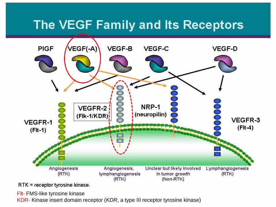
Flt- FMS-like tyrosine kinase
KDR- Kinase insert domain receptor (KDR, a type III receptor tyrosine kinase)

VEGF-VEGFR signaling
Angiogenesis
EC proliferation,
survival, migration and
invasion

VEGF Is a Key Mediator of Angiogenesis
Upstream activators of
VEGF synthesis
Downstream
signaling pathways

Hypoxia and Angiogenesis
• Hypoxia induces Angiogenesis: - during embryonic development - tumor growth - ischemia
• How??? Hypoxia induces Vascular Endothelial Growth Factor (VEGF)

Role of hypoxia in angiogenesis: (Hypoxia - HIF – VEGF module)
Like VEGF
VBC: Von Hippel-Lindau (VHL)-
containing VHL-elongin B/C and cullin-2
Angiogenic
Switch
E3-ubiquitin
Ligase
complex
Or
Mutant VHL

Role of hypoxia in angiogenesis: (Hypoxia - HIF – VEGF module) conti…
HIF: hypoxia inducible factor
VEGF: vascular endothelial growth factor
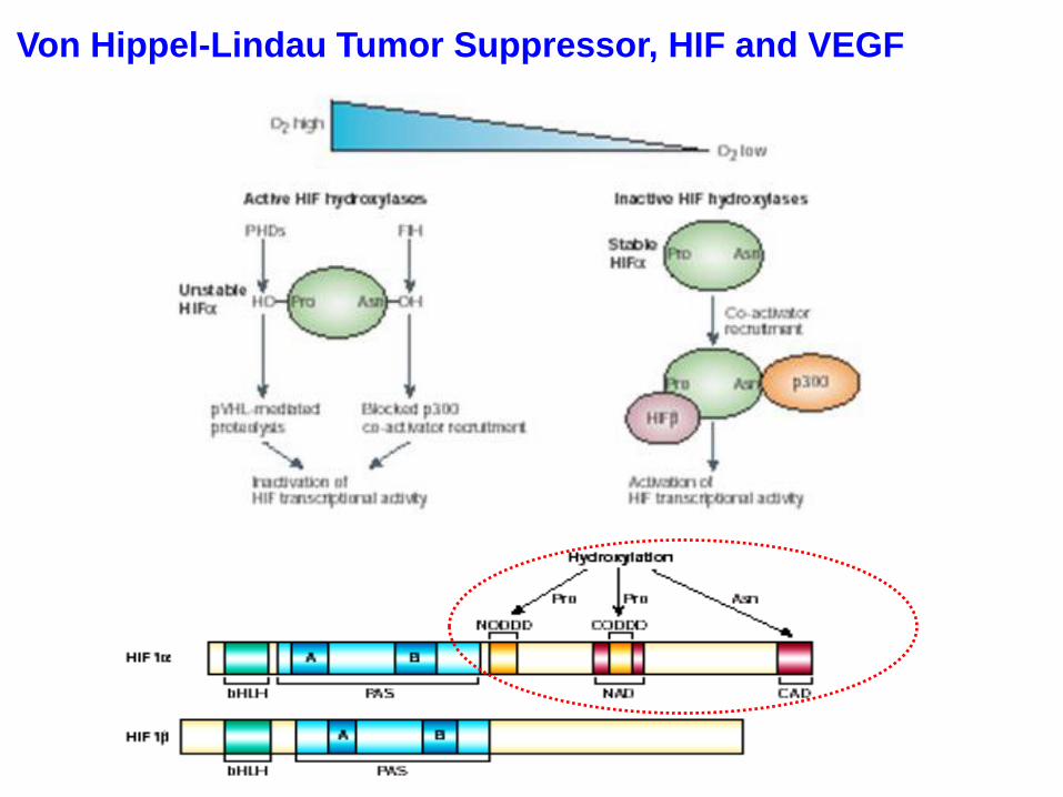
Von Hippel-Lindau Tumor Suppressor, HIF and VEGF

Angiogenesis-dependent diseases Excess:
• Cancer
• Infantile hemangiomas
• Autoimmune diseases, chronic inflammatory diseases:
• Rheumatoid arthritis
• Psoriasis
• Age-related macular degeneration
• Atherosclerosis
Deficiency:
• Limb ischemia
• Myocardial ischemia

Angiogenic inhibitors: • During the process of wound healing, the burst of angiogenesis must be shut down once the
newly formed capillaries have reached a certain density.
TSP-1 produced by stromal fibroblasts, ECs and immune cells
suppresses tumor progression by inhibiting angiogenesis through
direct effects on EC migration and survival and through indirect
effects on growth factor mobilization.


Inhibits EC migration, proliferation and induces EC apoptosis
Tumor angiogenesis
Judah Folkman (1971)- • Angiogenesis is a pre-requisite process for tumor growth and
metastatic progression
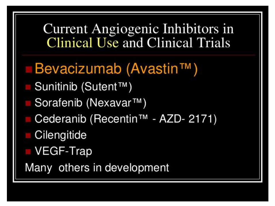
•Angioprevention- Targeting angiogenesis advocated for
cancer therapy


Tumor growth and angiogenesis Solid tumors can grow in size up to ~1-2 mm diameter by simple diffusion of
nutrients and gaseous exchange. However, beyond this size limit they require active
supply of such components for tumor growth and progression.
Angiogenic Switch
• Hypoxia
• Stabilization of HIF-alpha
• HR-gene expression (VGF)
• Growth, proliferation, survival and migration of ECs
• Sprouting, tube formation and tumor vasculature formation
• Tumor growth and metastatic progression

Carcinogenesis process

Features of tumor angiogenesis
• Extreme and chaotic expression of angiogenic factors
• Disorganized vascular structure and Low adhesion and pericyte coverage
• Hypoxic stress, metabolic changes, cancer cell intravasation and
lesser effects of chemotherapy

What Is Tumor Angiogenesis?
Tumor angiogenesis Proliferation of a network of blood
vessels that penetrates into cancerous growths.
Function Supplying nutrients and oxygen and
removing waste products. Mechanism Cancer cells release molecules that
send signals to surrounding normal host tissue. This signaling activates certain genes in the host tissue that, in turn, make proteins to encourage growth of new blood vessels.

Stroma contributes to tumor angiogenesis
Tumor Microenvironment (Tumor-associated stroma): induced by cytokines and chemokines secreted from tumor cells
• Macrophage: Tumor-‐Associated Macrophages (TAMs)
• Fibroblast: Carcinoma-‐Associated Fibroblasts (CAFs)
• Myeloid cell: Bone Marrow Derived Cells (BMDCs)
• Extracellular matrix (ECM)
Tumor microenvironment complexity and degree of
infiltration of various components correlates with the tumor
angiogenesis and invasiveness

Macrophage and tumor angiogenesis

Anti-angiogenic therapy Dr. Judah Folkman proposed the concept of anti-angiogenic therapy (NEJM.1971).


Antiangiogenic Therapies Potential Targets:
• Block pro-angiogenic molecules (e.g., VEGF)
• Using anti-angiogenic agents (e.g. angiostatin, endostatin,
TSP-1)
• Inhibit stroma-degrading enzymes (e.g., MMPIs)
• Target vascular antigens (e.g., avb3 integrin)
• Attack pericytes


Limitations of Anti-angiogenic therapy
• Resistance: expression of alternative angiogenic factors such as bFGF and
PDGF
• Toxicity and dosage (off target effects)
• Normalize disorganized tumor blood vessels
• Side effects (high blood pressure, bleeding and coronary artery disease)


Metastasis

When does metastasis begin?
Commitment to the metastatic phenotype: • How early does it occur?
• Can it be reversed?
Progenitor lesions: • What are the key progenitor lesions?
• What is the efficiency of transition to invasion?
• Are all metastasis precursors clonal?
What is the role of the host?
• Under what conditions does the host drive or suppress the process?
• Does the transition from pre-invasive to invasive lesions require host participation?
• If so what are the molecular and cellular players that are functionally important?
• The circuitry of the tumor host communication may be the key to prevention of invasion.

Physiologic basis of metastasis
• Is metastasis a normal physiologic program which is disregulated or inappropriately activated?
• Does a physiologic motility and invasion program exist for development, angiogenesis morphogenesis and wound healing?
• Is metastasis colony formation a natural ongoing process conducted by stem cells?

What is the driving force?
• Is the metastatic phenotype pre determined within the primary tumor? Within the host microenvironment?
• Are malignant cells a product of adaptation and selection?
• What is the selection factor? If malignant cells are survival of the fittest, then what is the fitness test?
• Is cell survival in a foreign (non home) tissue the ultimate selection factor?

Pre-cellular theory of
invasion and metastasis:
recognition of malignant
tumors and localized
versus metastatic disease
LeDran 1757: Noted that malignant tumors begin as localized disease, then
spread to regional lymph nodes and then enter the circulation to
subsequently appear in the lung
Bichat 1801: Tumors contain both parenchyma and stroma
Recamier 1829 : Used the term “Metastases”
Metastasis Pre-1900

Validation of the cellular theory of
cancer metastasis

Potential molecular mechanisms:
a) Preferential adhesion in the vessels of the target organ
b) Selective extravasation
c) Organ attractants
d) Organ specific survival and growth
The organ pattern of metastasis is characteristic of the tumor type
and tissue of origin.

Pre-metastatic niche formation Something secreted from primary tumor and changing the behavior of
host tissue at distant sites
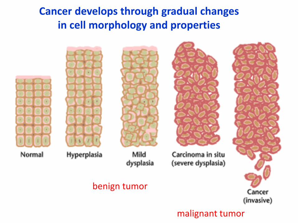
Cancer develops through gradual changes in cell morphology and properties
benign tumor
malignant tumor

Where do they go?
Figure 14.42 The Biology of Cancer (© Garland Science 2007)
Metastatic tropism

Figure 14.17b The Biology of Cancer (© Garland Science 2007)
Metastasis- a stepwise process

An organ is composed of several tissues
Epithelial cells
Connective tissue
Muscle tissue

Cancer cells change their epithelial properties, lose
their adhesion and penetrate through physical
barriers
basal lamina connective tissue

Intravasation

Once lodged in the blood vessels of various
tissues, cancer cells must escape from the lumina
of these vessels and penetrate into the surrounding
tissue- the step termed extravasation.

Formation of microthrombus (attachment of platelets) and
Proliferation in the lumen of the capillary
Platelet-mediated tumor cell extravasation

The blood: a hostile environment
Figure 14.7b The Biology of Cancer (© Garland Science 2007)
http://www.cancerquest.org/
- Cells are normally anchorage-dependent (anoikis) - Shear forces tear cells apart

After extravasation
what next???

Chemokines regulate leukocyte recirculation and trafficking to
sites of inflammation and infection
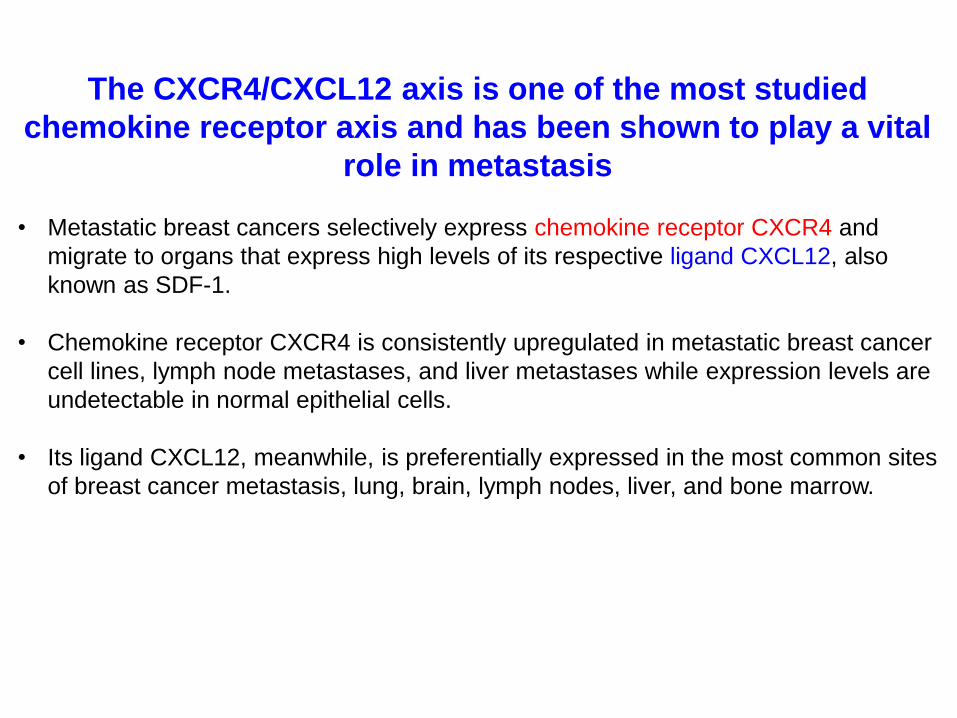
The CXCR4/CXCL12 axis is one of the most studied
chemokine receptor axis and has been shown to play a vital
role in metastasis
• Metastatic breast cancers selectively express chemokine receptor CXCR4 and
migrate to organs that express high levels of its respective ligand CXCL12, also
known as SDF-1.
• Chemokine receptor CXCR4 is consistently upregulated in metastatic breast cancer
cell lines, lymph node metastases, and liver metastases while expression levels are
undetectable in normal epithelial cells.
• Its ligand CXCL12, meanwhile, is preferentially expressed in the most common sites
of breast cancer metastasis, lung, brain, lymph nodes, liver, and bone marrow.
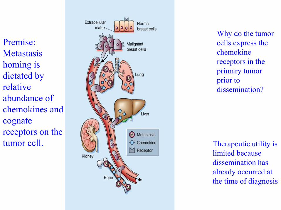
Premise:
Metastasis
homing is
dictated by
relative
abundance of
chemokines and
cognate
receptors on the
tumor cell.
Why do the tumor
cells express the
chemokine
receptors in the
primary tumor
prior to
dissemination?
Therapeutic utility is
limited because
dissemination has
already occurred at
the time of diagnosis

Figure 14.10a The Biology of Cancer (© Garland Science 2007)
Colonization
First, micrometastases

Figure 14.12 The Biology of Cancer (© Garland Science 2007)
Dormant micrometastases are viable
Figure 14.50a The Biology of Cancer (© Garland Science 2007)

Steeg Nature Med 06
Angiogenesis
Eventually: macrometastases
Intravasation
Latency
Colonization

Nguyen, Nature Rev. Cancer 2009

Figure 20-44 Molecular Biology of the Cell (© Garland Science 2008)
A sequence of inefficient steps
Metastatic inefficiency

Metastasis Promoting Genes - I
Gene Tissue Site Function
ARM-1 Lymphoma Promotes adhesion of tumor cells to the
endothelium
ATX Breast, Liver, Lung, Melanoma,
Teratocarcinoma
cytoskeletal reorganization and motility; G-
protein coupled receptor activation
CD44 Multiple sites cell-cell interactions; activates HGF/c-Met
pathway
Cox2 Breast, Colorectal, Gastric Prostaglandin synthase; induces VEGF
Cyr61 Breast Mediates adhesion; Erb-B2/3/4 pathway
Ezrin Liver, Ovary, Pancreas,
Prostate, Uterus
Membrane-cytoskeletal linker; RHO and RAC
interactions
HMG-I(Y) Breast, Cervical, Colorectal,
Prostate, Skin, Thyroid, Uterus
Regulated by EGF and MMP-9
Laminin-5 Multiple sites EGF and TGF-a induce expression of laminin
subunits; cell adhesion, motility
c-Met Multiple sites Activated by HGF; Modulates Ras and PI3
kinase

Metastasis Promoting Genes - II
Gene Tissue Site Function
MTA1 Breast, Cervix, Melanoma,
Ovary
Neucleosome remodeling; histone
deacetylase complex
Oncostatin M Lung Activates PKA-dependent pathway
PP2A Not determined Activated by p38/MAPK; inhibits MEK1,
MEK2, and MMP-1
RAGE Gastric, Lung, Pancreatic,
Renal
transmembrane receptor; activates p21,
MAPKs, NF-6B, cdc42/rac
S100A4 Breast, Colorectal, Prostate Calcium-binding protein; activates c-erbB-2
S100A9 Colon, Gastric, Skin Calcium-binding protein; Modulates Mac-1
integrin receptor through G-protein
Semaphorins Gastric, Leukemia, Lung, Skin cell-cell interactions; Receptor crosstalk with
c-Met binding semaphorin receptor, plexin
Thymosin-b15 Prostate actin binding; motility
Wnt-5a Breast, Colon, Lung,
Melanoma, Pancreas, Prostate
PKC activation with associated changes in
cytoskeleton, cell adhesion, and motility
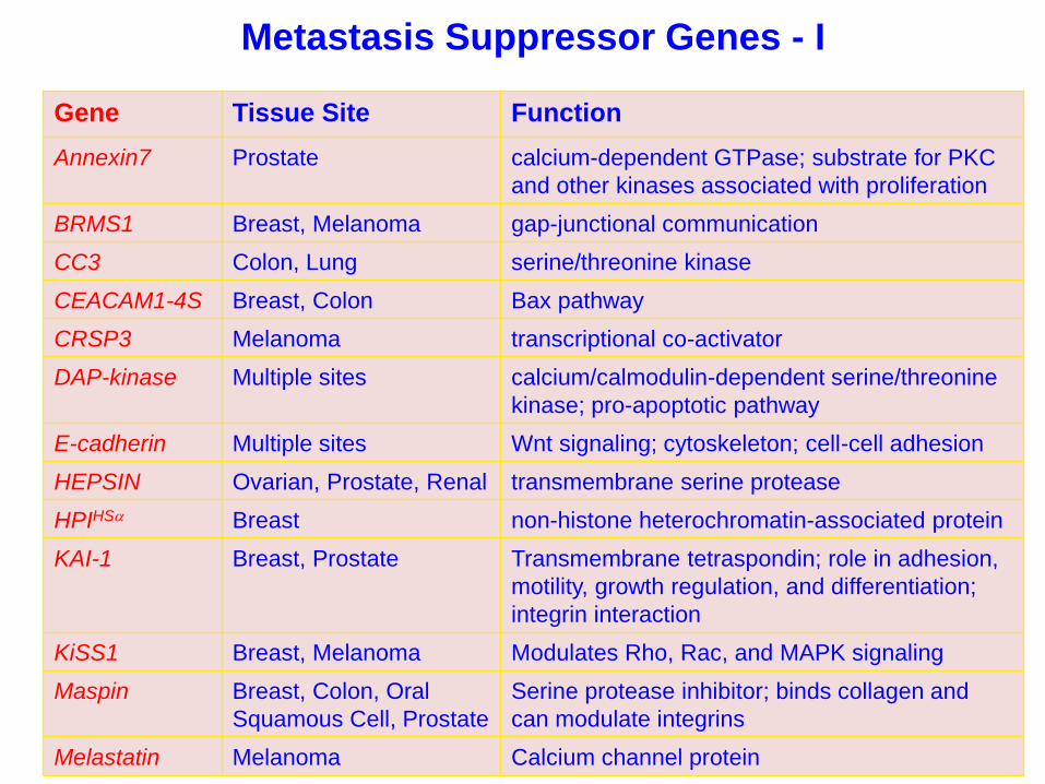
Metastasis Suppressor Genes - I
Gene Tissue Site Function
Annexin7 Prostate calcium-dependent GTPase; substrate for PKC
and other kinases associated with proliferation
BRMS1 Breast, Melanoma gap-junctional communication
CC3 Colon, Lung serine/threonine kinase
CEACAM1-4S Breast, Colon Bax pathway
CRSP3 Melanoma transcriptional co-activator
DAP-kinase Multiple sites calcium/calmodulin-dependent serine/threonine
kinase; pro-apoptotic pathway
E-cadherin Multiple sites Wnt signaling; cytoskeleton; cell-cell adhesion
HEPSIN Ovarian, Prostate, Renal transmembrane serine protease
HPIHSa Breast non-histone heterochromatin-associated protein
KAI-1 Breast, Prostate Transmembrane tetraspondin; role in adhesion,
motility, growth regulation, and differentiation;
integrin interaction
KiSS1 Breast, Melanoma Modulates Rho, Rac, and MAPK signaling
Maspin Breast, Colon, Oral
Squamous Cell, Prostate
Serine protease inhibitor; binds collagen and
can modulate integrins
Melastatin Melanoma Calcium channel protein

Metastasis Suppressor Genes - II
Gene Tissue Site Function
MKK4 Ovary, Prostate MAPK; phosphorylates and activates p38 and
JNK kinases
NESH Lung, Prostate src homology 3 adapter protein; down
regulates p21 pathway
NM23-H1 Breast, Colon, Melanoma,
Oral Squamous Cell
histidine kinase; phosphorylates KSR, which
might reduce ERK 1/2 activation
PTEN Multiple sites phosphatase; growth regulation, cell motility
RhoGD12 Bladder Inhibits GTP binding; regulates RHO and RAC
SFRP1 Breast, Colorectal Modulates Wnt signaling pathway
SHPS-1 Breast, Leukemia glycoprotein; may regulate RAS-MAPK
signaling; suppresses anchorage independent
growth
Syk Breast, Colon, Pancreas,
Skin
Tyrosine kinase; inhibits PI3 kinase; necessary
for MAPK activation
TSP-1 Multiple sites inhibits endothelial cell proliferation and
migration; c-Myc expression inhibits TSP-1
tropomyosins Breast interacts with e-cadherin/catenin complex
VDUP1 Melanoma Thioredoxin inhibitor; upregulates KiSS1;
interacts with CRSPs

How do cells become invasive???
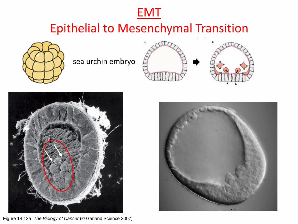
Figure 14.13a The Biology of Cancer (© Garland Science 2007)
EMT Epithelial to Mesenchymal Transition
sea urchin embryo

Major changes during EMT
- Loss of E-cadherin
- Cell shape changes driven by Rho GTPases
- MMPs


EMT in Tumor Progression

Figure 14.15b The Biology of Cancer (© Garland Science 2007)
Adopting changes typical to EMT Twist plays an essential role in cancer metastasis. Over-expression of Twist is
common in metastatic carcinomas.

MMPs (matrix metalloproteinases) help the cancer cells to invade the ECM

Signals from stroma controlling EMT

Epithelial Markers
E-cadherin Claudins Occludins
Desmoplakin Cytokeratins
Mesenchymal markers
Fibronectin Vitronectin Vimentin
Cell shape changes Cell movements, invasion
RhoB MMPs
Proliferation
Cyclin D CDK4
Rb phosph p21
Survival
PI3K activity ERK activity Caspases
P53 BID
Snail or Slug functions

Small GTPase family plays a key role of cancer
cell motility
Cytokines
Chemokines
Extracellular matrix
Rho family
•Rho
•Rac
•Cdc42
Stress fiber
Focal adhesion
Lamellipodia
Filopodia

EMT and cancer progression
Correlation between EMT inducing TFs with the
malignant behavior in cancer patients


Angiogenesis (role of VEGF etc.)
Tumor Angiogenesis
Metastasis and role of Tumor Angiogenesis
Invasion and migration
EMT process
Angioprevention OR anti-angiogenic therapy in preventing tumor metastasis
Targeting Tumor Metastasis
What we learn today!!!