Anemia - WSU MED · Investigations: Hgb
43
Anemia NABIL AL-KOURAINY, M.D. PGY1, INTERNAL MEDICINE 12.14.2017
Transcript of Anemia - WSU MED · Investigations: Hgb

AnemiaNABIL AL-KOURAINY, M.D.
PGY1, INTERNAL MEDICINE
12.14.2017

Definition
► Deficiency in O2-carrying capacity of blood 2/2 decreased RBC mass.
► May be due to RBC:
► Loss: bleeding, sequestration
► Decreased production: (hypoproliferative)
► Defective Hgb synthesis
► Defect in heme - iron, porphyrin ring or globin
► Deficiency in Fe, B12, Folate
► Impaired BM or stem cell function including any myeloid dysfunction or infiltrative process
► Leukemia
► Decreased EPO (fron renal failure) or decreased BM response to EPO
► Increased destruction: (hemolysis)
► SSA
► MAQHA

Symptoms of anemia
Decreased oxygenation
Exertional dyspnea
Dyspnea at rest
Fatigue
Bounding pulses
Lethargy, confusion
Decreased volume
Fatigue
Muscle cramps
Postural dizziness
syncope

Q: A 55-year-old woman is evaluated during a routine examination. She underwent biliopancreatic diversion with duodenal switch 8 years ago for treatment of obesity-related
complications and lost 68.0 kg (150.0 lb) in the first year following surgery. Her weight has been
relatively stable for the last year. She has generalized fatigue, involuntary muscle movement, mostly at night, dry skin, and brittle nails. Denies changes in vision or hearing. Her other medical
problems are type 2 diabetes mellitus and hypertension. Her prescription medications are metformin and lisinopril, and she also takes an over-the-counter multivitamin. Her last colonoscopy,
performed 5 years ago, was normal.
On physical examination, blood pressure is 140/79 mm Hg and pulse rate is 63/min. BMI is 25. The
examination is otherwise unremarkable.
Laboratory studies reveal a hemoglobin level of 10.5 g/dL (105 g/L) and a mean corpuscular
volume of 85 fL.
Which of the following deficiencies best explains this patient's current findings?
a) Copper
b) Iron
c) Vitamin A
d) Vitamin B12
Key points:- iron deficiency commonly occurs following
RY-Gastric Bypass, Patients with iron
deficiency are likely to have hypochromic,
microcytic anemia and may have brittle or deformed nails, cheilitis, pica, and restless
legs syndrome. Of note: Visual symptoms are
not seen with iron deficiency anemia.adapted from MKSAP 17

Clinical Presentation
Acute hemolysis
Intravascular hemolysis - acute back pain, free Hgb in plasma and urine, RF
Moderate anemia
Fatigue
Loss of stamina
Dyspnea
Tachycardia with exertion

o
Symptoms of known diseases causing anemia►Gastric ulceration►Rheumatoid arthritis►Renal failure
Duration of symptoms:►Hemoglobinopathies in longer duration
Treatment history►Medications for pain, supplementation: Fe,
B12, and folate►Nutritional history
Evaluation by History

Physical Exam
► Nourishment
► Signs of disease
► Vitals – fever, tachycardia, blood pressure
► Pallor
► Jaundice
► Lymphadenopathy
► Bone tenderness
► Petechiae
► CVS: Flow murmurs
► Resp: Dyspnea
► Abdomen: Splenomegaly

Note: the majority of patients seen in clinic who are diagnosed with anemia are asymptomatic
In the acutely anemic patient
who is volume depleted, it is
important to note that the
hemoglobin or hematocrit may be overestimated on the
initial blood sample, since
these are measures of blood
concentration
http://legacy.hopkinsilc.org/w/images/1/Anemia%202014/Anemia-table-history-and-physical-findings.png

Investigations:
Hgb
<12g/L (women)
<13g/L (men)
most* patients will experience some
symptoms related to anemia (no matter how slow the onset) when the
hemoglobin level reaches 7 g/dL.
* However, keep in mind pt’s baseline Hgb. This is why SSA pt can be asymptomatic at
a Hb of 6, while you or I will be symptomatic at a Hb of 11. The reason for
7 as a transfusion standard is for reducing the risk of cardiac complications of hypoxemia from anemia.
Laboratory definition of anemia
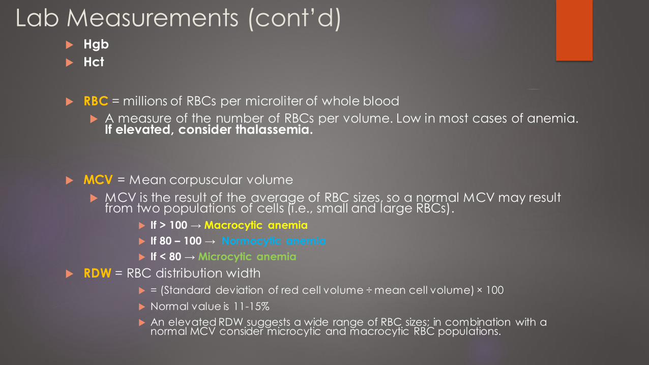
Lab Measurements (cont’d) Hgb
Hct
RBC = millions of RBCs per microliter of whole blood
A measure of the number of RBCs per volume. Low in most cases of anemia. If elevated, consider thalassemia.
MCV = Mean corpuscular volume
MCV is the result of the average of RBC sizes, so a normal MCV may result from two populations of cells (i.e., small and large RBCs).
If > 100 → Macrocytic anemia
If 80 – 100 → Normocytic anemia
If < 80 → Microcytic anemia
RDW = RBC distribution width
= (Standard deviation of red cell volume ÷ mean cell volume) × 100
Normal value is 11-15%
An elevated RDW suggests a wide range of RBC sizes; in combination with a normal MCV consider microcytic and macrocytic RBC populations.

Special Considerations in Determining
anemia
Acute BleedDrop in Hgb or Hct may not be shown until 36 to 48 hours
after acute bleed (even though patient may be hypotensive)
Pregnancy
In third trimester, RBC and plasma volume are expanded by 25 and 50%, respectively.
Labs will show reductions in Hgb, Hct, and RBC count, but according to RBC mass, are actually polycythemic
Volume Depletion
Patient’s who are severely volume depleted may not show anemia until after rehydrated

Symptoms of acute blood loss
In acute blood loss Hgb/Hct does not accurately reflect the volume of blood loss
Mild - asymptomatic - compensation thru enhanced
O2 delivery by changes in pH and increased CO2
10-15% - hypotension and decreased organ perfusion
>30% - postural hypotension, tachycardia
>40% - hypovolemic shock, confusion, diaphoresis, dyspnea
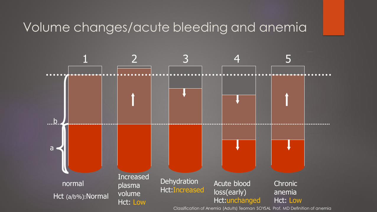
Volume changes/acute bleeding and anemia
normal
Hct (a/b%):Normal
Dehydration Hct:Increased
Acute blood loss(early) Hct:unchanged
Chronic anemia Hct: Low
1 2 3 4 5
Increased plasma volume Hct: Low
b
a
Classification of Anemia (Adults) Teoman SOYSAL Prof. MD Definition of anemia

Corrected reticulocyte count
Always correct for the retic ct. Why correct? Because men and women
have diff nl Hct. corrected reticulocyte count = reticulocyte % x (Pt Hct/nl Hct*).
*40% women, 45% men
C-retic > 2.0% suggest hemolysis or acute blood loss. < 2.0% suggest hypoproliferation.
The normal reticulocyte count is 0.5-1.5%. Insufficient response (not enough) hypo. Vs, adequate response (trying to correct for loss = hemolytic)

Vocabulary of anemia
http://legacy.hopkinsilc.org/w/images/1
/Anemia%202014/Anemia-table-
vocabulary.png

Let’s go through some common examples.
*note this is not comprehensive


Seen with: post-splenectomy or congenital absence of spleen as
well as: amyloidosis, severe HA, megaloblastic anemia,
hereditary spherocytosis, and MDS. Can also can be seen in
premature infants.
Aka Burr cells are RBC membrane defect characterized by
irregular spines - associated w ith severe liver disease
Aka schistocytes – seen in intravascular hemolysis as seen in
sepsis

Fe studies
Serum Iron: 50–150 µg/dL
TIBC: 300–360 μg/dL
Serum ferritin (also an acute phase reactant)
15-20 µg/dL – Lack of Iron stores
Women: ~30 µg/dL
Men: ~ 100 µg/dL
200 µg/dL – adequate iron stores
Serum Transferrin saturation: 25-50%
Ferritin is most important study for Fe deficiency. Serum iron
does not tell us much as it can
change acutely. Similarly, a
diabetic can have a normal serum glucose at any given
point.
Can obtain a soluble transferrin
receptor if ferritin is normal.

Bone Marrow Evaluation
Mandatory in patients with normal Fe status with hypoproliferative anemia
Can be used to diagnose primary bonemarrow diseases: myelofibrosis, infiltrativediseases

Q: A 52-year-old male presents to you with several months of fatigue. You order blood
work, which shows the following:
Of the causes listed, the most likely cause of this patient's anemia is:
•a) Autoimmune hemolytic anemia•b) Glucose-6-phosphate dehydrogenase deficiency•c) Thrombotic thrombocytopenic purpura•d) Vitamin B12 deficiency
Key points:- Low HCT with slight elevation in retic ct
- Corrected retic ct = 1.3 %
- (3% x 20%/45% = 1.3%)
- Hypoproliferative -> underproduction vs all other answer choices which are
hemolytic -> destruction
https://ilc.peaconline.org/m/1308

The MCV approach…

Index < 2.0 Index > 2.0
Adapted from Harrison’s 19th editionAdapted from Harrison’s 19th edition
1. Establish presence of anemia
2. Correct for reticulocyte count
• < 2.0 hypoproliferative
• > 2.0 hemolytic or
hemorrhage
- Vast majority will be normocytic 2/2 IDA
- Question then is why are the Fe
deficient? - Depends on age
- Be mindful of Red flag
symptoms. - 1) rectal bleeding, 2) iron-
deficiency anemia (IDA), 3) weight loss, 4) family
history of colon cancer, 5) fever, and 6) age of onset
after age 50.
- Make sure cancer screening is UTD,
especially CRC in
patients greater than 50y.
The CRC
approach

Index < 2.0 Index > 2.0
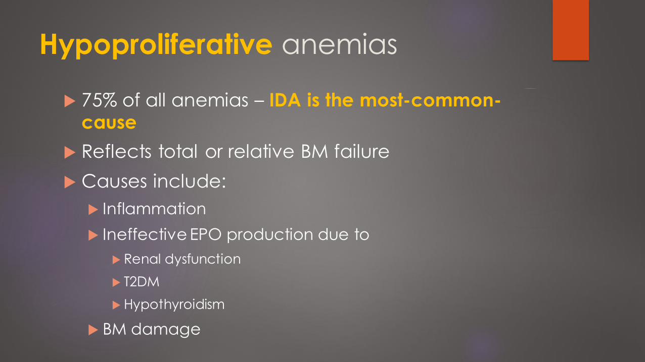
Hypoproliferative anemias
75% of all anemias – IDA is the most-common-cause
Reflects total or relative BM failure
Causes include:
Inflammation
Ineffective EPO production due to
Renal dysfunction
T2DM
Hypothyroidism
BM damage

Work-up
Fe studies
Ferritin
Bone Marrow biopsy/aspiration

Findings: AOCD vs IDA
AOCD:
Fe : Low
TIBC: normal or low
Soluble Transferrin Receptor (sTFR): Normal
Ferritin: Normal – High
IDA
Fe : Low
TIBC: High
Soluble Transferrin Receptor (sTFR): High
Ferritin: Low
- Ferritin is an acute phase reactant.
infectious processes may cause
ferritin to rise, even in the presence
of Fe deficiency.
- However, in true Fe deficiency,
ferritin <100ng/mL.- a ferritin >100ng/mL excludes
iron deficiency as a cause of
microcytic anemia.
- transferrin assay -> elevated in iron deficiency, vs normal in AOCD
Peripheral blood film with changes attributed to iron deficiency anemia.
https://openi.nlm.nih.gov/detailedresult.php?img=PMC3538607_1746-1596-7-168-1&query=&req=4&simCollection=PMC3538607_1746-1596-7-168-1&npos=1

Leukemia, Lymphoma and BM aplasia
Acute myelogenous leukemia - there is one lone
megakaryocyte at the right-center Aplastic anemia
https://upload.wikimedia.org/wikipedia/commons/3/31/Aplast ic_Anemia.jpghttps://library.med.utah.edu/WebPath/HEMEHTML/HEME031.html
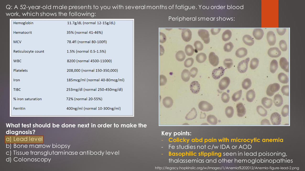
Q: A 52-year-old male presents to you with several months of fatigue. You order blood
work, which shows the following:
What test should be done next in order to make the diagnosis?
a) Lead level
b) Bone marrow biopsy
c) Tissue transglutaminase antibody leveld) Colonoscopy
Key points:- Colicky abd pain with microcytic anemia
- Fe studies not c/w IDA or AOD
- Basophilic stippling seen in lead poisoning,
thalassemias and other hemoglobinopathies
Peripheral smear shows:
http://legacy.hopkinsilc.org/w/images/1/Anemia%202012/Anemia-figure-lead-2.png
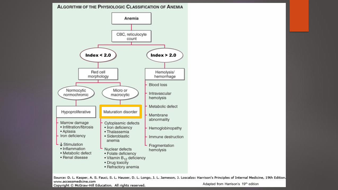
Index < 2.0 Index > 2.0
Adapted from Harrison’s 19th edition

Maturation disorders
CRC <2.0
Two types:
Macrocytic – nuclear abnormalities
Microcytic – cytoplasmic abnormalities
Poor erythropoiesis 2/2 marrow destruction
BM shows erythroid hyperplasia

Nuclear maturation disorders
B 12 deficiency
Folate deficiency
Toxins (MTX)
Alcohol - > folate deficiency

Cytoplasmic maturation disorders
Severe Fe deficiency
Thalassemias
Siderblastic
https://ilc.peaconline.org/sites/ilc.peaconline.org/files/modules/2017/Anemia/Anemia-table-thalassemia-vs-iron-deficiency.png

Sideroblastic anemia relative iron overload with ineffective
heme synthesis
BM stained with Prussian blue reveals characteristic ringed sideroblasts
Arrows point to two examples of ringed sideroblastshttps://ilc.peaconline.org/sites/ilc.peaconline.org/files/modules/2017/Anemia/Anemia-figure-ringed-sideroblast.png
https://ilc.peaconline.org/sites/ilc.peaconline.org/files/modules/2017/Anemia/Anemia-table-causes-of-acquired-sideroblastic-anemia.png

Lead poisoning
Microcytic
motor neuropathy and abdominal pain
basophilic stippling on PBS
http://legacy.hopkinsilc.org/w/images/1/Anemia%202012/Anemia-figure-lead-2.png

Testing to evaluate hypoproliferative
microcytic anemia
https://ilc.peaconline.org/sites/ilc.peaconline.org/files/modules/2017/Anemia/Anemia-table-tests-in-microcytic-anemia.png

Q: You are evaluating a 43-year-old male patient with normocytic anemia (hematocrit
31%; MCV 86fl). Renal function is normal, thyroid testing is normal, and there is no
evidence of anemia of chronic disease. Peripheral blood smear is as follows:
Which of the following statements is true?a) This patient most likely has myelodysplastic
syndrome.
b) This patient most likely has multiple myeloma.c) HIV infection is a potential cause of this patient's
anemia.d) Systemic lupus erythematosus is a potential cause
of this patient's anemia.
Key points
- aplastic anemia with pancytopenia
- absence of platelets
- Leukopenia
- lack of immature RBCs- HIV infection is a possible cause of aplastic
anemia.
https://ilc.peaconline.org/m/1308

Myelodysplasia
Features:
Macro or microcytosis
Iron ring in
mitochondria
Fe studies can be used
to help differentiate from other disorders
Bone marrow aspirate – MDS with excess blasts-2 (MDS-EB2). myeloblasts are smaller, with scant pale cytoplasm.
https://img.medscapestatic.com/pi/meds/ckb/78/13278tn.jpg

Index < 2.0 Index > 2.0
Adapted from Harrison’s 19th edition

Acute blood loss
cRetic > 2.0
Symptoms:
In acute blood loss Hgb/Hct does not accurately reflect the volume of
blood loss
Mild - asymptomatic - compensation thru enhanced O2 delivery by changes
in pH and increased CO2
10-15% - hypotension and decreased organ perfusion
>30% - postural hypotension, tachycardia
>40% - hypovolemic shock, confusion, diaphoresis, dyspnea
Not associated with increased erythrocytosis 2/2 time required for EPO production

Evaluation of Hemolytic Anemia
RBCs prematurely
removed from circulation by liver and spleen. Majority
of cases of HA.
RBCs lyse
within circulation
https://ilc.peaconline.org/m/1308

References• https://ilc.peaconline.org/m/1308*• Harrison’s Principles of Internal Medicine,19th Edition• adapted from MKSAP 17, (slide 4) • Bossi D, Russo M. Hemolytic anemias due to disorders of red cell membrane
skeleton. Mol Aspects Med 1996; 17:171.• https://emedicine.medscape.com/article/198475-overview• https://img.medscapestatic.com/pi/meds/ckb/78/13278tn.jpg• Classification of Anemia (Adults) Teoman SOYSAL Prof. MD Definition of anemia• https://upload.wikimedia.org/wikipedia/commons/3/31/Aplastic_Anemia.jpg• https://library.med.utah.edu/WebPath/HEMEHTML/HEME031.html