Andrea Romano Neuroradiology and headaches Valentina ...Neuroradiology and headaches TUTORIAL Andrea...
11
J Headache Pain (2006) 7:422–432 DOI 10.1007/s10194-006-0347-6 Neuroradiology and headaches TUTORIAL Andrea Romano Valentina Cipriani Alessandro Bozzao Abstract Headache is one of the most common outpatient pain con- ditions encountered in both physi- cian offices and emergency depart- ments. Establishment of an accurate diagnosis, accomplished only by a thorough history followed by a physical examination, is critical before treatment can be initiated. Many patients undergo evaluation with computed tomography and more recently magnetic resonance imaging to exclude important abnormalities. It is known that a lit- tle percentage of patients showed significant neuroradiologiacal abnormalities and the rate of signif- icant intracranial abnormalities in patients with headache and normal neurological examination exists. Keywords Headache • Magnetic resonance • Computed tomography Received: 25 October 2006 Accepted in revised form: 6 November 2006 Published online: 10 December 2006 A. Romano () • V. Cipriani • A. Bozzao Department of Neuroradiology, La Sapienza University of Rome Sant’Andrea Hospital Via di Grottarossa 1035 I-00189 Rome, Italy E-mail: [email protected] Introduction Headaches with attendant pain and debilitation have been noted throughout recorded medical history. New great sci- entific results have been obtained since the aetiologic the- ories included curses of the gods, evil spirits, imbalance of humours and many other equally imaginative ideas. Today, fortunately, some things have changed [1]. Headache is one of the most common outpatient pain conditions encountered in both physicians’ offices and emergency departments. Establishment of an accurate diagnosis, accomplished only by a thorough history fol- lowed by a physical examination, is critical before treat- ment can be initiated. Although the majority of patients who present with chronic or recurrent headache do not have neurological abnormalities, many patients undergo evaluation with computed tomography (CT) and more recently magnetic resonance imaging (MRI) to exclude eventual diseases responsible for the symptoms. MRI is more sensitive than CT in the detection of almost all intracranial abnor- malities. Despite this, limited data exist about the utility of head MRI in patients with headache. In a recent paper from Sempere et al. [2], which considered 1876 patients with headache (any type started 4 weeks before neu- roimaging), 1.2% of patients showed significant neuro- radiological abnormalities and the rate of significant intracranial abnormalities in patients with headache and normal neurological examination was 0.9%. The authors
Transcript of Andrea Romano Neuroradiology and headaches Valentina ...Neuroradiology and headaches TUTORIAL Andrea...

J Headache Pain (2006) 7:422–432DOI 10.1007/s10194-006-0347-6
Neuroradiology and headaches
T U T O R I A L
Andrea RomanoValentina CiprianiAlessandro Bozzao
Abstract Headache is one of themost common outpatient pain con-ditions encountered in both physi-cian offices and emergency depart-ments. Establishment of an accuratediagnosis, accomplished only by athorough history followed by aphysical examination, is criticalbefore treatment can be initiated.Many patients undergo evaluationwith computed tomography andmore recently magnetic resonanceimaging to exclude importantabnormalities. It is known that a lit-tle percentage of patients showedsignificant neuroradiologiacalabnormalities and the rate of signif-
icant intracranial abnormalities inpatients with headache and normalneurological examination exists.
Keywords Headache • Magneticresonance • Computed tomography
Received: 25 October 2006Accepted in revised form: 6 November 2006Published online: 10 December 2006
A. Romano (�) • V. Cipriani • A. BozzaoDepartment of Neuroradiology,La Sapienza University of RomeSant’Andrea HospitalVia di Grottarossa 1035I-00189 Rome, ItalyE-mail: [email protected]
Introduction
Headaches with attendant pain and debilitation have beennoted throughout recorded medical history. New great sci-entific results have been obtained since the aetiologic the-ories included curses of the gods, evil spirits, imbalanceof humours and many other equally imaginative ideas.Today, fortunately, some things have changed [1].
Headache is one of the most common outpatient painconditions encountered in both physicians’ offices andemergency departments. Establishment of an accuratediagnosis, accomplished only by a thorough history fol-lowed by a physical examination, is critical before treat-ment can be initiated.
Although the majority of patients who present withchronic or recurrent headache do not have neurologicalabnormalities, many patients undergo evaluation withcomputed tomography (CT) and more recently magneticresonance imaging (MRI) to exclude eventual diseasesresponsible for the symptoms. MRI is more sensitivethan CT in the detection of almost all intracranial abnor-malities. Despite this, limited data exist about the utilityof head MRI in patients with headache. In a recent paperfrom Sempere et al. [2], which considered 1876 patientswith headache (any type started 4 weeks before neu-roimaging), 1.2% of patients showed significant neuro-radiological abnormalities and the rate of significantintracranial abnormalities in patients with headache andnormal neurological examination was 0.9%. The authors
423
concluded that the proportion of patients with headacheand intracranial lesions is relatively small, but neitherneurological examination nor the features in the clinicalhistory permit us to rule out such abnormalities. There isgeneral agreement that imaging should be performed incase of important changes in the type of headache, incase of first or worst episode, when the onset is suddenor provokes awakening during sleeping, when neurolog-ical symptoms are associated, in very young or old peo-ple (over 5 and 50 years), in patients with cancer andpregnancy, when there are consciousness changes andwhen headache is related to Valsalva manoeuvres or sex-ual activity.
In the first part we will discuss the main neuroradio-logical findings that can be evaluated in primary and sec-ondary headaches by conventional imaging. In the secondpart of this tutorial we will report the more recent resultsin the applications of “functional” imaging in the study ofpathophysiology of primary headache.
“Conventional imaging” in primary headaches
Migraine
Migraine is thought to be a progressive inflammatory neu-rovascular disorder associated with considerable disabili-ty and impairment of quality of life. It is a disease accom-panied not only by characteristic throbbing pain, but alsoby associated symptoms and disability. Migraineheadache frequently is preceded by a prodrome, known asaura. The aura in migraine is a clearly defined neurologicdeficit, most often visual in nature, such as scotomas orvisual field changes [1].
The role of imaging and specifically of conventionalMRI is still debated. The association of migraine phenom-ena with neuroimaging abnormalities, as demonstrated byCT and MRI, has been the subject of much debate [3].
In a study by Ziegler et al. [4], 18 cases of migrainewith aura were considered. Only in four of the migrainepatients were areas of increased signal intensity in T2-weighted images identified. In three of the normal con-trols similar areas were seen as well. The authors con-cluded that these small areas, difficult to identify, may beof multiple aetiologies. Similar small unidentified brightareas have been seen not uncommonly in the asympto-matic older population.
In another study, by Igarashi et al. [5], the authorsassumed that repetition of migraine, with repeatedepisodes of cerebral hypoperfusion responsible for aura,could cause permanent ischaemic changes in the brain.The characteristics of MRI lesions in migraine were well
defined small foci on T2-weighted and proton density-weighted images in the white matter. These white matterlesions resemble so-called unidentified bright objects(UBOs) [6], which are often related to ageing, hyperten-sion and other atherosclerotic risk factors. In this study39.6% of patients with migraine showed small white mat-ter lesions on MRI. The incidence of white matter lesionswas not different between patients with and without aura.No significant links with risk factors were found. A groupof patients with migraine under 40 years old without riskfactors showed significantly higher incidence of positiveMRI changes (29.4%) than the controls (11.2%). The sideof the MRI lesions did not always correspond to the sideof usual aura or headache. The migraine may be associat-ed with early pathologic changes in the brain. MRI stud-ies in patients with migraine showed focal areas of highintensity on both T2-weighted and proton density-weight-ed MR images distributed bilaterally in the white matterof the brain [5].
These results are similar to those report by Soges et al.[7], who studied 24 patients with migraine and found 46%of them showed abnormal MRI studies. Their slightlyhigher percentage for positive MRI might be explained bythe slightly older mean age (36.8 years) than Igarashi etal.’s patients (34.0 years).
The most tempting speculation to explain brain lesionsin migraine is that repeated attacks of hypoperfusion dur-ing aura might cause permanent ischaemic changes [5]and frequency of attacks is an important indicator of exis-tence of white matter foci [8].
Ischaemia or an immune-based white matter demyelin-isation are other possible mechanisms for the white matterlesions [9].
Such high incidence of hyperintense foci in the whitematter of patients with migraine was not confirmed byother studies. Osborn et al. demonstrated parenchymalbrain lesions in only 12% of patients with migraine withaura [10].
In a recent study published in JAMA in 2004, no sig-nificant differences between patients with migraine andcontrols in overall infarct prevalence were found.However, in the cerebellar region, patients with migrainehad a higher prevalence of infarct than controls. Amongwomen, the risk for higher white matter lesion load wasincreased in patients with migraine compared with con-trols. This risk increased with attack frequency [11].
The same authors more recently underlined that braininfarction occurs far more frequently than expected inmigraine patients, being most pronounced in migrainewith aura (8% have subclinical cerebellar infarcts). Mostinfarcts remain clinically silent. Female migraine patientsare at increased risk of deep white matter lesions, inde-pendent of the effects of cardiovascular risk factors. The
424
influence of migraine severity (attack frequency) on therisk of both types of lesions suggests a causal relationshipbetween migraine severity and lesion load [12].
In a recent meta-analysis from Swartz and Kern, theauthors demonstrate that subjects with migraine are athigher risk of having white matter abnormalities on MRIthan those without migraine. This increased risk is presenteven in younger individuals who do not have co-occurringcerebrovascular disease risk factors [13].
Tension-type headache
Benedittis et al. showed that in the MRI of patients withtension-type headache there is a higher incidence of whitematter abnormalities compared to control subjects (33.3%vs. 7.4%). These lesions are distributed predominantly inthe frontal region and were shown to have a similar inci-dence compared with patients with migraine with aura(32.1%) [14].
Cluster headache (CH)
No specific abnormalities have been described in clusterheadache in conventional MRI. When an acute CH attackwas triggered with nitroglycerin (NTG), vasodilatationwas observed with PET occurring in the ipsilateral poste-rior inferior hypothalamic grey matter, in the contralater-al ventroposterior thalamus, in the anterior cingulate cor-tex, in the ipsilateral basal ganglia, in the right anteriorfrontal lobe and in both insulae. In patients out of thegroup who experienced only a mild NTG headache, acti-vation was seen bilaterally in the insulae and frontal cor-tices, the anterior cingulated cortex, the right thalamusand the left basal ganglia, but not in the hypothalamicgrey area. In addition, the authors found significant acti-vation (vasodilatation) in the region of the major basalarteries, which was caused in part by NTG but was alsoobserved in the spontaneous case and could be induced bycapsaicin injection into the forehead [15].
“Conventional imaging” in secondary headaches
Headaches are one of the most common symptoms thatneurologists evaluate. Although most are caused by pri-mary disorders, the list of differential diagnoses is one ofthe longest in all of medicine, with over 300 differenttypes and causes [16].
Headache attributed to head and/or neck trauma
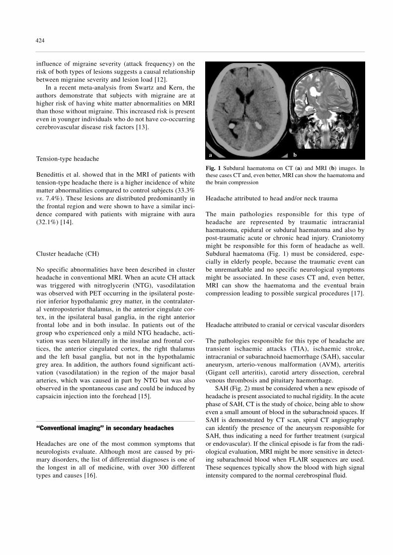
The main pathologies responsible for this type ofheadache are represented by traumatic intracranialhaematoma, epidural or subdural haematoma and also bypost-traumatic acute or chronic head injury. Craniotomymight be responsible for this form of headache as well.Subdural haematoma (Fig. 1) must be considered, espe-cially in elderly people, because the traumatic event canbe unremarkable and no specific neurological symptomsmight be associated. In these cases CT and, even better,MRI can show the haematoma and the eventual braincompression leading to possible surgical procedures [17].
Headache attributed to cranial or cervical vascular disorders
The pathologies responsible for this type of headache aretransient ischaemic attacks (TIA), ischaemic stroke,intracranial or subarachnoid haemorrhage (SAH), saccularaneurysm, arterio-venous malformation (AVM), arteritis(Gigant cell arteritis), carotid artery dissection, cerebralvenous thrombosis and pituitary haemorrhage.
SAH (Fig. 2) must be considered when a new episode ofheadache is present associated to nuchal rigidity. In the acutephase of SAH, CT is the study of choice, being able to showeven a small amount of blood in the subarachnoid spaces. IfSAH is demonstrated by CT scan, spiral CT angiographycan identify the presence of the aneurysm responsible forSAH, thus indicating a need for further treatment (surgicalor endovascular). If the clinical episode is far from the radi-ological evaluation, MRI might be more sensitive in detect-ing subarachnoid blood when FLAIR sequences are used.These sequences typically show the blood with high signalintensity compared to the normal cerebrospinal fluid.
Fig. 1 Subdural haematoma on CT (a) and MRI (b) images. Inthese cases CT and, even better, MRI can show the haematoma andthe brain compression
a b

425
Unruptured vascular malformations can be responsiblefor prolonged headache. Artero-venous and dural malfor-mations cause changes in brain vascular haemodynamics.MRA and CTA can easily show unruptured vascular mal-formations but conventional angiography is still mandatoryto understand the exact structure of the abnormality. Alsothe presence of arteriovenous (AV) shunts, such as patentforamen ovale (PFO), can represent a trigger for migrainewith aura attacks. A higher prevalence of PFO was demon-strated in migraineurs vs. the normal population [18].
Venous thrombosis (Fig. 3) should be considered whendealing with young women using hormone therapy [19].Venous thrombosis can be identified by means of MRI andMRA or by CT angiography. Parenchymal lesions (fre-quently haemorrhagic) can be associated as well.
Carotidynia is a syndrome encompassing many vari-eties of pain in the carotid region. It can be due to carotiddissection (Fig. 4). In case of suspect carotid dissection, ahigh signal on T1W can be demonstrated in the wall ofdistal internal carotid artery. The haematoma might pro-duce a reduction of the lumen size at MRA. Neurologicalsymptoms (either central deficit or hypoglossal nervecompression) can be associated [17]. Migraine may repre-sent a predisposing condition for spontaneous cervicalartery dissection [20].
Pituitary haemorrhage can cause a sudden and strongheadache. This may be related to previous therapy for pitu-itary adenoma or not. MRI can easily show the blood insidethe gland and follow-up can document partial or completeresolution of the disease (Fig. 5). Close follow-up must beconsidered to rule out a dangerous optic nerve compression.
Fig. 2 Subarachnoid haemorrhage (SAH). In (a) and (b) a SAH inthe acute phase is evident on the CT study. CT angiography study(c) demonstrates the presence of the aneurysm responsible for SAHin the M2 segment of middle cerebral artery (arrow). FLAIRsequence might be more sensitive than CT in detecting subarach-noid blood in the first days after the acute event. The blood isshown as a high signal intensity alteration compared to the normalcerebrospinal fluid (d, arrow)
a
c d
b
Fig. 3 Right transverse and sigmoid sinuses thrombosis. The T1hyperintensity inside the sinuses (a) and the occlusion of them inthe MR angiography study are evident (b). The high signal in MIPreconstruction of MRA is due to the intrinsic hyperintensity of theclot visible on conventional axial T1-weighted images (arrows)
a b
Fig. 4 Carotid dissection. High signal on T1-weighted image isobserved in the wall of left distal internal carotid artery (a,arrow). The haematoma produces a reduction of the lumen sizeat MRA (b, arrow)
a b
Fig. 5 Pituitary haemorrhage observed as a hyperintense areainside the gland in the acute phase (a) and as an area of reducedvascularisation post-contrast (b) in the follow-up
a b
426
Headache attributed to non-vascular intracranial disorder
There are many non-vascular intracranial disordersresponsible for headache-like idiopathic intracranialhypertension, high or low cerebrospinal fluid pressure,aseptic meningitis, intracranial neoplasm, epilepsy, hypo-thalamic or pituitary hyper- or hyposecretion.
Only a minority of patients who have headaches havebrain tumours. Some locations are more likely to produceheadache (e.g., a posterior fossa tumour causes headachemore often than a supratentorial tumour) [21].
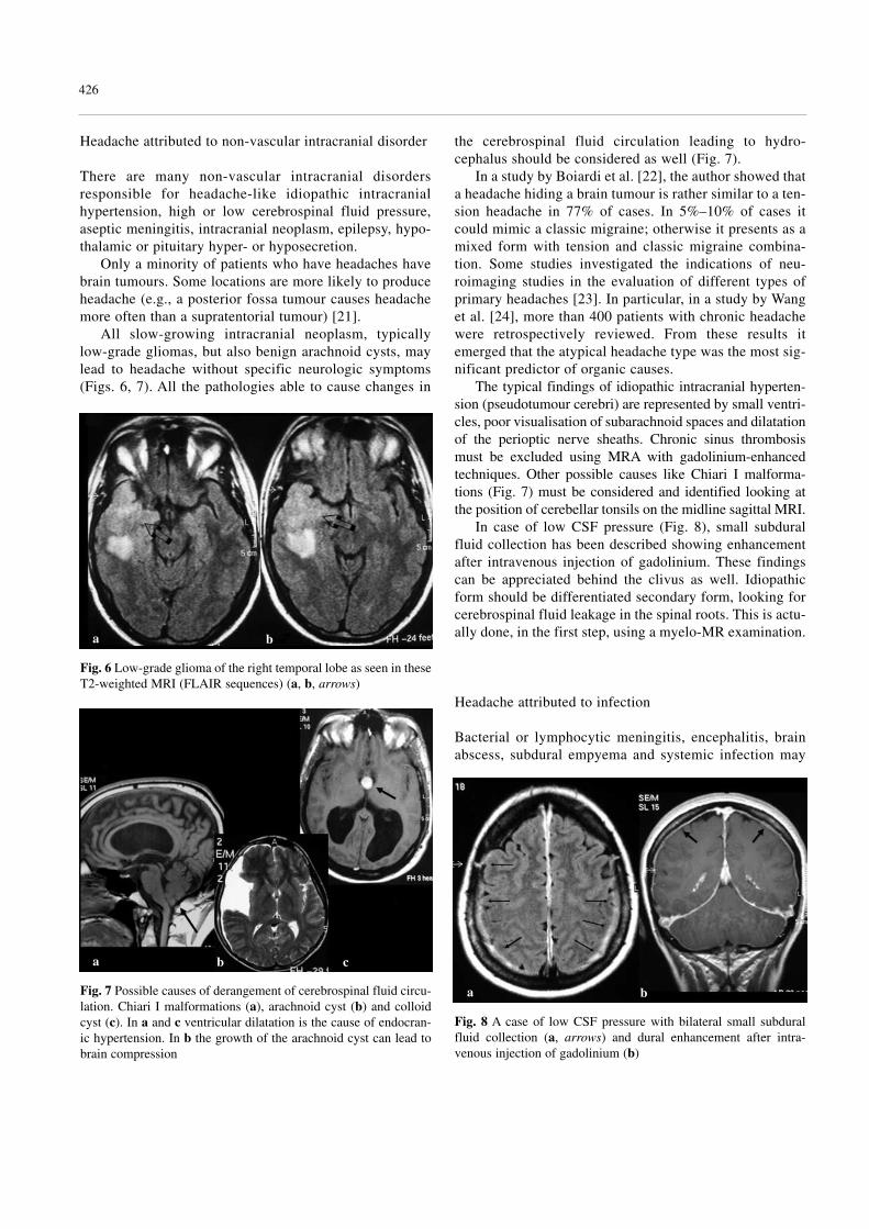
All slow-growing intracranial neoplasm, typicallylow-grade gliomas, but also benign arachnoid cysts, maylead to headache without specific neurologic symptoms(Figs. 6, 7). All the pathologies able to cause changes in
the cerebrospinal fluid circulation leading to hydro-cephalus should be considered as well (Fig. 7).
In a study by Boiardi et al. [22], the author showed thata headache hiding a brain tumour is rather similar to a ten-sion headache in 77% of cases. In 5%–10% of cases itcould mimic a classic migraine; otherwise it presents as amixed form with tension and classic migraine combina-tion. Some studies investigated the indications of neu-roimaging studies in the evaluation of different types ofprimary headaches [23]. In particular, in a study by Wanget al. [24], more than 400 patients with chronic headachewere retrospectively reviewed. From these results itemerged that the atypical headache type was the most sig-nificant predictor of organic causes.
The typical findings of idiopathic intracranial hyperten-sion (pseudotumour cerebri) are represented by small ventri-cles, poor visualisation of subarachnoid spaces and dilatationof the perioptic nerve sheaths. Chronic sinus thrombosismust be excluded using MRA with gadolinium-enhancedtechniques. Other possible causes like Chiari I malforma-tions (Fig. 7) must be considered and identified looking atthe position of cerebellar tonsils on the midline sagittal MRI.
In case of low CSF pressure (Fig. 8), small subduralfluid collection has been described showing enhancementafter intravenous injection of gadolinium. These findingscan be appreciated behind the clivus as well. Idiopathicform should be differentiated secondary form, looking forcerebrospinal fluid leakage in the spinal roots. This is actu-ally done, in the first step, using a myelo-MR examination.
Headache attributed to infection
Bacterial or lymphocytic meningitis, encephalitis, brainabscess, subdural empyema and systemic infection may
Fig. 6 Low-grade glioma of the right temporal lobe as seen in theseT2-weighted MRI (FLAIR sequences) (a, b, arrows)
a b
Fig. 8 A case of low CSF pressure with bilateral small subduralfluid collection (a, arrows) and dural enhancement after intra-venous injection of gadolinium (b)
a bFig. 7 Possible causes of derangement of cerebrospinal fluid circu-lation. Chiari I malformations (a), arachnoid cyst (b) and colloidcyst (c). In a and c ventricular dilatation is the cause of endocran-ic hypertension. In b the growth of the arachnoid cyst can lead tobrain compression
a b c

427
be responsible for headache. This type of headache ischaracterised by diffuse or continuous pain accompaniedby nausea and/or focal neurological symptoms and/orsigns. A direct space-occupying effect leading to raisedintracranial pressure and/or irritation of the meningeal orarterial structures are the mechanisms for causingheadache [17].
MRI with diffusion-weighted images (DWI) can beeasily characterised by the presence of a brain abscess dueto the typical restriction of diffusion inside the abscesscavity. Using these imaging characteristics we can easilydifferentiate an abscess from the presence of necrosisinside a primary (glioblastoma) or a secondary (metasta-sis) tumour of the brain.
Headache attributed to disorders of homeostasis
A lot of disorders of homeostasis cause headache, suchas hypoxia, hypercapnia, arterial hypertension, hypothy-roidism, hypertensive encephalopathy, pre-eclampsiaand eclampsia, anaemia, adrenocortical insufficiency,hyperaldosteronism, polycythemia, Cushing’s diseaseetc. [17].
In eclampsia MRI can demonstrate typical reversiblesignal alterations in the parieto-occipital regions.
Headache or facial pain attributed to disorders of cranium,neck, eyes, ears, nose, sinuses, teeth, mouth or other facialor cranial structures
Pain in the oro-facial region can be very distressing forthe patient, as it usually affects important daily livingfunctions, such as chewing, swallowing, talking andlaughing. Temporo-mandibular disorders constitute thesecond most common cause of oro-facial pain, followingdental pain.
Clinical examination of the oro-facial pain patientincludes assessment of cranial nerve function, cervicalspine evaluation, palpation of masticatory and neck mus-cles, temporo-mandibular joint examination and completeintra-oral and dental evaluation.
Cervicogenic headacheIn 16% of cases the lateral atlanto-axial joint is responsi-ble for headaches. Tumour of the same region may lead tocervicogenic pain as well (Fig. 9). There is no clinical cor-relation between changes seen MRI and common ‘abnor-malities’ such as disc degeneration, disc bulges, arthritisand potential sources of patient’s pain [25].
Trigeminal neuralgiaIn idiopathic form the typical clinical pattern is associatedwith a normal neurological and MRI examination. Themost common cause of this form is a compression oftrigeminal nerve root by an aberrant loop of blood vessels(Fig. 10). In a study by Robert et al. [26], MRI demon-strated that a compression of the fifth cranial nerve at theroot entry zone [27–29] by a vascular structure can beresponsible for trigeminal neuralgia. In another study byCharles et al. [30], a vascular conflict with trigeminalnerve at the root entry zone was seen on FISP images (atype of thin high-resolution imaging with high signalinside the vessels) in 10 of 13 (77%) symptomatic nervesand only in 8 of 113 (7%) asymptomatic nerves. MP-RAGE and FISP images demonstrated arterial contactequally well. The superior cerebellar artery is by far themost common vessel causing trigeminal compression(80% of cases). In case of nerve conflict the artery typi-
Fig. 9. Sarcoma of C2 in a patient with persistent pain in at the baseof the skull. (a) 3D reconstruction and (b) axial CT scan (arrow)
a b
Fig. 10 Neurovascular conflict. An aberrant loop of superior cere-bellar artery is the most common vessel causing trigeminal com-pression on its root entry zone (a, b, arrows). It is possible to see ahyperintensity on T2-weighted images in the intrapontine segmentof the trigeminal nerve (c, arrow)
a b
c

428
cally runs medially to the main trigeminal root. A T2 sig-nal alteration can be eventually present in the intra-pon-tine segment of the nerve (Fig. 10). In 15% of patientswith trigeminal neuralgia the cause of the disorder is rep-resented by multiple sclerosis (Fig. 11) (especially inyoung patients and when pain is bilateral), with a preva-lence of 2%. Rarely, other structural lesions, mainlylocalised in the pontine region, can lead to trigeminal neu-ralgia. These include vestibular or rarely trigeminalschwannoma, meningiomas, epidermoid or other cysts(Fig. 12). Vascular brainstem lesions, especially pontineinfarctions, angiomas or artero-venous malformations, arefurther causes of symptomatic trigeminal neuralgia [31].
Pain around the eyeThis is associated with disturbed vision and can be a pre-senting complaint for several ocular disorders. The com-mon neurological disorders that cause pain around the eyeare optic neuritis, inflammatory or infectious diseases,temporal arteritis and skull base fractures with lesions ofocular motor nerves.
In case of optic neuritis MRI can show a T2 signalhyperintensity in the affected, often oedematous nerve,
with increased gadolinium uptake in the acute phase of thedisease [31].
Temporo-mandibular disorders (TMD)The term refers to a variety of pathologic conditions thataffect the masticatory musculature, the temporo-mandibu-lar joints or both. TMD are classified into 3 main cate-gories: masticatory muscle disorders, articular discderangements and temporo-mandibular joint disorders.
MRI can be used to substantiate the clinical diagnosisconcerning articular disc displacement. Articular discderangements are usually characterised by displacementof the articular disc anteriorly and medially.
Disc displacement with reduction (Fig. 13) is charac-terised by improvement of the position of the displaced discduring opening. An opening and closing joint clicking can beheard but pain may or may not be present. Disc displacementwithout reduction refers to an altered disc-condyle structuralrelation that is not improved during mouth opening.
Fig. 12 Epidermoid lesion (a) and trigeminal schwannoma (b,arrow) leading to trigeminal neuralgia
a b
Fig. 13 Temporo-mandibular disorder. Articular disc displacementanteriorly (a). Reduction is characterised by improvement of the posi-tion of the displaced disc during opening (b). An opening and closingjoint clicking can be heard but pain may or may not be present
a b
Fig. 11 Young patient affected by multiple sclerosis with trigemi-nal neuralgia. An active plaque close to the intrapontine segment ofthe trigeminal nerve is evident in this MRI FLAIR image (arrow)
429
Pain is typically present in the acute condition, whilechronic disc dislocation may be non-painful [32, 33].
Rhinosinus-related headacheSinusitis is overdiagnosed as a cause of headache becauseof the belief that pain over the sinuses must be related tothe sinuses. In fact, frontal head pain more often is causedby migraine and tension-type headache [34].
The best diagnostic yield is obtained with CT. Axialand coronal reconstruction from volumetric data can easi-ly show the presence of sinus disease (Fig. 14), while MRIis mandatory in patients with signs of intracranial compli-cations. Standard radiographs are false negative in about aquarter of patients and should be avoided [31].
Idiopathic (Bell’s palsy)This is a common disorder and is often associated withfacial pain localised around the ear, jaw angle and neck.Symptomatic facial palsy due to different underlying con-ditions, most often inflammatory, infectious, compressive,infiltrative diseases or parotid tumours or lesions in thecerebellopontine angle like acoustic neurinoma, menin-gioma, cystic lesions or aneurysms, tend to be associatedwith hypoacusia, vertigo or facial palsy [31].
Functional neuroimaging of primary headaches
Introduction
The diagnosis of the various headache disorders is basedon clinical features as described by the InternationalHeadache Society criteria. Neuroimaging of headachepatients has dramatically changed our understanding ofthe pathophysiology of primary headaches and provided aunique insight into these syndromes [35]. Functional neu-roimaging of patients with headache is useful to study thepathophysiology rather than for diagnostic purposes. To
understand the functional imaging possibilities inheadaches, it is necessary to remember how pain struc-tures participate in painful conditions other thanheadache. In migraine with aura the primary event occursin the cortex and, including hypothalamus and thalamus,it finishes in the cortex, manifesting pain. In migrainewithout aura, brainstem findings suggest a dysfunctionalpain system [36].
Migraine with auraMigraine represents the classic primary headache and itis considered to have a primarily vascular pathogenesis.The pathophysiological concept of vascular headaches,however, is based on changes in vessel diameter or incerebral blood flow. These mechanisms would triggerpain. Regional cerebral blood flow (rCBF) studies haveemphasised a dysfunction of the cerebrovascular regula-tion in headache.
Changes in rCBF, shown by positron emission tomog-raphy (PET) studies, reflect variations in vessel diameterand synaptic activity (inhibition and excitation).Experimentally inducing cranial pain, activation (i.e.,increase in rCBF, bilaterally) in the anterior cingulate cor-tex, both insulae, the contralateral thalamus and the cere-bellum are evident [37].
Before these technological advances, knowledge of thepathogenesis of migraine was based largely on clinicalobservations. Harold G. Wolff [38] proposed that the neu-rological symptoms of the migraine aura were caused bycerebral vasoconstriction and the headache by vasodilata-tion. Lashley [39] assessed that cortical spreading depres-sion (CSD) of Leao was the primary cause, introducingthe concept of the neural theory of migraine [40].
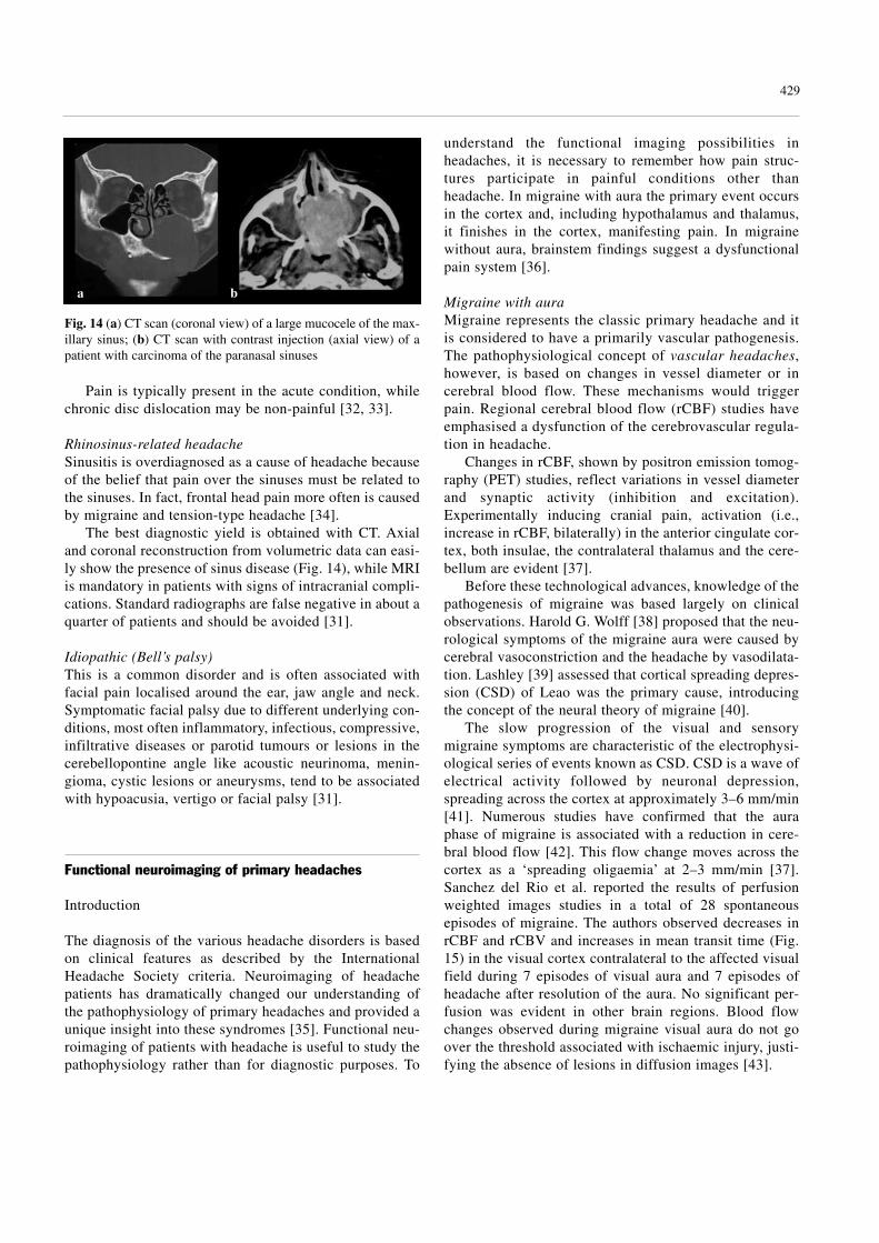
The slow progression of the visual and sensorymigraine symptoms are characteristic of the electrophysi-ological series of events known as CSD. CSD is a wave ofelectrical activity followed by neuronal depression,spreading across the cortex at approximately 3–6 mm/min[41]. Numerous studies have confirmed that the auraphase of migraine is associated with a reduction in cere-bral blood flow [42]. This flow change moves across thecortex as a ‘spreading oligaemia’ at 2–3 mm/min [37].Sanchez del Rio et al. reported the results of perfusionweighted images studies in a total of 28 spontaneousepisodes of migraine. The authors observed decreases inrCBF and rCBV and increases in mean transit time (Fig.15) in the visual cortex contralateral to the affected visualfield during 7 episodes of visual aura and 7 episodes ofheadache after resolution of the aura. No significant per-fusion was evident in other brain regions. Blood flowchanges observed during migraine visual aura do not goover the threshold associated with ischaemic injury, justi-fying the absence of lesions in diffusion images [43].
Fig. 14 (a) CT scan (coronal view) of a large mucocele of the max-illary sinus; (b) CT scan with contrast injection (axial view) of apatient with carcinoma of the paranasal sinuses
a b

430
Cerebral perfusion changes during migraine withaura have been described also by BOLD functional MRIstudies. In the typical visual aura of migraine, function-al MRI has revealed multiple neurovascular events in theoccipital cortex, resuming the CSD: (1) an initial hyper-aemia lasting 3.0–4.5 min, spreading at a rate of 3.5 mmper min, (2) followed by mild hypoperfusion lasting 1–2h, (3) an attenuated response to visual activation and (4)like CSD, in migraine aura, the first affected area is thefirst to recover [43].
In a recent case report [44] reversible changes located inthe right parieto-occipital cortex on DWI images weredescribed in a patient with acute onset of headache. Thepresence of positive DWI images, with the absence of lowapparent diffusion coefficient value, referred to vasogenicoedema, could be in accordance with focal prolonged hyper-perfusion associated with vasogenic leakage. The correlationand complete resolution of both clinical and neuroimagingabnormalities could validate a reversible neuronal inflam-matory pathology in migraine with typical aura. Anothersimilar finding has been described by Jacob et al., in whichthe author reported the case of a young woman affected bysporadic hemiplegic migraine (SHM) with reversiblechanges on MRI. In this case, both DWI and ADC mapswere positive for restricted diffusion of water; perfusionmaps showed increased vascularity and after gadolinium, anenhancement of grey matter in the affected regions was evi-dent. Spectroscopy study showed reduced NAA/Cr. Toexplain these results, in the absence of vascular occlusion,the authors suggested a metabolic alteration at cellular levelwith a spreading cortical depression leading to damage ofthe ATPase pump with altered membrane permeability [45].
In the last few years, several studies of magnetic reso-nance spectroscopy, a non-invasive technique that allowsthe investigation of variations in some cerebral metabo-lites, and in particular 31phosphorus, demonstrated a meta-bolic disturbance in the brain of migraine patients withaura and, to a lesser extent, of migraine patients withoutaura, which is evident even in the interictal period.
Such alterations concern energy metabolism and con-sist of increased inorganic phosphorus and ADP, reducedphosphocreatine and decreased phosphorylation potential.A mitochondrial dysfunction was hypothesised to be thebiochemical substrate that could contribute to brain corti-cal hyperexcitability [46].
In a recent 1H-MRS study, focused on visual cortex,before and after photic stimulation, a consistent decreasein the NAA signal in migraine with aura patients com-pared with migraine without aura patients and controlindividuals was noted [47]. In migraine with visual aurapatients associated with paraesthesia, paresis or dysphasia(Maplus), lactate increased only during stimulation, onlyin visual cortex; in migraine with aura patients, restinglactate was high in visual cortex, without further increaseduring stimulation [48].
In the interictal period Watanabe et al. found high lac-tate levels, speculating that anaerobic glycolysis occurs inpatients with migraine [49].
Migraine without auraFriberg and colleagues [41] demonstrated with SPECTthat interictally almost 50% of migraine without aura suf-ferers had abnormal interhemispherical asymmetries inrCBF. These asymmetries were discrete compared to thoseseen during the aura phase of a migraine attack. In a studyof the same group [50], middle cerebral artery (MCA)velocity studied by transcranial Doppler sonography onthe headache side was significantly lower than that on thenon-headache side. The authors concluded that in theheadache phase there might be a dilatation in the MCA onthe headache side [37].
Cluster headacheUsing PET, a possible site of the central origin of clusterheadache has been visualised in the hypothalamic greymatter [51]. During an acute attack, activation occurs infrontal areas, both insulae, contralateral thalamus and cin-gulate cortex. A specific activation was demonstrated inthe ipsilateral hypothalamus. This region is specific forcluster headache, as it has not been seen in migraine [37].
Structural imaging with voxel-based morphometry hasidentified an area in the posterior hypothalamic grey askey in understanding cluster headache. This area is subtlyenlarged in its grey matter volume, active during an acutecluster headache [52].
Fig. 15. Increases in MTT perfusion MRI map (a) in the visual cor-tex contralateral to the affected visual field during an episode ofvisual aura. Other brain regions do not show any perfusion changesduring the aura or during headache. Blood flow changes observedremain above the threshold associated with ischaemic injury, justi-fying the absence of lesions in diffusion-weighted images (b)
a b

431
In patients with cluster headache, both N-acetil aspar-tate/creatine and choline/creatine levels were significantlylower in comparison with either the control or chronicmigraine groups. Low levels of N-acetil aspartate/creatinerand choline/creatine suggest that cluster headache might berelated to both hypothalamic neuronal and myelin dysfunc-tion or loss in patients with cluster headache [52–54].
Brain 31P-MRS showed reduced phosphocreatine levelsand an increased ADP concentration, as in various types ofmigraine, suggesting the presence of similar pathogenicmechanisms between cluster headache and migraine [55].
Tension-type headacheUsing MRI and voxel-based morphometry, structuralabnormalities have been found in patients with chronictension-type headache (CTTH) for the first time. Pain pro-cessing areas such as dorsal rostral and ventral pons, ante-rior cingulate cortex, anterior and posterior insular cortex,right posterior temporal lobe, orbitofrontal cortex, parahippocampus bilaterally, and the right cerebellum werefound to have decreased grey matter in patients withCTTH compared with control subjects and patients withmedication overuse headache [56, 57].
References
1. Gallagher RM (2005) Headache pain. JAm Osteopath Assoc 105[Suppl4]:S7–S11
2. Sempere AP, Porta-Etessam J,Medrano V et al (2005) Neuroimagingin the evaluation of patients with non-acute headache. Cephalalgia 25:30–53
3. Cooney BS, Grossman RI, Farber REet al (1996) Frequency of magneticresonance imaging abnormalities inpatients with migraine. Headache36:616–621
4. Ziegler DK, Batnitzky S, Barter R et al(1991) Magnetic resonance imageabnormality in migraine with aura.Cephalalgia 11:147–150
5. Igarashi H, Sakai F, Kan S et al (1991)Magnetic resonance imaging of thebrain in patients with migraine.Cephalalgia 11:69–74
6. Bradley WG Jr (1988) Investigatorssolve mystery of unidentified brightspots. Diagn Imaging 57:322–326, 446
7. Soges LJ, Cacayorin ED,Ramachandran TS (1988) Migraine:evaluation by MR. AJNR Am JNeuroradiol 9:425–429
8. Gozke E, Ore O, Dortcan N et al(2004) Cranial magnetic resonanceimaging findings in patients withmigraine. Headache 44:166–169
9. Robbins L, Friedman H (1992) MRI inmigraineurs. Headache 32:507–508
10. Osborn RE, Alder DC, Mitchell CS(1991) Imaging of the brain in patientswith migraine headaches. AJNR Am JNeuroradiol 12:512–514
11. Kruit MC, van Buchem MA (2004)Migraine as a risk factor for subclinicalbrain lesions. JAMA 291:427–434
12. Kruit MC, Launer LJ, van BuchemMA et al (2005) MRI findings inmigraine. Rev Neurol (Paris)161:661–665
13. Swartz RH, Kern RZ (2004) Migraineis associated with magnetic resonanceimaging white matter abnormalities: ameta-analysis. Arch Neurol61:1366–1368
14. Benedittis G, Lorenzetti A, Sina C et al(1995) Magnetic resonance imaging inmigraine and tension-type headache.Headache 35:264–268
15. May A, Bahra A, Buchel C et al (2000)PET and MRI findings in clusterheadache and MRA in experimentalpain. Neurology 55:1328–1335
16. Evans RW (1996) Diagnostic testingfor the evaluation of headaches. NeurolClin 14:1–26
17. Nappi G (2004) The InternationalClassification of Headache Disorders,2nd edn. Headache classificationSubcommittee of the InternationalHeadache Society. Cephalalgia24[Suppl 1]:9–160
18. Ferrarini G, Malferrari G, Zucco R etal (2005) High prevalence of patentforamen ovale in migraine with aura. JHeadache Pain 6:71–76
19. Tessitore E, Schonauer C, Fera F(2001) Superior sagittal sinus thrombo-sis as unusual cause of headache: casereport. J Headache Pain 2:97–98
20. Pezzini A, Granella F, Grassi M et al(2005) History of migraine and the riskof spontaneous cervical artery dissec-tion. Cephalalgia 25:575–580
21. Purdy RA, Kirby S (2004) Headachesand brain tumors. Neurol Clin22:39–53
22. Boiardi A, Salmaggi A, Eoli M et al(2004) Headache in brain tumors: asymptom to reappraise critically.Neurol Sci 25:S143–S147
23. Bastue M, Gracia-Naya M, SantolariaL (2001) Reasons for requesting neu-roimaging studies in the evaluation ofprimary headache. Rev Neurol33:127–134
24. Wang HZ, Simonson TM, Greco WR etal (2001) Brain MR imaging in theevaluation of chronic headache inpatients without other neurologicsymptoms. Acad Radiol 8:405–408
25. Jensen S (2005) Neck related causes ofheadache. Aust Fam Physician34:635–639
26. Tash RR, Gordon ZE, Leslie DR et al(1989) Trigeminal neuralgia: MR imag-ing features. Radiology 172:767–770
27. Dandy WE (1934) Concerning thecause of trigeminal neuralgia. Am JSurg 24:447–455
28. Jannetta PJ (1980) Neurovascular com-pression in cranial nerve and systemicdisease. Ann Surg 192:518–523
29. Haines SJ, Jannetta PJ, Zorub DS(1980) Microvascular relations oftrigeminal nerve: an anatomical studywith clinical correlation. J Neurosurg52:381–386
30. Charles BL, Majoie M, Hulsman FJH,Verbeeten B et al (1997) Trigeminalneuralgia: comparison of two MRimaging techniques in the demonstra-tion of neurovascular contact.Radiology 204:455–460
31. Siccoli MM, Bassetti C, Sandor PS(2006) Facial pain :clinical differentialdiagnosis. Lancet Neurol 5:257–267

432
32. Sarlani E, Balciunas BA, Grace EG(2005) Oro-facial pain – Part I:Assessment and management of mus-culoskeletal and neuropathic causes.AACN Clin Issues 16:333–346
33. Sarlani E, Balciunas BA, Grace EG(2005) Orofacial pain – Part II:Assessment and management of vascu-lar, neurovascular, idiopathic, sec-ondary and psychogenic causes.AACN Clin Issues 16:347–358
34. Silberstein SD (2004) Headaches dueto nasal and paranasal sinus disease.Neurol Clin 22:1–19
35. May A (2006) A review of diagnosticand functional imaging in headache. JHeadache Pain 4:174–184
36. Sanchez del Rio M, Bakker D, Wu Oet al (1999) Perfusion weighted imag-ing during migraine: spontaneous visu-al aura and headache. Cephalalgia19:701–707
37. May A (2004) The contribution offunctional neuroimaging to primaryheadache. Neurol Sci 25:S85–S88
38. Wolff HG (1963) Headache and otherpain, 2nd Edn. Oxford UniversityPress, New York
39. Lashley KS (1941) Patterns of cerebralintegration indicated by the scotomasof migraine. Arch Neurol Psychiatry46:331–339
40. Aurora SK, Welch KMA (2000)Migraine: imaging the aura. CurrOpinion Neurol 13:273–276
41. Friberg L, Olesen J, Iversen H et al(1994) Interictal “patchy” regionalcerebral blood flow patterns inmigraine patients. A single photonemission computerized tomographicstudy. Eur J Neurol 1:35–43
42. May A, Kaube H, Büchel C et al(1998) Experimental cranial painelicited by Capsaicin: a PET study.Pain 74:61–66
43. Sanchez del Rio M, Bakker D, Wu Oet al (1999) Perfusion weighted imag-ing during migraine: spontaneous visu-al aura and headache. Cephalalgia19:701–707
44. Resnick S, Reyes-Iglesias Y, CarrerasR (2006) Migraine with aura associat-ed with reversible MRI abnormalities.Neurology 66:946–947
45. Jacob A, Mahavish K, Bowden A et al(2006) Imaging abnormalities in spo-radic hemiplegic migraine on conven-tional MRI, diffusion and perfusionMRI and MRS. Cephalalgia26:1004–1009
46. Montagna P (1995) Magnetic reso-nance spectroscopy in migraine.Cephalalgia 15:323–327
47. Sarchielli P, Tarducci R, Presciutti O etal (2005) Functional 1H-MRS findingsin migraine patients with and withoutaura assessed interictally. Neuroimage24:1025–1031
48. Sandor PS, Dydak U, Schoenen J et al(2005) MR-spectroscopic imaging dur-ing visual stimulation in subgroups ofmigraine with aura. Cephalalgia25:507–518
49. Watanabe H, Kuwabara T, Ohkubo M(1996) Elevation of cerebral lactatedetected by localized 1H-magnetic res-onance spectroscopy in migraine dur-ing the interictal period. Neurology47:1093–1095
50. Friberg L, Olesen J, Iversen HK,Sperling B (1991) Migraine pain asso-ciated with middle cerebral arterydilatation: reversal by sumatriptan.Lancet 338:13–17
51. May A, Bahra A, Büchel C et al (1998)Cerebral activation in cluster headachein and outside the bout: a PET study.Eur J Neurol 5:7–8
52. May A, Goadsby PJ (2001)Hypothalamic involvement and activa-tion in cluster headache. Curr PainHeadache Rep 5:60–66
53. Wang SJ, Lirng JF, Fuh JL et al (2006)Reduction in hypothalamic 1H-MRSmetabolite ratios in patients with clus-ter headache. J Neurol NeurosurgPsychiatry 77:622–625
54. Lodi R, Pierangeli G, Tonon C et al(2006) Study of hypothalamic metabo-lism in cluster headache by proton MRspectroscopy. Neurology 66:1264–1266
55. Montagna P, Lodi R, Cortelli P (1997)Phosphorus magnetic resonance spec-troscopy in cluster headache.Neurology 48:113–118
56. Mathew NT (2006) Tension-typeheadache. Curr Neurol Neurosci Rep6:100–105
57. Schmidt-Wilcke T, Leinisch E, StraubeA et al (2005) Gray matter decrease inpatients with chronic tension typeheadache. Neurology 65:1483–1486