SUSCEPTIBLE STAPHYLOCOCCUS AUREUS RESISTANT STAPHYLOCOCCUS …
Vol. 60, No. 11INFECrION AND IMMUNITY, Nov. 1992, p. 4838-48470019-9567/92/114838-10$02.00/0Copyright C 1992, American Society for Microbiology
Complement Activation by Polyclonal Immunoglobulin Giand G2 Antibodies against Staphylococcus aureus,
Haemophilus influenzae Type b, and Tetanus ToxoidROBBERT G. M. BREDIUS,12 PETER C. DRIEDIJK,1'2 MIREILLE F. J. SCHOUTEN,1
RON S. WEENING,2'3 ANnD THEO A. OUTl12*Clinical Immunology Laboratory, 1* and Department ofPediatrics, Emma Children's Hospital, 3 AcademicMedical Center, Meibergdreef 9, 1105AZ Amsterdam, and Laboratory for Experimental and Clinical
Immunology, University ofAmsterdam, 1006 AKAmsterdam, 2 The Netherlands
Received 18 May 1992/Accepted 31 August 1992
To obtain information on effector functions of human immunoglobulin G2 (IgG2), we have measured thecomplement-activating properties of polyclonal IgG subclass antibodies against bacterial antigens. IgGl andIgG2 were purified from serum samples from five healthy individuals, and complement activation wasmeasured with different bacterial antigens. We used Staphylococcus aureus Wood 46 (STAW), which is acommon antigen, Haemophilus influenzae type b (Hib), which is a common pathogenic microorganism inchildren, and formaldehyde-inactivated tetanus toxin (IT). Bacteria were incubated with antibodies and thenincubated with sera from agammaglobulinemic patients as a complement source, and C3c deposition wasmeasured by enzyme-linked immunosorbent assay. We found that anti-STAW IgG2 activated complement toa level similar to that of anti-STAW IgGl. Anti-Hib IgGl complement activation was as much as seven timeshigher than that of anti-Hib IgG2 in four individuals. In one individual, anti-Hib IgG2 was more effective incomplement activation than anti-Hib IgGl. Anti-TT antibodies showed patterns similar to those of anti-Hib.Our results indicate that IgG2 antibodies may contribute significantly to antibacterial defense. Also, individualdifferences in antibody effector functions should be taken into account when evaluating the immune status ofpatients and during early phase 1 studies of new vaccines.
Decreased concentrations of immunoglobulin G2 (IgG2)are often associated with recurrent bacterial infections (30,35, 37). A causal relationship is not clear, as IgG2 antibodiesare considered less effective in mediating complement acti-vation than IgGl antibodies, and their binding to Fc-y (con-stant fragment of IgG) receptors is supposed to be weakerthan that of IgGl (6).Much of the knowledge of effector functions of IgG
subclasses has been obtained from studies with aggregatedmyeloma proteins, showing that IgG2 binds complement lesseffectively than IgGl (23, 34). More recently, chimericmonoclonal antibodies (MAbs) with identical variable re-gions but different constant regions of human origin wereused to study Fc-mediated effector functions. Again, IgGlproved more effective than IgG2 in mediating binding of thefirst complement component (Clq), C4 activation, and com-plement-mediated cytolysis (5, 8, 14, 27). However, thesefindings may not reflect the physiologic activity of polyclonalhuman antibodies interacting with common bacterial anti-gens in vivo. In other studies, polyclonal antibodies from apool of hyperimmune sera were used, thus masking differ-ences between the donors (15, 43). Recently, Amir et al. (2)found that in pooled sera, affinity-purified IgGl against thecapsular polysaccharide (polyribosyl ribitol phosphate[PRP]) of Haemophilus influenzae type b (Hib) was moreactive than anti-PRP IgG2 in several test systems (bacteri-cidal, opsonization, and rat protection assays). However, insera from individual donors vaccinated with PRP vaccine,anti-PRP IgG2 preparations from two individuals were sim-
* Corresponding author.
ilar to the anti-PRP IgGl preparations from two otherindividuals.
Differences in the functional affinities of antibodies mayinfluence these analyses, since correlations between anti-body affinity and effector functions have been observed (1,17, 20).
In the present study, we have investigated whether IgG2antibodies have complement activation capacity, whichwould enlighten IgG2 deficiency-related disease. We havechosen a strategy that would reflect the physiologic situationas much as possible, by using polyclonal IgG subclassantibodies and common bacterial antigens. Human IgGl andIgG2 antibodies from five individuals were purified by affin-ity chromatography with Sepharose-protein A. DifferentG2m(n) allotypes were included for the investigation ofdifferences in complement-activating properties. The G2m(n)allotype substitution in the CH2 domain of the IgG moleculehas not yet been located precisely, but it may be close to thebinding and activation site of Clq (6, 38), the first componentof the complement cascade, and might thus influence com-plement-activating properties of IgG2. Complement-activat-ing properties of antibodies against Staphylococcus aureusWood 46 (STAW), Hib, and formaldehyde-inactivated teta-nus toxin (1T) were analyzed. We used a commensal non-encapsulated gram-positive microorganism, S. aureus,which is a common antigen, and against which most peopleshould have protective antibodies; an encapsulated gram-negative microorganism, Hib representative for the invasivedisease isolates in Europe (39); and 1T, as a protein andreference antigen, with which most individuals have beenimmunized and have protective IgGl and IgG2 antibodies.C3c deposition on the bacterial surface was measured byenzyme-linked immunosorbent assay (ELISA), using poly-
4838

COMPLEMENT ACTIVATION BY ANTIBACTERIAL IgG SUBCLASSES 4839
clonal anti-C3c, which recognizes native C3 and C3b, includ-ing C3bi. We show that anti-STAW IgG2 and IgGl activatedcomplement almost equally well. Anti-Hib IgG2 and anti-TTIgG2 showed interindividual differences: some IgG2 prepa-rations showed slightly more complement activation thanIgGl preparations, but most were less effective than IgGl.Our results indicate that IgG2 antibodies may have animportant role in defense against these bacteria.
MATERIALS AND METHODS
Materials. Sephacryl S-300 and protein A-SepharoseCL4B were from Pharmacia, Uppsala, Sweden. The Ami-con concentrator (cell model M-3) and Diaflo ultrafiltrationmembranes (YM10) were from Amicon, Danvers, Mass.Mouse MAbs specific for IgG subclasses (MH 161-1-MO1,MH 162-1-MO2, MH 163-1-MO2, and MH 164-4-MO2),horseradish peroxidase (HRP)-conjugated murine MAbsspecific for IgG subclasses (MH 161-1-ME2, MH 162-1-ME2, MH 163-1-ME3, and MH 164-4-ME3) and specific forhuman IgG (MH 16-1-ME) were from the Central Labora-tory of the Netherlands Red Cross Blood Transfusion Ser-vice (CLB), Amsterdam, The Netherlands. The specificity ofthese antibodies has been documented extensively (24, 25,29, 33). Polyclonal rabbit anti-IgA (KH 14-22-P), anti-IgM(KH 15-24-P), and anti-IgG (KH 16-109-P) antibodies werefrom the same institute. Human IgGl (clone 151) specific forTT was a gift from R. F. Tiebout, CLB. Rabbit anti-humanC3c (code no. A062), HRP-conjugated rabbit anti-humanC3c (code no. P213), HRP-conjugated rabbit anti-mouse IgG(code no. P260), and HRP-conjugated rabbit anti-human IgG(code no. P214) were purchased from Dako, Glostrup,Denmark. Mouse monoclonal anti-human SC5b-9 (neoanti-gen; code no. A239) was obtained from Sanbio bv, Uden,The Netherlands.
TI2 and purified PRP were obtained from the NationalInstitute for Health, Environment and Toxicology (RIVM;Bilthoven, The Netherlands). We used a representativeencapsulated Hib, strain 760705, which causes the majorityof invasive Hib disease in Europe (39). Hib and unencapsu-lated protein A-deficient STAW (270581) bacteria werekindly provided by L. van Alphen (Department of Microbi-ology, University of Amsterdam, Amsterdam, The Nether-lands). Hib was cultured in brain heart infusion broth con-taining hematin and NAD+, and STAW was cultured innutrient broth 2. Bacteria were harvested in log phase,washed three times with phosphate-buffered saline (PBS)(140 mM NaCl, 9.2 mM Na2HPO4, 1.3 mM NaH2PO4; pH7.4), and resuspended in coating buffer (0.05 M NaHCO3,pH 9.6). Phosphate buffer containing Ca2+ and Mg2+ (PiCMbuffer) (pH 7.2 to 7.4) consisted of 137 mM NaCl, 2.7 mMKCI, 8.1 mnM Na2HPO4, 1.5 mM KH2PO4, 1.0 mM MgCl2,0.6 mM CaCl2, 1% (wt/vol) glucose (all from Merck, Schu-chardt, Hohenbrunn, Germany), and 2.5% (vol/vol) humanserum albumin (from CLB, Amsterdam, The Netherlands).Tween 20, NaHCO3, citrate, and Na2HPO4 were also fromMerck. Tetramethyl-benzidine was purchased from Sigma,St. Louis, Mo. Flat-bottom, 96-well microtiter plates (Im-munolon M129A) were from Greiner, Kloten, Switzerland.Serum samples were obtained from healthy laboratory
personnel, and samples with large amounts of anti-Hib andanti-Sta IgGl and IgG2 were selected. Individuals withdifferent IgG2 G2m(n) allotypes were chosen. Sera frompatients with agammaglobulinemia (and with normal hemo-lytic complement activity) were used as source of comple-
ment. All agammaglobulinemic serum samples were storedat -80°C and thawed at 4°C just before use.
Purification of IgG subclasses. Serum samples were ob-tained from five healthy adults. Complement was heat inac-tivated (45 min at 56°C). IgG was separated from other serumproteins by gel filtration at 4°C, using Sephacryl S-300; PBSwas used as the elution buffer.
IgG-, IgM-, and IgA-rich fractions were pooled. The IgGantibodies were applied to a protein A-Sepharose column (20by 1.1 cm; 21-ml bed volume) at 4°C by the method ofDuhamel et al. (9). IgG was eluted with 0.02 M citratebrought to pH 5.0 with Na2HPO4 (approximately 0.04 MNa2HPO4) and then with a solution with a pH gradient frompH 5.0 to 3.0 (0.02 M citrate brought to pH 3.0 withNa2HPO4 [approximately 0.008 M Na2HPO4]). Fractionswere collected in tubes that contained 0.5-ml portions of 0.25M Na2HPO4 (pH 8.9) to neutralize the acid pH of thefractions immediately. IgG subclass content was measured(see below), and IgGl-, IgG2-, and IgG3-rich fractions fromeach individual were pooled. We purified IgG4 from oneindividual by immunoabsorption using Sepharose anti-IgG4(an anti-IgG4 MAb, MH 164-4-MO2), essentially as de-scribed by Nagelkerken et al. (28). The IgG subclass prepa-rations were concentrated with an Amicon concentrator anda YM10 membrane, diluted in PiCM buffer, and stored inaliquots at -80°C.IgG subclass assay. IgG subclasses were measured by
noncompetitive two-site ELISAs (29). Briefly, mouse MAbsspecific for IgG subclasses (MH 161-1-MO1, MH 162-1-M02, MH 163-1-MO2, and MH 164-4-MO2) were used tocoat microtiter plates. Uncoated sites were blocked, anddilutions of fractions or subclass preparations were added tothe wells and incubated (2 h at room temperature). TheDutch reference serum HOO-03 (CLB) was used as a stan-dard. Bound IgG antibody subclasses were detected byHRP-conjugated murine anti-human IgG MAb (MH 16-1-ME). The results of IgG subclass assays by the ELISAswere the same as those by radial immunodiffusion assay (29).IgG2 G2m(n) allotyping. The G2m(n) allotypes of the five
donors were determined by double immunodiffusion assayby the method of Rautonen et al. (32). Three of the selecteddonors were homozygous G2m(n) negative (n-/n-), one washomozygous G2m(n) positive (n+/n+), and one was hetero-zygous (n+/n-).Complement deposition assay. Deposition of complement
C3c on bacteria was measured by ELISA, essentially asdescribed earlier (16). Preparation and coating of STAW,Hib, and TT were performed as previously described forELISAs of antibodies to bacterial antigens (33). STAW andHib were coated (150 ,ul per well; 2 h at room temperature)at concentrations of approximately 5 x 10' and 1 x 10⁷ CFUper ml, respectively, and TT in a concentration of 1.5 Lf/ml.TT was coated at least 48 h before the assay. Free bindingsites were blocked with 150 ,ul of PiCM buffer. Plates werewashed first with PBS containing 0.05% (vol/vol) Tween 20(PBS-Tween) and then washed three times with PBS. Atleast four serial dilutions of heat-inactivated samples made inPiCM buffer were applied (100 pLI per well) and incubated for1 h at 37°C. Standard and control sera were applied in thesame way. Plates were washed again. For a source ofcomplement, we used serum obtained from an agammaglob-ulinemic patient; a 100-,ul portion of serum 1% (vol/vol) inPiCM buffer was added to each well and incubated (30 min,37°C). After the wells were washed, 100 pl of HRP-conju-gated rabbit anti-human C3c (4 ,ug/ml), diluted in PBScontaining 0.1% (wt/vol) gelatin and 0.02% (vol/vol) Tween
VOL. 60, 1992

4840 BREDIUS ET AL.
20, pH 7.3, was added to each well and incubated (2 h, 37°C).Plates were washed, and 100 RI of substrate was added toeach well: 0.11 M acetic acid, 0.01% (wtlvol) tetramethyl-benzidine, 1% (vol/vol) dimethyl sulfoxide, and 0.003%(vol/vol) H202, pH 5.4. The reaction was stopped by adding100 ,ul of 2 M H2SO4. The anti-C3c reagent recognizes theC3c part of native C3 and C3b, including C3bi (44). Comple-ment activation was expressed either in A450, measured witha Titertek Multiscan MC (Flow Laboratories, Irvine, UK),or in percentage of normal human serum. Complementactivation was also expressed per amount of antibacterialantibody bound to the antigen and expressed as a ratio (seebelow [Table 3]). To do so, antibacterial antibodies weremeasured by ELISA (33) and expressed in micrograms ofbound antibody per milliliter (see next section), and comple-ment activation in the C3c deposition ELISA, was expressedas percentage of normal human serum, measured in an A450range of 0.5 to 1.0. For each antigen, a different serumsample was used as the standard; therefore, the relativecomplement activation ratios for one antigen cannot becompared with those of another antigen. Normal human serawere selected as standards by using high titers of antibacte-rial IgG and titration curves relatively parallel to those of thesubclass fractions as criteria. The amount of C3c depositionwas quantitated as indicated below.To obtain an independent measure of the amount of C3c
that was deposited onto the immune complexes, we per-formed a two-site ELISA for C3c. Serial dilutions of astandard serum with known levels of C3c were added to theanti-C3c serum-coated wells (2 jig/ml), and the furtherprocedure was performed as described previously (40). Thesame amounts of HRP-conjugated anti-C3c as in the C3cdeposition ELISA were added. Incubation times and en-zyme reaction times were equal in both ELISAs. In thisway, we were able to compare theA450 in the C3c depositionELISA with the A450 in the C3c capture ELISA, in whichknown amounts of C3c were added. To demonstrate furthercomplement activation, we measured the formation of theterminal complement complex (22) in a manner similar tothat of the C3c deposition assay. In this study, we usedmouse anti-human SC5b-9 (166 ng/ml) and HRP-conjugatedrabbit anti-mouse IgG (0.5 p,g/ml).The intraassay coefficient of variation (CV) of the C3c
deposition ELISA was less than 10%. The interassay CVwas 25%. Therefore, to compare IgGl and IgG2 antibodyactivities, the fractions were applied to one microtiter plate,and the assays were repeated for confirmation of results.
Determination of antibodies. IgG subclass antibodiesagainst STAW, Hib, and TT were measured by ELISA (33).Briefly, whole bacteria and TT were coated in the same wayas described above for the C3c deposition ELISA. After thefree sites were blocked, samples were added to each well.Antibacterial IgG subclass antibodies in sera were detectedby HRP-conjugated MAbs to each of the subclasses (MH161-1-ME2, MH 162-1-ME2, MH 163-1-ME3, and MH 164-4-ME3). Anti-TT IgGl (clone 151) was used to calibrate thereference sera (33). Enzyme reaction times of the IgGsubclass ELISAs were standardized. Antibodies in the pu-rified IgG subclass fractions were analyzed in the same way,and parallel ELISAs in which the total amount of IgGantibodies bound were detected with HRP-conjugated anti-IgG (P214; Dako) were run. The reproducibility and CVs ofthese assays was as follows: intraassay CV, <4%; interassayCV, <9%; but 31% for anti-STAW IgG2 (33). The summedamounts of IgGl, IgG2, IgG3, and IgG4 antibodies did notalways match with total IgG antibodies. We assume, how-
E
cmaCD0C
.0ocn
2.0-IgG
1.5
1.0 IgG3 IG2 lG
0.5
010. A k#ydI1 5 35 55 120 160
Elution volume (ml)
*3.6
*2.4_
3- 0.63 .1. =- Q -3co0.0 003 e
I0.0 L 0.0 -200
FIG. 1. IgG subclass purification with Sepharose-protein A. IgG(approximately 60 mg of protein) obtained after Sephacryl S-300 gelfiltration was loaded onto the column at pH 5.0. After most of theunbound protein (55-ml elution volume) had run through the col-umn, the pH gradient from 5.0 to 3.0 was started. The solid lineshows Am8. IgGl, IgG2, IgG3, and IgG4 were measured by ELISA.
ever, that up-to-twofold differences are still within accept-able limits. Hib capsular polysaccharide PRP was tyrami-nated as described previously (3), and antibodies againstPRP were measured in the same way as in the whole-cellanti-Hib ELISA, after the tyrosylated plates were coatedwith PRP (5 jig/ml). Reference serum with known amountsof anti-PRP antibodies was used as standard (42) (providedby L. van Alphen). In this study, antibody amounts inmicrogram equivalents (pugeq) are shortly indicated as mi-crograms hereafter.
Statistics. Differences between the ratios of C3c depositionper microgram of antibodies of IgGl and IgG2 preparations(see Table 3) were tested by the Wilcoxon matched-pairsigned-rank test and Student's t test.
RESULTS
Purification of IgGl and IgG2 antibodies. IgGl and IgG2antibodies from five individuals were purified. From eachindividual the IgG-rich fractions, obtained after gel filtration,were loaded onto the Sepharose-protein A column at pH 5.0.A representative elution pattern is shown in Fig. 1. Gradientelution was started after the peak of unbound protein (IgG3)had eluted. The first peak was IgG2, whereas IgGl elutedfrom the column approximately 0.5 pH unit later. After gelfiltration, the mean recoveries of IgGl and IgG2 were 80(range, 64 to 100%) and 84% (65 to 100%), respectively.After affinity chromatography and concentration of antibod-ies, the mean recoveries were 52% (38 to 65%) for IgGl and48% (42 to 63%) for IgG2. Most of the IgG3 was lost in theIgA-rich pool obtained after gel filtration, and the meanrecovery of IgG3 was only 28%. IgGl-rich fractions con-tained 4 to 10% IgG2, and IgG2-rich fractions contained lessthan 5% IgGl, except for 12% in one preparation. IgG4 wasfound mainly in the IgGl-rich fractions (1 to 8%).
Specific antibodies. The values for recovery and purity ofantibacterial antibodies, measured with anti-IgG subclassmonoclonal antibodies, were similar to those for total IgGsubclass proteins. Table 1 shows the results of the purifica-tion of IgG subclass anti-Hib antibodies for each of the fiveserum samples. In the IgGl pools, anti-Hib IgGl rangedfrom 87 to 95% of the total anti-Hib. In the IgG2 pools,anti-Hib IgG2 was 84 to 100% of the total anti-Hib. We alsomeasured the anti-Hib antibodies with polyclonal HRP-conjugated anti-IgG. The anti-Hib IgG thus measured (Table1) corresponded to the sum of the subclass anti-Hib, with
INFECT. IMMUN.

COMPLEMENT ACTIVATION BY ANTIBACTERIAL IgG SUBCLASSES 4841
TABLE 1. IgG subclass antibodies against Hib
Data on antibodies from the following individual:
Source Antibodya A B C D EConcn tb Concn % Concn % Concn % Concn %(pg/ml) (ILg/nml) (pg/ml) (pg/ml) (,ug/ml)
Serum IgGl aHib 4.10 23 25.90 58 10.50 80 21.20 79 4.20 52IgG2 aHib 12.90 73 18.20 41 2.56 20 5.76 21 3.84 48IgG3 aHib 0.40 2 0.20 0 0.02 0 0.00 0 0.00 0IgG4 aHib 0.30 2 0.50 1 0.02 0 0.01 0 0.01 0
IgG aHib 15.2 40.3 13.7 21.0 13.8
IgGl fraction IgGl aHib 1.60 87 12.60 93 4.20 94 6.10 95 2.10 91IgG2 aHib 0.20 11 0.96 7 0.26 6 0.35 5 0.20 9IgG4 aHib 0.04 2 0.04 0 0.01 0 0.00 0 0.02 1
IgG aHib 3.7 14.7 5.3 5.5 4.0IgG aPRP 2.0 4.0 0.7 1.0 6.0
IgG2 fraction IgGl aHib 0.90 16 0.67 7 0.05 9 0.01 1 0.00 0IgG2 aHib 4.60 84 8.32 93 0.48 91 0.80 99 1.76 100IgG4 aHib 0.00 0 0.00 0 0.00 0 0.00 0 0.00 0
IgG aHib 6.0 10.0 1.0 1.3 2.3IgG aPRP 12.0 5.0 0.7 1.0 1.0
a IgGl aHib, IgG2 aHib, IgG3 aHib, and IgG4 aHib, Anti-Hib detected with HRP-conjugated anti-IgG subclass MAbs (see text). IgG aHib, Anti-Hib detectedwith HRP-conjugated polyclonal anti-IgG (see text). aPRP, Anti-PRP detected with HRP-conjugated polyclonal anti-IgG (see text).
b Percentage of total anti-Hib IgG in the sample.
minor discrepancies for the serum from individual E, theIgGl fraction from individual A, and the IgG2 fraction fromindividual C. The summary of the purification of anti-STAW,anti-Hib, and anti-TT antibodies from five serum samples isgiven in Table 2. In the IgGl fractions, the IgGl antibodieswere 85 92, and 96% of total anti-STAW, anti-Hib, andanti-TT, respectively. In the IgG2 fractions, the IgG2 anti-bodies were 93, 93, and 94% of total anti-STAW, anti-Hib,and anti-TT, respectively. The antibacterial IgG3 antibodyconcentrations were very low, and most IgG3 proteins werelost in the IgA-rich fractions after gel filtration, whichresulted in the lack of measurable IgG3 antibodies in thesubclass fractions. Antibacterial IgG4 antibodies were de-tected only in serum from one individual (A). The IgGlpreparation from this individual contained some anti-STAWIgG4 (0.15 jig/ml). Other IgGl (and IgG2) preparationscontained less than 2% antibacterial IgG4. Again, antibodyconcentrations measured with polyclonal HRP-conjugatedanti-IgG corresponded to the summed total of the IgGsubclass antibodies. We considered the purity to be suffi-cient to assess the effector functions of IgGl and IgG2antibodies separately.Complement activation assay. Figure 2 shows the C3c
deposition with Hib as coated antigen. Higher absorbancesignals were obtained when more antigen was coated ontothe plates (Fig. 2a) and when higher concentrations ofantibodies or complement source were used (Fig. 2b). Forfurther experiments, we used 5 x 10⁶ and 1 x 101 CFU/ml,for STAW and Hib, respectively, and 1.5 Lf/ml for TT forcoating the plate, which are the same conditions as in theantibacterial antibody ELISA. Different agammaglobuline-mic sera serving as complement source (and with normalhemolytic activity) were compared. An agammaglobuline-mic serum preparation with low background levels (no
antibodies added) was selected and used for further experi-ments. For the analysis of antibacterial antibody-dependentC3c deposition, the optimal complement concentration wasobtained when 1 ,ul of agammaglobulinemic serum in 100 ,ulof PiCM buffer (1% [vol/vol]) per well was used.
Information on the amount of C3c deposited was obtainedby performing a C3c capture ELISA at the same time as theC3c deposition assay.A450s of 0.1, 0.2, 0.3, 0.5, 0.7, 0.9, and1.1 were obtained when 100-pl portions of a solution con-taining 22, 46, 63, 103, 148, 188, and 228 nM C3c, respec-tively, were added in the C3c capture ELISA.Complement activation by IgGI and IgG2. The IgG sub-
class preparations from the five individuals were analyzed.When comparing IgGl and IgG2, we always used the sameagammaglobulinemic serum preparation. To assess the rela-tive complement activation properties of the IgG subclasspreparations, we applied the amounts of IgG subclass anti-bodies that had shown similar amounts of antibody bound tothe antigen, as determined in the antibacterial antibodyELISAs. Figure 3a shows the results of anti-STAW IgGland IgG2 antibodies from individual A. Bound anti-STAWIgGl and IgG2 antibodies showed almost equal complementactivation. The same was found for the antibodies from otherindividuals, except anti-STAW IgGl from individual C wasmore than twice as active as his anti-STAW IgG2 (see Table3). We calculated that there was no loss of biological activityduring the purification process by demonstrating that thebiological activities of the purified IgG subclass fractionswere the same as those of serum preparations and IgG poolafter Sephacryl S-300.
Figure 3b shows complement activation by anti-Hib IgGland IgG2 antibodies for two individuals. Anti-Hib IgGl fromindividual Awas more active than his anti-Hib IgG2 (see alsoTable 3). Similarly, three other IgGl preparations were
VOL. 60, 1992

TABLE 2. Summary of antibody results from five individuals after purification of IgG subclass antibodies against bacterial antigens
Anti-STAW Anti-Hib Anti-lTSource Antibody Concnb Concn Concn
Concn/ %b (C°g/ml) % (lLg/ml)
Serum IgGlc 0.73 34 13.2 58 3.06 87IgG2 1.91 63 8.7 40 0.30 13IgG3 0.02 1 0.1 1 0.00 0IgG4 0.05 2 0.2 1 0.00 0
IgGd 2.78 20.8 3.3
IgGl fraction IgGl 0.60 85 5.3 92 1.41 96IgG2 0.07 12 0.4 8 0.03 4IgG4 0.04 3 0.0 1 0.00 0
IgG 0.85 6.6 1.5aPRPe 2.7
IgG2 fraction IgGl 0.02 6 0.3 7 0.01 6IgG2 0.64 93 3.2 93 0.08 94IgG4 0.00 0 0.0 0 0.00 0
IgG 0.86 4.1 0.3aPRP 3.9
a Mean of concentrations from five individuals (A to E).b Percentage of total anti-STAW, anti-Hib, or anti-YT IgG in the sample.c Antibodies detected with HRP-conjugated monoclonal anti-IgG subclass antibodies (see text).d Antibodies detected with HRP-conjugated polyclonal anti-IgG (see text).I Antibodies against PRP measured with HRP-conjugated polyclonal anti-IgG (see text).
slightly more active than IgG2 from the same person. How-ever, anti-Hib IgG2 from individual E had higher activitythan anti-Hib IgGl (Fig. 3b). Table 3 summarizes the resultsof complement activation by anti-Hib antibodies for the fiveindividuals.
Since both anti-outer membrane protein antibodies and
a 1.2E0
w 0.80
n 0.40
00
anti-capsular polysaccharide (PRP) antibodies play a role inthe immune response to Hib (19, 21), we measured anti-PRP(see Table 1) in addition to antibodies against whole bacteria.This enabled us to express the results for C3c deposition onwhole Hib bacteria relative to the anti-PRP antibodies in theIgG subclass fractions (Table 3). Also, for C3c deposition
b 1.2E
Ln
0.80
0
c
D 0.4L-)o
0.00.0 0.4 0.8 1.2 1.6 2.0
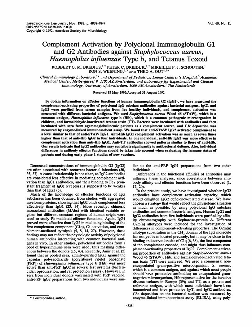
O i-10l 1_107 5.107W.W Wnt-Hlb%O ,uI.&. 1%P -.
Hib coating (cfu/ml) anti-Hib (;igeq/ml)FIG. 2. C3c deposition assay. (a) Titration of Hib coating and antibodies. Hib was coated at increasing amounts (1 x 10⁶, 5 x 10⁶ and 1
x 107 CFU/ml), as indicated on the abscissa. Different amounts of a serum preparation containing anti-Hib were incubated, and then serum
(1 ,ul per well in 100 ,ul of PiCM buffer) from an agammaglobulinemic patient as a complement source was added. C3c deposition was
expressed asA450. To each well 0.25 (0), 0.5 (Cl), 1.0 (A), or 2.0 (V) ,ul of anti-Hib serum was added. Without anti-Hib serum, no complementactivation was observed (*). (b) Titration of antibodies and complement. Hib was coated at 1 x 10⁷ CFU/ml. Serial dilutions of anti-Hibantibodies were incubated for 2 h at 37°C. Next, different concentrations of serum from an agammaglobulinemic patient were incubated for30 min at 37°C. C3c deposition was expressed as A450. To each well 0.3 (0), 1.0 (A), or 3.0 (0) pl of serum from an agammaglobulinemicpatient was added. No complement activation was observed without coated antigen with complement source or with coated antigen without
complement source (*). Similar titration curves with iT and STAW as antigens were observed.
I *
is * I
4842 BREDIUS ET AL. INFE+CT. IMMUN.
I II

COMPLEMENT ACTIVATION BY ANTIBACTERIAL IgG SUBCLASSES 4843
b
0.005 0.05IgG anti-Sta (pgeq/ml)
- 1.2c0
5 0.9-c0O._X
0.6
U,E 0.3-
--i E0
0.5 0 -
0.2 1IgG anti-Hib (pgeq/mi)
5
c- 2.0E
r_0. 1.6-
f; 1.24-
0
X 0.80-
0E) 0.4E0.0
Ao
V0.5 5 50
IgG anti-TT (ngeq/mI)
FIG. 3. (a) Complement activation by anti-STAW IgGl and IgG2 from individual A. Serial dilutions of IgGl (-) and IgG2 fraction, containinganti-Sta (0) were incubated for 2 h at 37C and then incubated with serum from an agammaglobulinemic patient (1 IL1 per well in 100 p.l of PiCMbuffer). C3c deposition was expressed as A450. (b) Complement activation by anti-Hib IgGl and IgG2 from two individuals. Serial dilutions ofIgGl (-) and IgG2 fraction containing anti-Hib (0) from individual A and IgGl (-) and anti-Hib IgG2 (0) from individual E were incubated for2 h at 37C and then incubated with serum from an agammaglobulinemic patient (1 p1 per well in 100 p1 of PiCM buffer). C3c deposition wasexpressed as A450. (c) Complement activation by anti-Tf IgGl and IgG2 from two individuals. Serial dilutions of IgGl (V) and IgG2 fractioncontaining anti-TT (V) from individual B and IgGl (0) and anti-TT IgG2 (0) from individual E were incubated for 2 h at 37C and then incubatedwith serum from an agammaglobulinemic patient (1 ,ul per well in 100 p1 of PiCM buffer). C3c deposition was expressed as A450.
per anti-PRP antibody, the IgGl antibodies from four serumsamples were two to seven times more active than the IgG2antibodies from the same individual. For individual E, theopposite was found. The IgGl preparation from individual Econtained predominantly anti-PRP antibodies, in contrast tothe IgGl fractions from other individuals. However, noaffinity-purified anti-PRP preparations were used to establishthe relative functions of the non-anti-PRP versus the anti-PRP antibody activities.
Figure 3c shows that anti-TT IgGl antibodies from indi-vidual B had lower C3c deposition activity than his anti-TTIgG2. Anti-TT IgGl from individual E was about 9 timesmore active than his anti-TT IgG2. In Table 3, it is shown
that in four of five persons, the activity of anti-TT IgGl wasone to nine times that of anti-TT IgG2.
It is important to note from Table 3 that the relativecomplement activation properties of IgGl and IgG2 prepa-rations depended on the antigen studied. Thus, in individualA anti-STAW IgGl showed slightly lower activity thananti-STAW IgG2, whereas in the same preparation anti-HibIgGl and anti-TT IgGl were more active than anti-Hib IgG2and anti-TT IgG2, respectively. In individual E, anti-STAWIgGl and IgG2 had equal activity, but anti-Hib IgG2 was 1.5times more active than IgGl and anti-TT IgG2 was lessactive than IgGl.G2m(n) aliotype differences and sources of complement. To
TABLE 3. Complement component C3c deposition by antibacterial IgG subclass antibodies
Individual C3c depositione(allotype) Fraction C3c/aSTAW C3c/aHib C3c/aTTA (n+/n+) IgGl 2.1 ± 0.6 0.5 + 0.1 (0.9 + 0.1) 1.3 ± 0.2
IgG2 3.4 + 0.4" 0.1 ± 0.Od (0.1 ± 0.0)" 1.1 + 0.2
B (n+/n-) IgGl 0.7 ± 0.2 0.7 ± 0.1 (2.8 ± 0.3) 0.6 + 0.1IgG2 0.7 ± 0.2 0.1 ± o.Od (0.2 ± 0.0)" 1.5 + 0.2c
C(n-/n-) IgGl 1.5 ± 0.5 0.4 + 0.1 (3.0 ± 0.4) 1.2 ± 0.2IgG2 0.6 ± 0.2c 0.2 ± o.Od (0.2 0.0) 0.7 O0.1"
D (n-/n-) IgGl 0.6 ± 0.1 0.7 + 0.1 (3.5 + 0.4) 4.6 ± 0.4IgG2 0.5 ± 0.1 0.3 ± o.Od (0.4 + 0.0)c 1.0 + 0.2c
E (n-/n-) IgGl 3.1 ± 0.6 0.4 + 0.1 (0.3 ± 0.0) 9.3 ± 0.9IgG2 2.9 ± 0.4 0.6 ± 0.1" (1.4 ± 0.2)" 1.0 ± 0.3"
a G2m(n) ailotype of IgG2.b Ratios (means + standard deviations of five experiments) of C3c deposition per microgram of antlbodies bound to the antigen; C3c in percentage of normal
human serum, as determined with HRP-conjugated anti-C3c (based on at least three serial dilutions of the antibody fractions), and bound antibacterial antibodiesin micrograms as determined with ELISA using HRP-conjugated polyclonal anti-IgG antibodies (see text). Ratios of C3c deposition per microgram of boundanti-PRP antibodies are shown in parentheses.
Significantly different from IgGl preparation by Wilcoxon matched-pair signed-rank test (P < 0.05) and Student's t test (P < 0.02).dSignificantly different from IgGl preparation by Wilcoxon matched-pair signed-rank test (P < 0.025) and Student's t test (P < 0.02).
a_1.0Ec
0
cw 0.8
0*-0.6
:11U
0e 0.4C
E.D 0.20.2E0c 0
i ,)Er
13 //l/ /
jo
Oll~~~VI',
500
VOL. 60, 1992

4844 BREDIUS ET AL.
1.2 -E
O08'0.8
-o
0.0 0.4 0.8 1.2 1.6 2.0
anti-Hib (,ugeq/ml)FIG. 4. Terminal complement complex (SC5b-9) formation in-
duced by anti-Hib antibodies. The conditions were the same as forthe C3c ELISA described in the legend to Fig. 2b. Mouse anti-human SC5b-9 MAb (166 ng/ml) and HRP-conjugated rabbit anti-mouse IgG (0.5 ,ug/ml) were used to detect terminal complementcomplex formation. SC5b-9 was expressed asA450. To each well 1.0(A), 3.0 (0), or 9.0 (V) ,ul serum from an agammaglobulinemicpatient was added. No complement activation was observed withoutcoated antigen with complement source or with coated antigenwithout complement source (*).
investigate whether there might be allotype-related differ-ences in complement activation, we used sera from individ-uals with different G2m(n) allotypes. Table 3 shows thatdifferences in complement activation properties betweenIgG2 preparations from the five individuals were not relatedto their G2m(n+/n+) and G2m(n-/n-) allotypes. Six differentserum samples from agammaglobulinemic patients, assources of complement, were compared to assess possibledifferences in interactions with IgGl and IgG2 antibodies.Some of the agammaglobulinemic serum samples had lowlevels of antibacterial antibodies, and high background levelsdisturbed the complement activation mediated by IgG sub-class preparations. Nevertheless, no particular preferencewas found for any of the agammaglobulinemic serum sam-ples, and relative differences in complement activation byIgG subclass antibodies remained similar with differentcomplement sources (data not shown).IgG4 antibodies. The IgGl preparation from individual A
contained antibacterial IgG4. IgG4 may serve as blockingantibody and possibly inhibit complement activation byother IgG subclasses (10, 41). However, the preparationfrom this individual did not show less active IgGl thananti-Hib or anti-YT IgG2. Complement activation mediatedby anti-STAW IgGl antibodies could be inhibited only byvery high concentrations of anti-STAW IgG4 antibodiesfrom this individual.SC5b-9 determination. We measured the formation of
terminal complement complexes with the use of MAbsagainst SC5b-9. Figure 4 shows terminal complement com-plex formation by anti-Hib antibodies in the presence ofdifferent concentrations of agammaglobulinemic serum. Op-timal anti-Hib dependent SC5b-9 formation and low back-ground levels (when no anti-Hib was added) was observedwith 3 pl of agammaglobulinemic serum per well (3% [vol!vol]), which is more than was needed for C3c deposition.Thus, full complement activation was established, also at arelatively low complement concentrations of 1% agamma-globulinemic serum. IgGl and IgG2 antibodies against both
bacterial antigens, STAW and Hib, were tested in this assayand the same results were obtained as in the C3c depositionELISA (data not shown). Thus, differences between com-plement activation by IgGl and IgGl antibodies extend tothe formation of the membrane attack complex.
DISCUSSION
In this study, we investigated complement activation asone of the effector functions of IgG subclass antibodies.IgGl and IgG2 antibodies were purified from serum fromhealthy individuals by gel filtration and Sepharose-protein Aaffinity chromatography. In contrast to most other studies, inthis study we separated IgGl and IgG2 from several subjectsto detect possible individual variations in activity. Comple-ment activation by polyclonal antibodies against three dif-ferent antigens was measured by ELISA. Anti-STAW IgG2activated complement as well as anti-STAW IgGl. Anti-HibIgGl showed better complement activation than anti-HibIgG2 in four individuals. In one individual, anti-Hib IgG2was more effective in complement activation than anti-HibIgGl. Also, anti-YT IgGl showed better complement activa-tion than anti-YT IgG2. However, again anti-YT IgG2 anti-bodies from one individual showed more effective comple-ment activation than anti-YT IgGl.The purification of IgG subclasses with Sepharose-protein
A was performed by the method of Duhamel et al. (9) (Fig.1). To purify IgGl and IgG2 antibodies from an IgG pool, weused a linear pH gradient. We first purified IgG from otherserum proteins with the intention of obtaining pure IgG3after Sepharose-protein A chromatography (6). However,most IgG3 was lost in the IgA-rich fraction after gel filtra-tion, and to assess functional properties of IgG3, largeramounts of specific IgG3 were needed. In addition, pureIgG4 could not be obtained with this procedure, as most ofthe IgG4 was found in the fractions that contained IgGl.IgG4 may serve as blocking antibody and possibly inhibit thecomplement activation by other IgG subclasses (10, 41).However, only one IgGl preparation contained antibacterialIgG4 antibodies, and this IgGl preparation did not show lessactivity than the IgG2 preparation. Apparently, the IgG4antibody amounts were too low to interfere.We obtained an average recovery of around 50% of the
total amount of IgGl and IgG2 subclass protein. Recoveriesof specific IgG subclass antibodies against the three anti-gens, measured with antigen-specific IgG subclass ELISAwere similar (Table 1). The total amount of purified IgG thatwas obtained by our procedure was relatively high comparedwith that obtained by Persson et al. (31), who used much lessserum (0.5 to 1 ml) to separate IgG subclasses by affinitychromatography with anti-IgG subclass MAbs. Most otherstudies did not show recoveries of IgG subclasses or givedata on recoveries of antigen-specific IgG subclasses (2, 15,31, 43). The recovery of biological activity was good. Puri-fication of IgG subclasses with acidic pH did not result in aloss of functional activity (2, 15, 43). All in all, this procedureis simple, allows large-scale separation of IgGl and IgG2antibodies, and enables the study of effector functions ofpurified IgG subclass antibodies against more than oneantigen. When necessary, additional purification can beobtained by depletion of contaminants with MAbs againstIgG subclasses. The disadvantage of loss of IgG3 antibodiesin the IgA fractions may be circumvented by applying otherpurification strategies, such as the use of caprylic acid in theinitial steps (18).
It should be realized that the S. aureus strain we used was
INFECT. IMMUN.

COMPLEMENT ACTIVATION BY ANTIBACTERIAL IgG SUBCLASSES 4845
unencapsulated, protein A deficient, and not a clinical iso-late. We used this strain to avoid interaction of protein Awith the Fc portion of the IgG antibodies, which wouldinterfere with specific antibody-antigen-mediated comple-ment activation (6, 9). The results obtained with this straindo not necessarily apply to all S. aureus strains.The relative amounts of C3c bound to the bacterial anti-
gens were measured by a modified ELISA based on earlierpublished procedures (12, 16, 44). In our assay, the comple-ment activation took place in the microtiter plate wellscoated with whole unopsonized bacteria or bacterial antigen.We cannot exclude the possibility that coating of the bacteriato the microtiter plate, and incubation with reagents such as(low concentrations of) Tween 20 may have disturbed theintegrity of the bacterial membranes. However, gram-nega-tive organisms are relatively resistant to treatment withdetergents (26). Also, we have observed that phagocytosis ofintact bacteria opsonized in solution correlated very wellwith complement activation properties of anti-Hib (andanti-STAW) antibody (data not shown).By detecting formation of terminal complement com-
plexes, we showed, in an independent way, that complementis activated and that activation of C3 led to further activationof the complement cascade (Fig. 2a and 4). We used thesame coating conditions in the antibody ELISA and the C3cdeposition ELISA, as differences in antigen concentrationsmay cause variations in detection and activity of boundantibodies (11, 17).
Opsonization and complement activation by IgG sub-classes has been studied extensively (2, 5, 6, 8, 14, 15, 23, 27,34, 43). Complement activation by anti-STAW IgG subclassantibodies has not been reported earlier. Our experimentsshowed that anti-STAW IgG2 and IgGl were equally activein most individuals (Table 3 and Fig. 3a). Observed statisti-cally significant differences (Wilcoxon matched-pair signed-rank test) were small.No earlier studies have been reported in which comple-
ment activation by anti-YT IgG2 antibodies was measured.The concentrations of anti-Yr IgG2 in serum and IgG2anti-TI in the purified fractions were very low or virtuallyabsent in some serum samples. After correction for antibodyamounts, individual differences were found in the comple-ment activation by IgG subclasses. Anti-YT IgGl showed anup-to-ninefold higher complement activation than IgG2 an-ti-YT in four individuals. In one individual, anti-YT IgG2 wasmore effective in complement activation than anti-TY IgGl(Table 3 and Fig. 3c).
Antibodies against Hib have been studied in detail for theirfunctional properties. We studied sera from healthy personsbefore any vaccination with Hib antigens and measuredbound antibodies and C3c deposition on whole bacteria. Inaddition, the contribution of anti-capsular polysaccharide(PRP) antibodies was examined separately after anti-PRPspecific ELISA. Our findings obtained by comparing com-plement activity by anti-Hib IgGl and IgG2, measured withwhole-cell ELISA were not different from activity by anti-PRP, measured with anti-PRP ELISA (Table 3). In healthyunvaccinated donors, both anti-outer membrane proteinantibodies and anti-capsular polysaccharide (PRP) antibod-ies play a role in the host defense against Hib (19, 21). Aftervaccination with PRP conjugates, the anti-PRP antibodiesmay dominate in the effector functions (21).Most studies on the effector functions of anti-Hib antibod-
ies included complement activation via the alternative path-way and consequently used much higher concentrations ofserum (up to 20%) as complement source (2, 7, 19). We used
1% agammaglobulinemic serum and found this sufficient forclassical pathway activation. Upon using higher concentra-tions of agammaglobulinemic serum as a source of comple-ment, high background levels were found, which werecaused by low levels of antibacterial antibodies still presentin the agammaglobulinemic serum.Weinberg et al. (43) prepared IgGl- and IgG2-depleted
fractions from IgG pools obtained from adults immunizedwith the Hib capsular polysaccharide PRP vaccine. Nosignificant differences were found in the ability to activatecomplement-mediated bacteriolysis or in protection of infantrats. More recently, anti-PRP IgG2 affinity-purified subclassfractions from pooled sera were shown to be less effectivethan anti-PRP IgGl in a complement-mediated bactericidalassay and in opsonic activity (2). In addition, anti-PRP IgGlfrom all individuals showed more bactericidal activity thantheir anti-PRP IgG2; however, some IgG2 preparations wereas active as IgGl preparations, when different individualswere compared (2). Our findings show that in most individ-uals, anti-Hib IgGl is more active than anti-Hib IgG2 andthat in some individuals the reverse can be found. We thusconfirm that anti-Hib IgG subclass antibodies are heteroge-neous with respect to complement activation and that IgG2can certainly be effective against Hib. Although we realizethat only (affinity-) purified anti-PRP and anti-"non-PRP"antibody preparations could further resolve the relative IgGsubclass activities, our results and those of others stronglysuggest that antibody specificity (19, 21) and possibly thefunctional affinity (17, 20) of the antibody, rather than thesubclass, determines the effectiveness of anti-Hib.
In conclusion, we have found that anti-STAW IgG2 is aneffective antibody in defense against this microorganism.IgG2 antibodies against Hib and TT showed interindividualdifferences: one IgG2 preparation showed better comple-ment activation than IgGl, but other preparations showedless complement activation. Thus, the relative complementactivation by IgG subclass antibodies is not strictly related tothe subclass. Other factors, such as epitope density (14, 27),antigenic specificity (19, 21), and affinity (4, 13, 17, 20, 36) ofthe antibody, seem to be of more importance to the relativeeffectiveness of IgGl and IgG2 antibodies. The observedinterindividual differences in functional activity of the IgGsubclasses has implications for the evaluation of early phase1 studies of new vaccines. Effector functions, such ascomplement activation and opsonization should be mea-sured for serum from each individual separately, rather thanfor pooled serum samples in which interindividual variationsare masked.With respect to the clinical significance of IgG2 deficien-
cies, our results indicate that IgG2 antibodies may certainlycontribute to the immune defense. General conclusions withregard to IgG2 effector functions have to be drawn with greatcare for the abovementioned reasons. The defense againstbacteria, however, involves not only complement activationbut also phagocytosis. Further studies to analyze the effectorfunctions of our IgG subclass preparations in phagocytosisassays are under way.
ACKNOWLEDGMENTS
We gratefully acknowledge L. van Alphen and R. Lutter for theircomments and helpful discussions. We also thank G. de Lange forG2m(n) allotyping and L. van Emmerik for providing the tyrosylatedHib capsular polysaccharide (PRP).
VOL. 60, 1992

4846 BREDIUS ET AL.
REFERENCES1. Amir, J., X. Liang, and D. M. Granoff. 1990. Variability in the
functional activity of vaccine-induced antibody to Haemophilusinfluenzae type b. Pediatr. Res. 27:358-364.
2. Amir, J., M. G. Scott, M. H. Nahm, and D. M. Granoff. 1990.Bactericidal and opsonic activity of IgGl and IgG2 anticapsularantibodies to Haemophilus influenzae type b. J. Infect. Dis.162:163-171.
3. Anthony, B. F., N. F. Concepcion, S. A. McGeary, J. I. Ward,D. C. Heiner, P. Shapshak, and R. A. Insel. 1982. Immunospec-ificity and quantitation of an enzyme-linked immunosorbentassay for group B streptococcal antibody. J. Clin. Microbiol.16:350-354.
4. Blank, S. E., G. A. Leslie, and L. W. Clem. 1972. Antibodyaffinity and valence in viral neutralization. J. Immunol. 108:665-673.
5. Brfiggemann, M., G. T. Williams, C. I. Bindon, M. R. Clark,M. R. Walker, R. Jefferis, H. Waidmann, and M. S. Neuberger.1987. Comparison of the effector functions of human immuno-globulins using a matched set of chimeric antibodies. J. Exp.Med. 166:1351-1360.
6. Burton, D. 1985. Review. Immunoglobulin G: functional sites.Mol. Immunol. 22:116-206.
7. Cates, K. L., K. H. Marsh, and D. M. Granoff. 1985. Serumopsonic activity after immunization of adults with Haemophilusinfluenzae type b-diphtheria toxoid conjugate vaccine. Infect.Immun. 48:183-189.
8. Dangi, J. L., T. G. Wensel, S. L. Morrison, L. Stryer, L. A.Herzenberg, and V. T. Oi. 1988. Segmental flexibility andcomplement fixation of genetically engineered chimeric human,rabbit and mouse antibodies. EMBO J. 7:1989-1994.
9. Duhamel, R. C., P. H. Schur, K. Brendel, and E. Meezan. 1979.pH gradient elution of human IgGl, IgG2 and IgG4 from proteinA-Sepharose. J. Immunol. Methods 31:211-217.
10. Eichler, I., L. Joris, Y. P. Hsu, J. van Wye, R. Bram, and R.Moss. 1989. Nonopsonic antibodies in cystic fibrosis. Pseudo-monas aeruginosa lipopolysaccharide-specific immunoglobu-lin-G antibodies from infected patient sera inhibit neutrophiloxidative responses. J. Clin. Invest. 84:1794-1804.
11. Eisen, H. N., and G. W. V. Siskind. 1964. Variations in affinityof antibodies during the immune response. Biochemistry 3:996-1008.
12. Engels, W., J. Endert, and C. P. A. van Boven. 1985. Aquantitative method for assessing the third complement factor(C3) attached to the surface of opsonized Pseudomonas aenrg-inosa: interrelationship between C3 fixation, phagocytosis andcomplement consumption. J. Immunol. Methods 81:43-53.
13. Fauci, A. S., M. M. Frank, and J. S. Johnson. 1970. Therelationship between antibody affinity and the efficiency ofcomplement fixation. J. Immunol. 105:215-220.
14. Garred, P., T. E. Michaelsen, and A. Aase. 1989. The IgGsubclass pattern of complement activation depends on epitopedensity and antibody and complement concentration. Scand. J.Immunol. 30:379-382.
15. Givner, L. B., C. J. Baker, and M. S. Edwards. 1987. Type IIIgroup B Streptococcus: functional interaction with IgG subclassantibodies. J. Infect. Dis. 155:532-539.
16. Gordon, D. L., J. Rice, J. J. Finlay-Jones, P. J. McDonald, andM. K. Hostetter. 1988. Analysis of C3 deposition and degrada-tion on bacterial surfaces after opsonization. J. Infect. Dis.157:697-704.
17. Griswold, W. R., A. H. Lucas, J. F. Bastian, and G. Garcia.1989. Functional affinity of antibody to Haemophilus influenzaetype b polysaccharide. J. Infect. Dis. 159:1083-1087.
18. Habeeb, A. F., and R. D. Francis. 1984. Preparation of humanimmunoglobulin by caprylic acid precipitation. Prep. Biochem.14:1-17.
19. Hetherington, S. V. 1989. Antibody to the outer membraneproteins is the dominant opsonic antibody in normal humanserum against H. influenzae type b. Immunology 67:87-91.
20. Hetherington, S. V., and M. L. Lepow. 1992. Correlation be-tween antibody affinity and serum bactericidal activity in in-fants. J. Infect. Dis. 165:753-756.
21. Hetherington, S. V., and C. C. Patrick. 1992. Complementcomponent 3 binding to Haemophilus influenzae type b in thepresence of anticapsular and anti-outer membrane antibodies.Infect. Immun. 60:19-24.
22. Hugo, F., S. Kramer, and S. Bhakdi. 1987. Sensitive ELISA forquantitating of the terminal membrane C5b-9 and fluid-phaseSC5b-9 complex of human complement. J. Immunol. Methods99:243-251.
23. Ishizaka, T., K Ishizaka, S. Salmon, and H. Fudenberg. 1967.Biologic activities of aggregated -y-globulin. VIII. Aggregatedimmunoglobulins of different classes. J. Immunol. 99:82-91.
24. Jefferis, R, C. B. Reimer, F. Skvaril, G. G. de Lange, D. M.Goodall, T. L. Bentley, D. J. Phillips, A. Vlug, S. Harada, J.Radl, E. Claassen, J. A. Boersma, and J. Coolen. 1992. Evalua-tion of monoclonal antibodies having specificity for human IgGsubclasses: results of the 2nd IUIS/WHO collaborative study.Immunol. Lett. 31:143-168.
25. Jefferis, R., C. B. Reimer, F. Skvaril, G. G. de Lange, N. R.Ling, J. Lowe, M. R. Walker, D. J. Phillips, C. H. Aloisio, T. W.Wells, J. P. Vaerman, C. G. Magnusson, H. Kubapwa, M.Cooper, F. Vartdal, B. Vandvik, J. J. Haa"man, 0. Makela, A.Sarnesto, Z. Lando, J. Gergely, E. Rajnav6lgyi, G. Lasl6, J.Radl, and G. A. Molinaro. 1985. Evaluation of monoclonalantibodies having specificity for human IgG subclasses: resultsof an IUIS/WHO collaborative study. Immunol. Lett. 10:223-252.
26. Lugtenberg, B., and L. van Alphen. 1983. Molecular architec-ture and functioning of the outer membrane of Escherichia coliand other Gram-negative bacteria. Biochim. Biophys. Acta737:51-115.
27. Michaelsen, T. E., P. Garred, and A. Aase. 1991. Human IgGsubclass pattern of inducing complement-mediated cytolysisdepends on antigen concentration and to a lesser extent onepitope patchiness, antibody affinity and complement concen-tration. Eur. J. Immunol. 21:11-16.
28. Nagelkerken, L. M., M. van Zoonen-van Exel, H. K. vanWalbeek, R. C. Aalberse, and T. A. Out. 1982. Analysis ofcerebrospinal fluid and serum of patients with multiple sclerosisby means of anti-idiotypic antisera. J. Immunol. 128:1102-1106.
29. Out, T. A., E. A. van de Graaf, N. J. van den Berg, and H. M.Jansen. 1991. IgG subclasses in bronchoalveolar lavage fluidfrom patients with asthma. Scand. J. Immunol. 33:719-727.
30. Oxelius, V. A. 1984. Immunoglobulin G (IgG) subclasses andhuman disease. Am. J. Med. 76:7-18.
31. Persson, M. A. A., S. E. Brown, M. W. Steward, L. Ham-marstrom, C. I. E. Smith, C. R. Howard, M. Wahl, B. Rynnel-Dagofi, G. Lefranc, and A. 0. Carbonara. 1988. IgG subclass-associated affinity differences of specific antibodies in humans.J. Immunol. 140:3875-3879.
32. Rautonen, N., I. Seppila, T. Hallberg, R. Grubb, and 0.Mikela. 1989. Determination of homozygosity or heterozygos-ity for the G2m(n) allotype by a monoclonal precipitatingantibody. Exp. Clin. Immunogenet. 6:31-38.
33. Ruths, S., P. C. DriediUk, R. S. Weening, and T. A. Out. 1991.ELISA procedures for the measurement of IgG subclass anti-bodies to bacterial antigens. J. Immunol. Methods 140:67-78.
34. Schumaker, V. N., M. A. Calcott, H. L. Spiegelberg, and H. J.Muller-Eberhard. 1976. Ultracentrifuge studies of the binding ofIgG of different subclasses to the Clq subunit of the firstcomponent of complement. Biochemistry 15:5175-5181.
35. Schur, P. H., H. Borel, E. W. Gelfand, C. A. Alper, and F. S.Rosen. 1970. Selective gamma-G globulin deficiencies in pa-tients with recurrent pyogenic infections. New Engl. J. Med.283:631-634.
36. Sennhauser, F. H., A. Balloch, R. A. Macdonald, M. J. Shelton,and D. M. Roberton. 1990. Maternofetal transfer of IgG anti-Escherichia coli antibodies with enhanced avidity and opsonicactivity in very premature neonates. Pediatr. Res. 27:365-371.
37. Shackelford, P. G., S. H. Polmar, J. L. Mayus, W. L. Johnson,J. M. Corry, and M. H. Nahm. 1986. Spectrum of IgG2 subclassdeficiency in children with recurrent infections: prospectivestudy. J. Pediatr. 108:647-653.
38. Tan, L. K, R. J. Shopes, V. T. Oi, and S. L. Morrison. 1990.
INFECT. IMMUN.

COMPLEMENT ACTIVATION BY ANTIBACTERIAL IgG SUBCLASSES 4847
Influence of the hinge region on complement activation, Clqbinding, and segmental flexibility in chimeric human immuno-globulins. Proc. Natl. Acad. Sci. USA 87:162-166.
39. Van Alphen, L., L. Geelen, K. J6nd6ttir, A. K. Takala, H.Kiyhty, and H. C. Zanen. 1987. Distinct geographic distributionof subtypes of Haemophilus influenzae type b in WesternEurope. J. Infect. Dis. 136:216-218.
40. Van de Graaf, E. A., H. M. Jansen, M. M. Bakker, C. Alberts,J. K. M. Eeftinck Schattenkerk, and T. A. Out. 1992. ELISA ofcomplement C3a in bronchoalveolar lavage fluid. J. Immunol.Methods 147:241-250.
41. Van der Zee, J. S., P. van Swieten, and R. C. Aalberse. 1986.Inhibition of complement activation by IgG4 antibodies. Clin.
Exp. Immunol. 64:415-422.42. Ward, J. I., D. P. Greenberg, P. W. Anderson, K. S. Burkart,
I. D. Christenson, L. K. Gordon, H. Kiiyhty, J. S. Kuo, and P.Vella. 1988. Variable quantitation of Haemophilus influenzaetype b anticapsular antibody by radioantigen binding assay. J.Clin. Microbiol. 26:72-78.
43. Weinberg, G. A., D. M. Granoff, M. H. Nahm, and P. G.Shackelford. 1986. Functional activity of different IgG subclassantibodies against type b capsular polysaccharide of Haemoph-ilus influenzae. J. Immunol. 136:4232-4236.
44. Whicher, J. T., J. Higginson, P. G. Riches, and S. Radford. 1980.Clinical applications of immunofixation: detection and quantita-tion of complement activation. J. Clin. Pathol. 33:781-785.
VOL. 60, 1992