
and CB Cannabinoid Receptor Antagonists Prevent ... and CB2 Cannabinoid... · 2 Cannabinoid...
11
CB 1 and CB 2 Cannabinoid Receptor Antagonists Prevent Minocycline-Induced Neuroprotection Following Traumatic Brain Injury in Mice Ana Belen Lopez-Rodriguez 1,5 , Eleni Siopi 2 , David P. Finn 4 , Catherine Marchand-Leroux 2 , Luis M. Garcia-Segura 5 , Mehrnaz Jafarian-Tehrani 2,3 and Maria-Paz Viveros 1 1 Faculty of Biology, Department of Animal Physiology (Animal Physiology II), Complutense University of Madrid—Instituto de Investigación Sanitaria del Hospital Clínico San Carlos (IdISSC), Madrid, Spain, 2 Faculté des Sciences Pharmaceutiques et Biologiques, Laboratoire de Pharmacologie de la Circulation Cérébrale (EA4475), 3 CNRS UMR 8194, UFR Biomédicale des Saints-Pères, Université Paris Descartes, Sorbonne Paris Cité, Paris, France, 4 Pharmacology and Therapeutics, School of Medicine, NCBES Neuroscience Cluster and Centre for Pain Research, National University of Ireland Galway, Galway, Ireland and 5 Instituto Cajal, Consejo Superior de Investigaciones Cientificas (CSIC), Madrid, Spain Address correspondence to Maria-Paz Viveros, Department of Animal Physiology (Animal Physiology II), Faculty of Biology, Complutense University of Madrid, Calle Jose Antonio Novais 2, Madrid 28040, Spain. Email: [email protected] Traumatic brain injury (TBI) and its consequences represent one of the leading causes of death in young adults. This lesion mediates glial activation and the release of harmful molecules and causes brain edema, axonal injury, and functional impairment. Since glial activation plays a key role in the development of this damage, it seems that controlling it could be beneficial and could lead to neuro- protective effects. Recent studies show that minocycline suppresses microglial activation, reduces the lesion volume, and decreases TBI- induced locomotor hyperactivity up to 3 months. The endocannabi- noid system (ECS) plays an important role in reparative mechanisms and inflammation under pathological situations by controlling some mechanisms that are shared with minocycline pathways. We hypoth- esized that the ECS could be involved in the neuroprotective effects of minocycline. To address this hypothesis, we used a murine TBI model in combination with selective CB1 and CB2 receptor antagon- ists (AM251 and AM630, respectively). The results provided the first evidence for the involvement of ECS in the neuroprotective action of minocycline on brain edema, neurological impairment, diffuse axonal injury, and microglial activation, since all these effects were pre- vented by the CB1 and CB2 receptor antagonists. Keywords: cannabinoid system, diffuse axonal injury, microglial activation, minocycline, neuroprotection, traumatic brain injury Introduction Traumatic brain injury (TBI) produces severe physical and neu- ropsychological consequences and is the primary cause of death in young adults (World Health Organization 2006; Boto et al. 2009). Due to the huge impact of TBI on society, more than 700 clinical trials are currently recruiting or active (clini- caltrials.gov). However, an effective therapeutic agent for TBI remains elusive (Wang et al. 2006; Beauchamp et al. 2008; Elliott et al. 2011). Approximately 85–89% of TBI patients show nonopen trauma injuries caused by traffic accidents (Masson et al. 2001; Wu et al. 2008), so, closed-head trauma animal models provide useful tools of clinical relevance to mimic and investigate the majority of TBI cases in humans. In the present study, we have used the weight-drop method which is a direct, nonpenetrating, impact acceleration model (Cernak 2005) that causes a controlled closed-head trauma (Hellal et al. 2003; Homsi et al. 2009, 2010; Siopi et al. 2011; Siopi, Calabria, et al. 2012; Siopi, Llufriu-Dabén, et al. 2012; Siopi et al. 2013). The weigh-drop method is one of the most frequently used rodent models of head injury (Cernak 2005). It reproduces many symptoms found in head-injured patients, such as brain edema, hemorrhage, inflammation, cell death, diffuse axonal injury (DAI), and functional impairment (Homsi et al. 2009, 2010), what makes this procedure very suitable for studying a wide range of outcomes after TBI. One of the hallmarks of TBI is edema formation that ac- counts for up to 50% of the mortality of TBI victims (Marmarou 2003). It occurs in the first few hours after TBI and persists up to 2 weeks (Hanstock et al. 1994; Barzó et al. 1997). In addition, TBI induces neurological impairment, behavioral al- terations (Emilien and Waltregny 1996; see Bales et al. 2009 for review), DAI caused by uncontrolled inertia rotational forces (Vanezis et al. 1987; Iwata et al. 2004), and the activation of glial cells, particularly microglia (Davalos et al. 2005; d’Avila et al. 2012), which may contribute to the development of sec- ondary damage in response to brain insults (Werner and Engel- hard 2000; Lucas et al. 2006). Therefore, inhibition of these processes may result in neuroprotective effects. Recent studies show that minocycline, a highly lipophilic derivative of the antibiotic tetracycline, suppresses microglial activation, reduces cerebral edema and lesion volume, and offers persistent neuroprotection in a TBI murine model (Homsi et al. 2009, 2010; Siopi et al. 2011; Siopi, Calabria, et al. 2012; Siopi, Llufriu-Dabén, et al. 2012). Minocycline exerts antiinflammatory, antiapoptotic, and antioxidative effects; however, the specific mechanism of action of this drug in the brain remains unclear (Sanchez Mejia et al. 2001; Chu et al. 2010; Plane et al. 2010). Some of the proposed mechan- isms of minocycline-mediated neuroprotection are related to the inhibition of microglial activation (Tikka et al. 2001), the attenuation of the activation of the p38 mitogen-activated protein kinase (Tikka and Koistinaho 2001), or the reduction of proinflammatory cytokines (Markovic et al. 2011). Minocy- cline also has antioxidant activity (Kraus et al. 2005), reduces the production of nitric oxide (Romero-Perez et al. 2008), con- trols glutamatergic calcium signaling (Tikka and Koistinaho 2001; González et al. 2007), and inhibits the expression of the arachidonic acid metabolism enzyme, 5-lipoxygenase (Chu et al. 2010). The endocannabinoid system (ECS) is comprised of the endo- genous ligands (endocannabinoids), the enzymatic machinery, and specific membrane receptors and transporters (Piomelli 2003; Mackie 2006). Endocannabinoids mainly act through the © The Author 2013. Published by Oxford University Press. All rights reserved. For Permissions, please e-mail: [email protected] Cerebral Cortex doi:10.1093/cercor/bht202 Cerebral Cortex Advance Access published August 19, 2013 by guest on May 18, 2015 http://cercor.oxfordjournals.org/ Downloaded from
Transcript of and CB Cannabinoid Receptor Antagonists Prevent ... and CB2 Cannabinoid... · 2 Cannabinoid...
CB1 and CB2 Cannabinoid Receptor Antagonists Prevent Minocycline-InducedNeuroprotection Following Traumatic Brain Injury in Mice
Ana Belen Lopez-Rodriguez1,5, Eleni Siopi2, David P. Finn4, Catherine Marchand-Leroux2, Luis M. Garcia-Segura5,Mehrnaz Jafarian-Tehrani2,3 and Maria-Paz Viveros1
1Faculty of Biology, Department of Animal Physiology (Animal Physiology II), Complutense University of Madrid—Instituto deInvestigación Sanitaria del Hospital Clínico San Carlos (IdISSC), Madrid, Spain, 2Faculté des Sciences Pharmaceutiques etBiologiques, Laboratoire de Pharmacologie de la Circulation Cérébrale (EA4475), 3CNRS UMR 8194, UFR Biomédicale desSaints-Pères, Université Paris Descartes, Sorbonne Paris Cité, Paris, France, 4Pharmacology and Therapeutics, School of Medicine,NCBES Neuroscience Cluster and Centre for Pain Research, National University of Ireland Galway, Galway, Ireland and 5InstitutoCajal, Consejo Superior de Investigaciones Cientificas (CSIC), Madrid, Spain
Address correspondence to Maria-Paz Viveros, Department of Animal Physiology (Animal Physiology II), Faculty of Biology, ComplutenseUniversity of Madrid, Calle Jose Antonio Novais 2, Madrid 28040, Spain. Email: [email protected]
Traumatic brain injury (TBI) and its consequences represent one ofthe leading causes of death in young adults. This lesion mediatesglial activation and the release of harmful molecules and causesbrain edema, axonal injury, and functional impairment. Since glialactivation plays a key role in the development of this damage, itseems that controlling it could be beneficial and could lead to neuro-protective effects. Recent studies show that minocycline suppressesmicroglial activation, reduces the lesion volume, and decreases TBI-induced locomotor hyperactivity up to 3 months. The endocannabi-noid system (ECS) plays an important role in reparative mechanismsand inflammation under pathological situations by controlling somemechanisms that are shared with minocycline pathways. We hypoth-esized that the ECS could be involved in the neuroprotective effectsof minocycline. To address this hypothesis, we used a murine TBImodel in combination with selective CB1 and CB2 receptor antagon-ists (AM251 and AM630, respectively). The results provided the firstevidence for the involvement of ECS in the neuroprotective action ofminocycline on brain edema, neurological impairment, diffuse axonalinjury, and microglial activation, since all these effects were pre-vented by the CB1 and CB2 receptor antagonists.
Keywords: cannabinoid system, diffuse axonal injury, microglial activation,minocycline, neuroprotection, traumatic brain injury
Introduction
Traumatic brain injury (TBI) produces severe physical and neu-ropsychological consequences and is the primary cause ofdeath in young adults (World Health Organization 2006; Botoet al. 2009). Due to the huge impact of TBI on society, morethan 700 clinical trials are currently recruiting or active (clini-caltrials.gov). However, an effective therapeutic agent for TBIremains elusive (Wang et al. 2006; Beauchamp et al. 2008;Elliott et al. 2011). Approximately 85–89% of TBI patientsshow nonopen trauma injuries caused by traffic accidents(Masson et al. 2001; Wu et al. 2008), so, closed-head traumaanimal models provide useful tools of clinical relevance tomimic and investigate the majority of TBI cases in humans. Inthe present study, we have used the weight-drop methodwhich is a direct, nonpenetrating, impact acceleration model(Cernak 2005) that causes a controlled closed-head trauma(Hellal et al. 2003; Homsi et al. 2009, 2010; Siopi et al. 2011;Siopi, Calabria, et al. 2012; Siopi, Llufriu-Dabén, et al. 2012;Siopi et al. 2013). The weigh-drop method is one of the most
frequently used rodent models of head injury (Cernak 2005). Itreproduces many symptoms found in head-injured patients,such as brain edema, hemorrhage, inflammation, cell death,diffuse axonal injury (DAI), and functional impairment (Homsiet al. 2009, 2010), what makes this procedure very suitable forstudying a wide range of outcomes after TBI.
One of the hallmarks of TBI is edema formation that ac-counts for up to 50% of the mortality of TBI victims (Marmarou2003). It occurs in the first few hours after TBI and persists upto 2 weeks (Hanstock et al. 1994; Barzó et al. 1997). Inaddition, TBI induces neurological impairment, behavioral al-terations (Emilien and Waltregny 1996; see Bales et al. 2009 forreview), DAI caused by uncontrolled inertia rotational forces(Vanezis et al. 1987; Iwata et al. 2004), and the activation ofglial cells, particularly microglia (Davalos et al. 2005; d’Avilaet al. 2012), which may contribute to the development of sec-ondary damage in response to brain insults (Werner and Engel-hard 2000; Lucas et al. 2006). Therefore, inhibition of theseprocesses may result in neuroprotective effects.
Recent studies show that minocycline, a highly lipophilicderivative of the antibiotic tetracycline, suppresses microglialactivation, reduces cerebral edema and lesion volume, andoffers persistent neuroprotection in a TBI murine model(Homsi et al. 2009, 2010; Siopi et al. 2011; Siopi, Calabria,et al. 2012; Siopi, Llufriu-Dabén, et al. 2012). Minocyclineexerts antiinflammatory, antiapoptotic, and antioxidativeeffects; however, the specific mechanism of action of this drugin the brain remains unclear (Sanchez Mejia et al. 2001; Chuet al. 2010; Plane et al. 2010). Some of the proposed mechan-isms of minocycline-mediated neuroprotection are related tothe inhibition of microglial activation (Tikka et al. 2001), theattenuation of the activation of the p38 mitogen-activatedprotein kinase (Tikka and Koistinaho 2001), or the reductionof proinflammatory cytokines (Markovic et al. 2011). Minocy-cline also has antioxidant activity (Kraus et al. 2005), reducesthe production of nitric oxide (Romero-Perez et al. 2008), con-trols glutamatergic calcium signaling (Tikka and Koistinaho2001; González et al. 2007), and inhibits the expression of thearachidonic acid metabolism enzyme, 5-lipoxygenase (Chuet al. 2010).
The endocannabinoid system (ECS) is comprised of the endo-genous ligands (endocannabinoids), the enzymatic machinery,and specific membrane receptors and transporters (Piomelli2003; Mackie 2006). Endocannabinoids mainly act through the
© The Author 2013. Published by Oxford University Press. All rights reserved.For Permissions, please e-mail: [email protected]
Cerebral Cortexdoi:10.1093/cercor/bht202
Cerebral Cortex Advance Access published August 19, 2013 by guest on M
ay 18, 2015http://cercor.oxfordjournals.org/
Dow
nloaded from
activation of specific metabotropic receptors coupled to Gαi/oproteins (cannabinoid receptor type 1 and 2, CB1R and CBR2,respectively). The CB1R is the predominant cannabinoid re-ceptor within the central nervous system and is highly ex-pressed in brain regions involved in emotional processing,motivation, motor activation, and cognitive function (Herken-ham 1992). It is expressed in neurons, astrocytes, microglia(Stella 2010), and oligodendrocytes (Molina-Holgado et al.2002). CB2R is found in the brain under pathological con-ditions (Chin et al. 2008), and more recent studies have de-scribed its presence under normal physiological conditionsas well (see Marco et al. 2011 for review). An ECS plays a keyrole in neuroprotection and neurotoxicity (Zhang et al. 2009;Fowler et al. 2010; Sarne et al. 2011), mediating compensatoryand reparatory mechanisms under pathological situations, likeneurodegenerative diseases (Micale et al. 2007), focal ischemicdamage (Nagayama et al. 1999), stab wound lesions (LópezRodríguez et al. 2011), and closed-head injuries (Panikashviliet al. 2001, 2006). Some of the mechanisms proposed toexplain the neuroprotective actions of endocannabinoidsappear to be shared by minocycline. These include the modifi-cation of calcium currents (Garcia-Martinez et al. 2010; Sarneet al. 2011), the regulation of caspase-3 expression (Chen andBuck 2000; Sarker et al. 2000), the control of NF-κβ pathways(Nikodemova et al. 2006; Kozela et al. 2010), and the regu-lation of nitric oxide production (Esposito et al. 2001; Romero-Perez et al. 2008). In view of these common mechanisms, wehypothesized that the ECS could be involved in the neuropro-tective effects of minocycline. To address this hypothesis, westudied the involvement of CB1R and CB2R in the effects ofminocycline on edema and neurological impairment in amouse TBI model. Moreover, we evaluated the activation ofglial cells and the induction of DAI to define the connectionbetween minocycline neuroprotective pathways and the ECS.
Materials and Methods
AnimalsAll experiments were performed on adult male Swiss mice (Janvier,Le Genet St Isle, France; 28–30 g body weight). Animals were group-housed (6 mice/cage) in a controlled temperature environment (22 ± 2°C), under a 12 h light/dark cycle, with access to food and waterad libitum. Animal care and experiments were approved by the Univer-sity of Paris Descartes Animal Ethics Committee (P2.MJT.049.08),and followed the French regulations and the European CommunitiesCouncil Directive (86/609/EEC) on the protection of animals forexperimental use.
Traumatic Brain InjuryThe mouse model of closed-head injury was performed as previouslydescribed (Hellal et al. 2003; Homsi et al. 2009, 2010). Prior to theexperiment, each mouse was randomly picked by the experimenterand instantly assigned to 1 of the 7 groups designed for the study. Themice were anesthetized with 2% halothane before being subjected toTBI. Closed-head trauma was induced by a 50-g weight dropped froma 36-cm height along a stainless steel rod, on the right frontal side ofthe head. This experimental paradigm creates DAI in the corpus callo-sum and a limited contra-coup lesion in the right hemisphere (orbito-frontal cortex and perirhinal cortex), accompanied with functionaldeficits and a 5–15% mortality rate within the first 5 min following theimpact (Homsi et al. 2009, 2010; Siopi et al. 2013). Initially, we used atotal of 60 animals and 49 of them sustained TBI. Among these lattermice, 4 individuals died after trauma, and these were the only subjectsexcluded from the study.
Pharmacological TreatmentsAnimals were randomly assigned to 1 of the 7 groups: Naïve; TBI;TBI-AM251; TBI-AM630; TBI-Mino; TBI-AM251-Mino; TBI-AM630-Mino. The CB1R antagonist AM251 (1 mg/kg; Tocris Bioscience,Bristol, UK), the CB2R receptor antagonist AM630 (1 mg/kg, Tocris),or their vehicle [ethanol:cremophor:saline (1:1:18), Cremophor® ELFluka, Sigma-Aldrich, Madrid, Spain] were intraperitoneally (i.p.) ad-ministered 1 h before the lesion. A cohort of animals undergoing thisprotocol was sacrificed 24 h after lesion by cervical dislocation andused for behavioral studies and brain edema assessment (n = 12 pergroup), and the other cohort was perfused intracardially 24 h after TBIand was used for immunohistochemistry studies (n = 8 per group).
The cannabinoid receptor antagonist and the doses used in thisexperiment have been previously described as receptor-specific andwas able to block estradiol neuroprotective effects (López Rodríguezet al. 2011); furthermore, although there is a paucity of data on the halflives of these 2 antagonists in mice, someworks suggest that the behav-ioral half life of AM251 in rats is approximately 22 h (McLaughlin et al.2003). Thus, it is possible that the antagonists do indeed antagonize theeffects of doses of minocycline administered up to 9 h later. Minocycline(minocycline hydrochloride; Sigma-Aldrich, Fallavier, France) or itsvehicle (filtered PBS 0.01 M, pH 7.4) were i.p. administered at 5 min (90mg/kg), and at 3 and 9 h (45 mg/kg) after TBI. This treatment protocolhas been previously described to be the most effective in reducingedema, lesion volume, and microglial activation after TBI (Homsi et al.2009, 2010) and to induce acute and persistent functional recovery in thesame model of TBI (Homsi et al. 2009, 2010; Siopi et al. 2011; Siopi,Calabria, et al. 2012; Siopi, Llufriu-Dabén, et al. 2012).
Acute Neurological AssessmentThe functional outcome was assessed by a person that was blind tothe experimental groups. A cumulative score was designed based on aprevious study (Flierl et al. 2009) and was performed at the peak ofacute neurological impairment 24 h post-TBI (Girgis et al. 2013; Siopiet al. 2013). Five phenotypical cues (5 points) were evaluated, as wellas the capacity of the animal to exit a circular arena within 2 min(3 points), adding up to a total score of 8 points (Girgis et al. 2013;Siopi et al. 2013). One point was attributed to every normal state ofalertness, posture, and exploration, as well as to the absence of ble-pharoptosis and lack of stereotypic movements. Otherwise, 0 pointswere accorded.
The test was conducted in an open circular plastic arena (16 cmheight and 30 cm diameter) that contained an exit aperture (2 × 2.5cm). The animal was placed in the center of the arena and was allowedto explore freely for 2 min. The behavior of the animal was evaluatedby its ability to exit the arena within 2 min after performing a few at-tempts, which were reflected by head-stretching movements at the exitaperture (3 points). A quotation of 2 points was attributed to the sameperformance when it surpassed the 2-min deadline. One point was at-tributed to a disinhibited behavior, depicted by an impulsive exit ofthe circle within 2 min without head-stretching. Finally, a score of 0point was accorded when the animal did not leave the device normade any attempts of exit within the 2-min time interval.
Evaluation of Cerebral EdemaCerebral edema was evaluated by the measurement of the brain watercontent (BWC), as previously described (Hellal et al. 2003; Homsi et al.2009; Siopi et al. 2013). Animals were sacrificed at the peak of cerebraledema occurred 24 h after TBI by cervical dislocation, and the brainwas quickly and gently removed. We took a punch of tissue from theright injured hemisphere (3–0 mm from bregma), and it was immedi-ately weighed in order to obtain the wet weight (WW) and heated at100 °C for 24 h. Then, samples were weighed again to obtain the dryweight (DW). BWC was calculated as follows: % H2O = [(WW−DW)/WW] × 100.
Tissue Fixation and ImmunohistochemistryFor histological analysis, 24 h after brain injury, animals were anesthe-tized with i.p. sodium pentobarbital (100 mg/kg, Cevá Santé Animale,France) and perfused intracardially, first with 50 mL saline (0.9% NaCl)and then with 100 mL fixative solution (4% paraformaldehyde in 0.1 M
2 Cannabinoid Receptor Antagonists Prevent Neuroprotection • Lopez-Rodriguez et al.
by guest on May 18, 2015
http://cercor.oxfordjournals.org/D
ownloaded from
phosphate buffer). Brains were removed and immersed overnight at 4°C in the same fixative solution and then rinsed with phosphate buffer.Coronal sections, 50 µm thick, were obtained using a Vibratome (VT1000 S, Leica Microsystems, Wetzlar, Germany).
Immunohistochemistry was carried out on free-floating sectionsunder moderate shaking. All washes and incubations were done in 0.1M phosphate buffer pH 7.4, containing 0.3% bovine serum albuminand 0.3% Triton X-100. The endogenous peroxidase activity wasquenched in a solution of 3% hydrogen peroxide in 30% methanol.Sections were incubated overnight at 4 °C with either a rabbit polyclo-nal antibody against beta-amyloid peptid (β-APP), a marker of DAI(diluted 1:1000, Invitrogen, Carlsbad, CA, USA), or a rabbit polyclonalantibody against Iba-1 (ionized calcium-binding adaptor molecule 1), amarker of microglia (diluted 1:2000; Wako Pure Chemical Industries,Japan). Next day, sections were incubated for 2 h with biotinylatedgoat anti-rabbit immunoglobulin G (diluted 1:300; Pierce, Rockford,IL, USA). After several washes, sections were incubated for 90 minwith avidin–biotin–peroxidase complex (diluted 1:250; ImmunoPureABC peroxidase staining kit, Pierce), and the reaction product was re-vealed with 2 µg/mL 3,3′-diaminobenzidine (Sigma-Aldrich, St Louis,MO, USA) and 0.01% hydrogen peroxide in 0.1 M phosphate buffer.Finally, sections were dehydrated, mounted on gelatinized slides, cov-erslipped, and examined with a Leitz Laborlux microscope (Leica Mi-crosystems).
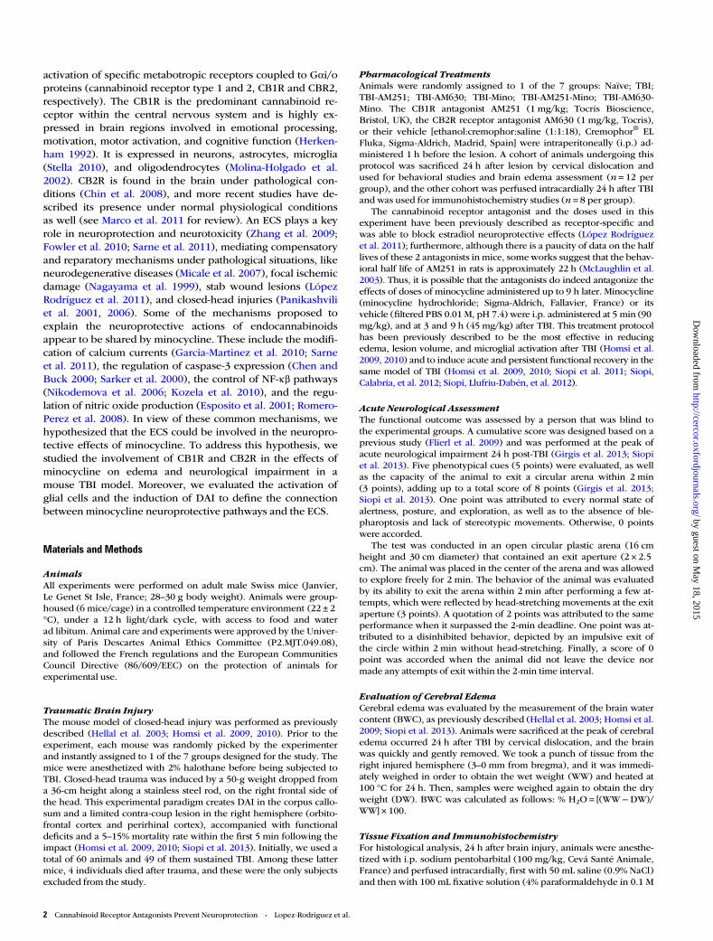
Morphometric AnalysisWe examined every section obtained from 2.5 to −2 mm bregma(Paxinos and Watson 1998) in a blind code. Within these sections, thelesion was found in every group except in naïve animals. Sections from8 animals in each experimental group were analyzed, all of them in theblind code. The number of Iba-1-positive cells within the area sur-rounding the lesion was counted with a morphometric grid under the×10 objective in a defining area of 350 × 350 µm; this grid was placedin the screen of a TV monitor connected to the microscope. The mor-phology of Iba-1 immunoreactive microglia was assessed with the ×40objective. Cells were classified in 5 morphological types: Type I,cells with few cellular processes (2 or less); Type II, cells showing 3–5short branches; Type III, cells with numerous (>5) and longer cell pro-cesses and a small cell body; Type IV, cells with large somas and re-tracted and thicker processes; and Type V, cells with ameboid cellbody, numerous short processes, and intense Iba-1 immunostaining.Iba1-immunoreactive cells with large somas and retracted and thickerprocesses and cells with amoeboid cell body, numerous shortprocesses, and intense Iba1 immunostaining were classified as reactivemicroglia (Diz-Chaves et al. 2012; see Fig. 1). For each animal, we ana-lyzed a total of 100 cells, within 4 different slices in the ventral cortexof the right hemisphere surrounding the lesion, as well as inthe “corpus callosum,” in the same region where we analyzed the DAI(interhemisphere area).
DAI was assessed by analyzing the surface density of β-APP-immunolabeled axons using a double square lattice test systemdefining an area of 80 × 80 µm on the grid C16 of Weibel (1979). Themorphometric grid was placed in the screen of a TV monitor connectedto the microscope. Four sections for each animal were examined usingthe ×40 objective, and the number of intersections of the lines of thegrid with β-APP-immunoreactive material profiles was counted withinthe interhemisphere area of the corpus callosum.
Statistical AnalysisData were analyzed using a 3-way analysis of variance (ANOVA), withfactors being TBI, cannabinoid receptor antagonists, and minocyclinetreatment. Data were not always normally distributed. Therefore, tosatisfy the assumption of normality for the ANOVA, we transformedthe data when necessary by the Neperian logarithm function. If trans-formed data were not normally distributed, nonparametric tests wereused (Kruskal–Wallis and post hoc pair-wise comparisons with Mann–Whitney U-test). When appropriate, 3-way ANOVAs were followed byseparate 2-way ANOVA and 1-way ANOVA split by the independentfactors to further analyze the data. Post hoc comparisons were per-formed with a level of significance set at P < 0.05. For data that werenormally distributed and homoscedastic, we used a standard parametricpost hoc test (Bonferroni’s test) and for those that were normally distrib-uted, but nonhomoscedastic, we performed nonparametric post hoccomparisons (Games–Howell’s test). Student’s t-test was used when2-group comparison was necessary. Statistical analyses were carried outwith the SPSS 19.0 software package (SPSS, Inc., Chicago, IL, USA). Dataare presented as mean + standard error of the mean (SEM).
Results
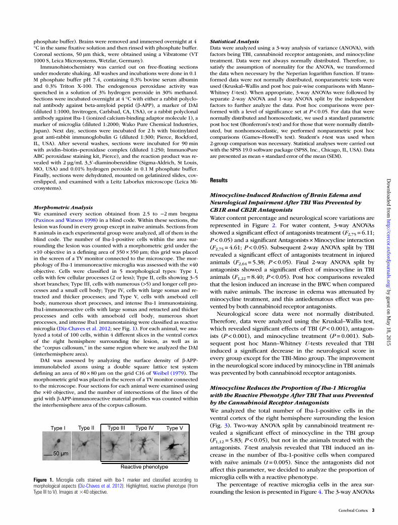
Minocycline-Induced Reduction of Brain Edema andNeurological Impairment After TBI Was Prevented byCB1R and CB2R AntagonistsWater content percentage and neurological score variations arerepresented in Figure 2. For water content, 3-way ANOVAsshowed a significant effect of antagonists treatment (F2,75 = 6.11;P < 0.05) and a significant Antagonists ×Minocycline interaction(F2,75 = 4.61; P < 0.05). Subsequent 2-way ANOVA split by TBIrevealed a significant effect of antagonists treatment in injuredanimals (F2,64 = 5.38; P < 0.05). Final 2-way ANOVA split byantagonists showed a significant effect of minocycline in TBIanimals (F1,22 = 8.40; P < 0.05). Post hoc comparisons revealedthat the lesion induced an increase in the BWC when comparedwith naïve animals. The increase in edema was attenuated byminocycline treatment, and this antiedematous effect was pre-vented by both cannabinoid receptor antagonists.
Neurological score data were not normally distributed.Therefore, data were analyzed using the Kruskal–Wallis test,which revealed significant effects of TBI (P < 0.001), antagon-ists (P < 0.001), and minocycline treatment (P = 0.001). Sub-sequent post hoc Mann–Whitney U-tests revealed that TBIinduced a significant decrease in the neurological score inevery group except for the TBI-Mino group. The improvementin the neurological score induced by minocycline in TBI animalswas prevented by both cannabinoid receptor antagonists.
Minocycline Reduces the Proportion of Iba-1 Microgliawith the Reactive Phenotype After TBI That was Preventedby the Cannabinoid Receptor AntagonistsWe analyzed the total number of Iba-1-positive cells in theventral cortex of the right hemisphere surrounding the lesion(Fig. 3). Two-way ANOVA split by cannabinoid treatment re-vealed a significant effect of minocycline in the TBI group(F1,12 = 5.83; P < 0.05), but not in the animals treated with theantagonists. T-test analysis revealed that TBI induced an in-crease in the number of Iba-1-positive cells when comparedwith naïve animals (t = 0.005). Since the antagonists did notaffect this parameter, we decided to analyze the proportion ofmicroglia cells with a reactive phenotype.
The percentage of reactive microglia cells in the area sur-rounding the lesion is presented in Figure 4. The 3-way ANOVAs
Figure 1. Microglia cells stained with Iba-1 marker and classified according tomorphological aspects (Diz-Chaves et al. 2012). Highlighted, reactive phenotype (fromType III to V). Images at ×40 objective.
Cerebral Cortex 3
by guest on May 18, 2015
http://cercor.oxfordjournals.org/D
ownloaded from
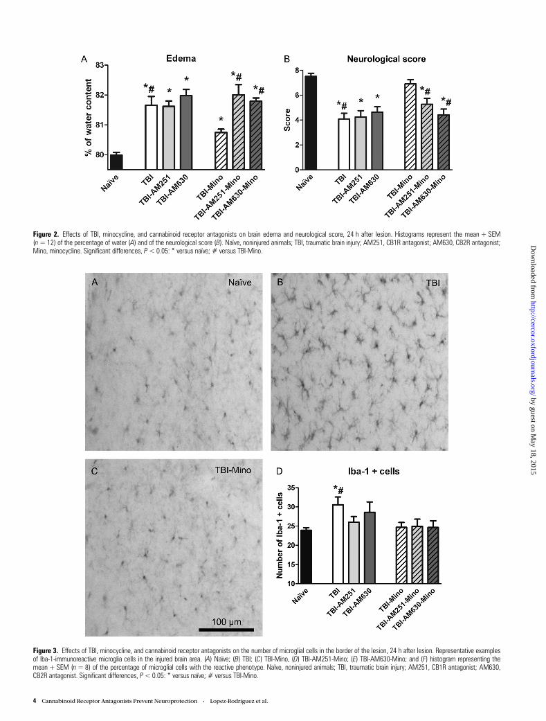
Figure 2. Effects of TBI, minocycline, and cannabinoid receptor antagonists on brain edema and neurological score, 24 h after lesion. Histograms represent the mean + SEM(n=12) of the percentage of water (A) and of the neurological score (B). Naïve, noninjured animals; TBI, traumatic brain injury; AM251, CB1R antagonist; AM630, CB2R antagonist;Mino, minocycline. Significant differences, P< 0.05: * versus naïve; # versus TBI-Mino.
Figure 3. Effects of TBI, minocycline, and cannabinoid receptor antagonists on the number of microglial cells in the border of the lesion, 24 h after lesion. Representative examplesof Iba-1-immunoreactive microglia cells in the injured brain area. (A) Naïve; (B) TBI; (C) TBI-Mino, (D) TBI-AM251-Mino; (E) TBI-AM630-Mino; and (F) histogram representing themean + SEM (n= 8) of the percentage of microglial cells with the reactive phenotype. Naïve, noninjured animals; TBI, traumatic brain injury; AM251, CB1R antagonist; AM630,CB2R antagonist. Significant differences, P<0.05: * versus naïve; # versus TBI-Mino.
4 Cannabinoid Receptor Antagonists Prevent Neuroprotection • Lopez-Rodriguez et al.
by guest on May 18, 2015
http://cercor.oxfordjournals.org/D
ownloaded from
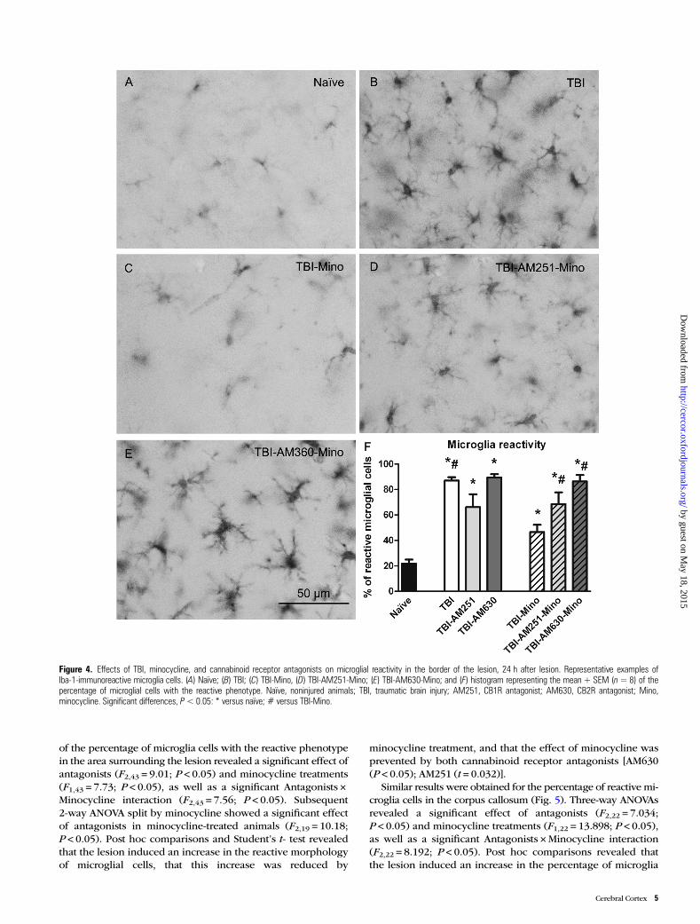
of the percentage of microglia cells with the reactive phenotypein the area surrounding the lesion revealed a significant effect ofantagonists (F2,43 = 9.01; P < 0.05) and minocycline treatments(F1,43 = 7.73; P < 0.05), as well as a significant Antagonists ×Minocycline interaction (F2,43 = 7.56; P < 0.05). Subsequent2-way ANOVA split by minocycline showed a significant effectof antagonists in minocycline-treated animals (F2,19 = 10.18;P < 0.05). Post hoc comparisons and Student’s t- test revealedthat the lesion induced an increase in the reactive morphologyof microglial cells, that this increase was reduced by
minocycline treatment, and that the effect of minocycline wasprevented by both cannabinoid receptor antagonists [AM630(P < 0.05); AM251 (t = 0.032)].
Similar results were obtained for the percentage of reactive mi-croglia cells in the corpus callosum (Fig. 5). Three-way ANOVAsrevealed a significant effect of antagonists (F2,22 = 7.034;P < 0.05) and minocycline treatments (F1,22 = 13.898; P < 0.05),as well as a significant Antagonists ×Minocycline interaction(F2,22 = 8.192; P < 0.05). Post hoc comparisons revealed thatthe lesion induced an increase in the percentage of microglia
Figure 4. Effects of TBI, minocycline, and cannabinoid receptor antagonists on microglial reactivity in the border of the lesion, 24 h after lesion. Representative examples ofIba-1-immunoreactive microglia cells. (A) Naïve; (B) TBI; (C) TBI-Mino, (D) TBI-AM251-Mino; (E) TBI-AM630-Mino; and (F) histogram representing the mean + SEM (n=8) of thepercentage of microglial cells with the reactive phenotype. Naïve, noninjured animals; TBI, traumatic brain injury; AM251, CB1R antagonist; AM630, CB2R antagonist; Mino,minocycline. Significant differences, P< 0.05: * versus naïve; # versus TBI-Mino.
Cerebral Cortex 5
by guest on May 18, 2015
http://cercor.oxfordjournals.org/D
ownloaded from
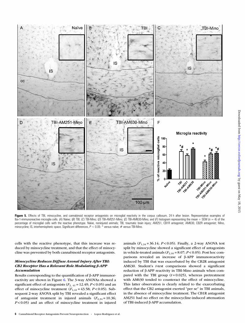
cells with the reactive phenotype, that this increase was re-duced by minocycline treatment, and that the effect of minocy-cline was prevented by both cannabinoid receptor antagonists.
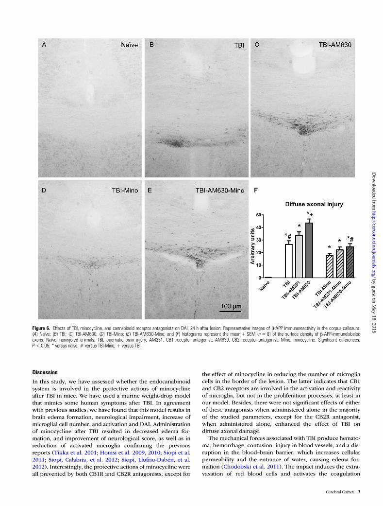
Minocycline Reduces Diffuse Axonal Injury After TBI:CB2 Receptor Has a Relevant Role Modulating β-APPAccumulationResults corresponding to the quantification of β-APP immunor-eactivity are shown in Figure 6. The 3-way ANOVAs showed asignificant effect of antagonists (F2, 41 = 12.49; P < 0.05) and aneffect of minocycline treatment (F1,41 = 43.58; P < 0.05). Sub-sequent 2-way ANOVA split by TBI revealed a significant effectof antagonist treatment in injured animals (F2,34 = 10.36;P < 0.05) and an effect of minocycline treatment in injured
animals (F1,34 = 36.14; P < 0.05). Finally, a 2-way ANOVA testsplit by minocycline showed a significant effect of antagonistsin vehicle-treated animals (F2,16 = 8.07; P < 0.05). Post hoc com-parisons revealed an increase of β-APP immunoreactivityinduced by TBI that was exacerbated by the CB2R antagonistAM630. Student’s t-test comparisons showed a significantreduction of β-APP reactivity in TBI-Mino animals when com-pared with the TBI group (t = 0.025), whereas pretreatmentwith AM630 tended to counteract the effect of minocycline.This latter observation is clearly related to the exarcerbatingeffect that the CB2 antagonist exerted “per se” in TBI animals,in the absence of minocycline treatment. The CB1R antagonistAM251 had no effect on the minocycline-induced attenuationof TBI-induced β-APP accumulation.
Figure 5. Effects of TBI, minocycline, and cannabinoid receptor antagonists on microglial reactivity in the corpus callosum, 24 h after lesion. Representative examples ofIba-1-immunoreactive microglia cells. (A) Naïve; (B) TBI; (C) TBI-Mino; (D) TBI-AM251-Mino; (E) TBI-AM630-Mino; and (F) histogram representing the mean + SEM (n= 4) of thepercentage of microglial cells with the reactive phenotype. Naïve, noninjured animals; TBI, traumatic brain injury; AM251, CB1R antagonist; AM630, CB2R antagonist; Mino,minocycline; IS, interhemispheric space. Significant differences, P<0.05: * versus naïve; # versus TBI-Mino.
6 Cannabinoid Receptor Antagonists Prevent Neuroprotection • Lopez-Rodriguez et al.
by guest on May 18, 2015
http://cercor.oxfordjournals.org/D
ownloaded from
Discussion
In this study, we have assessed whether the endocanabinoidsystem is involved in the protective actions of minocyclineafter TBI in mice. We have used a murine weight-drop modelthat mimics some human symptoms after TBI. In agreementwith previous studies, we have found that this model results inbrain edema formation, neurological impairment, increase ofmicroglial cell number, and activation and DAI. Administrationof minocycline after TBI resulted in decreased edema for-mation, and improvement of neurological score, as well as inreduction of activated microglia confirming the previousreports (Tikka et al. 2001; Homsi et al. 2009, 2010; Siopi et al.2011; Siopi, Calabria, et al. 2012; Siopi, Llufriu-Dabén, et al.2012). Interestingly, the protective actions of minocycline wereall prevented by both CB1R and CB2R antagonists, except for
the effect of minocycline in reducing the number of microgliacells in the border of the lesion. The latter indicates that CB1and CB2 receptors are involved in the activation and reactivityof microglia, but not in the proliferation processes, at least inour model. Besides, there were not significant effects of eitherof these antagonists when administered alone in the majorityof the studied parameters, except for the CB2R antagonist,when administered alone, enhanced the effect of TBI ondiffuse axonal damage.
The mechanical forces associated with TBI produce hemato-ma, hemorrhage, contusion, injury in blood vessels, and a dis-ruption in the blood–brain barrier, which increases cellularpermeability and the entrance of water, causing edema for-mation (Chodobski et al. 2011). The impact induces the extra-vasation of red blood cells and activates the coagulation
Figure 6. Effects of TBI, minocycline, and cannabinoid receptor antagonists on DAI, 24 h after lesion. Representative images of β-APP immunoreactivity in the corpus callosum.(A) Naïve; (B) TBI; (C) TBI-AM630; (D) TBI-Mino; (E) TBI-AM630-Mino; and (F) histograms represent the mean + SEM (n=8) of the surface density of β-APP-immunolabeledaxons. Naïve, noninjured animals; TBI, traumatic brain injury; AM251, CB1 receptor antagonist; AM630, CB2 receptor antagonist; Mino, minocycline. Significant differences,P<0.05: * versus naïve; # versus TBI-Mino; + versus TBI.
Cerebral Cortex 7
by guest on May 18, 2015
http://cercor.oxfordjournals.org/D
ownloaded from
cascade. Several studies have shown that coagulation factorscan affect the function of microglia, triggering an inflammatoryresponse around the injury and leading to the release of proin-flammatory cytokines, such as TNF-α, IL-6 (Möller et al. 2000),and IL-1β, which plays a crucial role in cerebral edema for-mation (Lazovic et al. 2005) by parenchymal cells (Soares et al.1995; Royo et al. 1999). Minocycline may reduce edema for-mation by decreasing microglial activation and the release ofproinflammatory cytokines, such as IL-1β (Lazovic et al. 2005;Homsi et al. 2009). The present results show that the cannabi-noid receptor antagonists inhibited the protective action ofminocycline, suggesting that the activation of CB receptorsmay contribute to this effect of minocycline. Several previousdata support this view. For instance, CB1R and CB2R agonistsinhibit the production of proinflammatory interleukins (Kleinet al. 2000; Smith et al. 2000; Molina-Holgado et al. 2002; DiFilippo et al. 2004; Kozela et al. 2010; Paulsen et al. 2011). Fur-thermore, the activation of CB2R controls the immuneresponse of microglial cells, increasing their migration to thelesion area or reducing the production of harmful molecules,such as TNF-α or free radicals (Walter et al. 2003; Eljasche-witsch et al. 2006; Dirikoc et al. 2007). Particularly, in TBImodels, the selective activation of CB2R leads to a reduction inthe activated microglia (Elliott et al. 2011), demonstrating apivotal role of this receptor in microglial activity and immuneresponse.
The enhanced microglial reactivity caused by TBI may alsocontribute to DAI, since activated microglia trigger oxidativestress responses and excitotoxicity mechanisms that causeaxonal damage (Hanisch and Kettenmann 2007). This possi-bility is supported by our findings in the corpus callosum,where microglial reactivity was increased by TBI and reducedby minocycline. These results suggest that the protective effectof minocycline on DAI may be, in part, related to the control ofreactive microglia in the corpus callosum. In addition, our find-ings indicate that minocycline decreases the accumulation ofβ-APP in the corpus callosum caused by TBI. The majority ofaxonal injuries occur in the zones of transition between thegray and white matter of the brain, for example, the corpuscallosum (Gennarelli 1994; Povlishock et al. 1994), and theycan be detected by the accumulation of β-APP in the damagedaxons (Roberts et al. 1994). This effect of minocycline may berelated to the control of amyloid precursor metabolism, bythe inhibition of β-site APP cleaving enzyme 1 activity and thedecrease in NFkappa B levels (Ferretti et al. 2012). Our find-ings suggest that the activation of CB2R by endogenousligands is also protective, since the CB2R antagonist, per se,enhanced the damage caused by TBI, increasing the β-APPaccumulation in the corpus callosum. Some in vitro exper-iments in human tissue and cell lines (Tolón et al. 2009) havedemonstrated that the activation of CB2 receptors withspecific agonists (JWH-015) stimulates the phagocytosis ofbeta-amyloid deposits. Our findings also support the viewthat neuroprotective effects derived from selective CB2R acti-vation may represent an avenue for further development ofnovel therapeutic agents in the treatment of TBI (Elliott et al.2011). This protective action mediated by the ECS mightinvolve the control of NFkappa B activity (Ferretti et al.2012), the reduction of excitotoxicity and oxidative stress(Kozela et al. 2011), and the control of beta-amyloid phagocy-tosis (Tolón et al. 2009).
As for brain edema, microglial activation, and diffuse axonaldamage, the protective action of minocycline on neurologicalimpairment after TBI was prevented by both CB1R and CB2Rantagonists. TBI produces cognitive impairments in humans(Wortzel and Arciniegas 2012) and in animal models (Rochatet al. 2010; Shenaq et al. 2012). For example, following TBI,humans have difficulty in remembering lists of numbers(Brooks 1972) or performing matching-to-sample tasks(McLean et al. 1983). In animal models, rats and mice showdeficits in spatial learning and memory (Shenaq et al. 2012)and impulsive behavior (Rochat et al. 2010). Many of these dys-functions are related to edema formation (Tominaga andOhnishi 1989), and the improvement in neurological functionproduced by minocycline correlates inversely to brain edema(Homsi et al. 2009). Therefore, CB1 and CB2 receptors maycontribute to the protective effect of minocycline on neurologi-cal impairment by decreasing brain edema.
In all the parameters that we have assessed, the cannabinoidreceptor antagonists partially prevented the protective effectsof minocycline. However, it seems that the CB1 receptor maybe less involved in the protective effects of this drug. Microglialcells undergo a process of differentiation and activation afterinjury, and during these stages, the pattern of CB1 and CB2receptor expression also changes. Cabral and Marciano-Cabralet al. (2005) have demonstrated that CB1 and CB2 receptors arefound constitutively at very low levels in microglia; however,after activation microglial cells express high levels of CB2 recep-tor, while the expression of CB1 receptor remains at low levels.This difference in expression may explain why the CB1 receptorantagonist was less effective than the CB2 receptor antagonist inreducing the effects of minocycline in our studies.
In summary, our findings confirm that minocycline de-creases brain damage caused by TBI and indicate for the firsttime, and that the activation of CB receptors is required for theneuroprotective actions of this compound. Further studies arenecessary to determine at which molecular points the signalingof minocycline and CB receptors interact to exert neuroprotec-tive actions.
Funding
This work has been supported by GRUPOS UCM-BSCH951579; Delegación del Gobierno para el Plan Nacional sobreDrogas (Orden SAS/1250/2009); Instituto de Salud Carlos III;Redes temáticas de Investigación Cooperativa en salud, Red deTrastornos Adictivos (RD06/0001/1013 and RD2012/0028/0021) to M.P.V.; Ministerio de Economía y Competividad,Spain (BFU2011-30217-C03-01) to L.M.G.S., and the nonprofi-table organization “Fondation des Gueules cassées” to E.S. andM.J.T.
NotesConflict of Interest: None declared.
ReferencesBales JW, Wagner AK, Kline AE, Dixon CE. 2009. Persistent cognitive
dysfunction after traumatic brain injury: a dopamine hypothesis.Neurosci Biobehav Rev. 33:981–1003.
8 Cannabinoid Receptor Antagonists Prevent Neuroprotection • Lopez-Rodriguez et al.
by guest on May 18, 2015
http://cercor.oxfordjournals.org/D
ownloaded from
Barzó P, Marmarou A, Fatouros P, Hayasaki K, Corwin F. 1997. Con-tribution of vasogenic and cellular edema to traumatic brainswelling measured by diffusion-weighted imaging. J Neurosurg.87:900–907.
Beauchamp K, Mutlak H, Smith WR, Shohami E, Stahel PF. 2008.Pharmacology of traumatic brain injury: where is the “goldenbullet”? Mol Med. 14:731–740.
Boto GR, Gómez PA, De la Cruz J, Lobato RD. 2009. A historical analy-sis of severe head injury. Neurosurg Rev. 32:343–353.
Brooks DN. 1972. Memory and head injury. J Nerv Ment Dis.155:350–355.
Cabral GA, Marciano-Cabral F. 2005. Cannabinoid receptors in micro-glia of the central nervous system: immune functional relevance.J Leukoc Biol. 78:1192–1197.
Cernak I. 2005. Animal models of head trauma. NeuroRx. 2:410–422.Chen Y, Buck J. 2000. Cannabinoids protect cells from oxidative cell
death: a receptor-independent mechanism. J Pharmacol Exp Ther.293:807–812.
Chin C-L, Tovcimak AE, Hradil VP, Seifert TR, Hollingsworth PR, Chan-dran P, Zhu CZ, Gauvin D, Pai M, Wetter J et al. 2008. Differentialeffects of cannabinoid receptor agonists on regional brain activityusing pharmacological MRI. Br J Pharmacol. 153:367–379.
Chodobski A, Zink BJ, Szmydynger-Chodobska J. 2011. Blood-brainbarrier pathophysiology in traumatic brain injury. Trans Stroke Res.2:492–516.
Chu L-S, Fang S-H, Zhou Y, Yin Y-J, Chen W-Y, Li J-H, Sun J, Wang M-L,Zhang W-P, Wei E-Q. 2010. Minocycline inhibits 5-lipoxygenaseexpression and accelerates functional recovery in chronic phase offocal cerebral ischemia in rats. Life Sci. 86:170–177.
Davalos D, Grutzendler J, Yang G, Kim JV, Zuo Y, Jung S, Littman DR,Dustin ML, Gan W-B. 2005. ATP mediates rapid microglial responseto local brain injury in vivo. Nat Neurosci. 8:752–758.
d’Avila JC, Lam TI, Bingham D, Shi J, Won SJ, Kauppinen TM, Massa S,Liu J, Swanson RA. 2012. Microglial activation induced by braintrauma is suppressed by post-injury treatment with a PARP inhibi-tor. J Neuroinflammation. 9:31.
Di Filippo C, Rossi F, Rossi S, Amico MD. 2004. Cannabinoid CB2 re-ceptor activation reduces mouse myocardial ischemia-reperfusioninjury: involvement of cytokine. J Leukoc Biol. 75:453–459.
Dirikoc S, Priola SA, Marella M, Zsürger N, Chabry J. 2007. Nonpsy-choactive cannabidiol prevents prion accumulation and protectsneurons against prion toxicity. J Neurosci. 27:9537–9544.
Diz-Chaves Y, Pernia O, Carrero P, Garcia-Segura LM. 2012. Prenatalstress causes alterations in the morphology of microglia and theinflammatory response of the hippocampus of adult female mice.J Neuroinflammation. 9:71.
Eljaschewitsch E, Witting A, Mawrin C, Lee T, Schmidt PM, Wolf S,Hoertnagl H, Raine CS, Schneider-Stock R, Nitsch R et al. 2006.The endocannabinoid anandamide protects neurons during CNSinflammation by induction of MKP-1 in microglial cells. Neuron.49:67–79.
Elliott MB, Tuma RF, Amenta PS, Barbe MF, Jallo JI. 2011. Acute effectsof a selective cannabinoid-2 receptor agonist on neuroinflammationin a model of traumatic brain injury. J Neurotrauma. 28:973–981.
Emilien G, Waltregny A. 1996. Traumatic brain injury, cognitive andemotional dysfunction. Impact of clinical neuropsychology re-search. Acta Neurol Belg. 96:89–101.
Esposito G, Izzo AA, Di Rosa M, Iuvone T. 2001. Selective cannabinoidCB1 receptor-mediated inhibition of inducible nitric oxide synthaseprotein expression in C6 rat glioma cells. J Neurochem. 78:835–841.
Ferretti MT, Allard S, Partridge V, Ducatenzeiler A, Cuello AC. 2012.Minocycline corrects early, pre-plaque neuroinflammation and in-hibits BACE-1 in a transgenic model of Alzheimer’s disease-likeamyloid pathology. J Neuroinflammation. 9:62.
Flierl Ma, Stahel PF, Beauchamp KM, Morgan SJ, Smith WR, ShohamiE. 2009. Mouse closed head injury model induced by a weight-dropdevice. Nat Protoc. 4:1328–1337.
Fowler CJ, Rojo ML, Rodriguez-Gaztelumendi A. 2010. Modulation ofthe endocannabinoid system: neuroprotection or neurotoxicity?Exp Neurol. 224:37–47.
Garcia-Martinez EM, Sanz-Blasco S, Karachitos A, Bandez MJ,Fernandez-Gomez FJ, Perez-Alvarez S, De Mera RMMF, Jordan MJ,Aguirre N, Galindo MF et al. 2010. Mitochondria and calcium fluxas targets of neuroprotection caused by minocycline in cerebellargranule cells. Biochem Pharmacol. 79:239–250.
Gennarelli TA. 1994. Animate models of human head injury. J Neuro-trauma. 11:357–368.
Girgis H, Palmier B, Croci N, Soustrat M, Plotkine M, Marchand-LerouxC. 2013. Effects of selective and non-selective cyclooxygenase inhi-bition against neurological deficit and brain oedema followingclosed head injury in mice. Brain Res. 1491:78–87.
González JC, Egea J, Del Carmen Godino M, Fernandez-Gomez FJ,Sánchez-Prieto J, Gandía L, García AG, Jordán J, Hernández-GuijoJM. 2007. Neuroprotectant minocycline depresses glutamatergicneurotransmission and Ca(2+) signalling in hippocampal neurons.Eur J Neurosci. 26:2481–2495.
Hanisch U-K, Kettenmann H. 2007. Microglia: active sensor and versa-tile effector cells in the normal and pathologic brain. Nat Neurosci.10:1387–1394.
Hanstock CC, Faden AI, Bendall MR, Vink R. 1994. Diffusion-weightedimaging differentiates ischemic tissue from traumatized tissue.Stroke. 25:843–848.
Hellal F, Pruneau D, Palmier B, Faye P, Croci N, Plotkine M, Marchand-Verrecchia C. 2003. Detrimental role of bradykinin B2 receptor in amurine model of diffuse brain injury. J Neurotrauma. 20:841–851.
Herkenham M. 1992. Cannabinoid receptor localization in brain:relationship to motor and reward systems. Ann N Y Acad Sci.654:19–32.
Homsi S, Federico F, Croci N, Palmier B, Plotkine M, Marchand-LerouxC, Jafarian-Tehrani M. 2009. Minocycline effects on cerebral edema:relations with inflammatory and oxidative stress markers followingtraumatic brain injury in mice. Brain Res. 1291:122–132.
Homsi S, Piaggio T, Croci N, Noble F, Plotkine M, Marchand-Leroux C,Jafarian-Tehrani M. 2010. Blockade of acute microglial activationby minocycline promotes neuroprotection and reduces locomotorhyperactivity after closed head injury in mice: a twelve-weekfollow-up study. J Neurotrauma. 27:911–921.
Iwata A, Stys PK, Wolf JA, Chen X-H, Taylor AG, Meaney DF, SmithDH. 2004. Traumatic axonal injury induces proteolytic cleavage ofthe voltage-gated sodium channels modulated by tetrodotoxin andprotease inhibitors. J Neurosci. 24:4605–4613.
Klein TW, Lane B, Newton CA, Friedman H. 2000. The cannabinoidsystem and cytokine network. Proc Soc Exp Biol Med. 225:1–8.
Kozela E, Lev N, Kaushansky N, Eilam R, Rimmerman N, Levy R,Ben-Nun A, Juknat A, Vogel Z. 2011. Cannabidiol inhibits patho-genic T cells, decreases spinal microglial activation and amelioratesmultiple sclerosis-like disease in C57BL/6 mice. Br J Pharmacol.163:1507–1519.
Kozela E, Pietr M, Juknat A, Rimmerman N, Levy R, Vogel Z. 2010. Can-nabinoids Δ9-tetrahydrocannabinol and cannabidiol differentiallyinhibit the lipopolysaccharide-activated NF-κB and Interferon-β/STAT proinflammatory pathways in BV-2 microglial cells. J BiolChem. 285:1616–1626.
Kraus RL, Pasieczny R, Lariosa-Willingham K, Turner MS, Jiang A,Trauger JW. 2005. Antioxidant properties of minocycline: neuro-protection in an oxidative stress assay and direct radical-scavengingactivity. J Neurochem. 94:819–827.
Lazovic J, Basu A, Lin H-W, Rothstein RP, Krady JK, Smith MB, LevisonSW. 2005. Neuroinflammation and both cytotoxic and vasogenicedema are reduced in interleukin-1 type 1 receptor-deficient miceconferring neuroprotection. Stroke. 36:2226–2231.
López Rodríguez AB, Mateos Vicente B, Romero-Zerbo SY,Rodriguez-Rodriguez N, Bellini MJ, Rodriguez de Fonseca F,Bermudez-Silva FJ, Azcoitia I, Garcia-Segura LM, Viveros M-P.2011. Estradiol decreases cortical reactive astrogliosis after braininjury by a mechanism involving cannabinoid receptors. CerebrCortex. 21:2046–2055.
Lucas S-M, Rothwell NJ, Gibson RM. 2006. The role of inflammation inCNS injury and disease. Br J Pharmacol. 147:S232–S240.
Mackie K. 2006. Cannabinoid receptors as therapeutic targets. Ann RevPharmacol Toxicol. 46:101–122.
Cerebral Cortex 9
by guest on May 18, 2015
http://cercor.oxfordjournals.org/D
ownloaded from
Marco EM, García-Gutiérrez MS, Bermúdez-Silva F-J, Moreira FA, Gui-marães F, Manzanares J, Viveros M-P. 2011. Endocannabinoidsystem and psychiatry: in search of a neurobiological basis for detri-mental and potential therapeutic effects. Front Behav Neurosci.5:63.
Markovic DS, Vinnakota K, Van Rooijen N, Kiwit J, Synowitz M, GlassR, Kettenmann H. 2011. Minocycline reduces glioma expansionand invasion by attenuating microglial MT1-MMP expression. BrainBehav Immun. 25:624–628.
Marmarou A. 2003. Pathophysiology of traumatic brain edema: currentconcepts. Acta Neurochir Suppl. 86:7–10.
Masson F, Thicoipe M, Aye P, Mokni T, Senjean P, Schmitt V, DessallesPH, Cazaugade M, Labadens P. 2001. Epidemiology of severebrain injuries: a prospective population-based study. J Trauma. 51:481–489.
McLaughlin PJ, Winston K, Swezey L, Wisniecki A, Aberman J, TardifDJ, Betz AJ, Ishiwari K, Makriyannis A, Salamone JD. 2003. Thecannabinoid CB1 antagonists SR 141716A and AM 251 suppressfood intake and food-reinforced behavior in a variety of tasks inrats. Behav Pharmacol. 14:583–588.
McLean A, Temkin NR, Dikmen S, Wyler AR. 1983. The behavioral se-quelae of head injury. J Clin Neuropsychol. 5:361–376.
Micale V, Mazzola C, Drago F. 2007. Endocannabinoids and neurode-generative diseases. Pharmacol Res. 56:382–392.
Molina-Holgado E, Vela JM, Arévalo-Martín A, Almazán G, Molina-Holgado F, Borrell J, Guaza C. 2002. Cannabinoids promoteoligodendrocyte progenitor survival: involvement of cannabinoidreceptors and phosphatidylinositol-3 kinase/Akt signaling. J Neuro-sci. 22:9742–9753.
Möller T, Hanisch UK, Ransom BR. 2000. Thrombin-induced activationof cultured rodent microglia. J Neurochem. 75:1539–1547.
Nagayama T, Sinor AD, Simon RP, Chen J, Graham SH, Jin K,Greenberg DA. 1999. Cannabinoids and neuroprotection in globaland focal cerebral ischemia and in neuronal cultures. J Neurosci.19:2987–2995.
Nikodemova M, Duncan ID, Watters JJ. 2006. Minocycline exertsinhibitory effects on multiple mitogen-activated protein kinasesand IkappaBalpha degradation in a stimulus-specific manner in mi-croglia. J Neurochem. 96:314–323.
Panikashvili D, Shein NA, Mechoulam R, Trembovler V, Kohen R,Alexandrovich A, Shohami E. 2006. The endocannabinoid 2-AGprotects the blood-brain barrier after closed head injury and inhi-bits mRNA expression of proinflammatory cytokines. NeurobiolDis. 22:257–264.
Panikashvili D, Simeonidou C, Ben-Shabat S, Hanus L, Breuer A,Mechoulam R, Shohami E. 2001. An endogenous cannabinoid(2-AG) is neuroprotective after brain injury. Nature. 413:527–531.
Paulsen K, Tauber S, Timm J, Goelz N, Dumrese C, Stolzing A, Hass R,Ullrich O. 2011. The cannabinoid receptors agonist WIN55212-2inhibits macrophageal differentiation and alters expression andphosphorylation of cell cycle control proteins. Cell Commun Signal.9:33.
Paxinos G, Watson C. 1998. The rat brain in stereotaxic coordinates.4th ed. New York: Elsevier.
Piomelli D. 2003. The molecular logic of endocannabinoid signalling.Nat Rev Neurosci. 4:873–884.
Plane JM, Shen Y, Pleasure DE, Deng W. 2010. Prospects for minocy-cline neuroprotection. Arch Neurol. 67:1442–1448.
Povlishock JT, Hayes RL, Michel ME, McIntosh TK. 1994. Workshop onanimal models of traumatic brain injury. J Neurotrauma.11:723–732.
Roberts GW, Gentleman SM, Lynch A, Murray L, Landon M, GrahamDI. 1994. Beta amyloid protein deposition in the brain after severehead injury: implications for the pathogenesis of Alzheimer’sdisease. J Neurol Neurosurg Psychiatry. 57:419–425.
Rochat L, Beni C, Billieux J, Azouvi P, Annoni J-M, Van der Linden M.2010. Assessment of impulsivity after moderate to severe traumaticbrain injury. Neuropsychol Rehabil. 20:778–797.
Romero-Perez D, Fricovsky E, Yamasaki KG, Griffin M, Barraza-Hidalgo M, Dillmann W, Villarreal F. 2008. Cardiac uptake of
minocycline and mechanisms for in vivo cardioprotection. J AmColl Cardiol. 52:1086–1094.
Royo NC, Wahl F, Stutzmann JM. 1999. Kinetics of polymorphonuclearneutrophil infiltration after a traumatic brain injury in rat. Neurore-port. 10:1363–1367.
Sanchez Mejia RO, Ona VO, Li M, Friedlander RM. 2001. Minocyclinereduces traumatic brain injury-mediated caspase-1 activation,tissue damage, and neurological dysfunction. Neurosurgery. 48:1393–1399.
Sarker KP, Obara S, Nakata M, Kitajima I, Maruyama I. 2000. Ananda-mide induces apoptosis of PC-12 cells: involvement of superoxideand caspase-3. FEBS Lett. 472:39–44.
Sarne Y, Asaf F, Fishbein M, Gafni M, Keren O. 2011. The dualneuroprotective-neurotoxic profile of cannabinoid drugs. Br J Phar-macol. 163:1391–1401.
Shenaq M, Kassem H, Peng C, Schafer S, Ding JY, Fredrickson V, Guthi-konda M, Kreipke CW, Rafols JA, Ding Y. 2012. Neuronal damageand functional deficits are ameliorated by inhibition of aquaporinand HIF1α after traumatic brain injury (TBI). J Neurol Sci. 323:134–140.
Siopi E, Calabria S, Plotkine M, Marchand-Leroux C, Jafarian-TehraniM. 2012. Minocycline restores olfactory bulb volume and olfactorybehavior after traumatic brain injury in mice. J Neurotrauma. 29:354–361.
Siopi E, Cho AH, Homsi S, Croci N, Plotkine M, Marchand-Leroux C,Jafarian-Tehrani M. 2011. Minocycline restores sAPPα levels andreduces the late histopathological consequences of traumatic braininjury in mice. J Neurotrauma. 28:2135–2143.
Siopi E, Llufriu-Dabén G, Cho AH, Vidal-Lletjós S, Plotkine M,Marchand-Leroux C, Jafarian-Tehrani M. 2013. Etazolate, anα-secretase activator, reduces neuroinflammation and offers persist-ent neuroprotection following traumatic brain injury in mice.Neuropharmacology. 67C:183–192.
Siopi E, Llufriu-Dabén G, Fanucchi F, Plotkine M, Marchand-Leroux C,Jafarian-Tehrani M. 2012. Evaluation of late cognitive impairmentand anxiety states following traumatic brain injury in mice: theeffect of minocycline. Neurosci Lett. 511:110–115.
Smith SR, Terminelli C, Denhardt G. 2000. Effects of cannabinoid recep-tor agonist and antagonist ligands on production of inflammatory cy-tokines and anti-inflammatory interleukin-10 in endotoxemic mice.J Pharmacol Exp Ther. 293:136–150.
Soares HD, Hicks RR, Smith D, McIntosh TK. 1995. Inflammatoryleukocytic recruitment and diffuse neuronal degeneration are sep-arate pathological processes resulting from traumatic brain injury.J Neurosci. 15:8223–8233.
Stella N. 2010. Cannabinoid and cannabinoid-like receptors in micro-glia, astrocytes, and astrocytomas. Glia. 58:1017–1030.
Tikka T, Fiebich BL, Goldsteins G, Keinanen R, Koistinaho J. 2001.Minocycline, a tetracycline derivative, is neuroprotective against ex-citotoxicity by inhibiting activation and proliferation of microglia.J Neurosci. 21:2580–2588.
Tikka TM, Koistinaho JE. 2001. Minocycline provides neuroprotectionagainst N-methyl-D-aspartate neurotoxicity by inhibiting microglia.J Immunol. 166:7527–7533.
Tolón RM, Núñez E, Pazos MR, Benito C, Castillo AI, Martínez-OrgadoJA, Romero J. 2009. The activation of cannabinoid CB2 receptorsstimulates in situ and in vitro beta-amyloid removal by humanmacrophages. Brain Res. 1283:148–154.
Tominaga T, Ohnishi ST. 1989. Interrelationship of brain edema,motor deficits, and memory impairment in rats exposed to focalischemia. Stroke. 20:513–518.
Vanezis P, Chan KK, Scholtz CL. 1987. White matter damage followingacute head injury. Forensic Sci Int. 35:1–10.
Walter L, Franklin A, Witting A, Wade C, Xie Y, Kunos G, Mackie K,Stella N. 2003. Nonpsychotropic cannabinoid receptors regulate mi-croglial cell migration. J Neurosci. 23:1398–1405.
Wang H, Gao J, Lassiter TF, McDonagh DL, Sheng H, Warner DS,Lynch JR, Laskowitz DT. 2006. Levetiracetam is neuroprotective inmurine models of closed head injury and subarachnoid hemor-rhage. Neurocrit Care. 5:71–78.
10 Cannabinoid Receptor Antagonists Prevent Neuroprotection • Lopez-Rodriguez et al.
by guest on May 18, 2015
http://cercor.oxfordjournals.org/D
ownloaded from
Weibel ER. 1979. Stereological methods. Vol. 1. Practical methods forbiological morphometry. London: Academic Press.
Werner C, Engelhard K. 2000. Pathophysiology of traumatic braininjury. Crit Care Nurs Q. 23:14–25.
World Health Organization. 2006. The world health report 2006:working together for health. Geneva: WHO Press.
Wortzel HS, Arciniegas DB. 2012. Treatment of post-traumatic cogni-tive impairments. Curr Treat Options Neurol. 14:493–508.
Wu X, Hu J, Zhuo L, Fu C, Hui G, Wang Y, Yang W, Teng L, Lu S,Xu G. 2008. Epidemiology of traumatic brain injury in easternChina, 2004: a prospective large case study. J Trauma. 64:1313–1319.
Zhang M, Martin BR, Adler MW, Razdan RJ, Kong W, Ganea D, TumaRF. 2009. Modulation of cannabinoid receptor activation as a neuro-protective strategy for EAE and stroke. J Neuroimmune Pharmacol.4:249–259.
Cerebral Cortex 11
by guest on May 18, 2015
http://cercor.oxfordjournals.org/D
ownloaded from