Anatomy revision course pt1
34
natomy Revision Session 1 slides are available online at Slideshare.net/ muradalshehry After this session The Upper Limb
-
Upload
murad-alshehry -
Category
Education
-
view
80 -
download
0
Transcript of Anatomy revision course pt1

Anatomy Revision Session 1
slides are available online atSlideshare.net/muradalshehry
After this session
The Upper Limb
Anatomy Revision Session 1
Objectives of this session
• To be able to Identify the muscles of the upper limb.• Know relations of muscles to important nerves.
• There will be a few pop Quiz…. Stay focused ☺
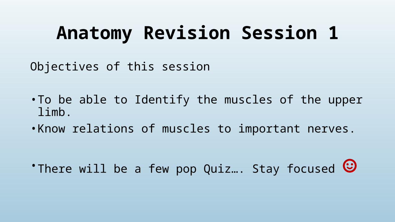
Orientation and Movement REVISION
adduction
abduction
flexionextension
circumduction
rotation

elevation
depression
supination
pronation
protraction
retraction
Orientation and Movement REVISION
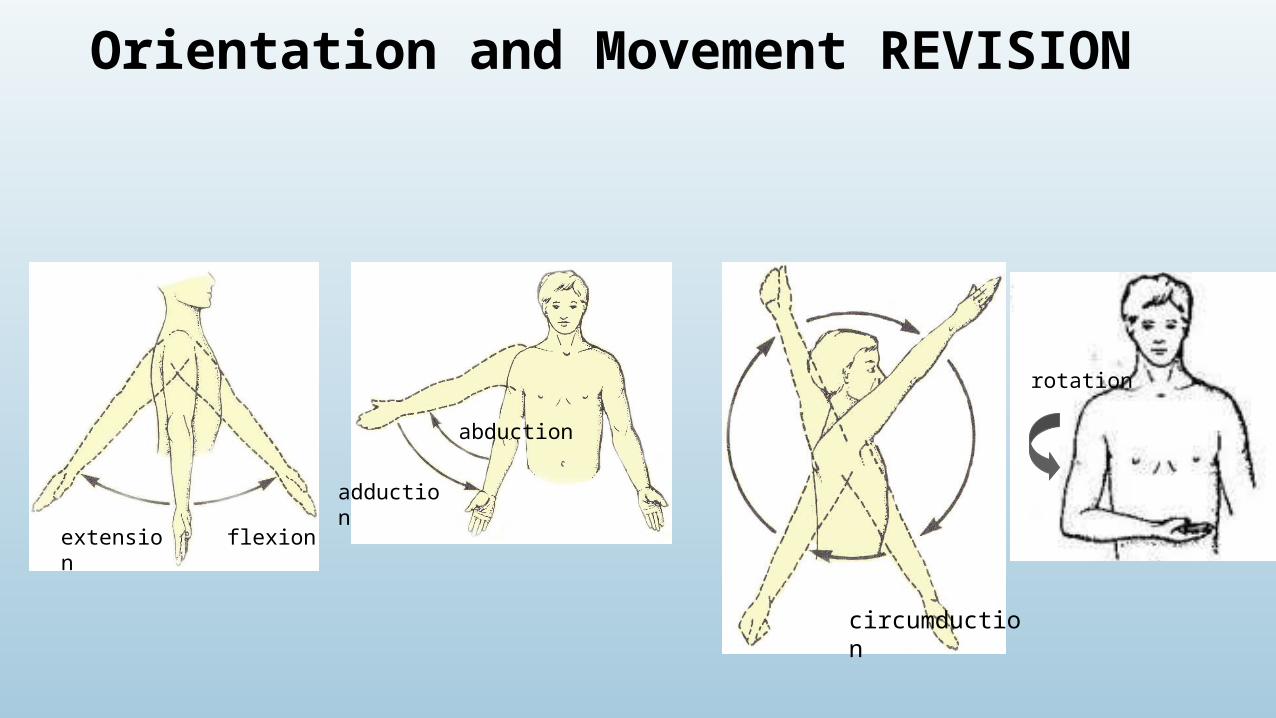
Pectoral girdle and shoulder joint
shoulder bones?
• Humerus • Scapula • Clavicle
The glenohumeral joint is a ball-and-socket joint
• Flexible• vulnerable
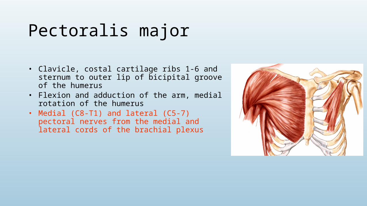
Pectoralis major
• Clavicle, costal cartilage ribs 1-6 and sternum to outer lip of bicipital groove of the humerus
• Flexion and adduction of the arm, medial rotation of the humerus
• Medial (C8-T1) and lateral (C5-7) pectoral nerves from the medial and lateral cords of the brachial plexus
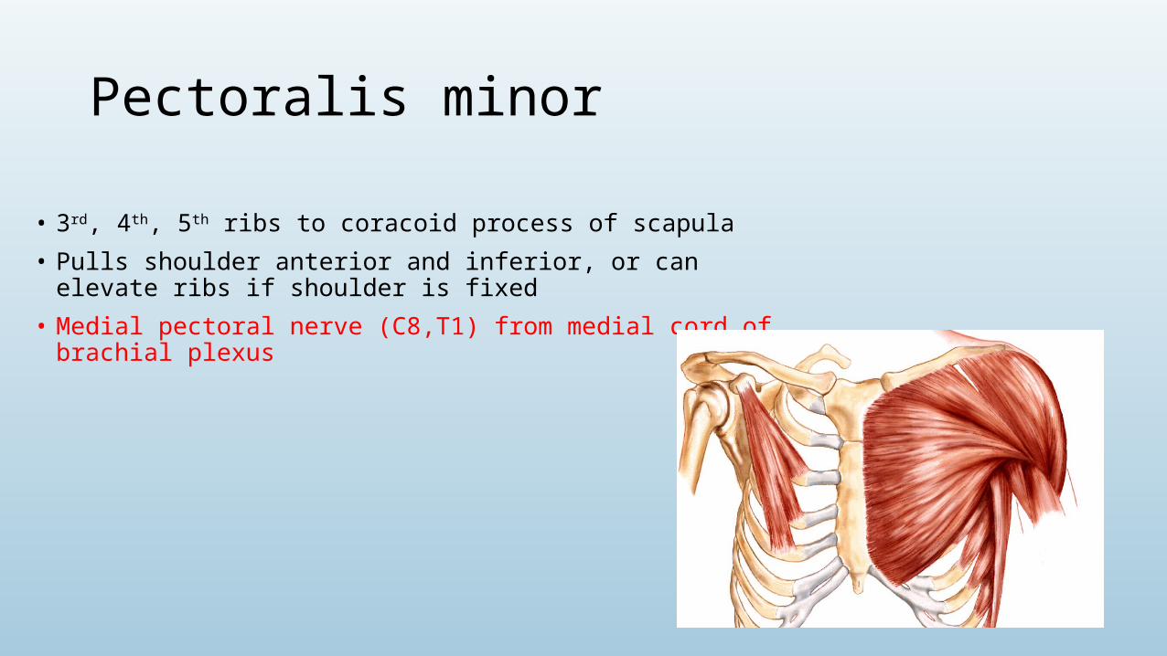
Pectoralis minor
• 3rd, 4th, 5th ribs to coracoid process of scapula
• Pulls shoulder anterior and inferior, or can elevate ribs if shoulder is fixed
• Medial pectoral nerve (C8,T1) from medial cord of brachial plexus

Serratus anterior
• Outer surface upper 8 ribs to medial border of the scapula
• Protraction of the scapula
• Long thoracic nerve (C5, 6, 7) of the brachial plexus
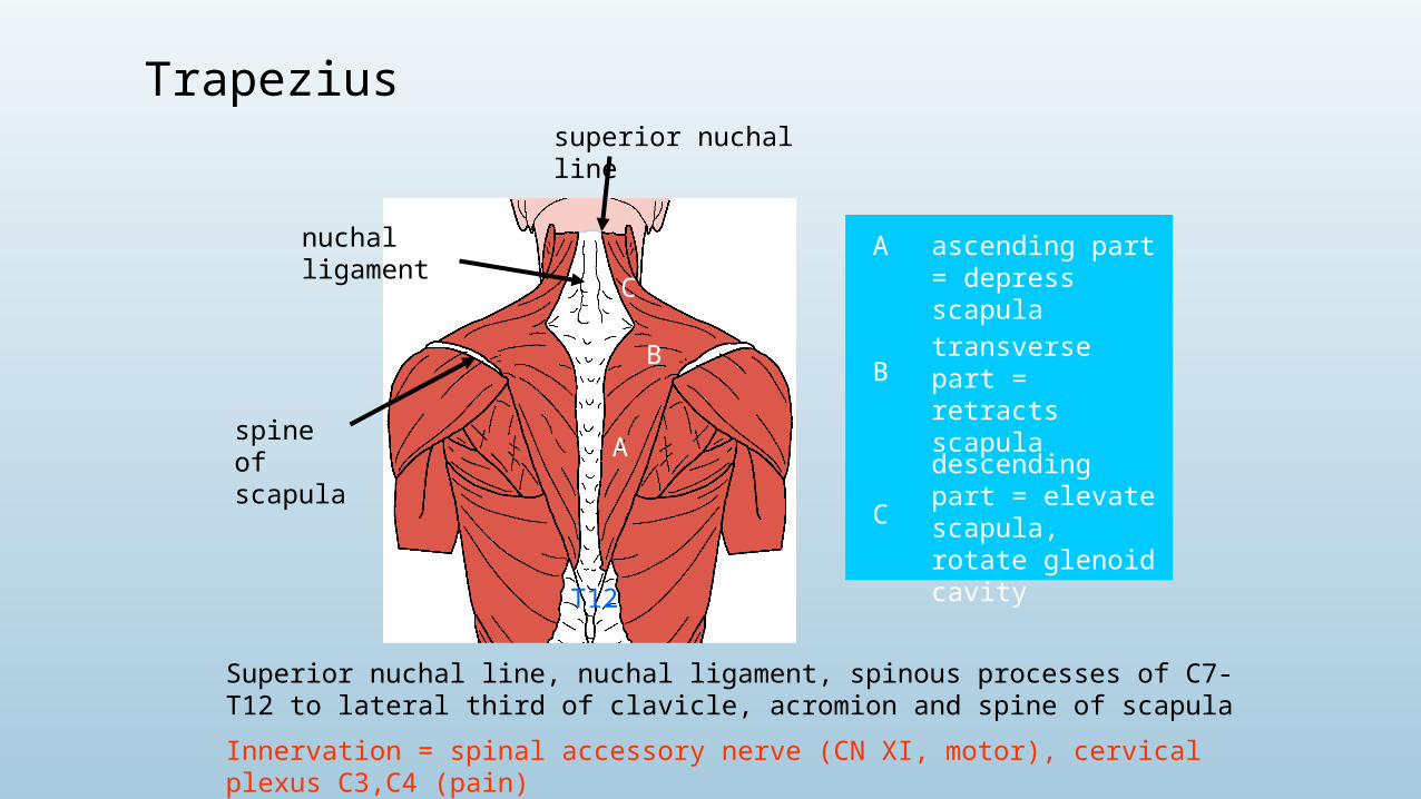
spine of scapula
nuchal ligament
A
B
C
Superior nuchal line, nuchal ligament, spinous processes of C7-T12 to lateral third of clavicle, acromion and spine of scapula
Innervation = spinal accessory nerve (CN XI, motor), cervical plexus C3,C4 (pain)
A ascending part = depress scapula
Btransverse part = retracts scapula
C
descending part = elevate scapula, rotate glenoid cavity
superior nuchal line
T12
Trapezius
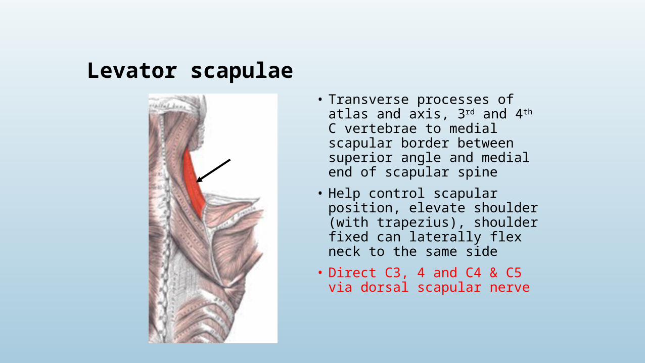
Levator scapulae• Transverse processes of atlas and axis,
3rd and 4th C vertebrae to medial scapular border between superior angle and medial end of scapular spine
• Help control scapular position, elevate shoulder (with trapezius), shoulder fixed can laterally flex neck to the same side
• Direct C3, 4 and C4 & C5 via dorsal scapular nerve
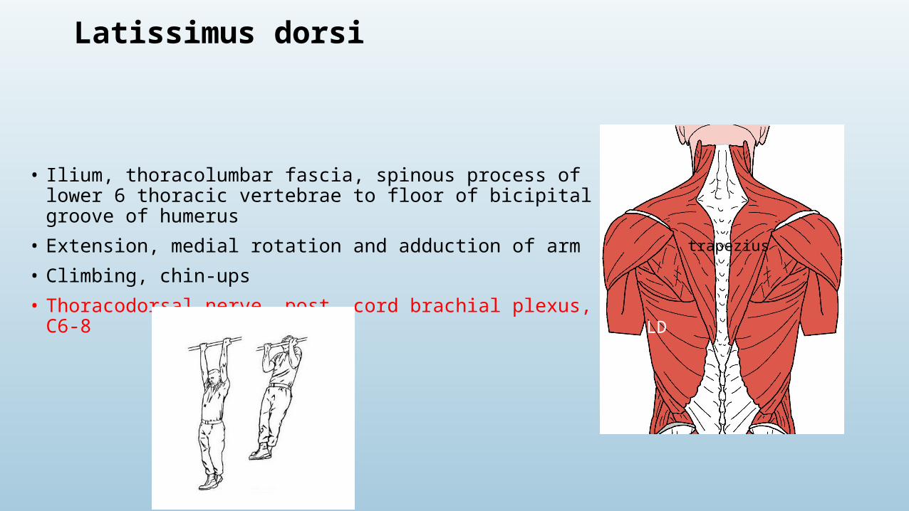
Latissimus dorsi
• Ilium, thoracolumbar fascia, spinous process of lower 6 thoracic vertebrae to floor of bicipital groove of humerus
• Extension, medial rotation and adduction of arm
• Climbing, chin-ups
• Thoracodorsal nerve, post. cord brachial plexus, C6-8
trapezius
LD

Levator scapulae• Transverse processes of atlas and axis,
3rd and 4th C vertebrae to medial scapular border between superior angle and medial end of scapular spine
• Help control scapular position, elevate shoulder (with trapezius), shoulder fixed can laterally flex neck to the same side
• Direct C3, 4 and C4 & C5 via dorsal scapular nerve
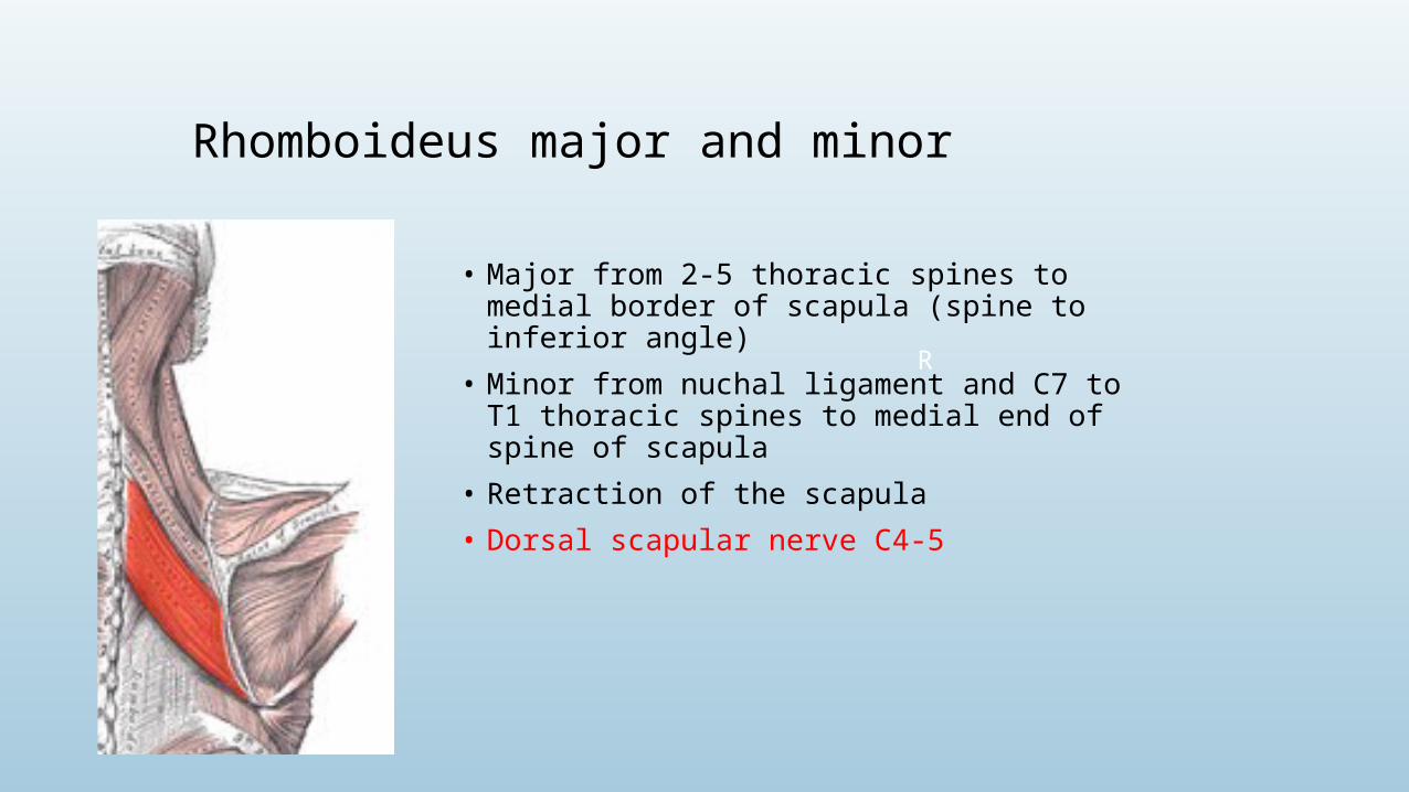
Rhomboideus major and minor
• Major from 2-5 thoracic spines to medial border of scapula (spine to inferior angle)
• Minor from nuchal ligament and C7 to T1 thoracic spines to medial end of spine of scapula
• Retraction of the scapula
• Dorsal scapular nerve C4-5
R

Deltoid
• Lateral 1/3rd clavicle, acromion, spine of scapula to deltoid tuberosity of humerus
1. Abduction of the arm from 10o to 110o mostly middle fibres
2. Extension and lateral rotation of arm from posterior fibres
3. flexion and medial rotation of arm from anterior fibres
• Axillary nerve (C5,6) from posterior cord of brachial plexusdeltoid
POST
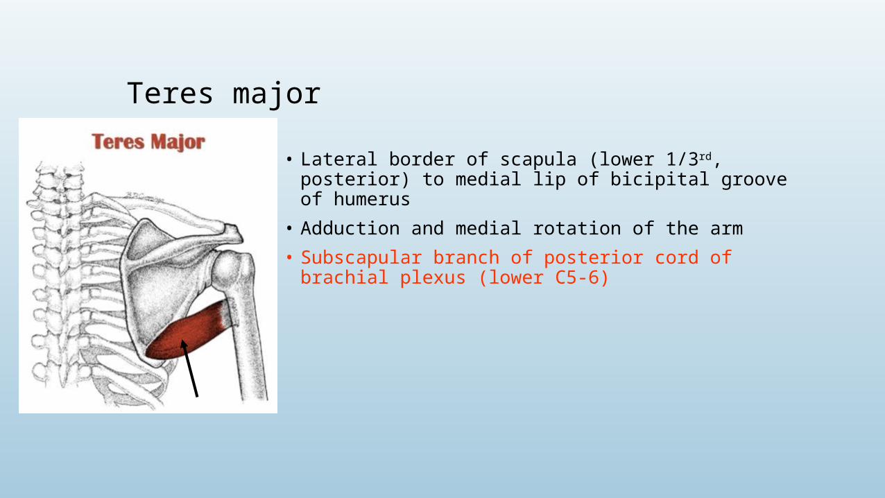
Teres major
• Lateral border of scapula (lower 1/3rd, posterior) to medial lip of bicipital groove of humerus
• Adduction and medial rotation of the arm
• Subscapular branch of posterior cord of brachial plexus (lower C5-6)
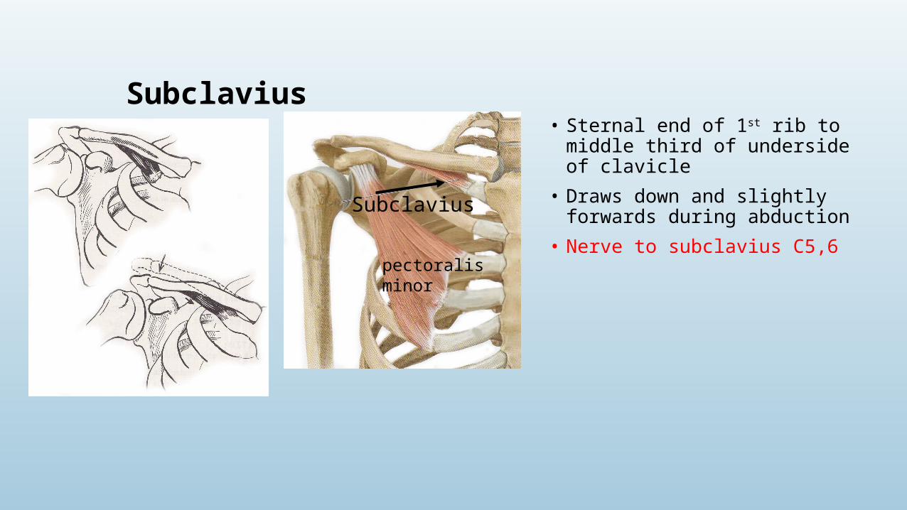
Subclavius• Sternal end of 1st rib to middle third of
underside of clavicle
• Draws down and slightly forwards during abduction
• Nerve to subclavius C5,6
pectoralis minor
Subclavius
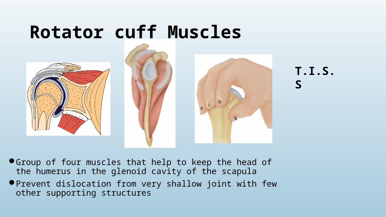
Rotator cuff Muscles
Group of four muscles that help to keep the head of the humerus in the glenoid cavity of the scapula
Prevent dislocation from very shallow joint with few other supporting structures
T.I.S.S
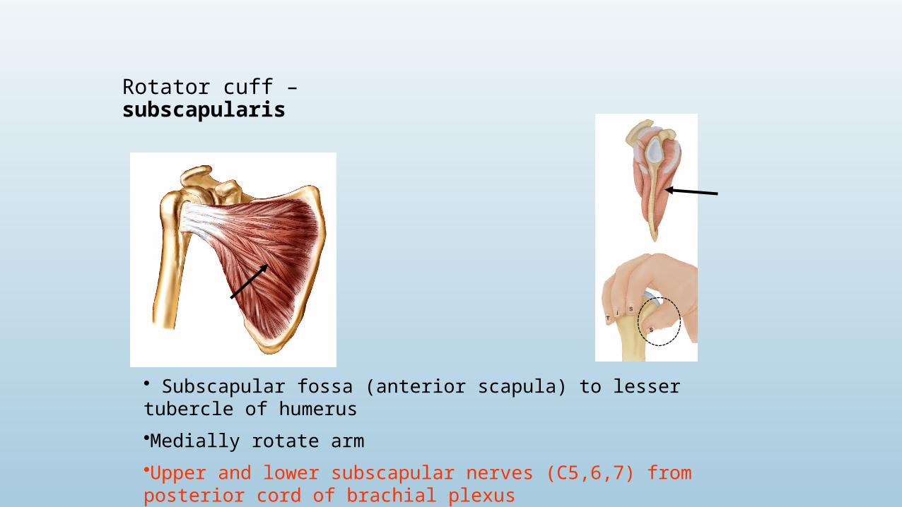
Rotator cuff – subscapularis
• Subscapular fossa (anterior scapula) to lesser tubercle of humerus
•Medially rotate arm
•Upper and lower subscapular nerves (C5,6,7) from posterior cord of brachial plexus
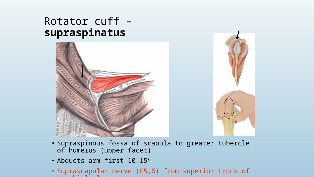
Rotator cuff – supraspinatus
• Supraspinous fossa of scapula to greater tubercle of humerus (upper facet)
• Abducts arm first 10-150
• Suprascapular nerve (C5,6) from superior trunk of brachial plexus
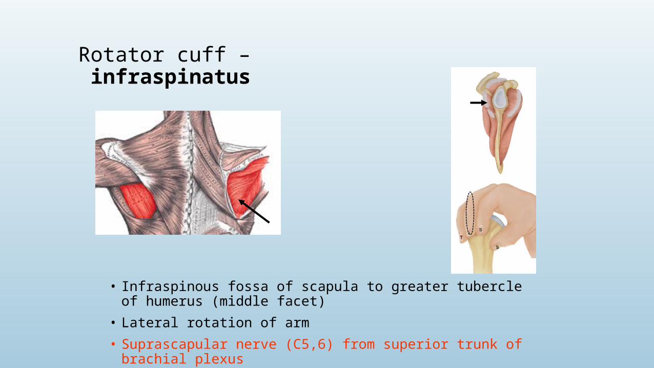
Rotator cuff – infraspinatus
• Infraspinous fossa of scapula to greater tubercle of humerus (middle facet)
• Lateral rotation of arm
• Suprascapular nerve (C5,6) from superior trunk of brachial plexus

Rotator cuff – teres minor
• Upper 2/3rds lateral border of scapula to greater tubercle of humerus (lower facet)
• Lateral rotation of arm
• Axillary nerve (C5,6) branch of posterior cord of the brachial plexus
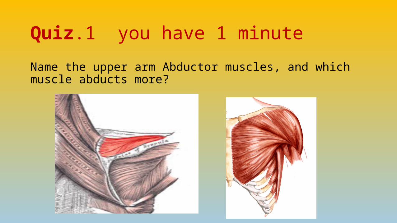
Quiz.1 you have 1 minute
Name the upper arm Abductor muscles, and which muscle abducts more?


Anterior compartment of the arm
• Flexors of arm and elbow
• Musculocutaneous nerve
• Biceps brachii
• Coracobrachialis
• Brachialis
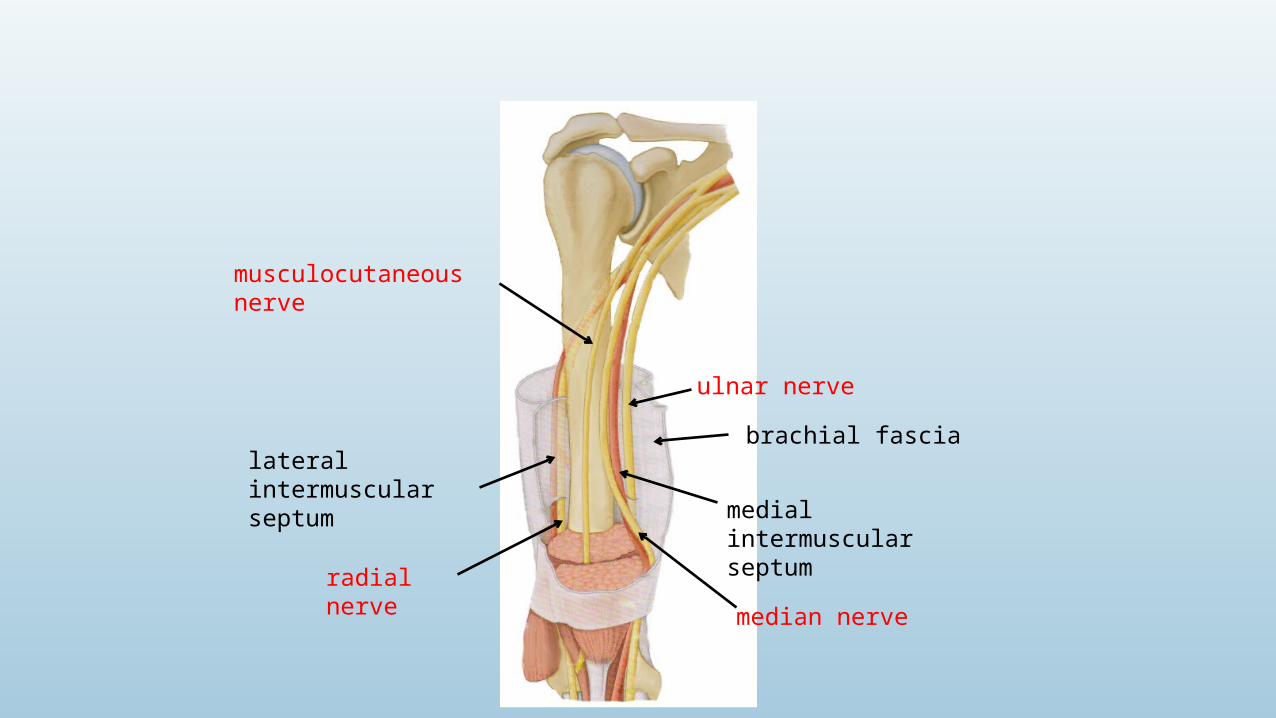
brachial fascia
medial intermuscular septum
lateral intermuscular septum
radial nerve
ulnar nerve
median nerve
musculocutaneous nerve
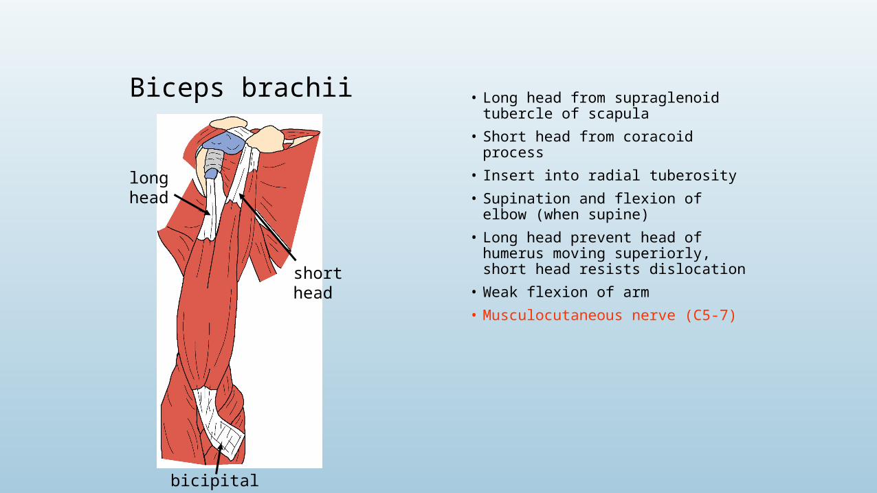
Biceps brachii • Long head from supraglenoid tubercle of scapula
• Short head from coracoid process• Insert into radial tuberosity• Supination and flexion of elbow
(when supine)• Long head prevent head of
humerus moving superiorly, short head resists dislocation
• Weak flexion of arm• Musculocutaneous nerve (C5-7)
long head
short head
bicipital aponeurosis
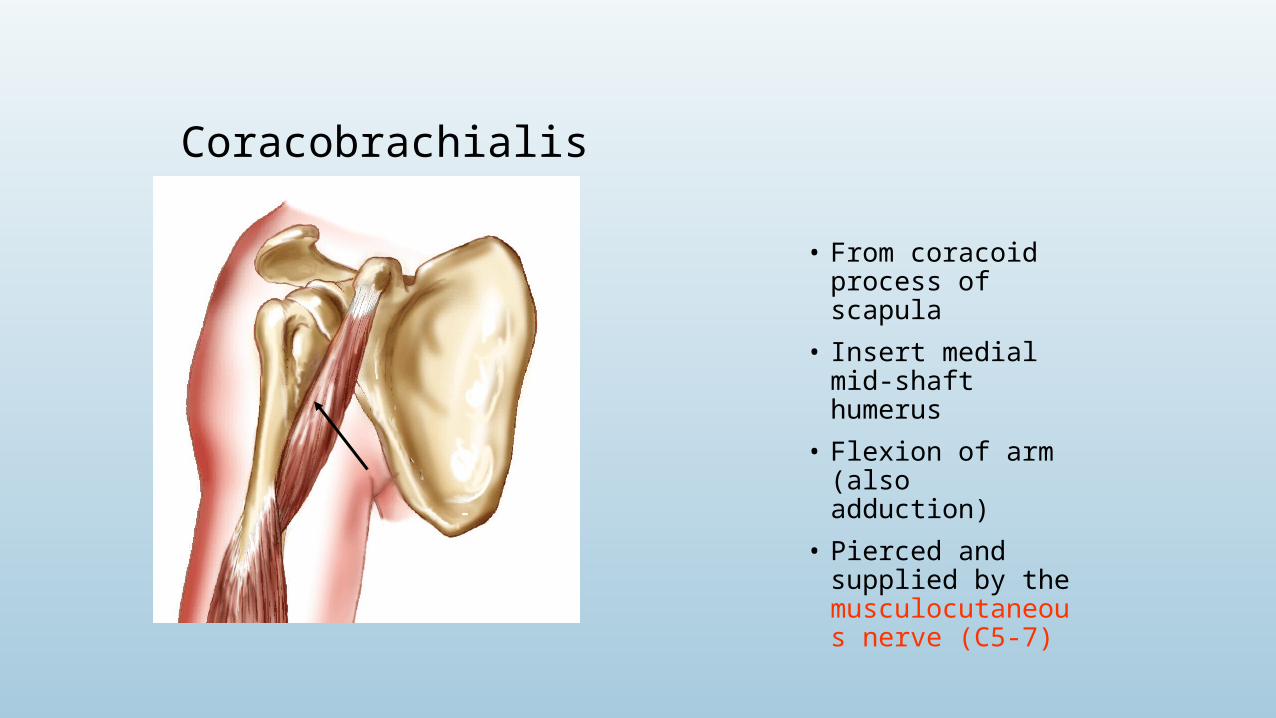
Coracobrachialis
• From coracoid process of scapula
• Insert medial mid-shaft humerus
• Flexion of arm (also adduction)
• Pierced and supplied by the musculocutaneous nerve (C5-7)
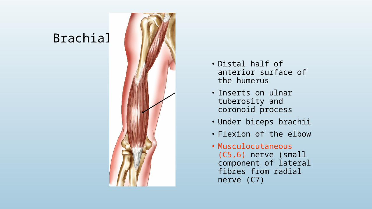
Brachialis
• Distal half of anterior surface of the humerus
• Inserts on ulnar tuberosity and coronoid process
• Under biceps brachii
• Flexion of the elbow
• Musculocutaneous (C5,6) nerve (small component of lateral fibres from radial nerve (C7)
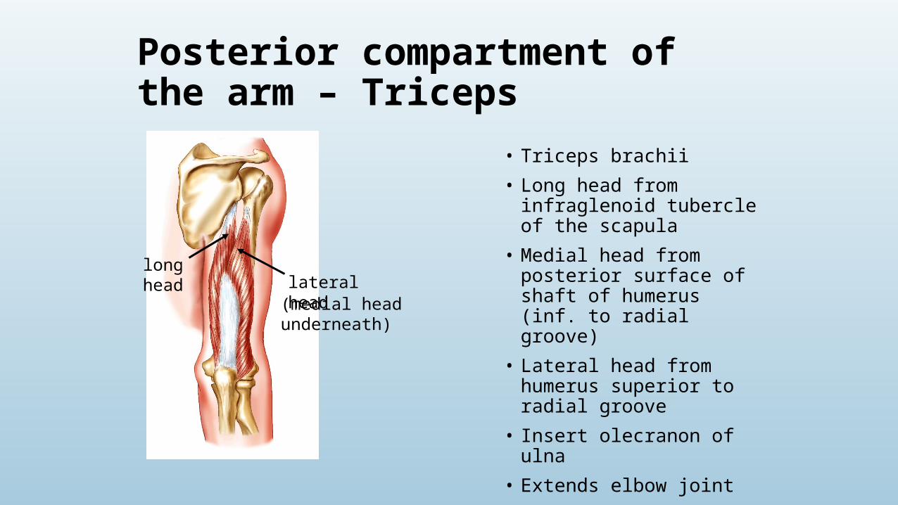
Posterior compartment of the arm – Triceps
• Triceps brachii
• Long head from infraglenoid tubercle of the scapula
• Medial head from posterior surface of shaft of humerus (inf. to radial groove)
• Lateral head from humerus superior to radial groove
• Insert olecranon of ulna
• Extends elbow joint
• Long head resists dislocation (especially during adduction)
• Radial nerve (C6-8)
long head lateral head
(medial head underneath)

Forearm
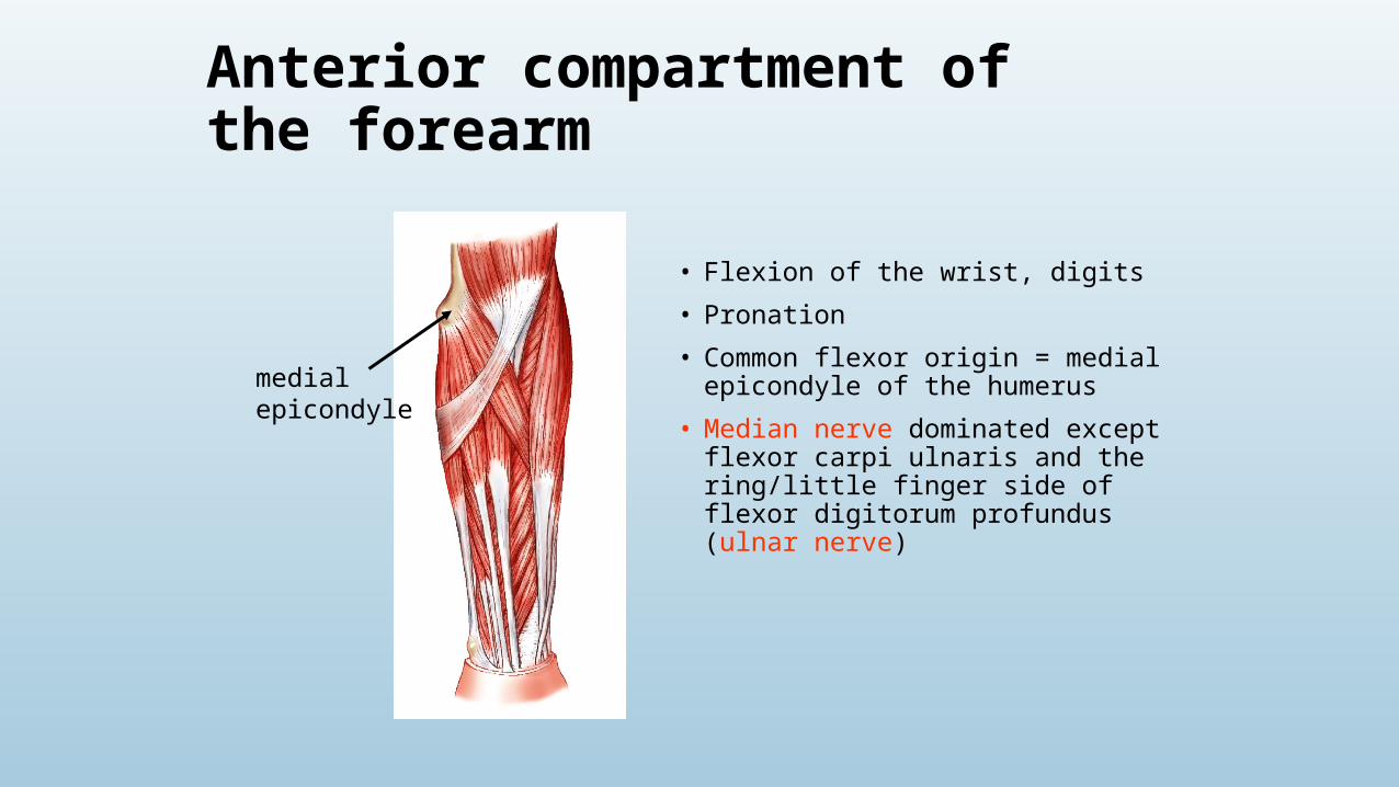
Anterior compartment of the forearm
• Flexion of the wrist, digits
• Pronation
• Common flexor origin = medial epicondyle of the humerus
• Median nerve dominated except flexor carpi ulnaris and the ring/little finger side of flexor digitorum profundus (ulnar nerve)
medial epicondyle
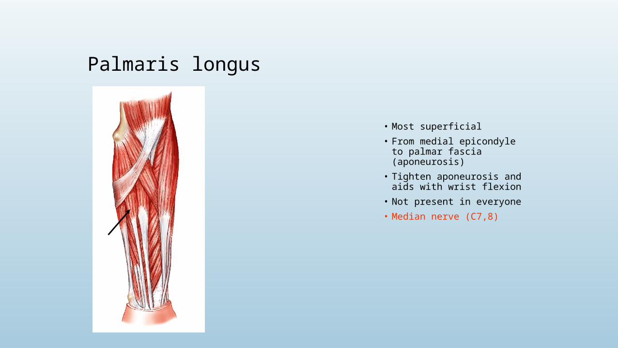
Palmaris longus
• Most superficial
• From medial epicondyle to palmar fascia (aponeurosis)
• Tighten aponeurosis and aids with wrist flexion
• Not present in everyone
• Median nerve (C7,8)
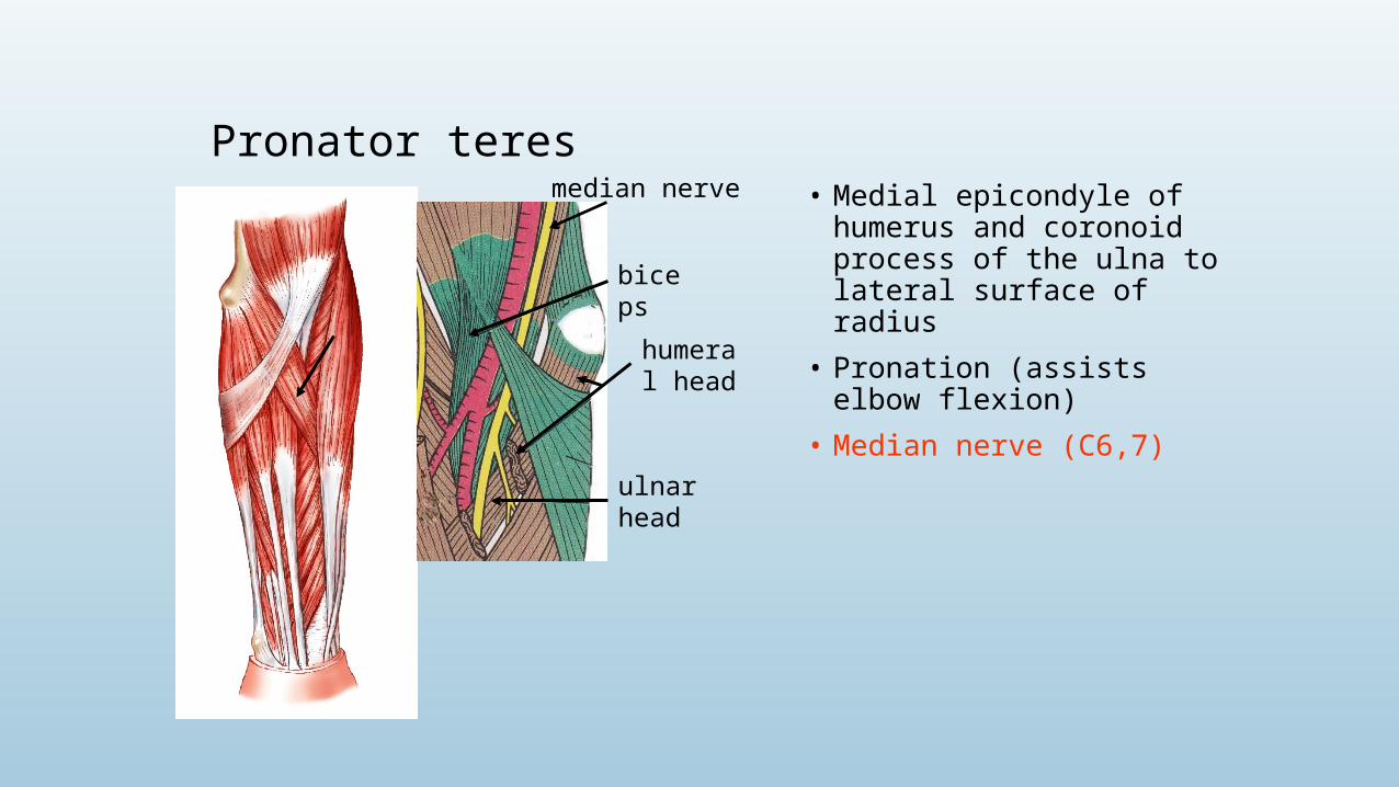
Pronator teres• Medial epicondyle of humerus and
coronoid process of the ulna to lateral surface of radius
• Pronation (assists elbow flexion)
• Median nerve (C6,7)
biceps
humeral head
ulnar head
median nerve
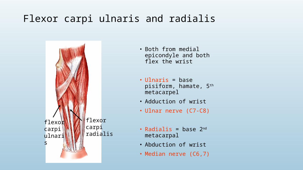
Flexor carpi ulnaris and radialis
• Both from medial epicondyle and both flex the wrist
• Ulnaris = base pisiform, hamate, 5th metacarpel
• Adduction of wrist
• Ulnar nerve (C7-C8)
• Radialis = base 2nd metacarpal
• Abduction of wrist
• Median nerve (C6,7)
flexor carpi ulnaris
flexor carpi radialis