Anatomy & Physiology I Lab BSC1085L Dr. Jason Schwartz BSC 2093 L.
65
Anatomy & Physiology I Lab BSC1085L Dr. Jason Dr. Jason Schwartz Schwartz BSC 2093 L
-
Upload
brice-west -
Category
Documents
-
view
217 -
download
0
Transcript of Anatomy & Physiology I Lab BSC1085L Dr. Jason Schwartz BSC 2093 L.

Anatomy & Physiology I LabBSC1085L
Dr. Jason SchwartzDr. Jason Schwartz
BSC 2093 L
Overview
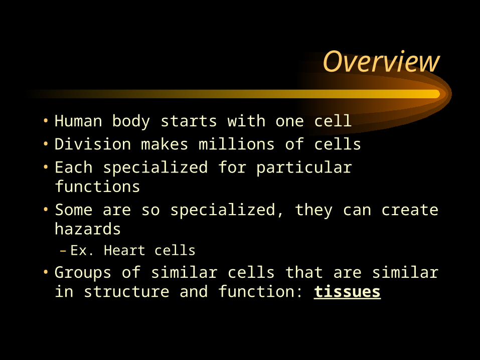
• Human body starts with one cell
• Division makes millions of cells
• Each specialized for particular functions
• Some are so specialized, they can create hazards– Ex. Heart cells
• Groups of similar cells that are similar in structure and function: tissues

•Histology = The study of tissues•Tissue = A collection of cells that perform related functions, and are similar in structure
•The Four Primary Tissue TypesEpithelialConnectiveMuscularNervous
Classification of Tissue
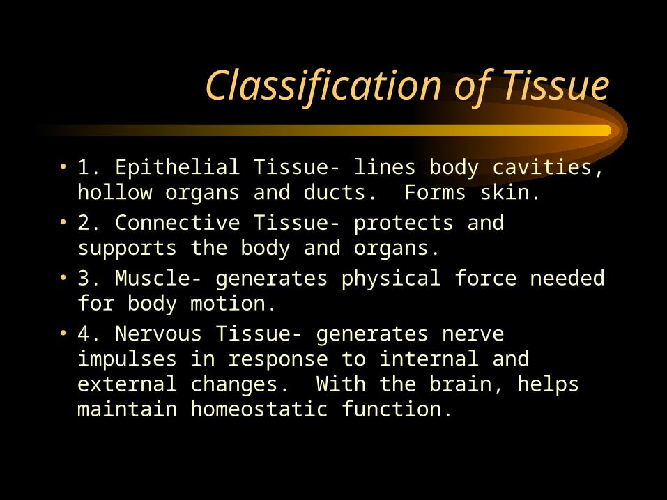
• 1. Epithelial Tissue- lines body cavities, hollow organs and ducts. Forms skin.
• 2. Connective Tissue- protects and supports the body and organs.
• 3. Muscle- generates physical force needed for body motion.
• 4. Nervous Tissue- generates nerve impulses in response to internal and external changes. With the brain, helps maintain homeostatic function.

Epithelial Tissue -- General Features• Cover surfaces, line cavities and form glands.• Attached to underlying connective tissue by a
basement membrane• Avascular---without blood vessels
– nutrients diffuse in from blood vessels in underlying connective tissue
– What does this mean for especially thick epithelia?
• Good nerve supply• Rapid cell division; responsive to environmental
stresses• Named according to the shape and arrangement of
cells
Special Characteristics
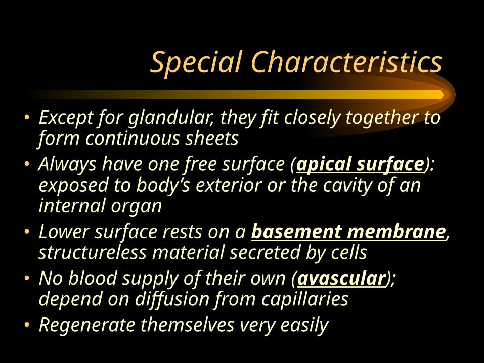
• Except for glandular, they fit closely together to form continuous sheets
• Always have one free surface (apical surface): exposed to body’s exterior or the cavity of an internal organ
• Lower surface rests on a basement membrane, structureless material secreted by cells
• No blood supply of their own (avascular); depend on diffusion from capillaries
• Regenerate themselves very easily
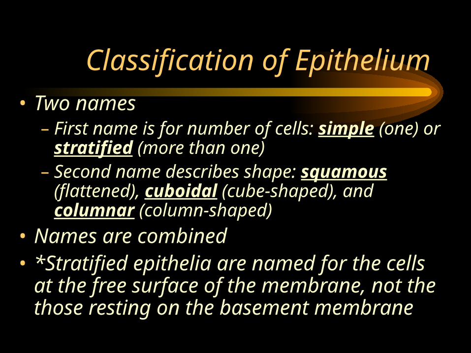
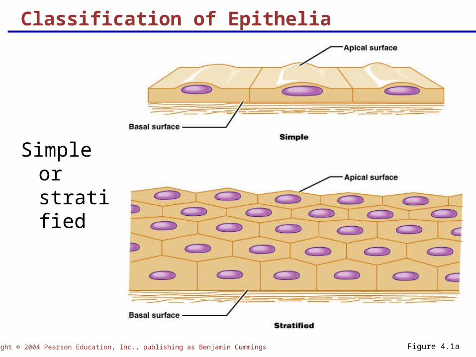
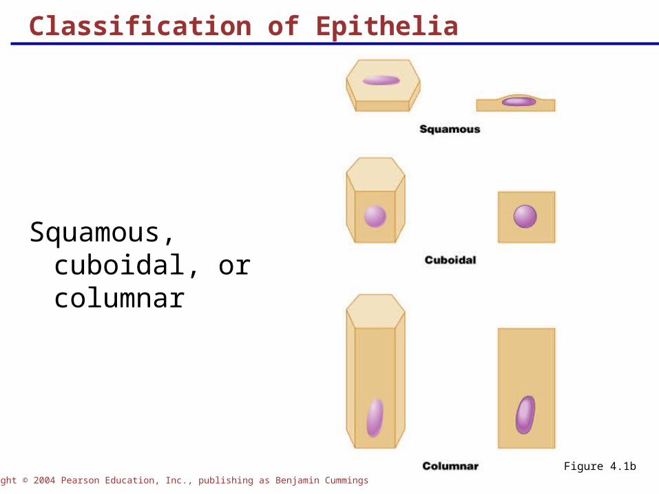
Classification of Epithelium• Two names
– First name is for number of cells: simple (one) or stratified (more than one)
– Second name describes shape: squamous (flattened), cuboidal (cube-shaped), and columnar (column-shaped)
• Names are combined• *Stratified epithelia are named for the cells at
the free surface of the membrane, not the those resting on the basement membrane
Copyright © 2004 Pearson Education, Inc., publishing as Benjamin Cummings
Classification of Epithelia
Simple or stratified
Figure 4.1a

Copyright © 2004 Pearson Education, Inc., publishing as Benjamin Cummings
Classification of Epithelia
Squamous, cuboidal, or columnar
Figure 4.1b
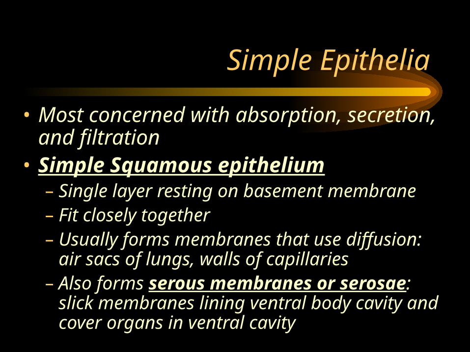
Simple Epithelia
• Most concerned with absorption, secretion, and filtration
• Simple Squamous epithelium– Single layer resting on basement membrane– Fit closely together– Usually forms membranes that use diffusion: air
sacs of lungs, walls of capillaries– Also forms serous membranes or serosae: slick
membranes lining ventral body cavity and cover organs in ventral cavity
Copyright © 2004 Pearson Education, Inc., publishing as Benjamin Cummings
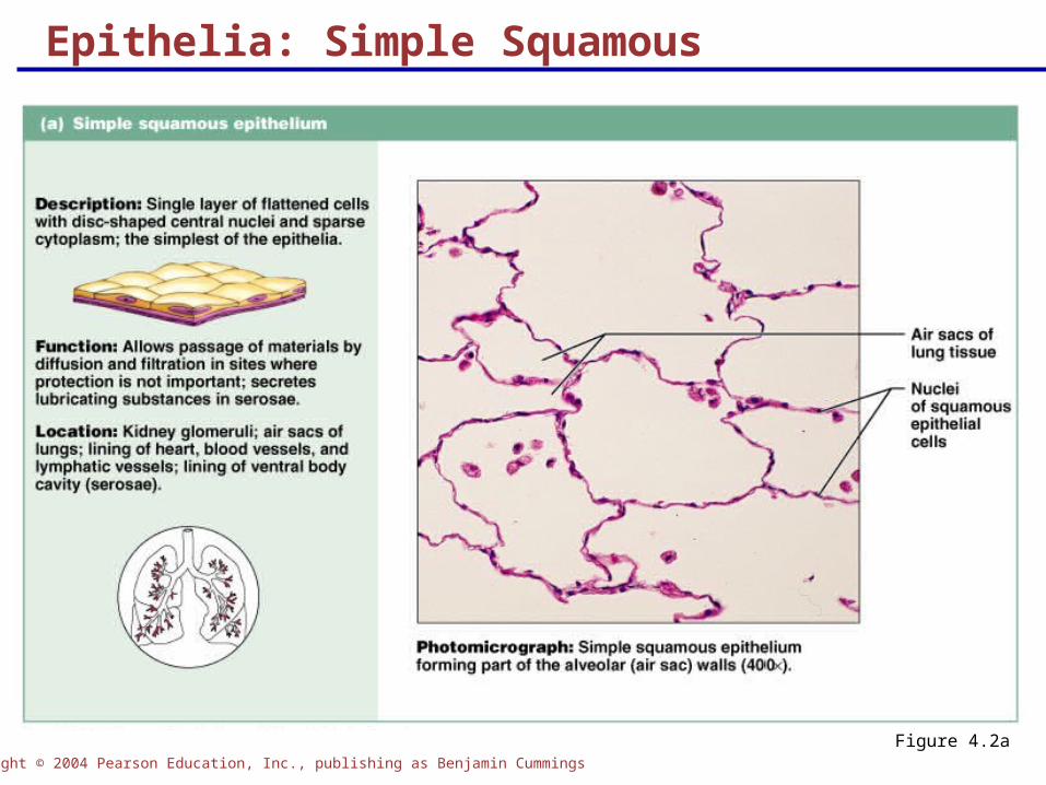
Epithelia: Simple Squamous
Figure 4.2a


Squamous Epithelium (surface view)
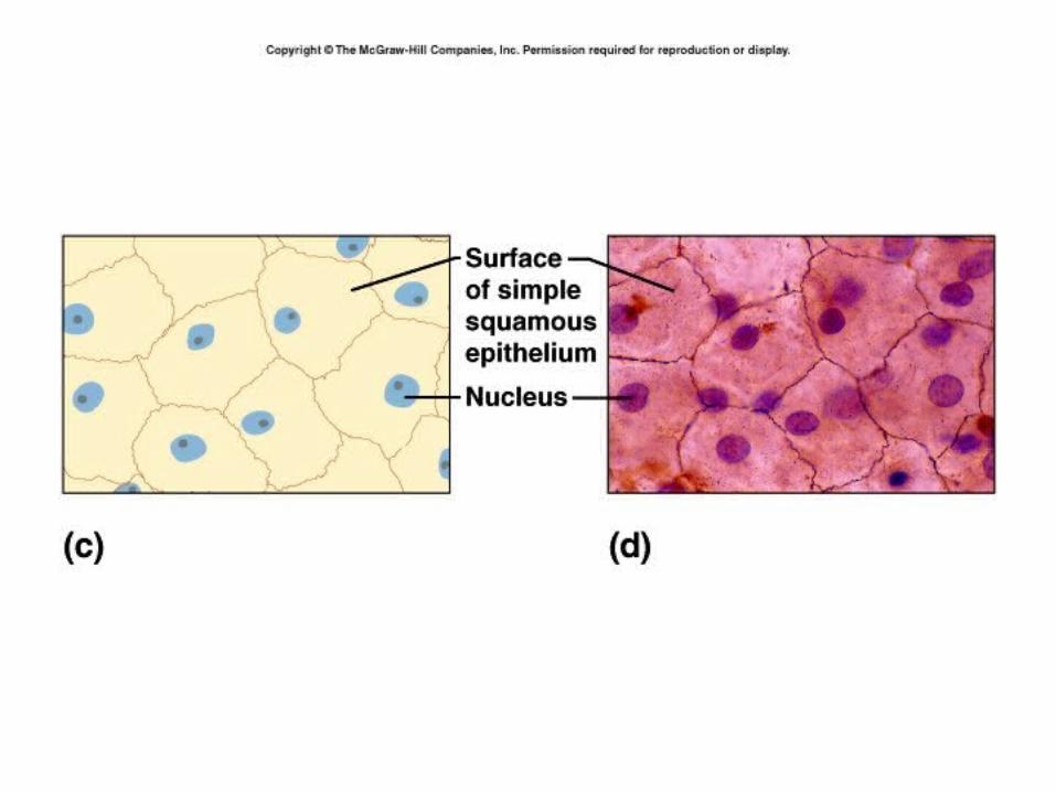

Simple Epithelial Tissue

Squamous Epithelium (surface view)
Copyright © 2004 Pearson Education, Inc., publishing as Benjamin Cummings
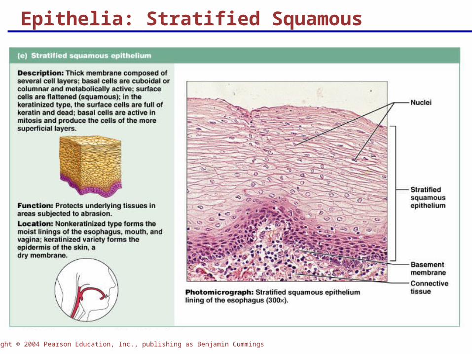
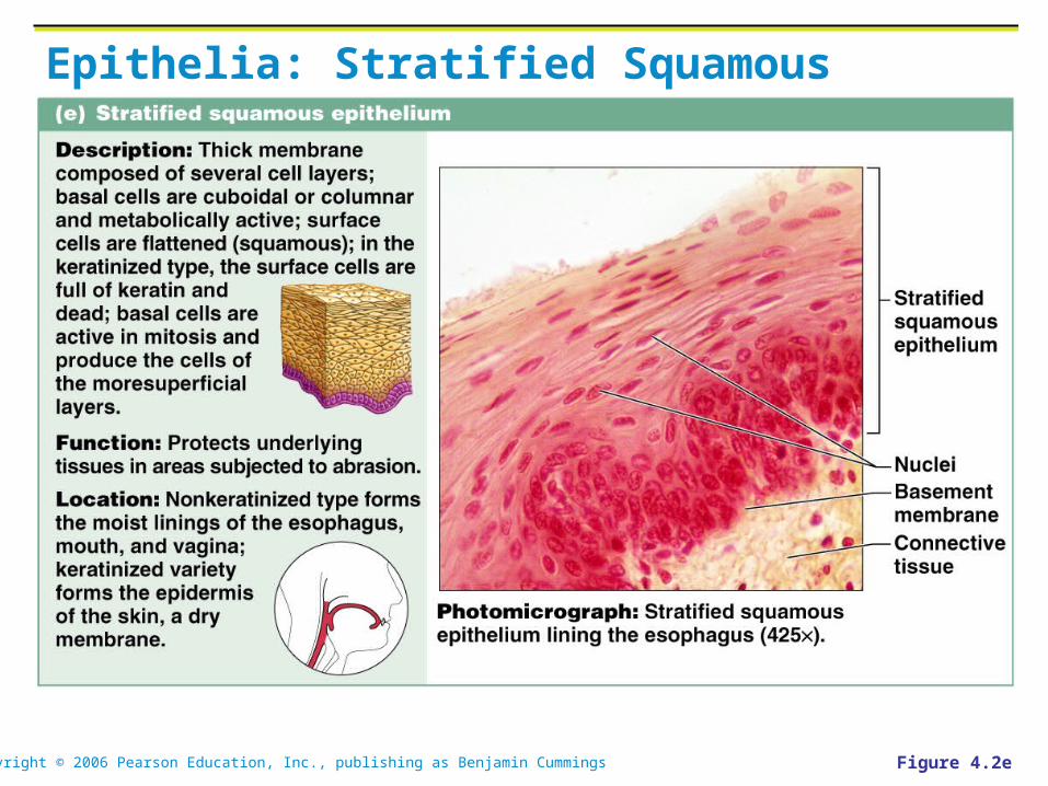
Epithelia: Stratified Squamous
Thick membrane composed of several layers of cells
Function in protection of underlying areas subjected to abrasion
Forms the external part of the skin’s epidermis (keratinized cells), and linings of the esophagus, mouth, and vagina (nonkeratinized cells)
Copyright © 2006 Pearson Education, Inc., publishing as Benjamin Cummings
Epithelia: Stratified Squamous
Figure 4.2e

Simple Cuboidal & Squamous Epithelium
Squamous
Cuboidal
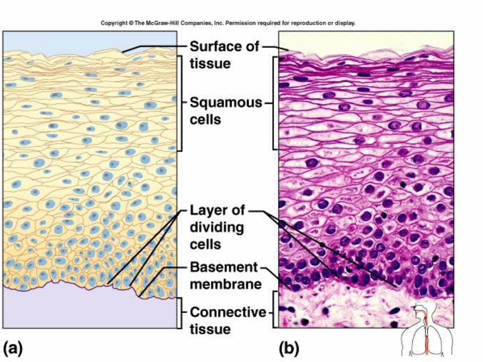

Fig. 5.9
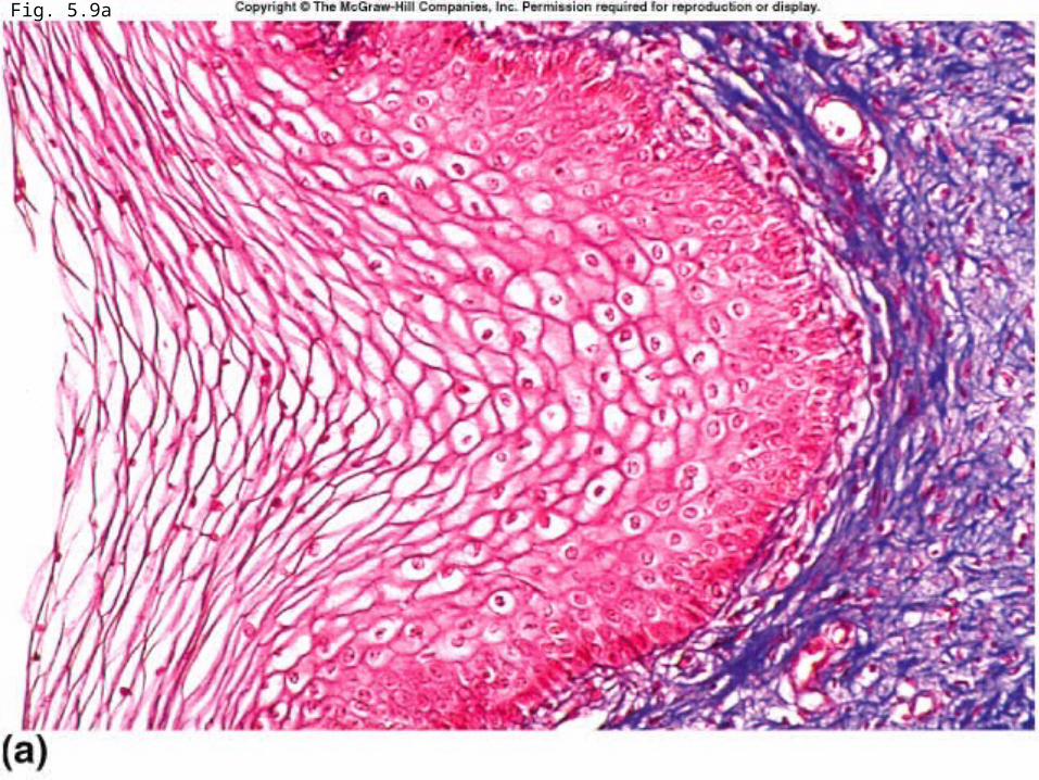
Fig. 5.9a
Copyright © 2006 Pearson Education, Inc., publishing as Benjamin Cummings
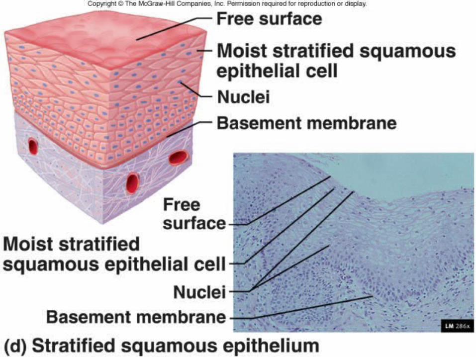
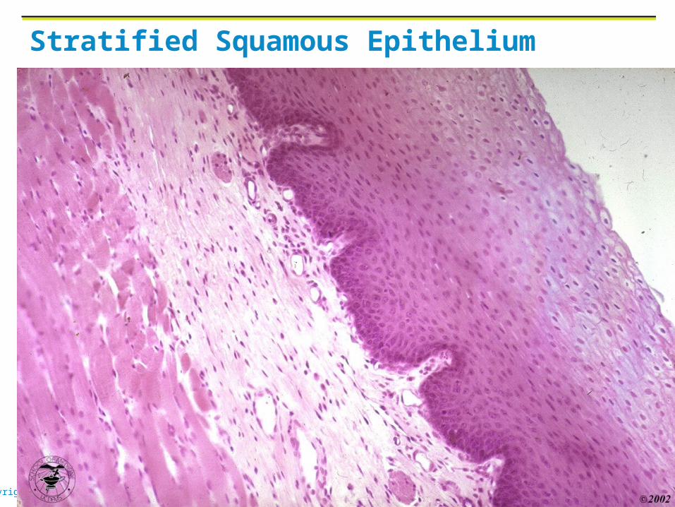
Stratified Squamous Epithelium
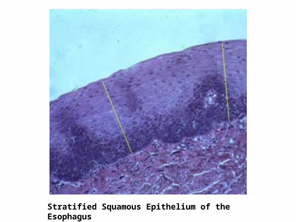
Stratified Squamous Epithelium of the Esophagus

Simple Epithelia contd.
• Simple Cuboidal Epi.– One layer resting on basement membrane– Common in glands and ducts: salivary glands and
pancreas– Forms walls of the kidney tubules, and covers
surface of the ovaries

Simple Cuboidal & Squamous Epithelium
Squamous
Cuboidal
Copyright © 2004 Pearson Education, Inc., publishing as Benjamin Cummings
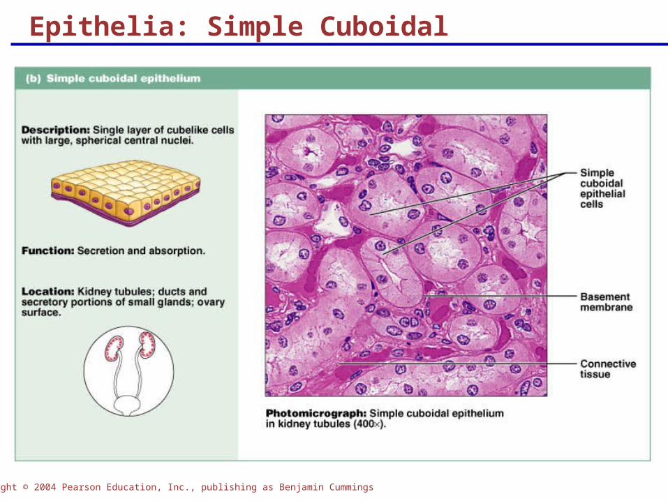
Epithelia: Simple Cuboidal
Single layer of cubelike cells with large, spherical central nuclei
Function in secretion and absorption
Present in kidney tubules, ducts and secretory portions of small glands, and ovary surface
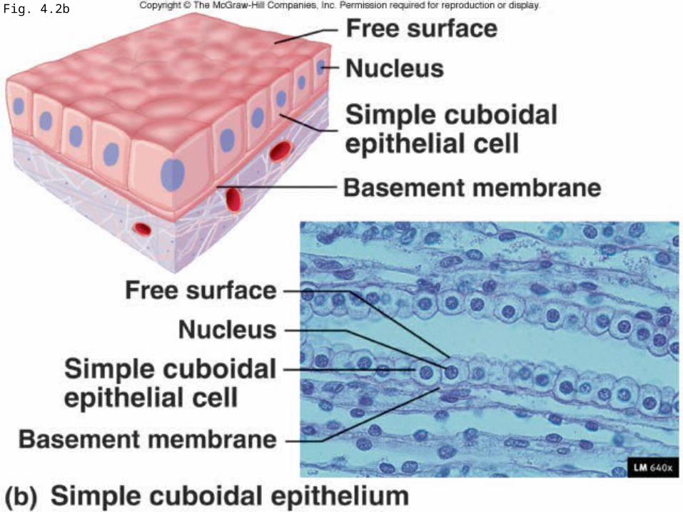
Fig. 4.2b
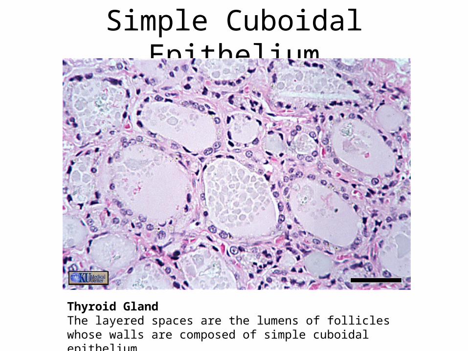
Simple Cuboidal Epithelium
Thyroid GlandThe layered spaces are the lumens of follicles whose walls are composed of simple cuboidal epithelium.

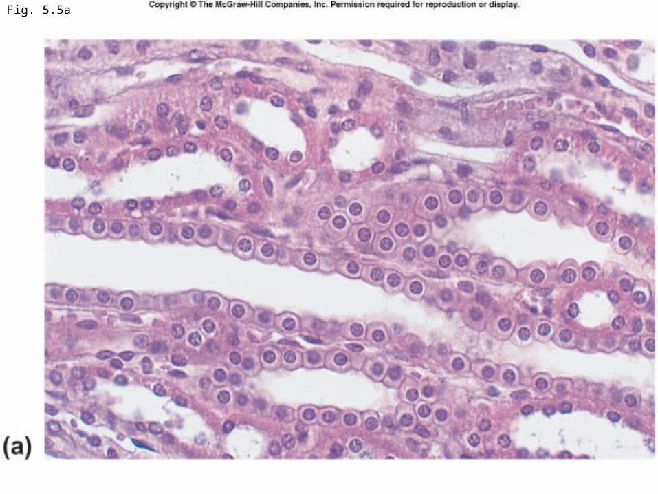
Fig. 5.5a

Fig. 5.5b
Simple Epithelia contd.
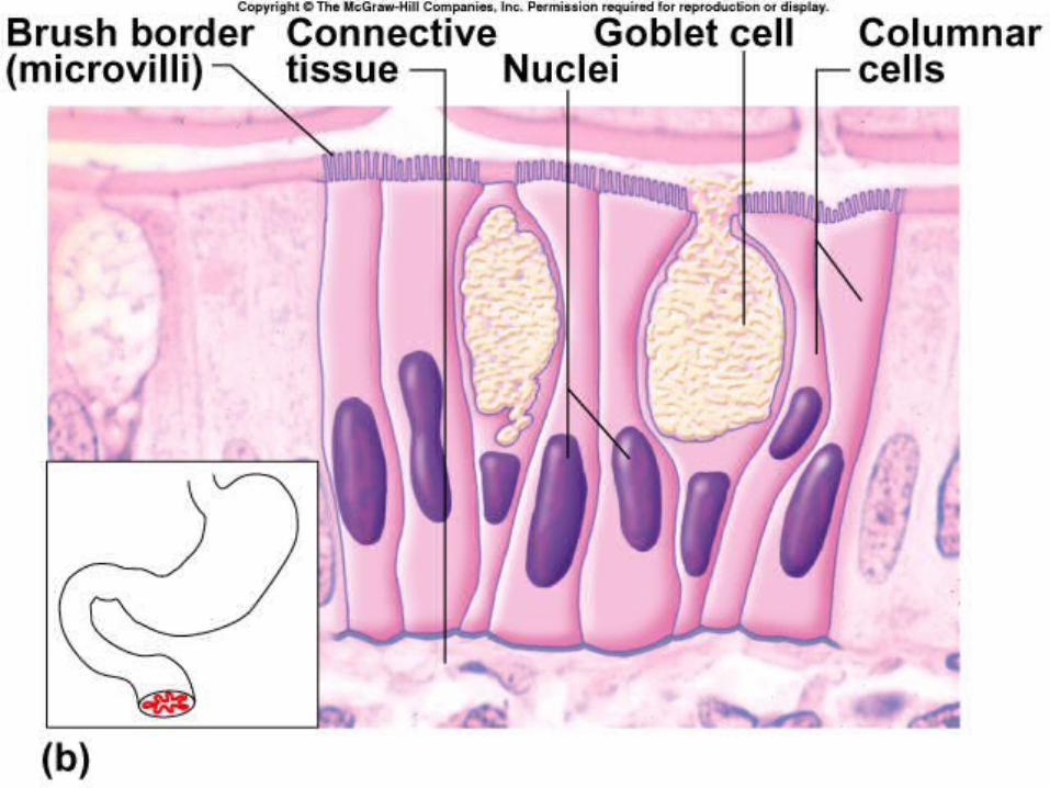
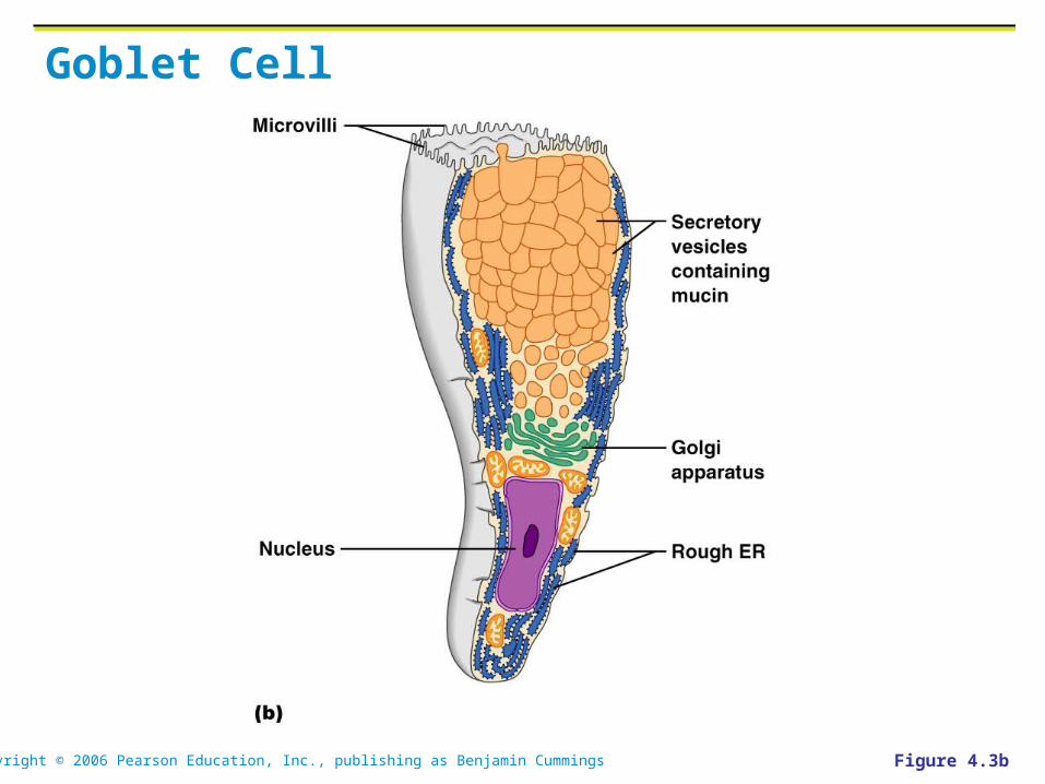
• Simple Columnar Epi.– One layer of tall cells– Fit close together– Goblet cells: produce a lubricating mucus, often seen in
this type of epithelium– Lines entire length of digestive tract from stomach to
anus
• *Epithelial membranes that line body cavities open to the exterior are called mucous membranes or mucosae
Copyright © 2006 Pearson Education, Inc., publishing as Benjamin Cummings
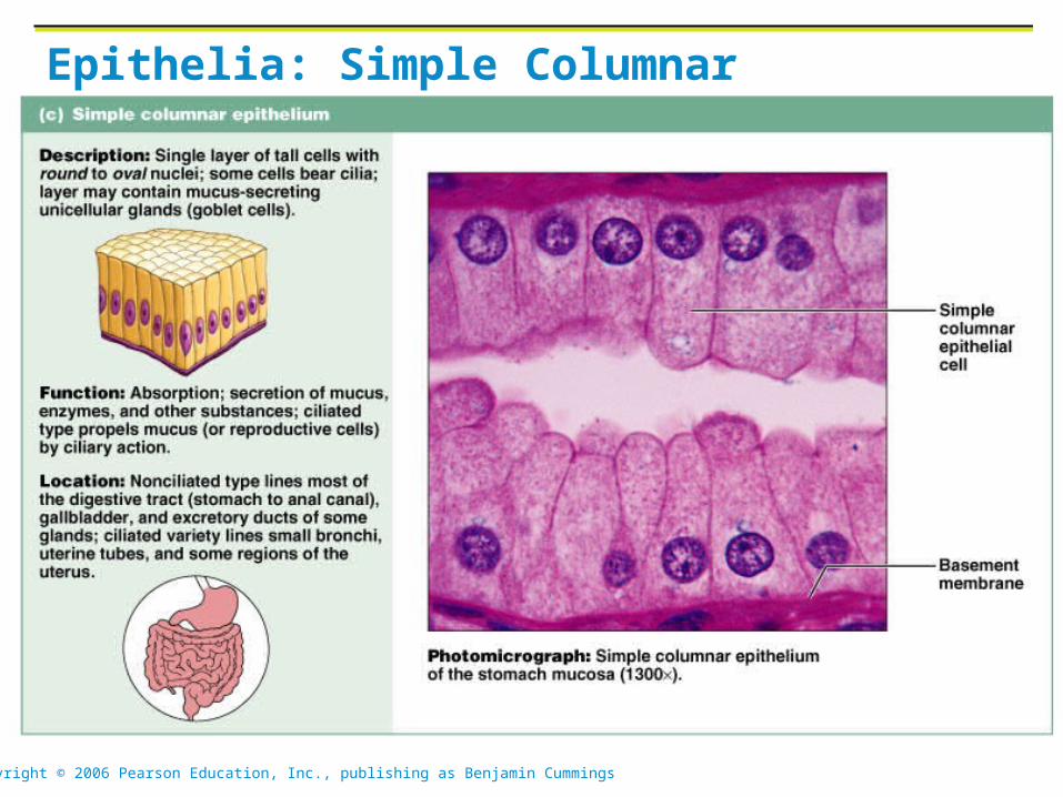
Epithelia: Simple Columnar
Copyright © 2004 Pearson Education, Inc., publishing as Benjamin Cummings

Fig. 4.2c
Copyright © 2006 Pearson Education, Inc., publishing as Benjamin Cummings
Goblet Cell
Figure 4.3b
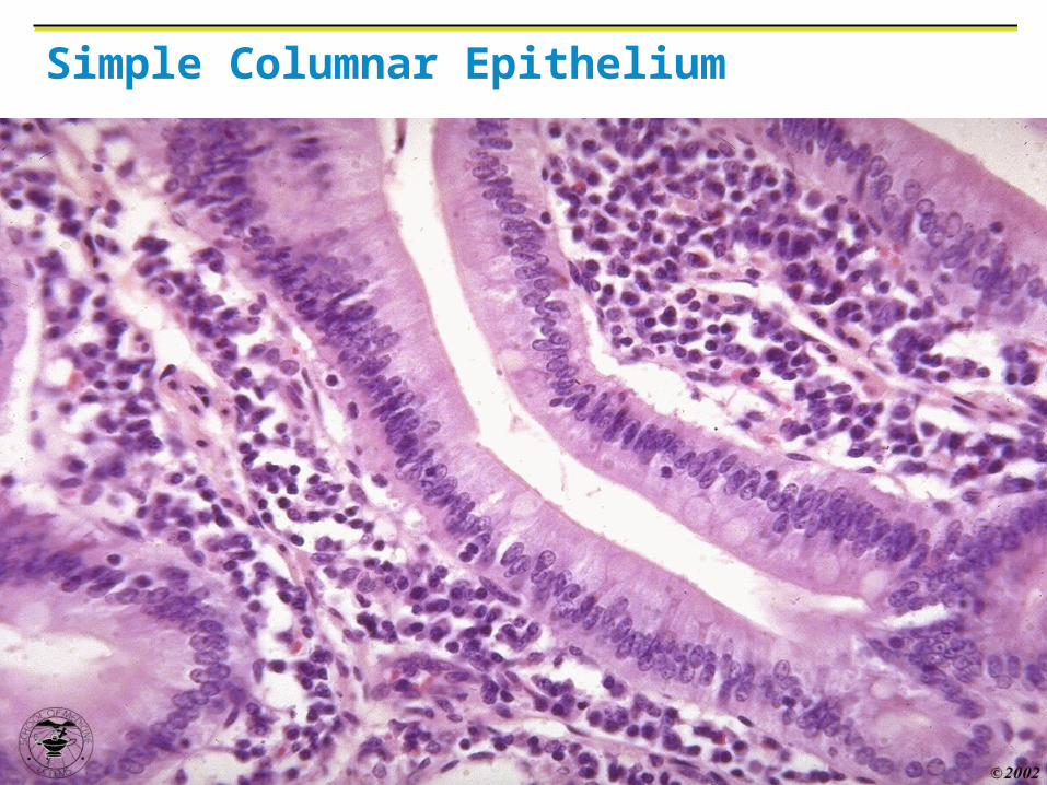
Simple Columnar Epithelium
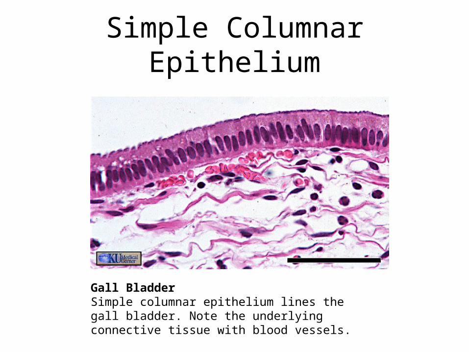
Gall BladderSimple columnar epithelium lines the gall bladder. Note the underlying connective tissue with blood vessels.
Copyright © 2006 Pearson Education, Inc., publishing as Benjamin Cummings
Simple Columnar Epithelium
Copyright © 2006 Pearson Education, Inc., publishing as Benjamin Cummings
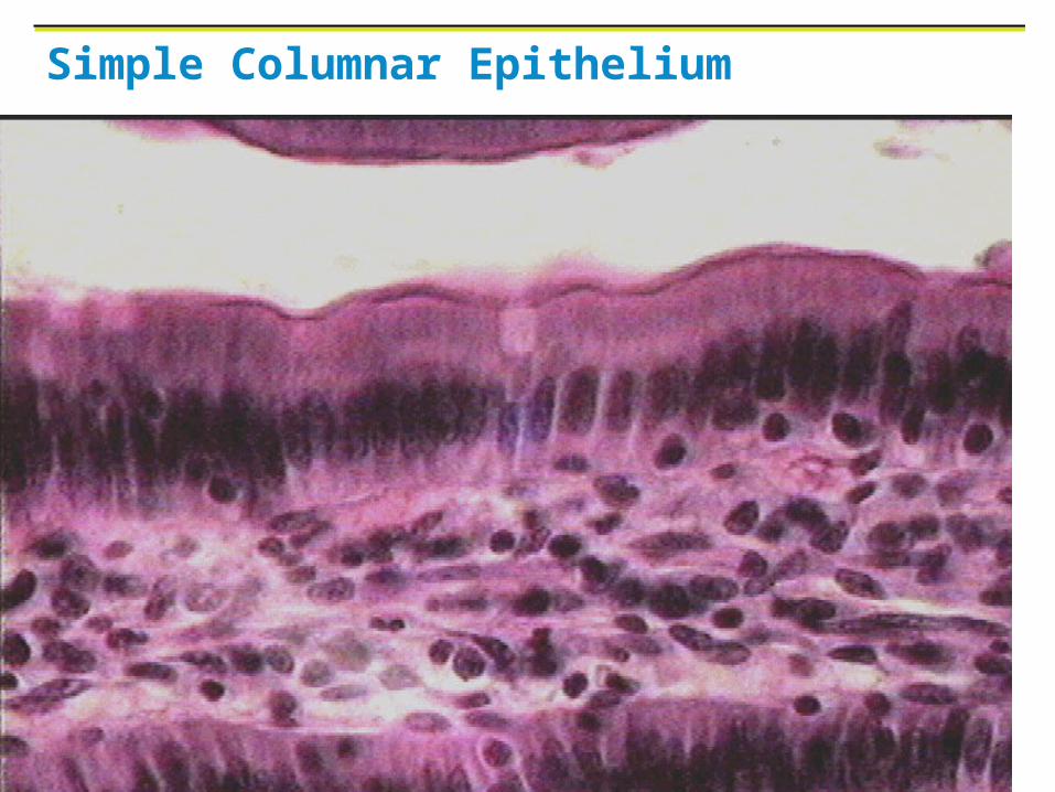
Simple Columnar Epithelium

Simple Epithelia contd.
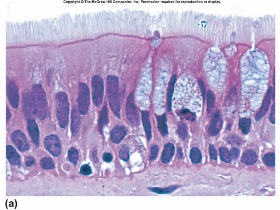
• Pseudostratified Columnar Epi.– Rest on basement membrane– Cells are different heights, and nuclei appear at
different heights above basement– Give false (pseudo) impression of stratified– Mainly absorption and secretion– Pseudostratified ciliated columnar epithelium
• Ciliated, lines most of respiratory tract
• Goblet cells produce mucus to trap dust and debris
Copyright © 2006 Pearson Education, Inc., publishing as Benjamin Cummings
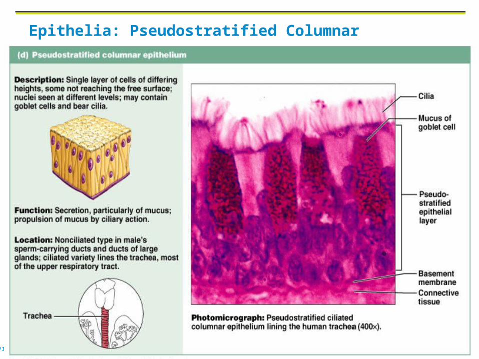
Epithelia: Pseudostratified Columnar
Single layer of cells with different heights; some do not reach the free surface
Nuclei are seen at different layers
Function in secretion and propulsion of mucus
Present in the male sperm-carrying ducts (nonciliated) and trachea (ciliated)
Stratified Epithelia
• Two or more cell layers• More durable than simple• Primarily for protection• Stratified Squamous Epithelium
– Most common stratified– Usually several layers– Free edge are squamous, closer to basement are
cuboidal or columnar– Found in “high friction” areas: esophagus, mouth,
other parts of skin
Stratified Epithelia
• Stratified Cuboidal and Columnar Epithelia– Usually just two cell layers with (at least) the
surface cells being cuboidal– Surface cells of stratified columnar are columnar,
but its basal cells vary in size and shape– Both are fairly rare– Found mainly in the ducts or large glands
Stratified Epithelia
• Transitional Epithelium– Highly modified– Stratified squamous epithelium– Forms lining of only a few organs: bladder, the ureters,
and part of the urethra– All part of the urinary system and undergo considerable
stretching– Basal layer are cuboidal or columnar; those at the free
surface vary in appearance– Stretching changes shape, cells can flatten and become
squamous-like

Fig. 4.2g


Simple Columnar Epithelium
Gall BladderSimple columnar epithelium lines the gall bladder. Note the underlying connective tissue with blood vessels.

Simple Columnar Epithelium
Gall BladderSimple columnar epithelium lines the gall bladder. Note the underlying connective tissue with blood vessels.
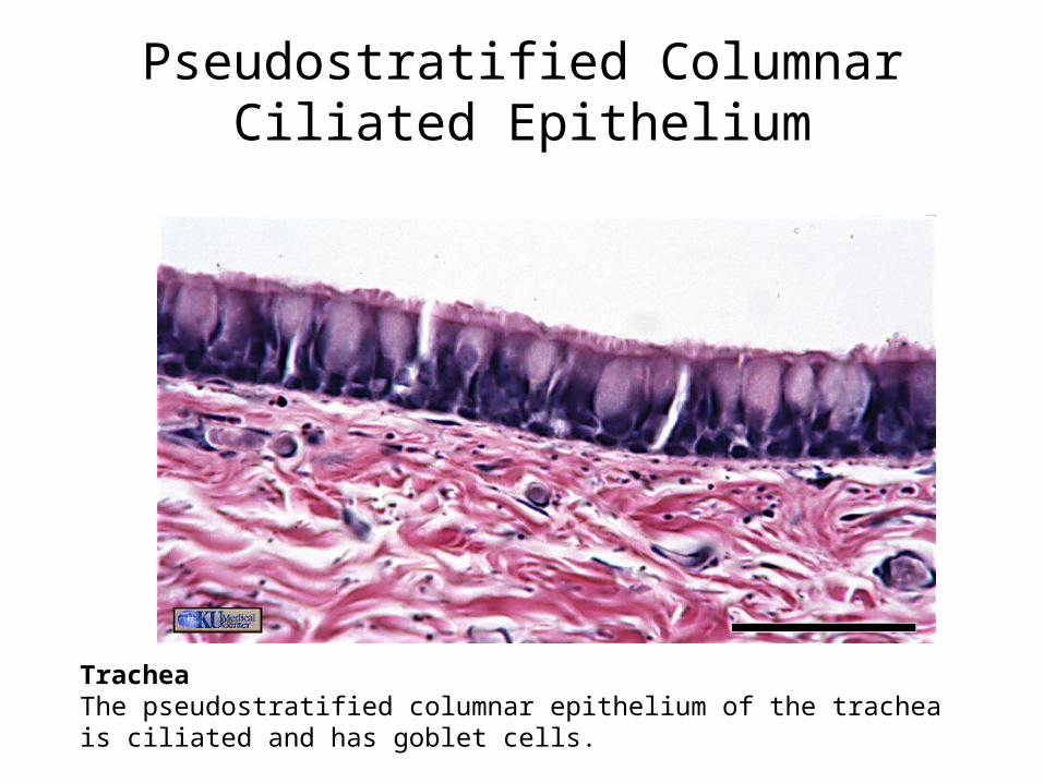
Pseudostratified Columnar Ciliated Epithelium
TracheaThe pseudostratified columnar epithelium of the trachea is ciliated and has goblet cells.

Pseudostratified Columnar Ciliated Epithelium
Copyright © 2006 Pearson Education, Inc., publishing as Benjamin Cummings
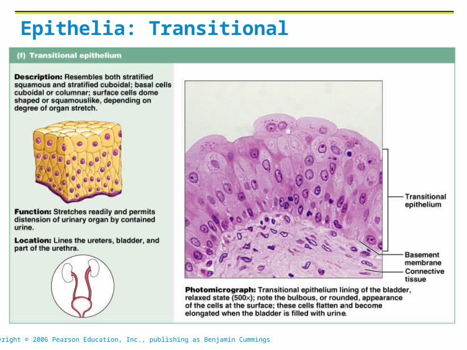
Epithelia: Transitional
Several cell layers, basal cells are cuboidal, surface cells are dome shaped
Stretches to permit the distension of the urinary bladder
Lines the urinary bladder, ureters, and part of the urethra

Copyright © 2004 Pearson Education, Inc., publishing as Benjamin Cummings
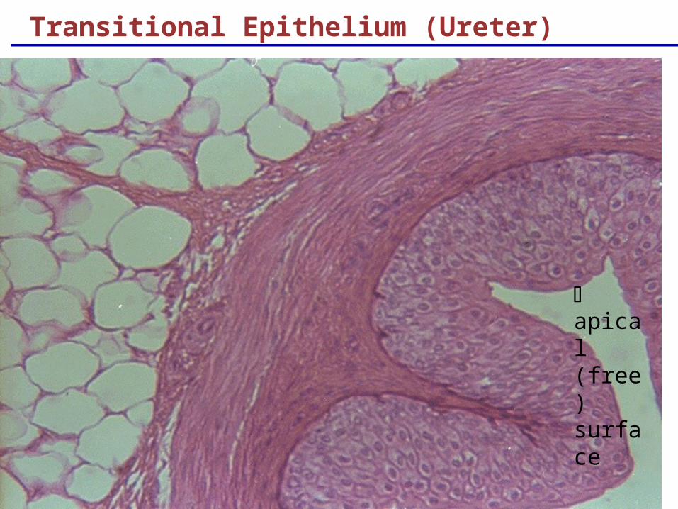
Transitional Epithelium (Ureter)
apical (free) surface
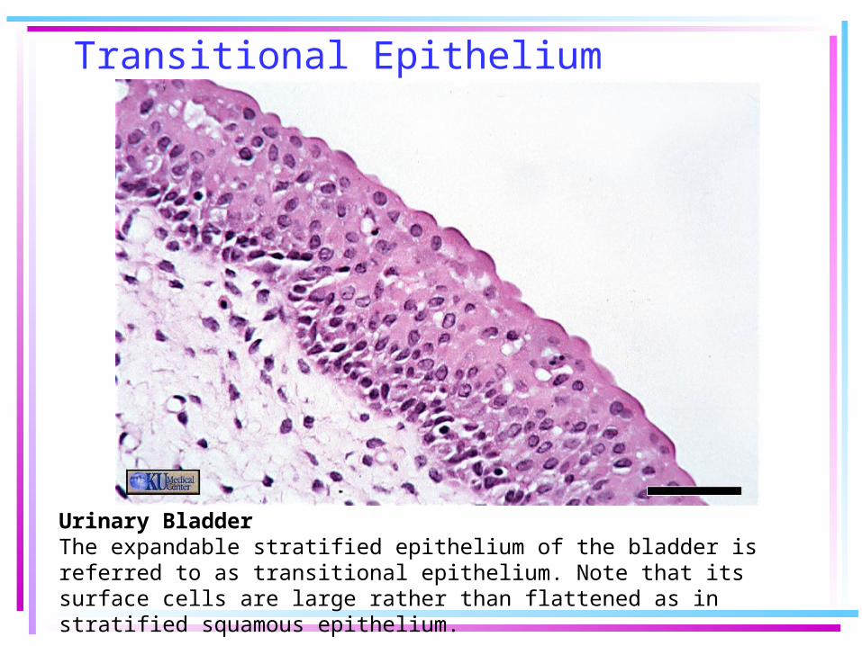
Transitional Epithelium
Urinary BladderThe expandable stratified epithelium of the bladder is referred to as transitional epithelium. Note that its surface cells are large rather than flattened as in stratified squamous epithelium.
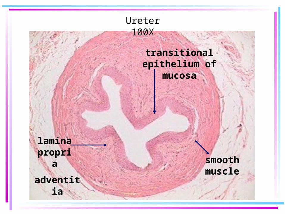
Ureter 100X
transitional epithelium of mucosa
smooth muscle
adventitia
lamina propria

* Ureter 400X
lamina propria
transitional epithelium
smooth muscle
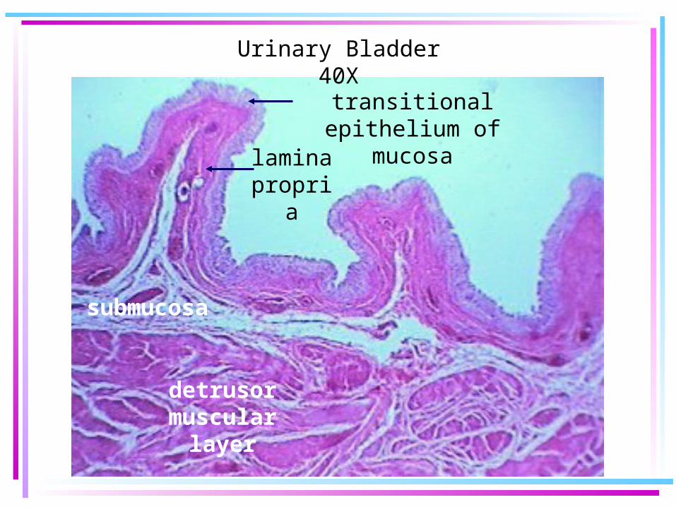
Urinary Bladder 40X
transitional epithelium of mucosa
detrusor muscular layer
lamina propria
submucosa
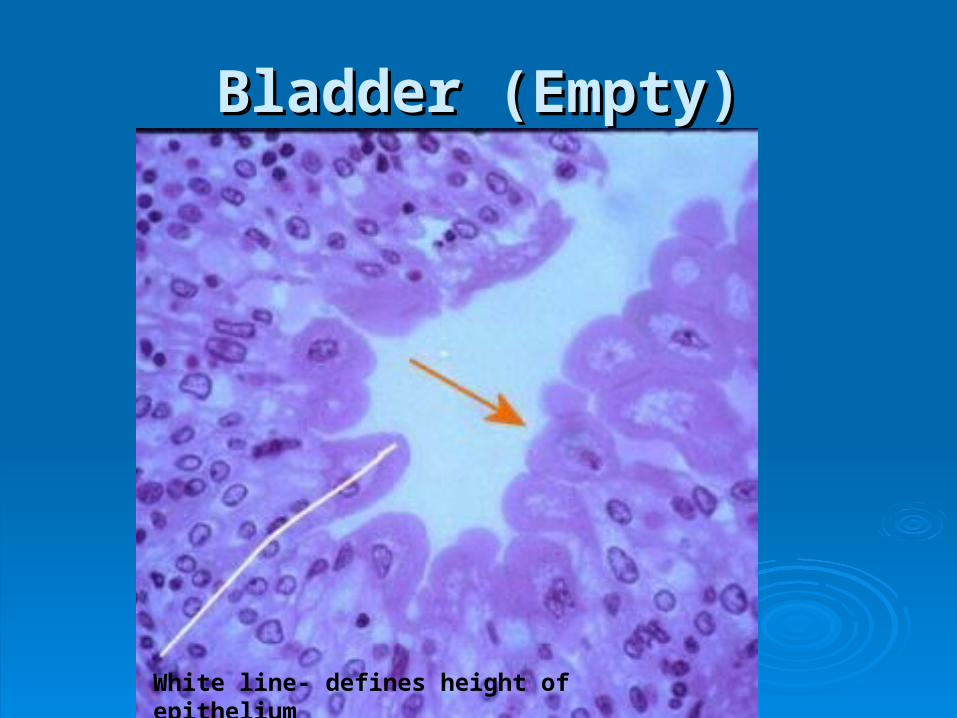
Bladder (Empty)Bladder (Empty)
White line- defines height of epithelium
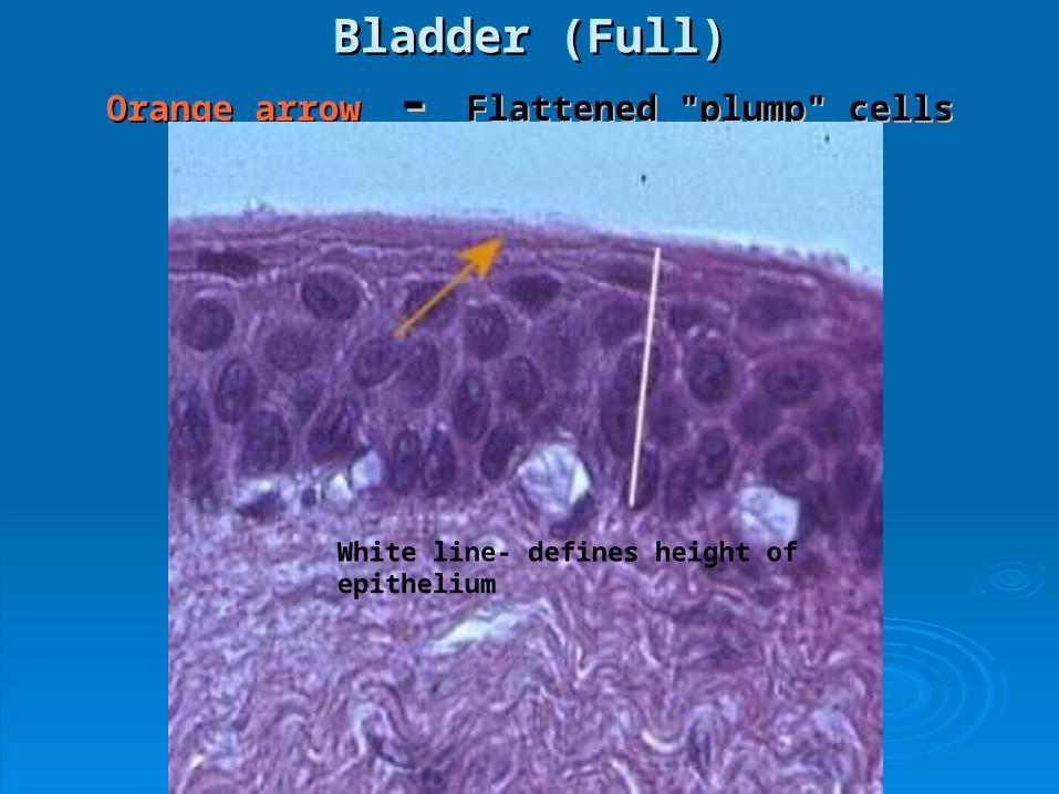
Bladder (Full)Bladder (Full)
Orange arrowOrange arrow - - Flattened "plump" cellsFlattened "plump" cells
White line- defines height of epithelium
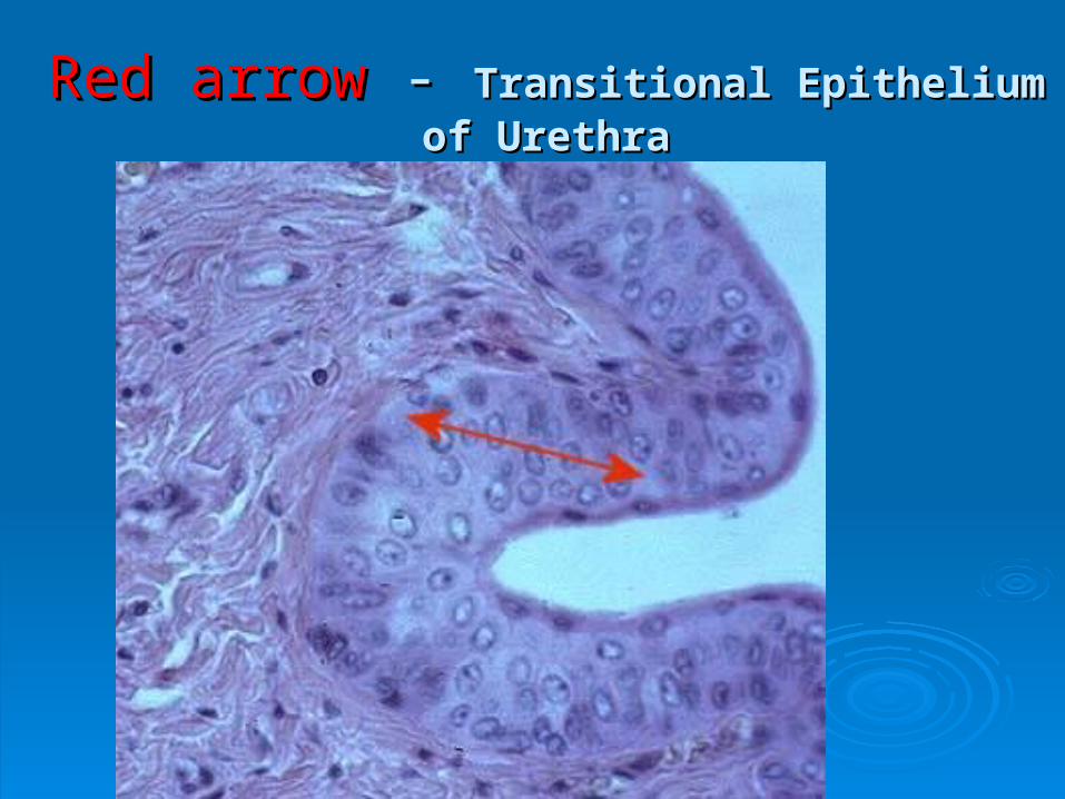
Red arrowRed arrow - - Transitional Epithelium of UrethraTransitional Epithelium of Urethra