Anatomy of the Middle Cerebral Artery: The Temporal...
8
Anatomy of the Middle Cerebral Artery: The Temporal Branches BY W. BRADFORD DeLONG, M.D., F.A.C.S. Abstract: Anatomy of the Middle Cerebral Artery: The Temporal Branches • Nineteen out of 23 middle cerebral arterial specimens had as the first major branch of the middle cerebral artery a sizable anterior temporal artery; a trunk forming the anterior and middle temporal branches; a trunk forming the anterior, middle, and posterior temporal arteries; or a trunk forming temporal and angular arterial branches. Patients harboring middle cerebral stenoses or occlusions who have correlating cerebral ischemic symptoms may be considered as candidates for microsurgical cerebral revascularization. However, if such patients undergo intracranial surgery, the superficial temporal artery should probably be joined to a supra-Sylvian arterial branch, rather than to a temporal arterial branch, in order to avoid delivering the new blood supply proximal to the stenotic or occluded segment. Additional Key Words cerebral atherosclerosis cerebral ischemia cerebral embolism and thrombosis microsurgical cerebral revascularization temporal lobe Introduction • Ring and Waddington 1 - 3 have described the terminal configuration of the middle cerebral artery, as well as the branching pattern of the artery within the Sylvian fissure. The configuration of the lenticulostriate arteries has been described by many authors, including Kaplan, 4 Stephens and Stilwell, 5 and Jain. 6 Foix and Levy 7 described an anterior temporal branch coursing from the middle cerebral artery near its origin, and Vander Eecken 8 mentions an anterior temporal artery as well as an inconstant temporopolar artery. The origins of the temporopo- lar artery and the anterior temporal artery have been illustrated by Stephens and Stilwell 9 in their meticulous photographic study of the cerebral vasculature, and the angiographical anatomy of the temporopolar branches has been illustrated by Dahlstrom et al. 10 However, descriptions of the middle cerebral temporal branches which we found in the literature did not correlate with our preliminary observations of these arterial branches. We undertook this study to define further the arterial anatomy of the temporal lobe, hoping to find immediate application of our From the Cardiovascular Research Laboratory, St. Joseph's Hospital, and the Department of Neurosurgery, University of California, San Francisco, California. Reprint requests to Dr. DeLong, 4141 Geary Boule- vard, San Francisco, California 94118. Presented at the Microneurosurgery Symposium, Good Samaritan Hospital, Cincinnati, Ohio, June 8-10, 1972. This study was funded in part by the St. Joseph's Hospital Research Foundation, San Francisco, California. 412 findings in the field of microsurgical cerebral revascularization. Methods Twelve human brain were injected, fixed, and dissected. Twenty-three middle cerebral arterial specimens were obtained. (One specimen was inadvertently discarded after only one middle cerebral artery had been dissected.) Both middle cerebral arteries of each fresh brain were injected with an acrylic compound after the brain had been removed from the cranial cavity.* The brains were then fixed in formalin. Each middle cerebral arterial complex was then dissected and mapped from the origin to the terminal branches over the convolu- tions. We used the arterial nomenclature of Ring and Waddington, and attempted to define grossly the central sulcus, in order to relate the arterial branches to this convolutional landmark. The 23 middle cerebral artery specimens fell into nine groups, oriented to the anatomical configuration of the arterial supply of the temporal lobe. Obviously, the grouping of the specimens would have been different if they had been oriented to the configuration of the central sulcus arteries, the angular artery, or some other branch of the middle cerebral arterial complex. Results In 19 out of the 23 middle cerebral arterial specimens, the first major branch of the middle cerebral arterial complex was either an artery supplying the anterior temporal lobe or a large trunk *Batson's #17 Anatomical Corrosion Compound, Polysci- ences, Inc., Paul Valley Industrial Park, Warrington, Pennsylvania, 18976. Stroke, Vol. 4. May-June 1973 by guest on May 18, 2018 http://stroke.ahajournals.org/ Downloaded from
Transcript of Anatomy of the Middle Cerebral Artery: The Temporal...

Anatomy of the Middle Cerebral Artery:
The Temporal BranchesBY W. BRADFORD DeLONG, M.D., F.A.C.S.
Abstract:Anatomy ofthe MiddleCerebral Artery:The TemporalBranches
• Nineteen out of 23 middle cerebral arterial specimens had as the first major branch ofthe middle cerebral artery a sizable anterior temporal artery; a trunk forming theanterior and middle temporal branches; a trunk forming the anterior, middle, andposterior temporal arteries; or a trunk forming temporal and angular arterial branches.
Patients harboring middle cerebral stenoses or occlusions who have correlatingcerebral ischemic symptoms may be considered as candidates for microsurgical cerebralrevascularization. However, if such patients undergo intracranial surgery, the superficialtemporal artery should probably be joined to a supra-Sylvian arterial branch, rather thanto a temporal arterial branch, in order to avoid delivering the new blood supply proximalto the stenotic or occluded segment.
Additional Key Words cerebral atherosclerosis cerebral ischemiacerebral embolism and thrombosis microsurgical cerebral revascularizationtemporal lobe
Introduction• Ring and Waddington1-3 have described theterminal configuration of the middle cerebral artery,as well as the branching pattern of the artery withinthe Sylvian fissure. The configuration of thelenticulostriate arteries has been described by manyauthors, including Kaplan,4 Stephens and Stilwell,5
and Jain.6 Foix and Levy7 described an anteriortemporal branch coursing from the middle cerebralartery near its origin, and Vander Eecken8 mentionsan anterior temporal artery as well as an inconstanttemporopolar artery. The origins of the temporopo-lar artery and the anterior temporal artery have beenillustrated by Stephens and Stilwell9 in theirmeticulous photographic study of the cerebralvasculature, and the angiographical anatomy of thetemporopolar branches has been illustrated byDahlstrom et al.10
However, descriptions of the middle cerebraltemporal branches which we found in the literaturedid not correlate with our preliminary observationsof these arterial branches. We undertook this studyto define further the arterial anatomy of the temporallobe, hoping to find immediate application of our
From the Cardiovascular Research Laboratory, St. Joseph'sHospital, and the Department of Neurosurgery, Universityof California, San Francisco, California.
Reprint requests to Dr. DeLong, 4141 Geary Boule-vard, San Francisco, California 94118.
Presented at the Microneurosurgery Symposium, GoodSamaritan Hospital, Cincinnati, Ohio, June 8-10, 1972.
This study was funded in part by the St. Joseph'sHospital Research Foundation, San Francisco, California.
412
findings in the field of microsurgical cerebralrevascularization.
MethodsTwelve human brain were injected, fixed, and dissected.Twenty-three middle cerebral arterial specimens wereobtained. (One specimen was inadvertently discardedafter only one middle cerebral artery had beendissected.) Both middle cerebral arteries of each freshbrain were injected with an acrylic compound after thebrain had been removed from the cranial cavity.* Thebrains were then fixed in formalin. Each middle cerebralarterial complex was then dissected and mapped fromthe origin to the terminal branches over the convolu-tions. We used the arterial nomenclature of Ring andWaddington, and attempted to define grossly the centralsulcus, in order to relate the arterial branches to thisconvolutional landmark.
The 23 middle cerebral artery specimens fell intonine groups, oriented to the anatomical configuration ofthe arterial supply of the temporal lobe. Obviously, thegrouping of the specimens would have been different ifthey had been oriented to the configuration of thecentral sulcus arteries, the angular artery, or some otherbranch of the middle cerebral arterial complex.
ResultsIn 19 out of the 23 middle cerebral arterialspecimens, the first major branch of the middlecerebral arterial complex was either an arterysupplying the anterior temporal lobe or a large trunk
*Batson's #17 Anatomical Corrosion Compound, Polysci-ences, Inc., Paul Valley Industrial Park, Warrington,Pennsylvania, 18976.
Stroke, Vol. 4. May-June 1973
by guest on May 18, 2018
http://stroke.ahajournals.org/D
ownloaded from

MIDDLE CEREBRAL ARTERY
FIGURE 1
The first large branch of the middle cerebral artery (MCA) is a large arterial trunk which supplies the entire temporal lobeby forming the temporopolar artery (TPA), anterior temporal artery (ATA), middle temporal artery (MTA), and posteriortemporal artery (PTA). The lenticulostriate arteries (LSA) arise from the main middle cerebral trunk.
which divided into several temporal arterialbranches. In some cases this large trunk terminatedas the anterior and middle temporal arteries; in othercases it continued posteriorly to terminate as the
posterior temporal artery or the angular artery. Thisbranch or trunk arose from the middle cerebralartery proximal or opposite to the lenticulostriatearteries in 12 of the 19 cases, and distal to the
TEMPORAL LOBE ARTERIAL ZONES
ATA MTA
FIGURE 2
Temporal lobe arterial zones, determined by the distance from the tip of the temporal lobe at which the temporal arterialbranches emerged from the Sylvian fissure.
Stroke, Vol. 4, May-June 1973 413
by guest on May 18, 2018
http://stroke.ahajournals.org/D
ownloaded from

DeLONG
OPF
ARTERIAL ZONES
CSA
PPA
OBF
ANG
TPAATA MTA
PTA
FIGURE 3
Arterial zones of the middle cerebral artery. OBF: orbitofrontal arterial complex; OPF: operculofrontal arterial complex;CSA: central sulcus arteries; PPA: posterior parietal artery; and ANG: angular artery.
lenticulostriate arteries in seven cases. After arisingfrom the middle cerebral artery, this arterial channelcoursed over the superior surface of the temporal
PPA
ACA
TPAVMTA
Group I. Seven specimens. First major MCA branch formedATA, MTA and PTA. ACA: anterior cerebral artery; ACH:anterior choroidal artery; ACM: anterior communicatingartery; AHB: recurrent artery of Heubner; and PCM: pos-terior communicating artery.
pole and anterior temporal lobe. Individual branchesof this artery then emerged from the Sylvian fissureand ran posteroinferiorly over the superior temporalgyrus to supply the anterior, middle, and posterioraspects of the temporal lobe (fig. 1).
The temporopolar artery arose either as aseparate branch from the middle cerebral artery or
OPF
TPAPTA
SATA MTA
FIGURE 5
Group II. Five specimens. First major MCA branch formedATA, MTA, PTA, and ANG.
414 Stroke, Vol. 4, May-June 1973
by guest on May 18, 2018
http://stroke.ahajournals.org/D
ownloaded from

MIDDLE CEREBRAL ARTERY
as a branch of the temporal trunk, and traveled inthe pia-arachnoid anteroinferiorly over the anteriorand medial aspects of the temporal pole.
The course of the angular artery as it emergedfrom the Sylvian fissure was defined approximatelyby a line extended horizontally from the posteriortermination of the Sylvian fissure. In the intacthuman, this line is approximately parallel to theanthropological baseline, mentioned by Taveras andWood.11
During the arterial dissections, the arterialbranches emerging from the Sylvian fissure werearbitrarily grouped into anterior, middle, or posteri-or temporal branches. In some specimens, thedistance between the emerging arterial branch andthe tip of the temporal pole was measured.
Figure 2 illustrates that the arterial branchestended to fall into three groups defined by thedistance between the temporal pole and the point atwhich they emerged from the Sylvian fissure. Thesemeasurements formed the basis for the arterial zonesof the temporal lobe illustrated in figure 3. Figure 3also illustrates the other middle cerebral arterialzones of the hemisphere, adapted from Ring andWaddington.
Figures 4 through 12 illustrate the nine groupsinto which these specimens could be classified. Onespecimen from each group is illustrated, oriented forclarity as though it were the left middle cerebralarterial complex. Such right-left inversion in the caseof some specimens may represent an anatomicaloversimplification, since the studies of LeMay andCulebras12 and of Geschwind and Levitsky13 have
PPA
ANG
TPA'PTA
FIGURE 6
Croup III. Three specimens. First major MCA branch waslarge ATA; next large trunk formed MTA, PTA, and ANG.
Stroke, Vol. A, May-June 1973
PPA
TPAATA* V\MTA
FIGURE 7
Group IV. Two specimens. First major MCA branch formedOBF and OFF; next branch was large ATA, then largebranch forming MTA and PTA.
indicated that there are specific arterial and corticaldifferences between the dominant and the nondomi-nant cerebral hemispheres.
The total number of middle cerebral arteriesdissected (23) is small and doubtless a number ofother variations would have become evident if morespecimens had been included in this series. In 14specimens the first major branch of the middlecerebral artery was an anterior temporal-middletemporal-posterior temporal trunk, a temporal-angular trunk, or an anterior temporal-middletemporal trunk (Groups I, II, VI, and VII) .
FIGURE 8
Group V. Two specimens. First major MCA branch formedOBF and OPF; next large trunk formed ATA, MTA, PTA,and ANG.
415
by guest on May 18, 2018
http://stroke.ahajournals.org/D
ownloaded from

DcLONG
OPF
ANG
MTA
FIGURE 9
Croup VI. One specimen. First major MCA branch formedATA and MTA. MCA terminated as ANG, PTA, and PPA.
PPA
ANG
PTAMTA
FIGURE 10
Group VII. One specimen. First major MCA branch formedATA and MTA. ANG and PTA arose from MCA beforeits termination as PPA and CSA.
In three specimens, the first major branch wasthe anterior temporal artery which was followedimmediately by a middle temporal-posterior tempo-ral-angular trunk (Group III). In two specimens thefirst major branch was the anterior temporal artery,with the middle and posterior temporal branchesarising more distally from the middle cerebralcomplex (Groups VIII and IX). In the remainingfour specimens, the orbitofrontal and operculofron-tal complexes arose proximal to the origins of thetemporal branches (Groups IV and V).
The configurations of the two middle cerebralarteries in any one brain tended to be asymmetrical.Table 1 illustrates the anatomical groupings of theleft or right middle cerebral arterial complex fromeach numbered anatomical specimen.
DiscussionIn 1967 Donaghy and Yasargil, working indepen-dently at that time, each constructed in a patient asuperficial temporal artery-cortical artery vascular
TABLE 1
Anatomical Groupings of Middle Cerebral Specimens
I A71-69L»A71-69 RA72-1 LA72-1 RA72-4 LA72-5 LA72-10 R
IV A71-70 RA72-7 R*
VII A72-4 R*
A72-2 L*A72-2 RA72-3 RA72-7 LUnnumb L
A72-5 RA72-10 LUnnumb R*
V A71-70 L VI A71-68 L*A72-3 L*
VIII A71-73 R* IX A71-73 L*
•Illustrated specimens.
416
bypass in an effort to relieve symptoms ofcerebrovascular insufficiency. This procedure hasnow been done by many neurosurgeons throughoutthe world, but evaluation of the procedure as ameans of mitigating the effects of cerebrovasculardisease remains in the embryonic stage. The studywe have presented here indicates that neurosurgeonsmust proceed with great caution as they choosevessels on the surface of the cerebral hemisphere toreceive a new blood supply from the scalp arteries.Yasargil has recommended that an arterial branchlying on the temporal lobe be used as a convenientrecipient for such microsurgical anastomoses.14-15
However, in cases of middle cerebral artery stenosisor occlusion, such an artery may not be a suitablerecipient. Figure 13 illustrates the cerebral angio-gram of a patient who harbored an occlusion of themiddle cerebral artery. This patient was not deemeda candidate for microsurgical revascularization.However, if this patient had been subjected to suchsurgery, and if an artery lying on the anteriortemporal lobe had been used as the recipient vessel,the angiogram demonstrates that any new collateralblood delivered from the superficial temporal arteryvia the anastomosis either would have been carriedinto the middle cerebral artery proximal to theocclusion, or would have been carried distally intothe terminal branches of the temporal arteries. Nonewould have been delivered to the ischemic area ofthe cerebral hemisphere distal to the middle cerebralartery occlusion.
It should be noted that Reichman18 hasanastomosed the superficial temporal artery tosupra-Sylvian cortical arteries with good technicalsuccess.
Stroke, Vol. 4, May-June 1973
by guest on May 18, 2018
http://stroke.ahajournals.org/D
ownloaded from
MIDDLE CEREBRAL ARTERY
CSA
ANG
OPF CSA
nx:PPA
TPA1
FIGURE 11
Group VIII. One specimen. First major MCA branch wasATA. First branch after OBF formed MTA and PTA.
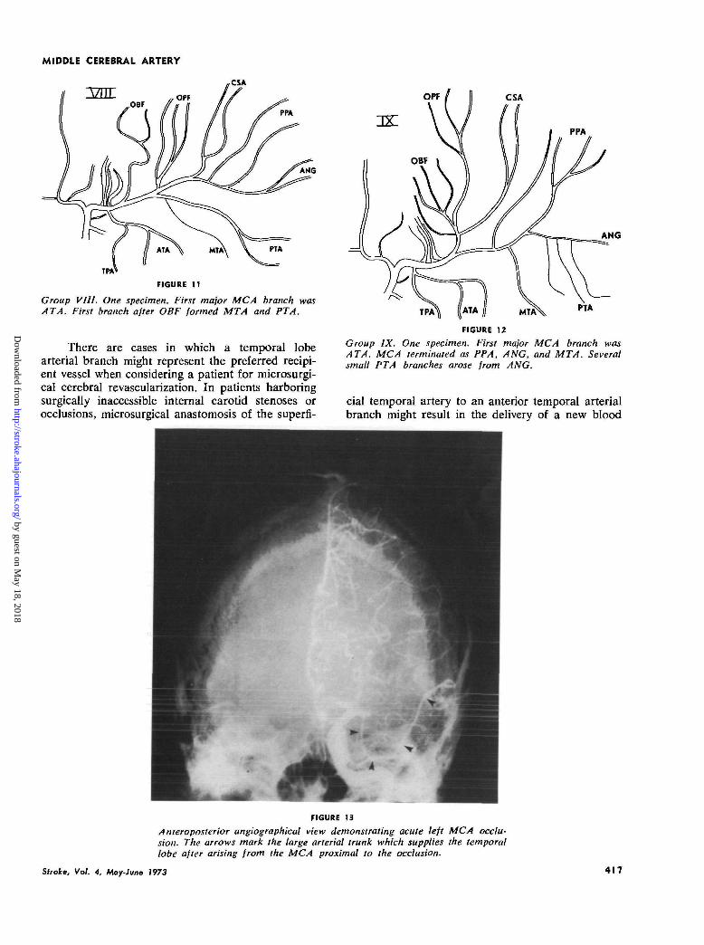
There are cases in which a temporal lobearterial branch might represent the preferred recipi-ent vessel when considering a patient for microsurgi-cal cerebral revascularization. In patients harboringsurgically inaccessible internal carotid stenoses orocclusions, microsurgical anastomosis of the superfi-
ANG
PTA
FIGURE 12
Group IX. One specimen. First major MCA branch wasATA. MCA terminated as PPA, ANG, and MTA. Severalsmall PTA branches arose from ANG.
cial temporal artery to an anterior temporal arterialbranch might result in the delivery of a new blood
FIGURE 13
Anteroposterior angiographical view demonstrating acute left MCA occlu-sion. The arrows mark the large arterial trunk which supplies the temporallobe after arising from the MCA proximal to the occlusion.
Stroke, Vol. 4, May-June 1973 41 f
by guest on May 18, 2018
http://stroke.ahajournals.org/D
ownloaded from
DeLONG
supply proximal to the lenticulostriate arteries closeto the origin of the middle cerebral artery. Such ananastomosis in these cases would be ideal forpotentially perfusing the ischemic middle cerebralarterial tree.
The hemodynamic consequences of each mid-dle cerebral arterial configuration remain to beassessed. The basic investigation of pulsatile cerebralhemodynamics has barely begun.
AcknowledgmentsI want to thank my neurosurgical associates, Dr. Glen O.Cross and Dr. Patrick E. Taylor, for their support of thisinvestigation. The Franciscan Sisters of the Sacred Heartwho sponsor St. Joseph's Hospital and the CardiovascularResearch Laboratory also deserve my thanks, and I wouldlike to acknowledge with gratitude the indispensable help ofDr. Thomas E. Wynn and Mr. Alva L. Bartges of theDepartment of Pathology at the St. Joseph's Hospital, SanFrancisco, California.
References1. Ring BA: Middle cerebral artery: Anatomical and
radiographic study. Acta Radiol 57: 289-300, 19622. Ring BA, Waddington M : Ascending frontal branch
of middle cerebral artery. Acta Radiol [Diagn](Stockholm) 6 :209-220, 1967
3. Ring BA: The Neglected Cause of Stroke. St. Louis,Warren H Green, Inc, 1969
4. Kaplan HA: The lateral perforating branches of theanterior and middle cerebral arteries. J Neurosurg23: 305-310, 1965
5. Stephens RB, Stilwell DL: Arteries and Veins of theHuman Brain. Springfield, Illinois, Charles C Thom-as, p 33-70, 1969
6. Jain KK: Some observations on the anatomy of themiddle cerebral artery. Canad J Surg 7: 134-139,1964
7. Foix C, Levy M: Les ramollissements sylviens. RevNeurol 2 : 1-51, 1927
8. Eecken HN Vander: Anastomoses Between Leptomen-ingeal Arteries. Springfield, Illinois, Charles CThomas, 1959
9. Stephens RB, Stilwell DL: Arteries and Veins of theHuman Brain. Springfield, Illinois, Charles C Thom-as, p 2 7 , 35, 45, 1969
10. Dahlstrom L, Fagerberg G, Lanner L, et a l :Anatomical and ongiographic studies of arteriessupplying anterior part of temporal lobe: A prelimi-nary report. Acta Radiol [Diagn] (Stockholm) 9:257-263, 1969
11. Taveras JM, Wood EH: Diagnostic Neuroradiology.Baltimore, Williams and Wilkins Co, p 1.6, 1964
12. LeMay M, Culebras A: Human brain: Differences inhemispheres demonstrable by arteriography. NewEng J Med 287: 168-170, 1972
13. Geschwind N, Levitsky W: Human brain: Left-rightasymmetries in temporal speech region. Science 161 :186-187, 1968
14. Yasargil MG: Microsurgery Applied to Neurosurgery.New York, Academic Press, p 105, 1969
15. Yasargil MG, Krayenbuhl HA, Jacobson JH: Micro-neurosurgical arterial reconstruction. Surgery 67:221-233, 1970
16. Reichman OH, Davis DO, Roberts TS, et a l : Collateralcirculation to the middle cerebral territory bysurgical anastomosis. Presented at the annual meet-ing, American Association of Neurological Surgeons,Statler Hilton Hotel, Boston, Massachusetts, April 16-19, 1972
418 Sfroke, Vol. 4. May-June 1973
by guest on May 18, 2018
http://stroke.ahajournals.org/D
ownloaded from

W. BRADFORD DELONGAnatomy of the Middle Cerebral Artery: The Temporal Branches
Print ISSN: 0039-2499. Online ISSN: 1524-4628 Copyright © 1973 American Heart Association, Inc. All rights reserved.
is published by the American Heart Association, 7272 Greenville Avenue, Dallas, TX 75231Stroke doi: 10.1161/01.STR.4.3.412
1973;4:412-418Stroke.
http://stroke.ahajournals.org/content/4/3/412World Wide Web at:
The online version of this article, along with updated information and services, is located on the
http://stroke.ahajournals.org//subscriptions/
is online at: Stroke Information about subscribing to Subscriptions:
http://www.lww.com/reprints Information about reprints can be found online at: Reprints:
document. Permissions and Rights Question and Answer about this process is available in the
located, click Request Permissions in the middle column of the Web page under Services. Further informationEditorial Office. Once the online version of the published article for which permission is being requested is
can be obtained via RightsLink, a service of the Copyright Clearance Center, not theStrokepublished in Requests for permissions to reproduce figures, tables, or portions of articles originallyPermissions:
by guest on May 18, 2018
http://stroke.ahajournals.org/D
ownloaded from