Anatomy II: Body Systems and Energy for Physical Activity Year 10 PASS Mater Dei Catholic College.
43
Anatomy II: Body Systems and Energy for Physical Activity Year 10 PASS Mater Dei Catholic College
-
Upload
francine-flowers -
Category
Documents
-
view
214 -
download
1
Transcript of Anatomy II: Body Systems and Energy for Physical Activity Year 10 PASS Mater Dei Catholic College.
Anatomy II: Body Systems and Energy for Physical Activity
Year 10 PASSMater Dei Catholic College
The Skeletal System
Skeletal Bones
Whack a Bonehttp://www.anatomyarcade.com/games/WAB/WAB.html
Poke a Musclehttp://www.anatomyarcade.com/games/PAM/PAM.html
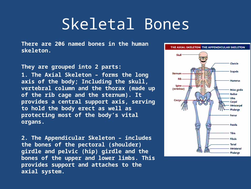
Skeletal BonesThere are 206 named bones in the human skeleton.
They are grouped into 2 parts:1. The Axial Skeleton – forms the long axis of the body; Including the skull, vertebral column and the thorax (made up of the rib cage and the sternum). It provides a central support axis, serving to hold the body erect as well as protecting most of the body’s vital organs.
2. The Appendicular Skeleton – includes the bones of the pectoral (shoulder) girdle and pelvic (hip) girdle and the bones of the upper and lower limbs. This provides support and attaches to the axial system.
Types of BonesThere are many different shapes and sizes of bone each designed to best perform its special function. Almost all types of bone can be classified into four main categories depending on their shape. They are classified as:•Long bones•Short bones•Flat bones•Irregular bones
The other 2 types of bones are classified by location not shape•Sesamoid bones – small bones embedded in tendons (e.g. patella)•Sutural Bones – small bones located between joints of some cranial bones
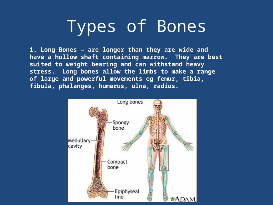
Types of Bones1. Long Bones – are longer than they are wide and have a hollow shaft containing marrow. They are best suited to weight bearing and can withstand heavy stress. Long bones allow the limbs to make a range of large and powerful movements eg femur, tibia, fibula, phalanges, humerus, ulna, radius.
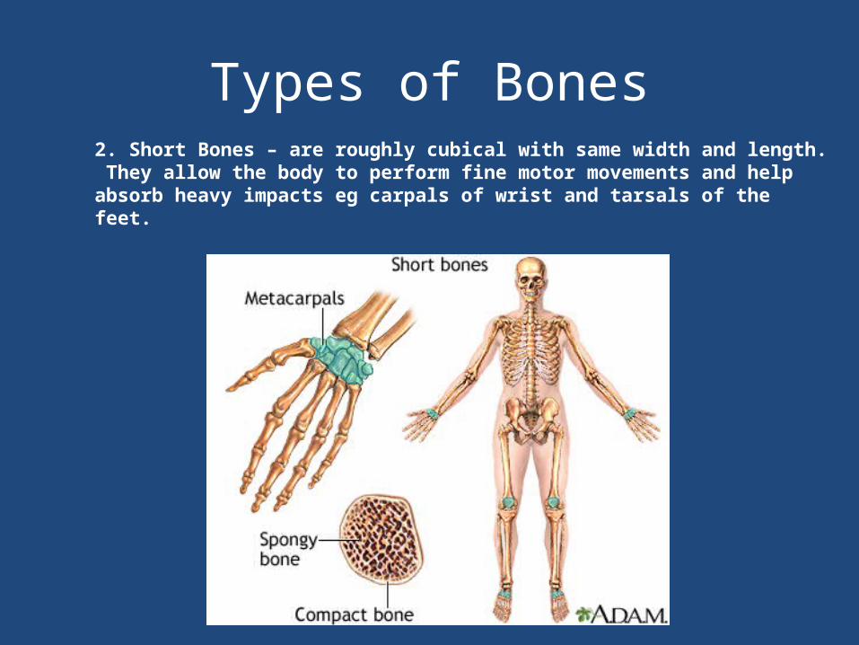
Types of Bones2. Short Bones – are roughly cubical with same width and length. They allow the body to perform fine motor movements and help absorb heavy impacts eg carpals of wrist and tarsals of the feet.
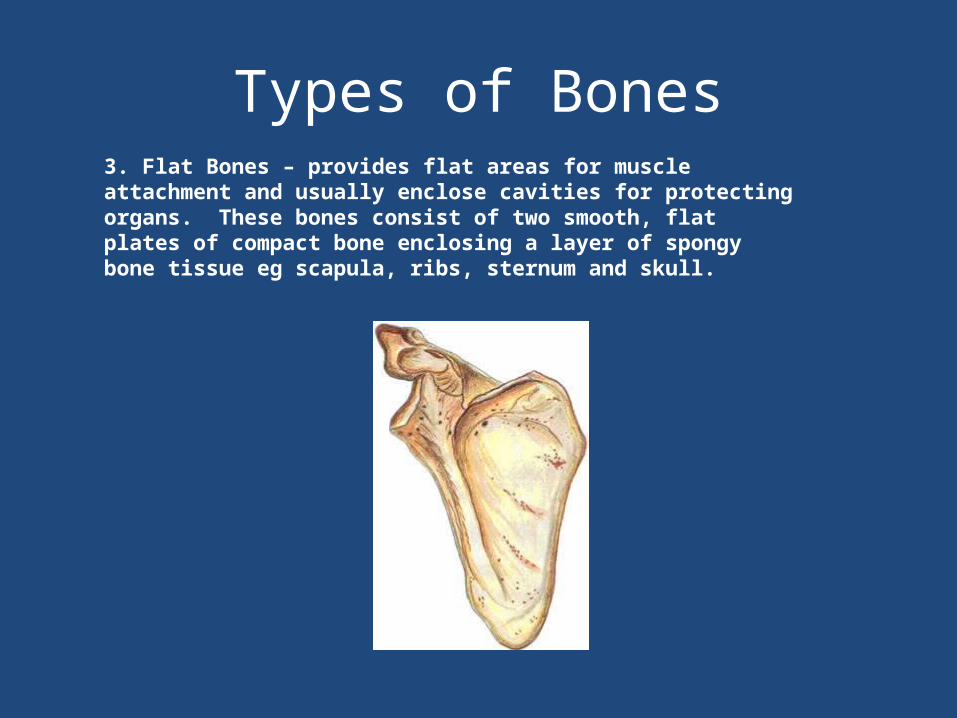
Types of Bones3. Flat Bones – provides flat areas for muscle attachment and usually enclose cavities for protecting organs. These bones consist of two smooth, flat plates of compact bone enclosing a layer of spongy bone tissue eg scapula, ribs, sternum and skull.
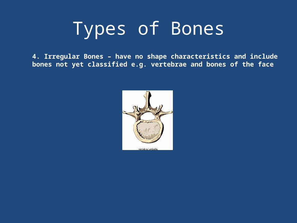
Types of Bones4. Irregular Bones – have no shape characteristics and include bones not yet classified e.g. vertebrae and bones of the face
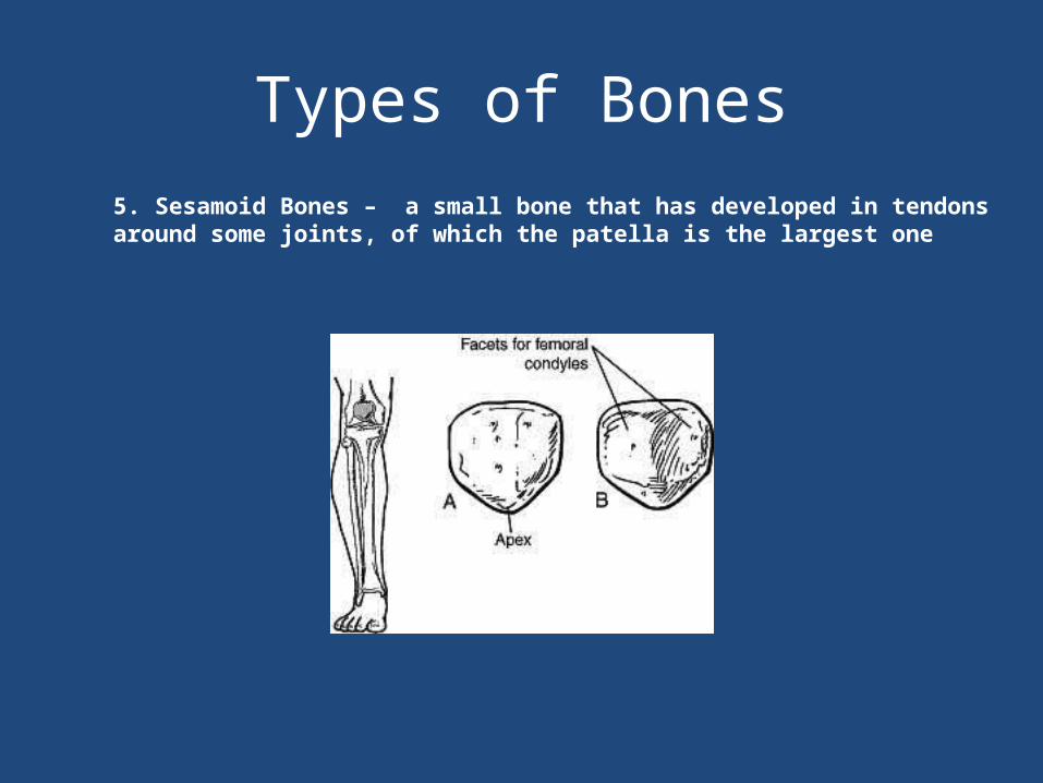
Types of Bones5. Sesamoid Bones – a small bone that has developed in tendons around some joints, of which the patella is the largest one
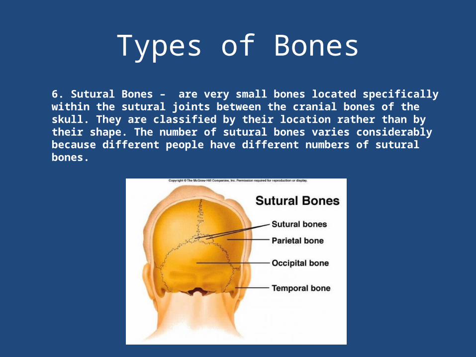
Types of Bones6. Sutural Bones – are very small bones located specifically within the sutural joints between the cranial bones of the skull. They are classified by their location rather than by their shape. The number of sutural bones varies considerably because different people have different numbers of sutural bones.
BonesIdentify each of the following bones on a partner:-Long Bones: Humerus, Femur, Radius, Tibia, Fibula
-Short Bones: Carpals, Tarsals, Phalanges, Metacarpals, Metatarsals
-Flat Bones: Cranium, Pelvis, Scapula
-Irregular Bones: Vertebrae
-Sesamoid Bones: Patella
-Sutural: Cranium
Bone TissueThere are two main types of tissue found in bone:
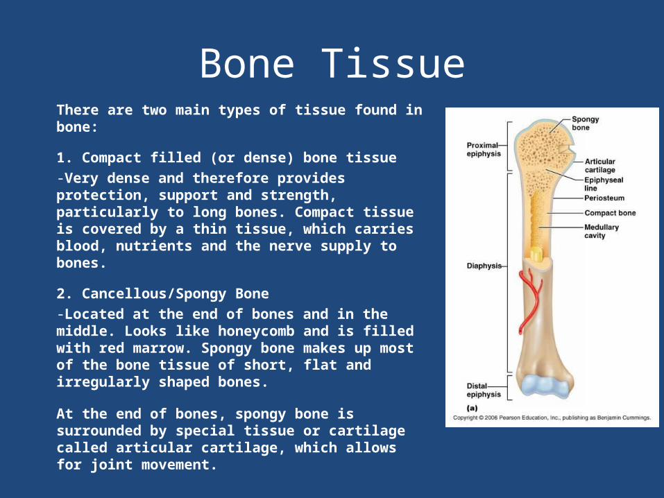
1. Compact filled (or dense) bone tissue-Very dense and therefore provides protection, support and strength, particularly to long bones. Compact tissue is covered by a thin tissue, which carries blood, nutrients and the nerve supply to bones.
2. Cancellous/Spongy Bone-Located at the end of bones and in the middle. Looks like honeycomb and is filled with red marrow. Spongy bone makes up most of the bone tissue of short, flat and irregularly shaped bones.
At the end of bones, spongy bone is surrounded by special tissue or cartilage called articular cartilage, which allows for joint movement.

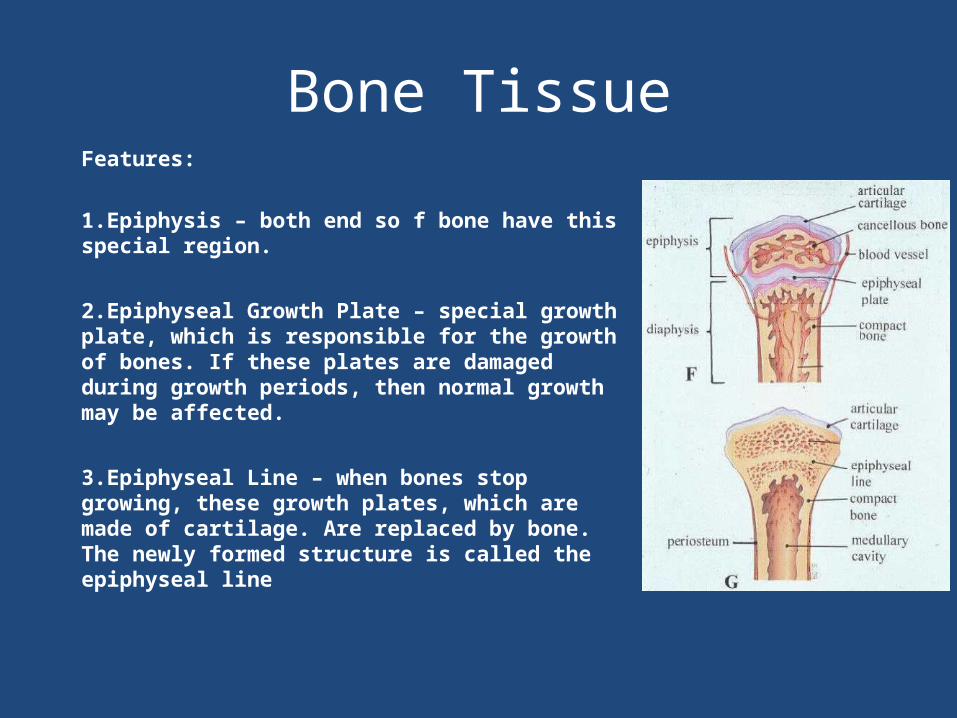
Bone TissueFeatures:
1.Epiphysis – both end so f bone have this special region.
2.Epiphyseal Growth Plate – special growth plate, which is responsible for the growth of bones. If these plates are damaged during growth periods, then normal growth may be affected.
3.Epiphyseal Line – when bones stop growing, these growth plates, which are made of cartilage. Are replaced by bone. The newly formed structure is called the epiphyseal line
Joint Structure and Actions
• Bones are very rigid – to allow mobility in the body we have joints.
• A joint (or articulation) is the point at which bones meet and articulate about each other.
• The function and stability of a joint are determined by:– The way in which the articulating bones fit together– The flexibility of the connective tissue binding the joint– The position of the muscles, tendons and ligaments around
the joint
Types of Joints
• Joints – occur where one or more bones meet. Joints can be fixed, for example Rib Cage, or immovable for example your elbow. Joints are classified according to their degree of movement.
• Joints may be classified as:– Fibrous or Immovable– Cartilaginous or slightly movable– Synovial or freely movable
Types of Joints
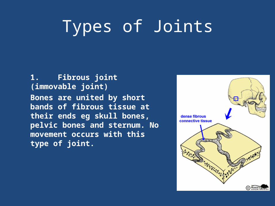
1. Fibrous joint (immovable joint) Bones are united by short bands of fibrous tissue at their ends eg skull bones, pelvic bones and sternum. No movement occurs with this type of joint.
Types of Joints
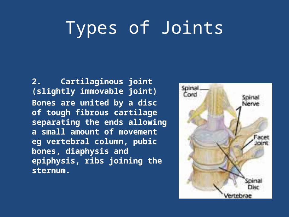
2. Cartilaginous joint (slightly immovable joint)Bones are united by a disc of tough fibrous cartilage separating the ends allowing a small amount of movement eg vertebral column, pubic bones, diaphysis and epiphysis, ribs joining the sternum.

Types of Joints
3. Synovial joints (moveable joints)All synovial joints have a capsule that encloses the joint spaceSynovial membrane lines the inner surface of the capsule and secretes synovial fluid into the joint cavity to keep it lubricated.Shock absorbent articular cartilage is found on the ends of the bones to protect then from wear and tear.Eg knee, hip, elbow joints
Types of Joints
Features of Synovial Joints1.Synovial Membrane – lines the interior cavity of the joint capsule. Produces synovial fluid which lubricates and provides nutrition for the joint.2.Ligaments – fibrous bands that surround and reinforce the joint. Joins bone to bone and are relatively inelastic.3.Hyaline Cartilage – protective covering to the ends of bones that form the joint. Gives the bone a smooth, white, shining appearance. Helps to reduce friction between the bones.4.Joint Capsule – Specialized covering which binds the ends of bones together.5.Bursae – small sacs filled with synovial fluid, found in joints where friction is likely to occur, eg the knee and ankle joints. These structures may become inflamed causing movements to be restricted, this is called bursitis.
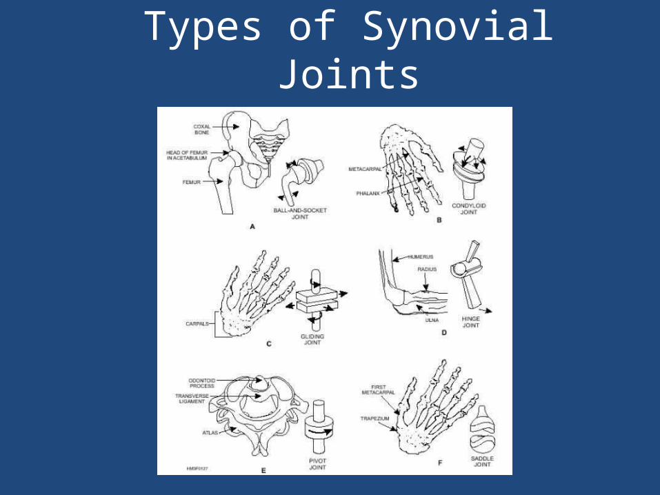
Types of Synovial Joints
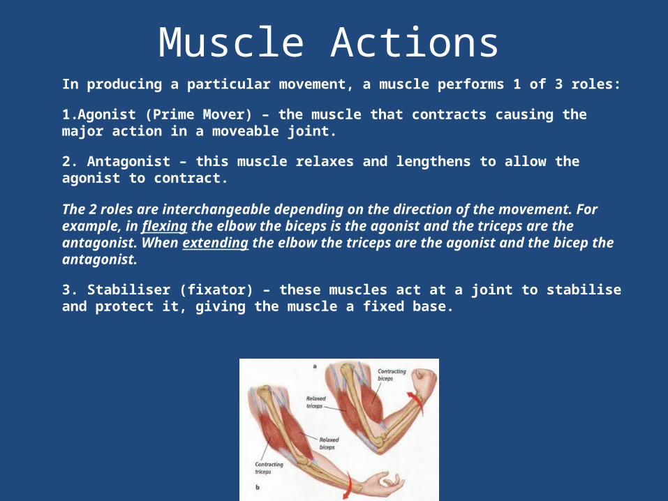
Muscle ActionsIn producing a particular movement, a muscle performs 1 of 3 roles:
1.Agonist (Prime Mover) – the muscle that contracts causing the major action in a moveable joint.
2. Antagonist – this muscle relaxes and lengthens to allow the agonist to contract.
The 2 roles are interchangeable depending on the direction of the movement. For example, in flexing the elbow the biceps is the agonist and the triceps are the antagonist. When extending the elbow the triceps are the agonist and the bicep the antagonist.
3. Stabiliser (fixator) – these muscles act at a joint to stabilise and protect it, giving the muscle a fixed base.
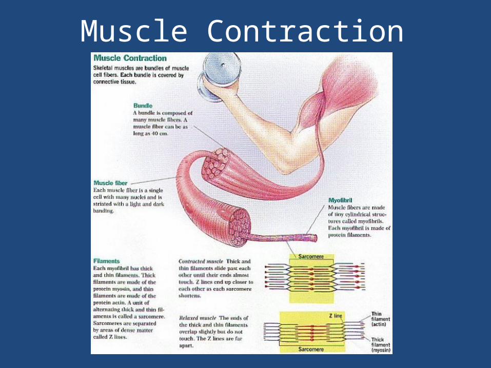
Muscle Contraction
Types of Contractions
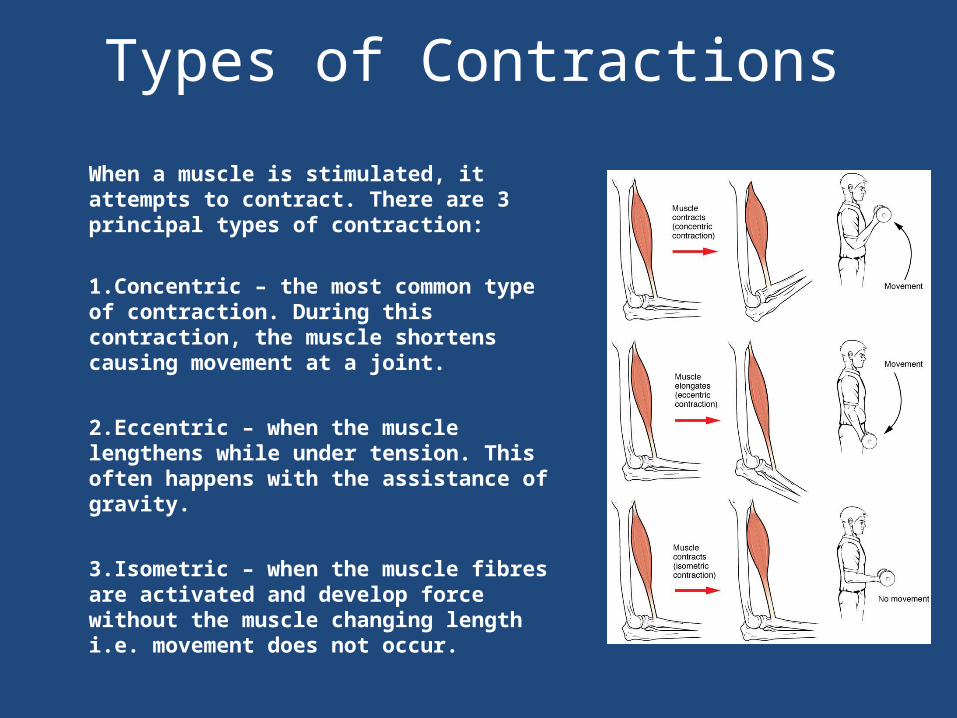
When a muscle is stimulated, it attempts to contract. There are 3 principal types of contraction:
1.Concentric – the most common type of contraction. During this contraction, the muscle shortens causing movement at a joint.
2.Eccentric – when the muscle lengthens while under tension. This often happens with the assistance of gravity.
3.Isometric – when the muscle fibres are activated and develop force without the muscle changing length i.e. movement does not occur.
Muscles
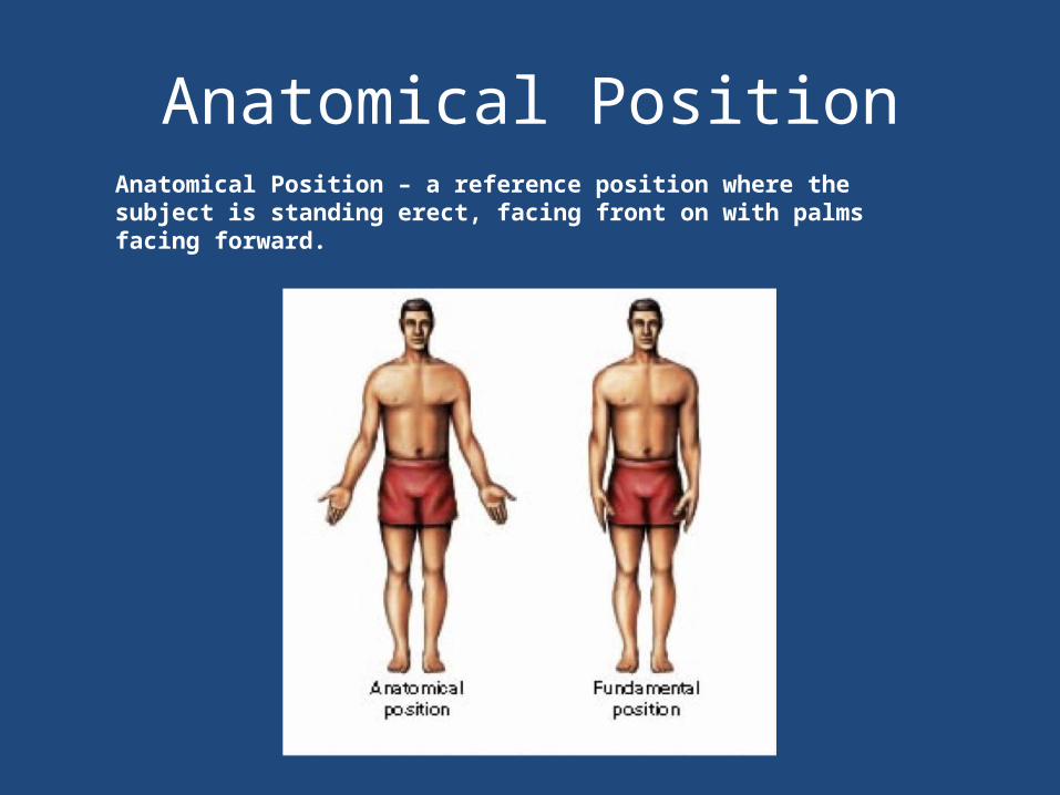
Anatomical PositionAnatomical Position – a reference position where the subject is standing erect, facing front on with palms facing forward.
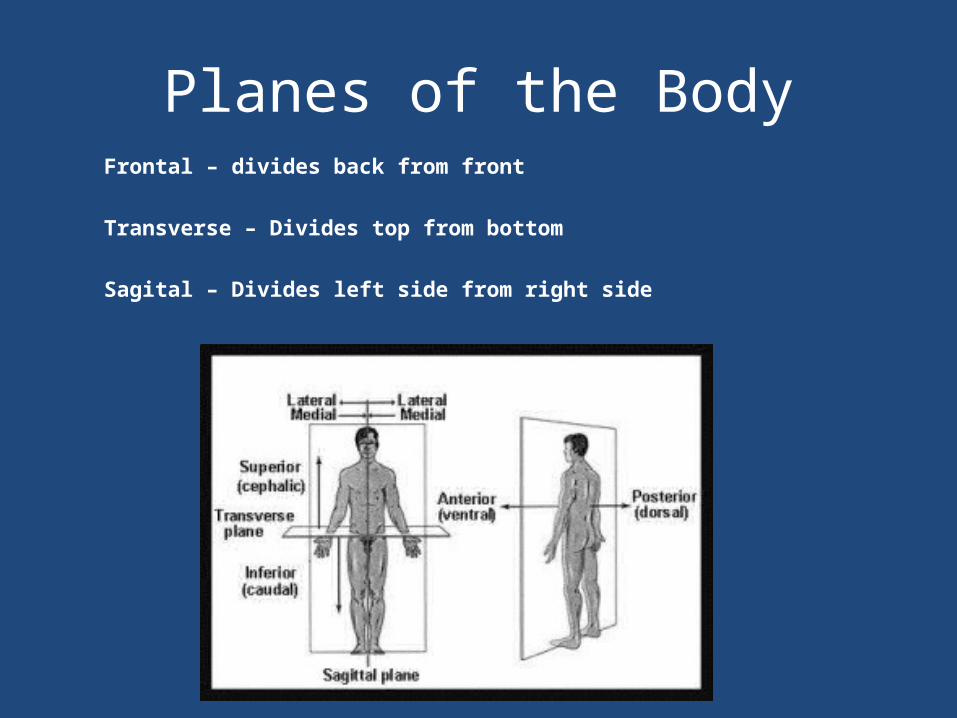
Planes of the BodyFrontal – divides back from front
Transverse – Divides top from bottom
Sagital – Divides left side from right side
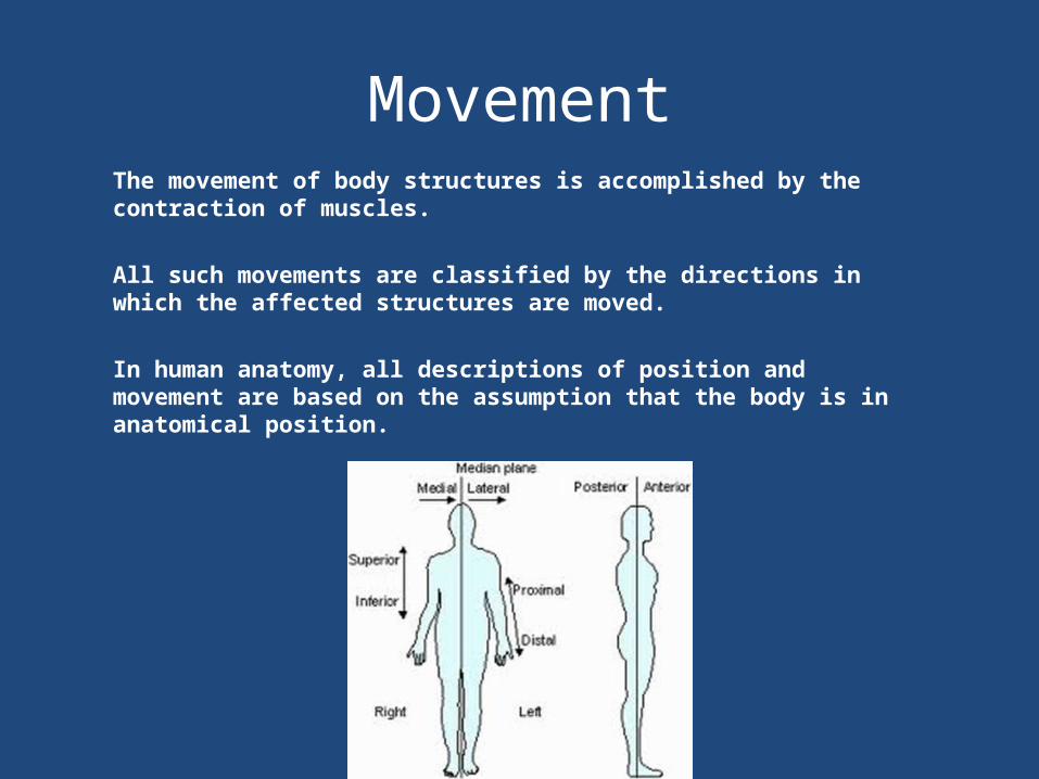
MovementThe movement of body structures is accomplished by the contraction of muscles.
All such movements are classified by the directions in which the affected structures are moved.
In human anatomy, all descriptions of position and movement are based on the assumption that the body is in anatomical position.
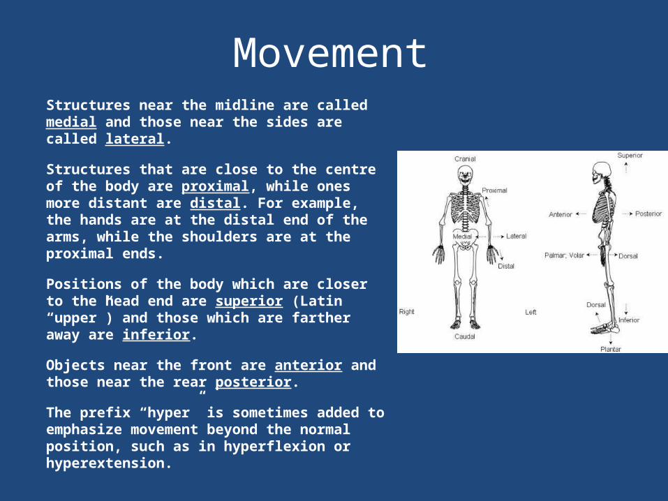
MovementStructures near the midline are called medial and those near the sides are called lateral.
Structures that are close to the centre of the body are proximal, while ones more distant are distal. For example, the hands are at the distal end of the arms, while the shoulders are at the proximal ends.
Positions of the body which are closer to the head end are superior (Latin “upper”) and those which are farther away are inferior.
Objects near the front are anterior and those near the rear posterior.
The prefix “hyper” is sometimes added to emphasize movement beyond the normal position, such as in hyperflexion or hyperextension.
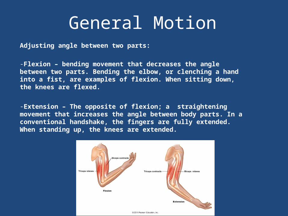
General MotionAdjusting angle between two parts:
-Flexion – bending movement that decreases the angle between two parts. Bending the elbow, or clenching a hand into a fist, are examples of flexion. When sitting down, the knees are flexed.
-Extension – The opposite of flexion; a straightening movement that increases the angle between body parts. In a conventional handshake, the fingers are fully extended. When standing up, the knees are extended.
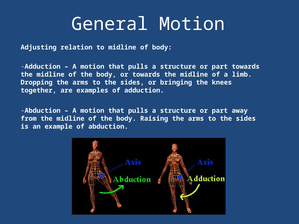
General MotionAdjusting relation to midline of body:
-Adduction – A motion that pulls a structure or part towards the midline of the body, or towards the midline of a limb. Dropping the arms to the sides, or bringing the knees together, are examples of adduction.
-Abduction – A motion that pulls a structure or part away from the midline of the body. Raising the arms to the sides is an example of abduction.
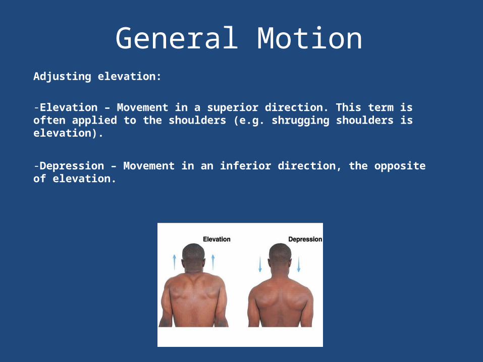
General MotionAdjusting elevation:
-Elevation – Movement in a superior direction. This term is often applied to the shoulders (e.g. shrugging shoulders is elevation).
-Depression – Movement in an inferior direction, the opposite of elevation.
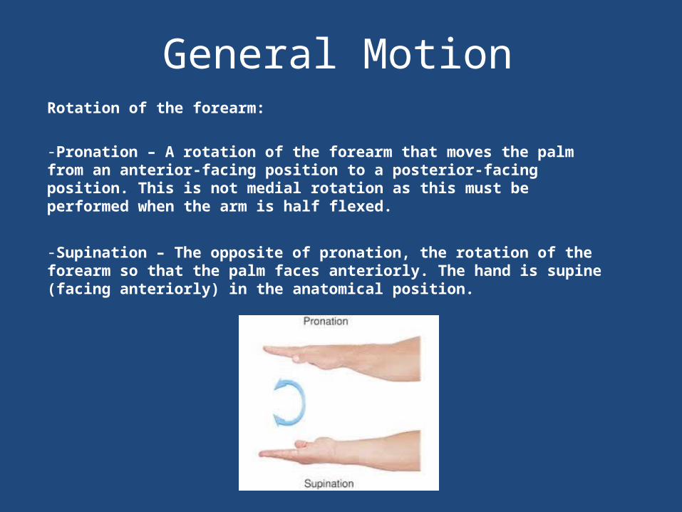
General MotionRotation of the forearm:
-Pronation – A rotation of the forearm that moves the palm from an anterior-facing position to a posterior-facing position. This is not medial rotation as this must be performed when the arm is half flexed.
-Supination – The opposite of pronation, the rotation of the forearm so that the palm faces anteriorly. The hand is supine (facing anteriorly) in the anatomical position.
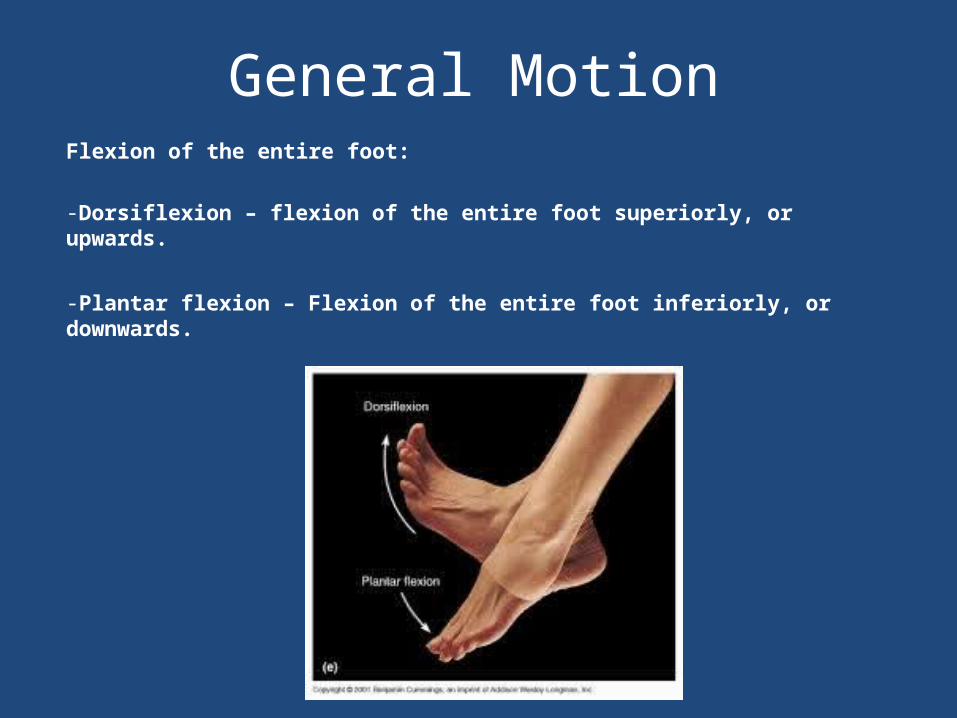
General MotionFlexion of the entire foot:
-Dorsiflexion – flexion of the entire foot superiorly, or upwards.
-Plantar flexion – Flexion of the entire foot inferiorly, or downwards.
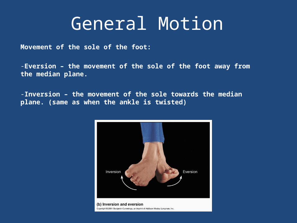
General MotionMovement of the sole of the foot:
-Eversion – the movement of the sole of the foot away from the median plane.
-Inversion – the movement of the sole towards the median plane. (same as when the ankle is twisted)
General Motion
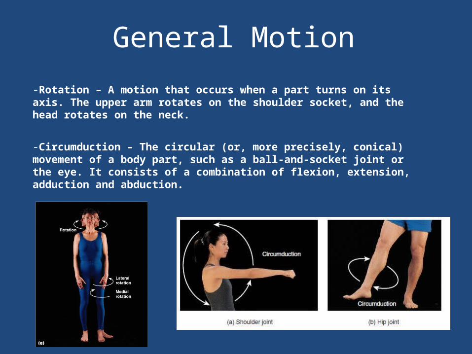
-Rotation – A motion that occurs when a part turns on its axis. The upper arm rotates on the shoulder socket, and the head rotates on the neck.
-Circumduction – The circular (or, more precisely, conical) movement of a body part, such as a ball-and-socket joint or the eye. It consists of a combination of flexion, extension, adduction and abduction.
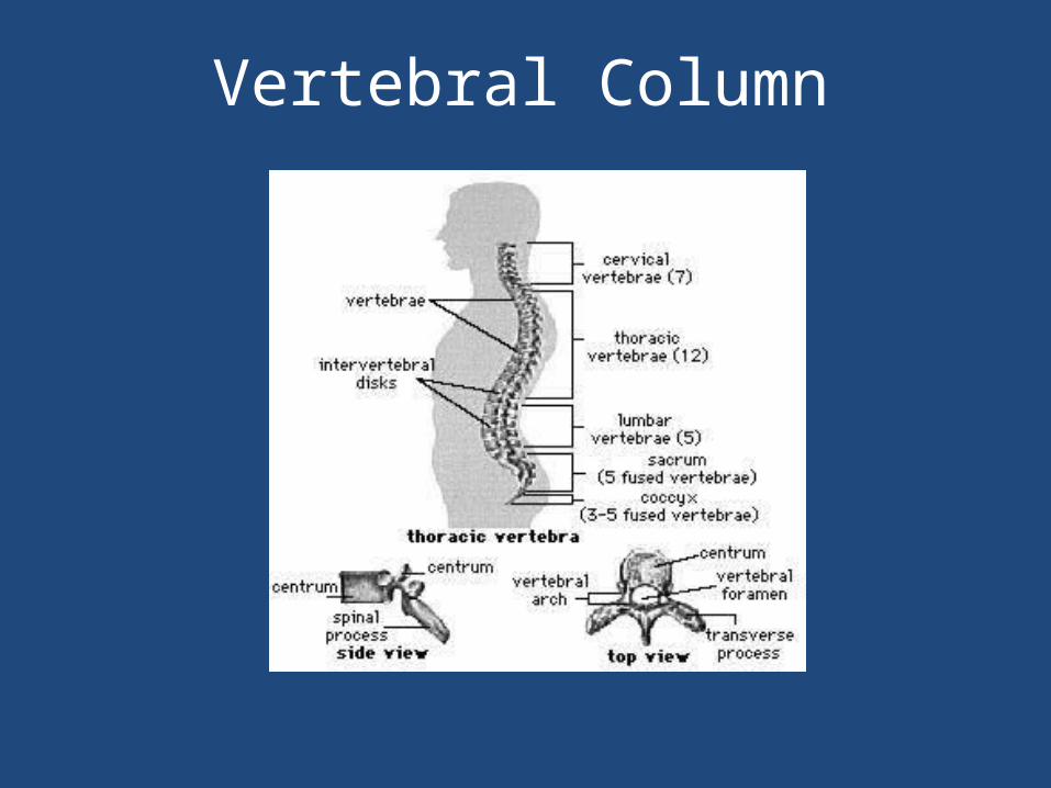
Vertebral Column
Body Systems
-Respiratory System
-Endocrine System
-Lymphatic System
-Nervous System
-Digestive System
-Urinary Tract

Respiratory System
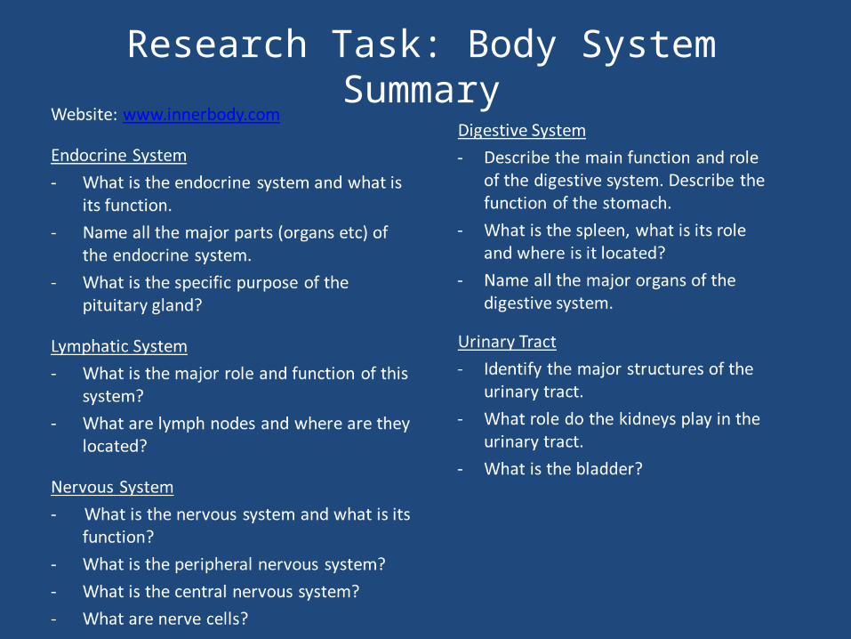
Research Task: Body System Summary