
Anatomy for Yogis - Zach Beach
68
Anatomy for Yogis By Zach Beach www.zachbeach.com Here is an amazement: once I was twenty years old and in every motion of my body there was a delicious ease, and in every motion of the green earth there was a hint of paradise, and now I am sixty years old, and it is the same. - Mary Oliver
Transcript of Anatomy for Yogis - Zach Beach

Anatomy for Yogis
By Zach Beach www.zachbeach.com
Here is an amazement: once I was twenty years old and in every motion of my body there was a delicious ease, and in every motion of the green earth there was a hint of paradise, and now I
am sixty years old, and it is the same. - Mary Oliver
Introduction The following is a 10 lesson (taking approximately 15 hours) anatomy course designed for aspiring yoga teachers going through their first 200-hr, as well as yoga practitioners looking for a deeper understanding of anatomy as it applies to their asana practices. Each lesson is organized as follows:
Lesson 1: Introduction, Planes of Motion, Basic Terms 3
Lesson 2: Bones, Spine, Movement of the Spine, Posture 11
Lesson 3: Soft Tissues, Muscles 101 18
Lesson 4: Pelvic Bowl, Hamstring and Psoas, Ribs and Breathing 23
Lesson 5: Skeletal System, Gross Muscles, and Muscle Movements 28
Lesson 6: Joint Types, The Hip Joint 34
Lesson 7: The Shoulder 39
Lesson 8: The Foot, Ankle, and Knee 45
Lesson 9: Putting it All Together: The Anatomy of a Backbend 53
Lesson 10: Flexibility and Stretching Principles 59 Let this course be the beginning of a lifetime of learning. The human body is as beautiful as it is complex, as much a miracle as it is impossible to fully understand. 10 lessons is very little compared to a multi-year physical therapy or medical program, and much shorter than many massage schools. Use the knowledge gained here as a solid foundation for teaching yoga in a safe and anatomically informed manner, and continue your studies as you continue your practice.
Anatomy for Yogis, By Zach Beach, www.zachbeach.com 1

Recommended reading:
● Andrew Biel, Trail Guide to the Body: How to Locate Muscles, Bones and More
● Blandine Calais-Germain, The Anatomy of Movement
● Elaine N. Marieb, Anatomy and Physiology Coloring Workbook: A Complete Study
Guide
● H. David Coulter, Anatomy of Hatha Yoga
● Kelly Solloway, The Yoga Anatomy Coloring Book (Elizabeth J. Rochester also has
another Yoga Anatomy Coloring Book)
● Leslie Kaminoff, Yoga Anatomy
● Ray Long, MD, The Key Muscles of Yoga, The Key Poses of Yoga, The Yoga Mat
Companion Series
● Wynn Kapit, Lawrence M. Elson, The Anatomy Coloring Book
Online Resources:
● bandhayoga.com
● pranamaya.com
● teachmeanatomy.info
● yoganatomy.com
Anatomy Apps
● Complete Anatomy by 3D4Medical
● Visible Body
Anatomy for Yogis, By Zach Beach, www.zachbeach.com 2
Lesson 1: Introduction, Planes of Motion, Basic Terms Before we discuss anatomy, let us consider the fundamental question of epistemology: how do we know what we know? Just as any spiritual path begins with a search for deeper truth and a questioning of conventional wisdom and potentially limiting beliefs, our discussion on the truth of our anatomy can begin by acknowledging the set of assumptions we are operating under. Modern anatomy and modern medicine is primarily based on the method of scientific reductionism. Science, in this case, is an empirical practice based on the belief that hypotheses and theories must be tested against observations of the natural world. Any hypothesis must have a strong compendium of evidence to make it a theory and even then, it is continuously tested as new evidence and findings are uncovered. Reductionism is one way of understanding complex phenomena and a common way to develop scientific models of the way the universe works. Reductionism attempts to describe a system by breaking it down into constituent or fundamental parts, which are then analyzed and understood in order to gain insight into how the system works as a whole. Reductionism is like taking apart a clock or a watch into all of its individual functioning parts. By understanding each piece, you can begin to understand how the clock works as a whole. When it comes to our understanding of anatomy, the extraordinarily beautiful and infinitely complex body is divided up into parts that we call systems. If you take the average anatomy book, for example, its chapters might be divided up as such:
1. The nervous system 2. The endocrine system 3. The cardiovascular system 4. The lymphatic system 5. The respiratory system 6. The digestive system 7. The urinary system 8. The reproductive systems
Each system tends to be a collection of organs and structures sharing a common function and/or the same fundamental properties.
Anatomy for Yogis, By Zach Beach, www.zachbeach.com 3

These systems are then further broken down. For example, the skeletal system is divided into the axial skeleton (head, rib cage, vertebrae, and sacrum) and the appendicular skeleton (bones of the limbs, shoulder, and pelvic girdle). These skeletons are then further divided into the 206 bones of the body. The digestive system may be broken down organ by organ, layer by layer, and eventually, cell type by cell type. While learning about the systems listed above are important to understanding how a body works, for the purpose of practicing and teaching yoga, we will primarily focus on one system: the musculoskeletal system. The musculoskeletal system includes all the bones, muscles, and connective tissues that make up the body, as well as the nervous system that controls them. These are the parts of the body that matter the most for a strong anatomical understanding of our asana practice. Because the human brain is not an all-knowing entity, breaking down complex systems into their parts and understanding those parts allows us to learn in an easy and methodical way. But it is important to note that all of these systems are part of a whole body. You’ll never see just a nervous system existing in the wild! Also, learning the parts of a watch won’t necessarily teach you how to know the time. That’s why it is important to stay rooted in your yoga practice and maintain a holistic view of the body. As you learn about the different parts and systems of the body, think about how they make up and interact with the whole. Spirit teaches us that everything in this universe is connected. This is especially true for our bodies. These lessons will not be mentioning anything about the energetic body, light body, subtle bodies, seed body, auric bodies, koshas, bandhas, nor their constituent chakras, nadis, meridians, prana, vayus, or kundalini energy. While countless sages, mystics, healers, and embodiment practitioners can speak to the subjective reality of such systems, we have yet to find any scientific or empirical evidence for their existence. Perhaps in the future we will develop the tools to measure the energetic and the subtle. But for now, we will simply focus on the physical body made up of physical matter: on what can be seen, verified, and measured. Nevertheless, remember to keep your mind free and your heart open to the infinite everything that is possible beyond our limited conceptual understanding, and to the mystery of all that is. Namaste.
Anatomy for Yogis, By Zach Beach, www.zachbeach.com 4

Remember: the bones of the _______________, linked together at the _______________, are
moved by the actions of the _______________.
Basic terms
Directional terms used to describe ORIENTATION in the ANATOMICAL POSITION
(i.e. where is...?) Front to back:
Anterior: toward the front of body Posterior: toward the back of body
Left to right: Medial: toward the midline Lateral: away from the midline
Top to bottom: Superior: toward the head Inferior: away from head
Center to outer (for the legs and arms) Distal: toward extremities Proximal: toward center of body
Inside to outside: Superficial: near the skin Deep: deeper into the body
Examples:
● Pectoralis major lies anterior to pectoralis minor. ● The triceps are posterior to biceps brachii. ● The lungs are superior to the liver. ● The stomach is inferior to the heart. ● The wrist joint is distal to the elbow joint. ● The scaphoid lies in the proximal row of carpal bones. ● The cornea is on the superficial surface of the eye. ● The bones are deep to the skin.
Anatomy for Yogis, By Zach Beach, www.zachbeach.com 5

Now, fill out an example for each one to help you remember:
1. The ________________________ is/are anterior to the ________________________.
2. The ________________________ is/are posterior to the ________________________.
3. The ________________________ is/are medial to the ________________________.
4. The ________________________ is/are lateral to the ________________________.
5. The ________________________ is/are superior to the ________________________.
6. The ________________________ is/are inferior to the ________________________.
7. The ________________________ is/are distal to the ________________________.
8. The ________________________ is/are proximal to the ________________________.
9. The ________________________ is/are superficial to the ________________________.
10. The ________________________ is/are deep to the ________________________.
Anatomy for Yogis, By Zach Beach, www.zachbeach.com 6

Terms used to describe MOVEMENT from the ANATOMICAL POSITION The Three Cardinal Planes of Motion:
Sagittal: vertical plane, runs anterior-posterior (forward-back), divides the body into left and right sides Frontal (Coronal): vertical plane, runs side to side, divides the body into forward and back (anterior-posterior) Transverse: horizontal plane, any movement along a central axis
List at least 5 yoga poses that are primarily in one plane of motion:
Sagittal:
_____________________________________________________________________________
_____________________________________________________________________________
Frontal:
_____________________________________________________________________________
_____________________________________________________________________________
Transverse:
_____________________________________________________________________________
_____________________________________________________________________________
How might we use this knowledge to increase the quality of our yoga classes?
_____________________________________________________________________________
_____________________________________________________________________________
Anatomy for Yogis, By Zach Beach, www.zachbeach.com 7

Joint Motions In Sagittal Plane:
Flexion: bones move toward each other (the angle decreases, bending the knee or elbow) OR anterior (like reaching the arm or leg forward)
Extension: bones move away from each other (the angle increases, like hyperextension) OR posterior (like reaching the arm or leg backward)
In Frontal Plane:
Abduction: bones move away from midline Adduction: bones move toward midline
In Transverse Plane:
Internal/medial rotation: rotation in External/lateral rotation: rotation out
Circumduction: Movement in two or three planes of motion
Special Joint motions Ankle Joint:
Plantar flexion: standing on toes, heels lifted (pointing foot) Dorsiflexion: standing on heels, toes and ball of foot lifted (flexing foot) Pronation: rotation foot inward and downward (outer arch up, inner arch flattens) Supination: the outward roll of the foot (inner arch up)
Elbow joint (these are sometimes referred to as forearm motions): Supination: palm up (palm is anterior, sometimes called external rotation by yogis) Pronation: palm down (palm is posterior, sometimes called internal rotation by yogis)
Shoulder joint: Elevation: shoulders up Depression: shoulders down Protraction: shoulder blades move away from the spine Retraction: shoulder blades move toward spine
Anatomy for Yogis, By Zach Beach, www.zachbeach.com 8

Pose Analysis Now, analyze a yoga pose and specify what joint motions from the anatomical position are required to get into the pose. Specify the plane(s) of motion (sagittal, frontal, transverse) and joint motions (abduction, adduction, flexion, extension, medial rotation, lateral rotation, etc.) for the ankle, knee, hip, spine, shoulder, elbow, and wrist. Suggested poses include: Chair, Warrior III, High Lunge, Bow, Triangle, Forward Fold, Downward Dog, Warrior II, Sphinx, Cross Legged). Note: many answers will be blank. Yoga pose: ______________________________________________________________
Plane(s) of motion: _______________________________________________________
Specify the joint motion for: Ankle:______________________________
Knee:_______________________________ Hip:________________________________
Spine:_______________________________ Shoulder:____________________________
Elbow:______________________________ Wrist:_______________________________
Yoga pose: ______________________________________________________________
Plane(s) of motion: _______________________________________________________
Specify the joint motion for: Ankle:______________________________
Knee:_______________________________ Hip:________________________________
Spine:_______________________________ Shoulder:____________________________
Elbow:______________________________ Wrist:_______________________________
Anatomy for Yogis, By Zach Beach, www.zachbeach.com 9

Yoga pose: ______________________________________________________________
Plane(s) of motion: _______________________________________________________
Specify the joint motion for: Ankle:______________________________
Knee:_______________________________ Hip:________________________________
Spine:_______________________________ Shoulder:____________________________
Elbow:______________________________ Wrist:_______________________________
Anatomy for Yogis, By Zach Beach, www.zachbeach.com 10

Lesson 2: Bones, Spine, Movement of the Spine, Posture The musculoskeletal system provides the body with support and movement. It consists of bones, joints, and soft tissues, which include muscles, tendons, ligaments, and fascia.
Bones and Joints Basics Bones are living, growing tissue made mostly of collagen. Collagen is a protein that provides a soft framework, and calcium phosphate is a mineral that adds strength and hardens that framework. This combination of collagen and calcium makes bone strong and flexible enough to withstand stress. Human beings have 270 bones at birth, some of which eventually fuse together, and we have 206 bones by adulthood. Our skeletal system serves five primary functions:
1. __________________________________________________
2. __________________________________________________
3. __________________________________________________
4. __________________________________________________
5. __________________________________________________
There are four main types of bones: long (femur), flat (scapula, cranium, ribs), short (talus,
carpal, tarsal) and irregular (vertebra). (Note: some textbooks will include a fifth type of bone,
sesamoid, like the patella).
Anatomy for Yogis, By Zach Beach, www.zachbeach.com 11

A joint is a point where any two or more bones meet. There are three main types of joints: fibrous (immovable), cartilaginous (partially movable) and synovial (freely movable) joint. Fibrous (synarthrodial) joints: This type of joint is held together only by ligaments. They have no joint cavity and do not move. All 22 bones of the skull except for the mandible connect at fibrous joints. Cartilaginous (synchondrosis and symphysis) joints: These joints occur where the connection between the articulating bones is made up of cartilage. Examples of cartilaginous joints include the intervertebral discs in the spine, the ribs to the sternum, the pubic symphysis, and the manubriosternal joint (between the manubrium and the sternum). These are “semi-movable.” Synovial (diarthrosis) joints: These joints are by far the most common classification of joint within the human body and will be our primary concern when it comes to yoga, since synovial joints are where most of our movement happens. All synovial joints have a synovial capsule (collagenous structure) surrounding the entire joint, a synovial membrane (the inner layer of the capsule) which secretes synovial fluid (a lubricating liquid), and cartilage known as hyaline cartilage which pads the ends of the articulating bones (think of the white/glossy part at the end of a chicken bone). The structure of the synovial joint can be seen below:
Anatomy for Yogis, By Zach Beach, www.zachbeach.com 12

The Spine In yoga there is a saying: you are as young as your spine. Whether or not that is literally true, our spine is our literal backbone and the center of all of our movement. Before we build the rest of our skeleton, let us discuss and get to know our spine.
Written into our spine is the evolution of our species. From fish who moved side to side with their head but never had to consider the force of gravity, to reptiles who developed a “primary curve” or arch to carry their weight, to mammals who then lifted their heads up to bring a new curve to the neck, and finally humans being the only species that mainly walks on two feet, a third curve was introduced. We now refer to these arches as thoracic (ribs), cervical (neck), and lumbar (lower back) curves. Patanjali writes in the yoga sutras: sthira sukham asanam. This sutra is most commonly translated as: seated posture should be steady and comfortable. We see this delicate balance of sthira, meaning weight bearing, strong, passive, or stable, and sukham, meaning easy, agreeable, flexible, active, or moving, in every living thing. An organism must be flexible enough to move around its environment, but not so flexible that it falls apart at the least amount of stress. A tree, for example, must be strong enough to withstand intense winds, but not so rigid that it snaps in half at the slightest stress. A bacterium must be rigid enough to separate itself from its environment, while also being able to move and be permeable enough for nutrients to come in and waste to come out. Our spine balances the necessity of stability with strong vertebrae, along with the necessity of flexibility with spongy, intervertebral discs. The structure of our spine is as follows:
Anatomy for Yogis, By Zach Beach, www.zachbeach.com 13

While every vertebrae is different, and every body is different from another, there are some similar structures across all vertebrae, as shown in the picture on the left. In the picture on the right, we can see the nucleus pulposus, which covers 15% of the disc space and consists of a gelatinous material made up of water and collagen fibers. The annulus fibrosus surrounding the nucleus is made up of concentric rings of fibrous cartilage. Notice also how the spinal cord is heavily protected, as it runs through the tunnel known as the vertebral foramen. Things can also go wrong with these discs, as shown below:
An estimated 80% of people will experience some form of debilitating back pain in their lives. This is why it becomes so important to maintain a healthy spine throughout one’s entire life, which yoga can greatly help with. Since there is no blood flow in the discs, a spine must move to stay healthy. Some people and even professionals will use the term “slipped disc,” but this term is a misnomer, and is usually referring to a herniated disc.
Anatomy for Yogis, By Zach Beach, www.zachbeach.com
14

Now, let us look at the structure of the entire spine, which consists of 24 vertebrae, with discs in between each one: There are 4 types of spinal movement, each with a general range of motion, as shown in the table below (note: some yogic textbooks will include a 5th type of movement, axial extension, for straightening the spine, like in tadasana, mountain pose, or navasana, boat pose).
Looking at the above table,
1. Where would you adjust a twist? 2. Which parts of the spine are the most and least movable? Why do you think that is? 3. Why have many studios stopped teaching shoulderstand?
Anatomy for Yogis, By Zach Beach, www.zachbeach.com 15
Movement Breath Yoga Plane of motion Cervical Thoracic Lumbar Total
Flexion Exhale cat Sagittal 40° 45° 60° 145°
Extension Inhale cow Sagittal 75° 25° 35° 135°
Axial rotation Exhale twist Transverse 50° 35° 5° 90°
Lateral flexion Exhale side-stretch Frontal 35° 20° 20° 75°

The spinal cord runs down the posterior to the vertebral body and branches out to specific areas of the body along the way. A break in the spinal cord will result in paralysis in all the areas inferior to the break. The autonomic nervous system has two branches: the sympathetic nervous system (fight, flight, freeze) and the parasympathetic nervous system (rest and digest, or feed and breed). Some textbooks will also include a third branch: the enteric nervous system.
The vagus nerve is of considerable interest to yogis and scientists alike, as it represents the physiological connection between the mind and the heart. This nerve, the longest cranial nerve, regulates our heart rate, lungs, and digestive tract. It also modulates inflammation levels within the body and is a marker of our overall state of health. High vagal tone is found in yogis, meditators, and people that show a concern for the well-being of others.
Anatomy for Yogis, By Zach Beach, www.zachbeach.com 16

When abnormalities occur in the spine, it may be evident by looking at that person’s posture. In latin, the prefix kyph means bent, and the thoracic curve is sometimes referred to as a kyphotic curve. Lord means bent backwards, and the term lordotic is sometimes used to describe both the cervical and lumbar curves (think of bending a metal bar towards you). The suffix -osis means abnormal. Combining these words together, we see the spine can have the following abnormalities:
1. Lumbar kyphosis - swayback, posterior tilt so the lumbar curve disappears 2. Lumbar lordosis - butt out, belly forward, large anterior tilt 3. Thoracic kyphosis - humpback, an abnormally rounded back 4. Scoliosis - abnormal lateral curvature of the spine.
Exercise: Posture test
Anatomy for Yogis, By Zach Beach, www.zachbeach.com 17

Lesson 3: Soft Tissues, Muscles 101 Now that we know what bones are and how they are structured, we can add literal flesh to our skeleton with the four soft tissue types.
Soft Tissue Types ● Ligament: Fibrous tissue, joins bone to bone. Helps stabilize joint, and allows
movement. Mostly collagen. ● Tendon: Fibrous tissue, joins muscle to bone. Mostly collagen. ● Fascia: Fibrous tissue that lifts, connects, attaches everything to everything else. May be
microscopic or form large bands. More elastin than collagen, allows muscles to “snap back” after stretching.
● Muscle: Contractile tissue that moves, stabilizes and supports bones. A sprain is a stretched, strained or torn ligament. Most common sprains are in the ankles, wrists, and thumbs. Anyone who has torn their ACL (Anterior Cruciate Ligament) knows the important roles that ligaments play in our joint stability. A strain is a stretched or torn muscle or tendon. When this occurs, scar tissue is formed upon healing. To prevent these injuries from happening, it helps for the body to be warm before any physical activity which softens the connective tissues. Lastly, fascia can get inflamed, as in the case of plantar fasciitis and iliotibial band syndrome (ITBS or IT band syndrome). We have a number of ligaments running up and down the spine as well, which help to stabilize the spine while still allowing for enough range of motion:
Anatomy for Yogis, By Zach Beach, www.zachbeach.com 18

Connective tissue test Give yourself a point if you can:
1. Bring a finger past 90° ____ 2. Hyperextend your knees ____ 3. Hyperextend your elbows ____ 4. Get your palms to the ground in a forward fold ____ 5. Touch your thumb to your forearm ____ 6. Bring your chin to chest with less than two fingers between ____ 7. Get your knee to the wall when toes are one-hand-length away from the wall ____
Total = ____ If you got a high score (more than 4), don’t celebrate too quickly. While a high number means it will often be easier for you to get into deeper expressions of yoga pose, it also means you will have to work to stabilize poses with muscles because the ligaments/tendons can get easily overstretched.
Muscles 101 The human body has at least 400 muscles, sometimes counted as more than 600 depending on what is referred to as a muscle. Muscles take up 40-45% of the average human’s body weight, and are responsible for all of our actions. From breathing to walking to speaking, none of what we do in this world could be accomplished without the actions of our muscles. There are three main types of muscles:
Smooth: Involuntary muscle, stretch and maintain tension found in the digestive system, airways, walls of internal organs Cardiac: Located in the heart, good for consistency and automaticity Skeletal: What we can see and feel, responsible for locomotion, breathing, posture and heat.
Skeletal muscle is our primary interest when it comes to yoga practice, because our skeletal muscles are what play a role in our movement and fitness. For our brain to move our bodies, it has to convert the electrical signals of the brain into physical movement of the muscles. This happens through our motor neurons, as the signals from our dendrites pass along our nerves and across the neuromuscular junction.
Anatomy for Yogis, By Zach Beach, www.zachbeach.com 19

Once the signal successfully reaches the muscle, the muscle structure as bundles upon bundles upon bundles. You can have more than 100,000 fibers in your bicep alone.
1. Muscles have the following characteristics: contractibility, extensibility, excitability,
and elasticity. This means: a. Muscles only generate force by contracting. b. Myofibrils can relax, elongate, and change length over time c. Signal is synchronized over entire muscle fiber, triggered by electrical impulses
via the motor neurons. 2. To get stronger:
a. Muscle hypertrophy - bigger muscle fibers i. Damage myofibrils through stress
ii. Repair, create more, and thicken iii. Body-builders, weight trainers iv. Muscles shorten in this process (they also shorten when chronically
contracted) b. Muscle hyperplasia - more muscle fibers
i. Rock climbers, gymnasts, ninjas, and yogis c. Change recruitment patterns - increase efficiency
i. Recruit more fibers in the right way, release other ones ii. This is why technique and form (aka alignment) is so important!
Anatomy for Yogis, By Zach Beach, www.zachbeach.com 20

IMPORTANT MUSCLE TERMS
Muscle identifiers:
Origin: Attachment which is less moveable or proximal bone
Insertion: Attachment which is more moveable or distal bone
Action: what a muscle does
Types of muscle contraction:
Concentric: shortening contraction (bicep curl or doing a pull-up)
Isometric: muscle is working but doesn’t change length (holding plank pose)
Eccentric: lengthening contraction (lowering down from a pull-up, folding forward)
Muscle movements:
Agonists/Prime mover: main muscle(s) doing a given action
Synergists: muscles that work as a team to perform a given action
Antagonist: muscle doing opposite action to a given muscle
Anatomy for Yogis, By Zach Beach, www.zachbeach.com 21

Case study: Elbow Flexion and Chaturanga Dandasana Antagonist and agonist muscles often occur in pairs, called antagonistic pairs. As one muscle contracts, the other relaxes. An example of an antagonistic pair is the biceps and triceps.
Anatomy for Yogis, By Zach Beach, www.zachbeach.com 22

Lesson 4: Pelvic Bowl, Hamstring and Psoas, Ribs and Breathing Now that we have learned the structure of the spine, and we know the properties of our soft and connective tissues, we can continue to build our body by adding some more bones and muscles. The pelvic bowl consists of three sections on each side: the ilium, pubis and the ischium. In babies these are separate bones that become fused early on in life, and all connect to form the hip socket, the acetabulum. The ischial tuberosities are what yogis call the sit (or sitz) bones, and when people say hip points or hip bones, they most likely mean the anterior-superior-iliac-spine, or AS-IS bones. The illium connects to the sacrum at the sacroiliac, or SI, joint. There is a dense network of sacroiliac ligaments that keep this joint in place. This joint can become inflamed and those who have lower back problems may need to work on stabilizing (or to stop de-stabilizing) their SI joint.
Anatomy for Yogis, By Zach Beach, www.zachbeach.com 23

Case study: Hip flexion and extension Consider what roles the iliopsoas (hip flexors) and hamstring (hip extensors) play in:
1. One legged and two legged leg lifts
2. Sitting
3. Correct Posture
4. Sitting and standing head to knee
5. Standing Forward fold
6. High lunge, Warrior I, and King Arthur’s pose
Remember: A muscle that does an action in one direction will often inhibit movement in the opposite direction. Similarly, to strengthen a muscle, perform its action. To stretch the
same muscle, perform the opposite action.
Anatomy for Yogis, By Zach Beach, www.zachbeach.com 24

Breathing Anatomy To begin our discussion on breathing anatomy, let us first discuss the rib cage. The ribs partially enclose and protect the chest cavity, where many vital organs (including the heart and the lungs) are located. The rib cage is collectively made up of long, curved individual bones with joint-connections to the spinal vertebrae. At the chest, many rib bones connect to the sternum via the costal cartilage, segments of hyaline cartilage that allow the rib cage to expand during respiration. Although fixed into place, these ribs do allow for some outward movement, and this helps stabilize the chest during inhalation and exhalation. The rib cage is made up of 12 paired rib bones; each are symmetrically paired on a right and left side. Of all 24 ribs, the first seven pairs are often labeled as true ribs. These bones are connected to the costal cartilage, while the five pairs of false ribs are not. Three of those connect to non-costal cartilage, and two pairs are deemed to be floating ribs, which means they only connect to the spine.
While there are some cases of minor anatomical variation, men and women generally have the same amount of ribs. A differing rib count between the genders is largely a medical myth.
Anatomy for Yogis, By Zach Beach, www.zachbeach.com 25

The main breathing muscle is the diaphragm, a dome/umbrella/jellyfish shaped muscle that lies between the thoracic and abdominal cavities. It originates along the low ribs, xiphoid process, side bodies of the lower lumbar, and the 5 arcuate ligaments, and connects to the cloverleaf shaped central tendon. The central tendon also leaves openings for the inferior vena cava, esophagus, and aorta. The thoracic cavity contains the lungs and the heart. It is compressible, like an accordion or empty plastic coke bottle. The abdominal cavity contains our digestive organs. It is liquid filled and changes shape, but not volume. Therefore, for us to breathe more deeply, we must allow the belly to draw out, or the ribs to expand and draw up.
Anatomy for Yogis, By Zach Beach, www.zachbeach.com 26

There are also a few muscles beyond that diaphragm that can help us breathe more deeply, or to force more rapid breathing.
Consider: What is happening in three-part yogic breath? What is happening with more active pranayama like shining skull, fire’s breath, breath of joy? How do babies breathe? How do many adults breathe? What can we do to improve our breath?
Anatomy for Yogis, By Zach Beach, www.zachbeach.com 27

Lesson 5: Skeletal System, Gross Muscles, and Muscle Movements All together, a human adult has 206 bones. This can seem like a lot to memorize. Fortunately, we can group many of these bones into helpful categories. More than half of the bones in our bodies are located in the hands, feet, and head. We have 27 bones in each hand, 26 bones in each foot, and 22 bones that are cranial and facial bones. Another 24 of our bones are our ribs, with 12 on each side (contrary to popular belief, both men and women have the same number of ribs), and another 24 bones are the vertebrae in the spine. When it comes to gross movements of the body, there are only a handful of bones we can focus on learning now (note: more advanced anatomy and most 500-hr trainings will go more in depth into bone structure, particularly in the ankle and foot). These are pictured below:
Anatomy for Yogis, By Zach Beach, www.zachbeach.com 28

Next, we can add in the main muscles of movement and their primary functions, including the associated antagonists. Remember:
Agonist / prime mover – muscle directly responsible for the movement at a joint. Antagonist – muscle that is relaxing and has the opposite action to the agonist.
Anatomy for Yogis, By Zach Beach, www.zachbeach.com 29
MOVEMENT AGONIST (prime mover) ANTAGONIST (relaxed)
Wrist flexion Flexor Digitorum Extensor Digitorum
Wrist extension Extensor Digitorum Flexor Digitorum
Elbow flexion Biceps Brachii Triceps Brachii
Elbow extension Triceps Brachii Biceps Brachii
Shoulder flexion
Anterior Deltoid Pectoralis Major Biceps Brachii
Posterior Deltoid Latissimus Dorsi Triceps Brachii
Shoulder extension
Posterior deltoid Latissimus Dorsi
Anterior deltoid Pectoralis Major
Shoulder adduction Latissimus Dorsi Pectoralis major
Deltoid (middle)
Shoulder abduction
Deltoid (middle) Latissimus Dorsi Pectoralis major
Spine/ Trunk flexion Rectus Abdominis Erector Spinae
Spine/ Trunk extension Erector Spinae Rectus Abdominis
Hip flexion
Iliopsoas Quadriceps
Gluteus Maximus Hamstrings
Hip extension
Gluteus Maximus Hamstrings
Iliopsoas Quadriceps
Knee flexion Hamstrings Quadriceps
Knee extension Quadriceps Hamstrings
Dorsiflexion Tibialis Anterior Gastrocnemius Soleus
Plantar Flexion Gastrocnemius Soleus
Tibialis Anterior

Let us add the rest of the muscles to the body. While this is not an exhaustive list of every muscle in the body and everything that they do, this list is a good reference guide for the main muscles of yoga and movement.
Gross Muscle Identification List
Muscles of mastication Masseter – elevates mandible Temporalis – elevates and retracts mandible Buccinator - compresses cheek Neck muscles
Sternocleidomastoid – prime mover of head and neck flexion Abdominal and spinal muscles
Rectus abdominis – flex and rotate lumbar region of spinal cord External oblique - aids rectus abdominis to increase intra-abdominal pressure, flex the spinal cord, rotate
spine Internal oblique – same as external Transversus abdominis – compresses abdominal contents Erector spinae group – prime mover of back extension
Respiratory muscles Diaphragm – prime mover of inspiration External intercostals – elevate rib cage to aid in inspiration Internal intercostals – depress rib cage and aid in forced expiration
Muscles that move the pectoral girdle Pectoralis minor– moves scapula forward and down or rib cage superiorly Serratus anterior – important role in abduction and raising of arm movement “boxers muscle” Trapezius – adduct or elevate scapula as in shrugging shoulders Rhomboid – retract scapula “squaring shoulders”
Muscles that move the arm Pectoralis major– prime mover of arm flexion or pulls rib cage up Latissimus dorsi – prime mover of arm extension (arm power stroke) Deltoid – prime mover of arm abduction antagonist of pectoralis major and latissimus dorsi
Subscapularis – medial rotator of humerus / stabilizes shoulder Supraspinatus – shoulder stabilization prevents downward dislocation of humerus Infraspinatus – stabilizes the shoulder / rotates humerus laterally Teres major - synergist of latissimus dorsi to medially rotate and adduct humerus Teres minor– same as infraspinatus to adduct arm
Muscles that move the forearm Biceps brachii – flexes elbow and supinates forearm Triceps brachii – forearm extensor / antagonist of flexors Brachialis – major forearm flexor
Muscles that move the wrist Anterior compartment
Pronator teres – pronates forearm Flexor carpi radialis – wrist flexor / abducts hand Palmaris longus – tenses skin and fascia of palm during hand movement
Anatomy for Yogis, By Zach Beach, www.zachbeach.com 30

Flexor carpi ulnaris – wrist flexor / adducts hand Brachioradialis – synergist in forearm flexion / stabilizes elbow
Posterior compartment Extensor carpi radialis longus – extends wrist synergist with extensor carpi ulnaris Extensor carpi radialis brevis – extends and abducts wrist with extensor carpi radialis longus Extensor digitorum – prime mover of finger extension Extensor carpi ulnaris – extends wrist with extensor carpi radialis
Muscles that move the thigh Iliopsoas
Iliacus – prime mover for flexing thigh Psoas major – flexes thigh and important in posture
Gluteus maximus – major extensor of thigh Gluteus medius – major abductor of thigh, also medially rotates Sartorius – flex, adduct, and laterally rotates thigh Tensor fasciae latae – flex and abducts thigh Gracilis – adducts, flexes, and medially rotates thigh / flexes knee Adductor magnus – anterior part adducts and medially rotates or flexes thigh/ posterior part synergist with
hamstrings on thigh extension Adductor longus – adduct, flex, medially rotate thigh Adductor brevis – adduct, medially rotates thigh
Muscles that move the leg Quadriceps femoris
Rectus femoris – extends knee and flexes thigh at hip Vastus lateralis – extends and stabilizes knee Vastus medialis – extends knee and stabilizes patella
Hamstrings Biceps femoris (lateral) – extends thigh and flexes knee Semitendinosus (medial) – extends thigh at hip / flexes knee Semimembranosus (medial) – extends thigh and flexes knee
Muscles that move the foot Gastrocnemius – plantar flexes foot Soleus– plantar flexes foot, important in walking and running Tibialis anterior – prime mover of lifting the foot (dorsiflexion) Extensor digitorum longus – prime mover of toe extension Fibularis (peronius) longus – plantar flexes and everts foot Fibularis (peronius) brevis – plantar flexes and everts foot Flexor digitorum longus – plantar flexes and inverts foot
Anatomy for Yogis, By Zach Beach, www.zachbeach.com 31

Anatomy for Yogis, By Zach Beach, www.zachbeach.com 32

Anatomy for Yogis, By Zach Beach, www.zachbeach.com 33

Lesson 6: Joint Types, The Hip Joint Now that we have puft together the entire skeleton and added the muscles, let’s continue to break down how everything works together. A joint is anywhere two bones meet. We have a nice round number of 360 joints in our bodies, each one roughly categorized as either a gliding, hinge, pivot, ellipsoid, saddle, or ball-and-socket joint, as shown below.
Anatomy for Yogis, By Zach Beach, www.zachbeach.com 34

Although there are 360 joints in the body, and it is helpful for some professions to know every single one, as beginning yoga teachers, we can limit our focus to some of the most important ones: the ankle, knee, hip, spine, shoulder, elbow, and wrist joints. Let’s start with the hip joint.
Case study: The Hip Joint
The Basics:
Ball and socket joint that connects the femur to the pelvis
Restrictions in range of motion (ROM) can affect lower back, knees, and feet
Difficult to feel and locate due to its depth and surrounding muscles
Strong bundle of ligaments bring stability to joint (people rarely dislocate their hips)
Movement:
Flexion - passive flexion is greater (it’s easy to hug knee to chest)
Extension - less ROM than flexion
Internal/medial rotation - needed for hero’s pose (even moreso with dorsiflexion)
External/lateral rotation - needed for lotus pose
Adduction - need to flex or extend legs to get in this position
Abduction - limited to 40 degrees before superior femoral neck hits the acetabulum
Circumduction - movement through all three planes
Anatomy for Yogis, By Zach Beach, www.zachbeach.com 35

Bone Structure:
The greater trochanter is an important insertion point for a lot of important muscles, like the gluteus medius and piriformis. The lesser trochanter also serves as an important insertion point, such as for the iliopsoas. It’s important to know that the acetabulum is oriented laterally, anteriorly, and inferiorly (meaning it points to the side, down, and forward). While this orientation is universal, the specific angles the femur meets the acetabulum can greatly vary amongst individuals and with age. The angle the neck of the femur meets the shaft can greatly vary too. Some examples are shown below (photos from Paul Grilley):
Anatomy for Yogis, By Zach Beach, www.zachbeach.com 36
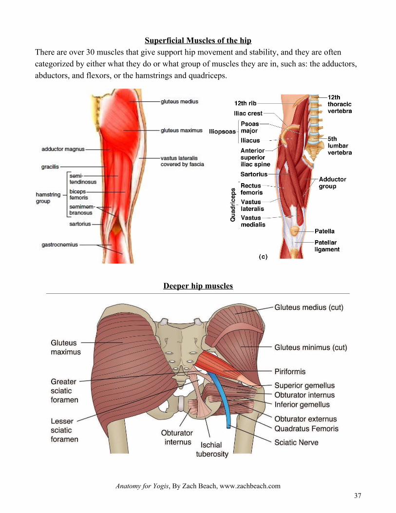
Superficial Muscles of the hip There are over 30 muscles that give support hip movement and stability, and they are often categorized by either what they do or what group of muscles they are in, such as: the adductors, abductors, and flexors, or the hamstrings and quadriceps.
Deeper hip muscles
Anatomy for Yogis, By Zach Beach, www.zachbeach.com 37

Around-the-hip joint yoga stretch sequence Many people will say they have “tight hips,” but are often unclear or uncertain about what muscles are actually tight. If a student asks you to do hip openers in class, you might want to ask them if they mean the front, back, inside, or outside of the hip, or deeper in the pelvic bowl.
Teacher challenge: Design a yoga class around a peak pose that requires open hips.
Anatomy for Yogis, By Zach Beach, www.zachbeach.com 38
To stretch the: Bring the hip into By coming into
hip flexors (anterior thigh)
Extension
abductors (lateral thigh) Adduction
hamstrings (posterior thigh) Flexion
adductors (medial thigh) Abduction
deep hip muscles External Rotation

Lesson 7: The Shoulder There are two ball and socket joints in the body. We have already covered the hip joint, and now we can look at the shoulder, which is a structure consisting of three bones: the humerus (upper arm bone), clavicle (collarbone), and scapula (shoulder blade) all tied together with numerous muscles, tendons and ligaments. The shoulder joint is more complex than the hip, because there are actually three joints that interact to create the movement of the shoulders. Those three joints are the:
● glenohumeral joint, where the humerus of the arm meets the scapula ● acromioclavicular joint, between the distal clavicle and acromion process of the
scapula, ● sternoclavicular joint, between the medial clavicle and manubrium of the sternum, as
shown below:
The shoulder joint is much more mobile than the hip joint and is in fact the most mobile joint in the body, allowing our arm to go into almost any position we want. We have a wide array of both superficial and deep muscles that work together to both stabilize the shoulder and allow us to swing from branches, do a handstand, and reach for things we need. This mobility comes at a cost however, as anyone who has dislocated their shoulder can attest. Let us first examine the glenohumeral joint. Unlike the head of the femur, which fits snugly into the acetabulum, the head of the scapula only covers a small portion of the head of the humerus, and is often described of as a golf ball on a tee. Four muscles are employed to keep the humeral head in the scapular socket during shoulder movements, together these are known as the rotator cuff: supraspinatus, infraspinatus, teres
Anatomy for Yogis, By Zach Beach, www.zachbeach.com 39

minor, and subscapularis (SITS muscles). The supraspinatus is the one most commonly torn or injured, as it can be worn down through mechanical abrasion and impingement on the acromion bone.
The acromioclavicular joint is at the top of the shoulder and its movement allows the arm to rise above the head, helping the movement of the scapula and giving the arm greater rotation for asanas such as Downward Facing Dog. At the medial end of the clavicle is the sternoclavicular joint, where the convex shape of the manubrium interfaces with the rounded end of the clavicle. Beyond the rotator cuff, there are then larger and more superficial muscles that cover the arm, shoulders and back, to keep the scapula in place, allow for dynamic range of movement, and give strength and stability to the upper body.
Anatomy for Yogis, By Zach Beach, www.zachbeach.com 40

Anatomy for Yogis, By Zach Beach, www.zachbeach.com 41

Anatomy for Yogis, By Zach Beach, www.zachbeach.com 42
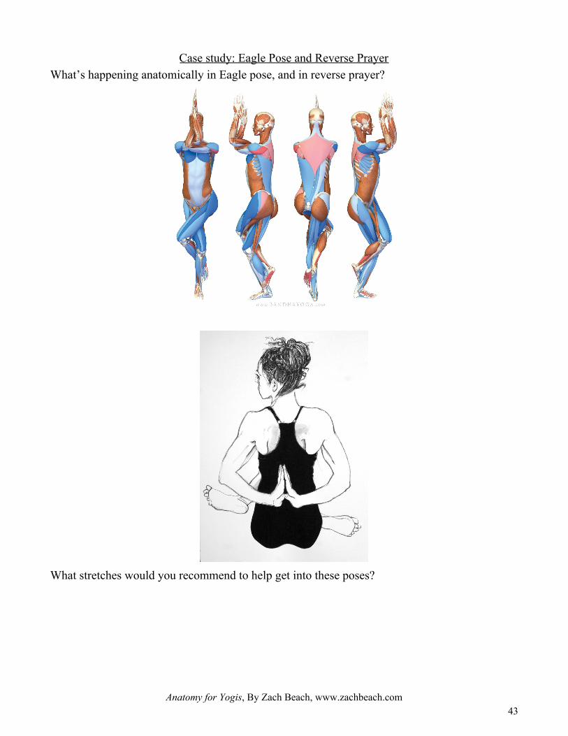
Case study: Eagle Pose and Reverse Prayer What’s happening anatomically in Eagle pose, and in reverse prayer?
What stretches would you recommend to help get into these poses?
Anatomy for Yogis, By Zach Beach, www.zachbeach.com 43

Shoulder Questions What muscles play a role in shoulder strength and stability in:
● Upward Dog
● Handstand
● Forearm stand
Lastly, consider the plight of the modern office worker:
Anatomically, what’s happening in the shoulders? Which muscles might need to be stretched? Which muscles might need to be strengthened? What yoga poses might you recommend for your corporate private yoga client?
Anatomy for Yogis, By Zach Beach, www.zachbeach.com 44

Lesson 8: The Foot, Ankle, and Knee Now that we have covered most of the major joints close to the axial skeleton, let us focus more on the appendicular skeleton, focusing on the foot, ankle, and knee. This part of the body is of course, our lowest part, and thus provides the foundation for almost everything we do, from standing, walking, running, and simply moving about our lives. In yoga, our feet are the base for all of our standing postures, and will form an intimate connection with the knee when it bends to come into poses like Warrior II or High Lunge. The ability for the foot and ankle to quickly adapt to changing forces immediately comes to play once we balance on one leg.
The Foot Every single day, we walk on our feet. They are the foundation of all of our standing postures. Each foot is made up of 26 bones, 30 joints and more than 100 muscles, tendons and ligaments, all of which work together to provide support, balance, and mobility. Bones of the feet Nearly one-fourth of the body's bones are in our feet. The 26 bones of the foot consist of eight distinct types, including the tarsals, metatarsals, phalanges, cuneiforms, talus, navicular, and cuboid bones.
● Calcaneus (the heel bone): The largest bone of the foot, it is commonly referred to as the heel of the foot. It points upward, while the remaining bones of the feet point downward.
● Talus (the ankle bone): This irregularly shaped bone creates the lower portion of the ankle joint. It is the second largest bone in the foot.
Anatomy for Yogis, By Zach Beach, www.zachbeach.com 45

● Navicular: This curved bone sits between the talus and cuneiforms. Tight shoes can irritate the navicular bones.
● Cuneiforms: These three small bones are closest to the five metatarsal bones. They sit in a row that begins at the inside of the foot, and moves toward the cuboid on the outside of the foot.
● Cuboid: This multifaceted bone sits on the outside of the foot near the fifth phalanx (little toe). The cuboid is easy to locate on the outer edge of the foot.
● Metatarsals – five bones (labeled one through five, starting with the big toe) that make up the forefoot. Sometimes the metatarsals are called the “rays” of the foot like sun rays that fan out.
● Phalanges (singular: phalanx) – the 14 bones that make up the toes. The big toe consists of two phalanges – the distal and proximal. The other toes have three.
● Sesamoids – two small, pea-shaped bones that lie beneath the head of the first metatarsal in the ball of the foot.
The cuboid, navicular and medial, intermediate and lateral cuneiforms are all considered tarsal bones, the five irregularly shaped bones of the midfoot that form the foot's arch. Muscles of the feet Twenty muscles give the foot its shape, support and the ability to move. The main muscles of the foot are:
● the tibialis posterior, which supports the foot's arch
● the tibilias anterior, which allows the foot to move upward
● the tibilias peroneal, which controls movement on the outside of the ankle
● the extensors, which help raise the toes, making it possible to take a step
● the flexors, which help stabilize the toes. These flexors include digitorum longus, fibularis longus, fibularis tertius, fibularis brevis.
Tendons and Ligaments of the feet Many tendons attach these muscles to the bones and ligaments that hold the bones together to maintain the foot's arch. The main tendon of the foot is the Achilles tendon, which runs from the
Anatomy for Yogis, By Zach Beach, www.zachbeach.com 46

calf muscle to the heel. The Achilles tendon makes it possible to run, jump, climb stairs and stand on your toes. The main ligaments of the foot are:
● plantar fascia – the longest ligament of the foot. The ligament, which runs along the sole of the foot, from the heel to the toes, forms the arch. By stretching and contracting, the plantar fascia helps us balance and gives the foot strength for walking.
● plantar calcaneonavicular ligament – a ligament of the sole of the foot that connects the calcaneus and navicular and supports the head of the talus.
● calcaneocuboid ligament – the ligament that connects the calcaneus and the tarsal bones and helps the plantar fascia support the arch of the foot.
The Arches of the Foot. The foot has three arches: two longitudinal (medial and lateral) arches and one anterior transverse arch. They are formed by the tarsal and metatarsal bones, and supported by ligaments and tendons in the foot. The medial arch is relatively more elastic than the other arches, gaining additional support from the tiabilias posterior and peroneus longus muscles from above. The lateral arch posses a special locking mechanism, allowing much less movement. Common issues of the foot include leaning forward, leaning back, and a flattening of the arches.
Anatomy for Yogis, By Zach Beach, www.zachbeach.com 47

The Ankle
The ankle joint is formed by the bones of the leg (tibia and fibula) and the foot (talus). Functionally, it is a hinge type joint, permitting dorsiflexion and plantarflexion of the foot. Plantar and Dorsiflexion In yoga, many of our prone backbends tend to use plantarflexion. Forward folds tend to be dorsiflexion. There's one muscle on the front of the leg for dorsiflexion, tibialis anterior. There are three on the back of the leg for plantar flexion: the gastrocnemius, soleus, and plantaris.
Anatomy for Yogis, By Zach Beach, www.zachbeach.com 48

Inversion and Eversion
It is very easy to invert the foot. In many poses like tabletop and upward dog we unconsciously do invert the foot. Lotus pose and many marici variations have deep inversion, as a result some people do sometimes feel pain in ankle. Ankle Sprains An ankle sprain refers to partial or complete tears in the ligaments of the ankle joint. It usually occurs via excessive inversion to a plantarflexed and weight-bearing foot. The lateral ligament is more likely to be damaged for two main reasons:
● The lateral ligament is weaker than the medial ligament. ● The lateral ligament resists inversion.
Anatomy for Yogis, By Zach Beach, www.zachbeach.com 49

The anterior talofibular ligament is the lateral ligament most at risk of irreversible damage.
The Knee The knee represents the incredibly important connection between the bottom and top halves of the legs, and receives considerable force from both sides. The joint surfaces are lined with hyaline cartilage, and are enclosed within a single joint cavity. The knee joint is formed by articulations between the patella, femur and tibia. Upon closer examination we can see that it is actually two joins, tibiofemoral and patellofemoral joints:
● Tibiofemoral – medial and lateral condyles of the femur articulate with the tibial condyles. It is the weight-bearing component of the knee joint. It is a hinge type synovial joint (also a bicondylar joint), which mainly allows for flexion and extension (and a small degree of medial and lateral rotation).
● Patellofemoral – anterior aspect of the distal femur articulates with the patella. It allows the tendon of the quadriceps femoris (knee extensor) to be inserted directly over the knee
Anatomy for Yogis, By Zach Beach, www.zachbeach.com 50

– increasing the efficiency of the muscle. As the patella is both formed and resides within the quadriceps femoris tendon, it provides a fulcrum to increase the power of the knee extensor, and serves as a stabilising structure that reduces frictional forces placed on femoral condyles.
Ligaments of the Knee
The medial and lateral collateral ligaments (MCL and LCL) run along the sides of the knee and limit sideways motion. The MCL protects the medial side of the knee, while the LCL protects the lateral side from an inside bending force, such as when a yogi places their foot on their knee in tree pose. Inside the knee joint are two cruciate ligaments: the ACL and PCL. The ACL, connected the tibia to the femur at the center of the knee, limits rotation and forward motion of the tibia away from the femur, and is a crucial source of stability. This is where ankle over knee alignment is so critical. The PCL, limits hyperextension, and not often innjured in yoga. Meniscus Tears Each of your knees has two menisci — C-shaped pieces of cartilage that act like a cushion between your shinbone and your thighbone. A torn meniscus causes pain, swelling and stiffness. You also might feel a block to knee motion and have trouble extending your knee fully. A torn meniscus is one of the most common knee injuries. Any activity that causes you to forcefully twist or rotate your knee, especially when putting your full weight on it, can lead to a torn meniscus.
Anatomy for Yogis, By Zach Beach, www.zachbeach.com 51

Meniscus tears can occur in all age groups. Traumatic tears are most common in active people from age 10-45. Degenerative tears are most common in people from age 40 upward.
● Degenerative Meniscal Tears - Degenerative meniscal tears are thought to occur as part of the aging process when the collagen fibers within the meniscus start to break down and lend less support to the structure of the meniscus. Degenerative tears are usually horizontal in the meniscus producing both an upper and lower segment of meniscus.
● Traumatic Meniscal Tears - Most traumatic meniscal tears occur as a result of a twisting injury when the knee rotates but the foot stays fixed in position. The meniscus can also tear from extreme bending of the knee. The combination of bend, rotation, and sudden kick that occurs in some forms of martial arts is associated with lateral meniscal tears.
Tears in the medial meniscus are the most common in yoga, whether originally injured during an asana or exacerbated in an asana such as Lotus pose or others where forced rotation at the hip joint can transfer stress into the knee when the foot is held in place by the floor or another part of the body.
Anatomy for Yogis, By Zach Beach, www.zachbeach.com 52

Lesson 9: Putting it All Together: The Anatomy of a Backbend If you want to go deeper into an expression of a pose, it helps to know what your muscles need to do. For the muscles that are working--the agonists--it will help to get them stronger. For the muscles that are lengthening, the antagonists, they will need to become more flexible and relaxed. Getting deeper into our backbends offers a nice case study for learning about key muscles in the body. If we take a look at the following variation of bow pose,
we can see that the entire back body is contracting to get the body off of the ground, while the front body is lengthening. Anatomically from the bottom up we see:
● plantarflexion ● knee flexion ● hip extension ● spinal extension ● shoulder flexion ● elbow flexion.
To get deeper into this pose and most other backbends, we need to strengthen the agonists for these movements and stretch the antagonists.
Anatomy for Yogis, By Zach Beach, www.zachbeach.com 53

Basic Backbend Principles 1. Strong legs: rotating the inner thighs inwards and back. 2. Core Engagement: support your back by pulling the belly in and up, protecting the
lower back. 3. Shoulders down: firm the bottom tips of the shoulder-blades in towards each other and
(as if your shoulder-blades are hands gently pressing into your upper back). 4. Long neck: Keep the neck long and happy and aim for an even arch over the whole
spine. 5. Counterposes: Always do a counterpose, like child’s pose, forward fold, or rabbit, after
1-3 backbends. 6. Courage and vulnerability: People do not usually enter into backbends in their day to
day activities, so even coming into one can be scary. Backbends also open the heart, and can bring up challenging emotions. This is why courage and vulnerability is key.
Being a Yoga Practitioner vs. Being a Yoga Teacher As yoga practitioners, we can follow Sri. K. Pattabhi Jois’s advice, “Do your practice and all is Coming.” It doesn’t matter if you have a PhD in philosophy, are an experienced doctor, or brand new to any athletic activity, the practice of yoga challenges us all in the exact places we need to grow the most. From an anatomical perspective, we call this the “peeling the onion approach.” In every yoga posture we move until we become inhibited in some way. Without perhaps knowing exactly which muscle is tight or which muscle is weak, we come back to the pose the next day and the next day until--often suddenly--an opening occurs and we are able to reach a deeper expression of the pose.
Anatomy for Yogis, By Zach Beach, www.zachbeach.com 54

However, after peeling away one restriction in one muscle, we simply encounter another. So we then work on peeling the next layer of the onion away. This is a perfectly acceptable approach to practicing yoga. But as yoga teachers, if we want to be of the best help to our students, it will help to be able to identify exactly what their bodies might need, in order to specifically target the correct issues. In this vein, it will greatly help the yoga teacher to know what muscles are playing what role in a pose. The next two pages cover the main agonists and antagonists we find in our backbends. As you continue to explore these poses in your own practice and with your students, try to identify how these muscles interact to create the desired anatomical position.
Anatomy for Yogis, By Zach Beach, www.zachbeach.com 55

Back body muscles to Strengthen
Anatomy for Yogis, By Zach Beach, www.zachbeach.com 56
Movement Muscle Origin Insertion Yoga poses
Hip extension, Knee flexion
Gluteus Maximus Ilium, sacrum, coccyx Gluteal tuberosity of femur Warrior III Upward Plank Locust (trdtnl)
Biceps Femoris Long head: Ischial tuberosity Short head: Posterior mid-shaft of femur
Head of fibula
Semimembranosus Ischial tuberosity Posterior medial condyle of tibia
Semitendinosus Ischial tuberosity Medial surface of proximal tibia
Adductor Magnus Inferior ischial ramus Linea aspera of femur
Spinal Extension iliocostalis iliac crest and sacrum angles of the ribs Cobra Peacock Locust (arms up)
longissimus transverse process at inferior vertebral levels
transverse process at superior vertebral levels and mastoid process
spinalis spinous processes at inferior vertebral levels
spinous processes at superior vertebral levels and base of the skull
Semispinalis capitis transverse processes of C7-T12
capitis: back of skull between nuchal lines;
Shoulder Flexion, elbow flexion
Anterior deltoid lateral one-third of the clavicle, acromion, the lower lip of the crest of the spine of the scapula
deltoid tuberosity of the humerus Chair pose
Downward dog Dolphin Puppy Biceps brachii short head: tip of the
coracoid process of the scapula; long head: supraglenoid tubercle of the scapula
tuberosity of the radius

Front body muscles to stretch
Anatomy for Yogis, By Zach Beach, www.zachbeach.com
57
Movement Muscle Origin Insertion Yoga poses
Hip extension, Knee flexion
Rectus femoris straight head: anterior inferior iliac spine; reflected head: above the superior rim of the acetabulum
patella and tibial tuberosity (via the patellar ligament)
Hero’s pose Lunges Splits
Vastus lateralis lateral intermuscular septum, lateral lip of the linea aspera and the gluteal tuberosity
patella and medial patellar retinaculum
Vastus medialis medial intermuscular septum, medial lip of the linea aspera
patella and medial patellar retinaculum
Vastus intermedius
anterior and lateral surface of the femur
patella
Sartorius anterior superior iliac spine medial surface of the tibia (pes anserinus)
Spinal Extension
Rectus abdominis
pubis and the pubic symphysis xiphoid process of the sternum and costal cartilages 5-7
Cobra Cow Upward dog Fish Side-bend
Intercostals upper borders of a rib fibers course up and medially to insert on the inferior margin of the rib above
Serratus anterior ribs 1-8 or 9 medial border of the scapula on its costal (deep) surface
Shoulder Flexion, elbow flexion
Latissimus dorsi vertebral spines from T7 to the sacrum, posterior third of the iliac crest, lower 3 or 4 ribs, sometimes from the inferior angle of the scapula
floor of the intertubercular groove
Puppy pose Wall down dog Cow-face arms
Pectoralis major medial 1/2 of the clavicle, manubrium & body of sternum, costal cartilages of ribs 2-6, sometimes from the rectus sheath of the upper abdominal wall
crest of the greater tubercle of the humerus
Pectoralis minor ribs 3-5 coracoid process of the scapula
Subscapularis medial two-thirds of the costal surface of the scapula (subscapular fossa)
lesser tubercle of the humerus
Triceps brachii long head: infraglenoid tubercle of the scapula; lateral head: posterolateral humerus & lateral intermuscular septum; medial head: posteromedial surface of the inferior 1/2 of the humerus
olecranon process of the ulna

Anatomy for Yogis, By Zach Beach, www.zachbeach.com
58

Lesson 10: Flexibility and Stretching Principles Before we close out our final lesson, let us consider a few more intermediate stretching principles to consider in your yoga practice and teaching. This lesson will cover different types of flexibility and stretching.
What is flexibility? Flexibility is defined as: the absolute range of movement in a joint or series of joints that is attainable in a momentary effort with the help of a partner or a piece of equipment. This definition tells us that flexibility is not something general but is specific to a particular joint or set of joints. It is a myth that some people are innately flexible throughout their entire body. Being flexible in one particular area or joint does not necessarily imply being flexible in another. Being "loose" in the upper body does not mean you will have a "loose" lower body. Furthermore, flexibility in a joint is also specific to the action performed at the joint. The ability to do front splits doesn't imply the ability to do side splits even though both actions occur at the hip.
Types of Flexibility
Many people are unaware of the fact that there are different types of flexibility. These different types of flexibility are grouped according to the various types of activities involved in athletic training. The ones which involve motion are called dynamic and the ones which do not are called static. The different types of flexibility are: Dynamic flexibility (also called kinetic flexibility) is the ability to perform dynamic (or kinetic) movements of the muscles to bring a limb through its full range of motion in the joints. Static-active flexibility (also called active flexibility) is the ability to assume and maintain extended positions using only the tension of the agonists and synergists while the antagonists are being stretched. For example, lifting the leg and keeping it high without any external support (other than from your own leg muscles). Static-passive flexibility (also called passive flexibility) is the ability to assume extended positions and then maintain them using only your weight, the support of your limbs, or some other apparatus (such as a chair or a barre). Being able to perform the splits is an example of
Anatomy for Yogis, By Zach Beach, www.zachbeach.com 59

static-passive flexibility. Active flexibility is harder to develop than passive flexibility (which is what most people think of as "flexibility"); not only does active flexibility require passive flexibility in order to assume an initial extended position, it also requires muscle strength to be able to hold and maintain that position.
Factors affecting flexibility Internal influences
● the type of joint (some joints simply aren't meant to be flexible) ● the internal resistance within a joint ● bony structures which limit movement ● the elasticity of muscle tissue (muscle tissue that is scarred due to a previous injury is not
very elastic) ● the elasticity of tendons and ligaments (ligaments do not stretch much and tendons should
not stretch at all) ● the elasticity of skin (skin actually has some degree of elasticity, but not much) ● the ability of a muscle to relax and contract to achieve the greatest range of movement ● the temperature of the joint and associated tissues (joints and muscles offer better
flexibility at body temperatures that are 1 to 2 degrees higher than normal) External influences
● the temperature of the place where one is training (a warmer temperature is more conducive to increased flexibility)
● the time of day (most people are more flexible in the afternoon than in the morning, being the most flexible around 2:30pm-4pm)
● the stage in the recovery process of a joint (or muscle) after injury (injured joints and muscles will usually offer a lesser degree of flexibility than healthy ones)
● age (pre-adolescents are generally more flexible than adults) ● gender (females are generally more flexible than males) ● one's ability to perform a particular exercise (practice makes perfect) ● one's commitment to achieving flexibility ● the restrictions of any clothing or equipment ● Some sources also the suggest that water is an important dietary element with regard to
flexibility. Increased water intake is believed to contribute to increased mobility, as well as increased total body relaxation.
Anatomy for Yogis, By Zach Beach, www.zachbeach.com 60

Types of Stretching Just as there are different types of flexibility, there are also different types of stretching. Stretches are either dynamic (meaning they involve motion) or static (meaning they involve no motion). Dynamic stretches affect dynamic flexibility and static stretches affect static flexibility. The different types of stretching are:
1. Ballistic stretching 2. Dynamic stretching 3. Static, passive, or relaxed stretching 4. Active stretching 5. PNF stretching
Ballistic stretching uses the momentum of a moving body or a limb in an attempt to force it beyond its normal range of motion. This is stretching, or "warming up", by bouncing into (or out of) a stretched position, using the stretched muscles as a spring which pulls you out of the stretched position. This type of stretching is not considered useful and can lead to injury. Dynamic stretching consists of controlled leg and arm swings that take you gently to the limits of your range of motion. An example of dynamic stretching would be slow, controlled leg swings, arm swings, or torso twists. While dynamic stretching is useful for many athletes, it is not traditionally part of a yoga practice (although some teachers do argue that Sun Salutations—or any yoga movement that moves in and out of poses with the breath—is a kind of dynamic stretching). As practitioners interested in gaining flexibility, we are primarily concerned with static stretching, active stretching, isometric stretching, and PNF stretching. Let’s go through each of these one by one.
Static Stretching In a static-passive stretch, the body is placed with mindful alignment into a position where a load, usually the weight of the body under the force of gravity, pulls on a targeted muscle and is held a minimum of twenty seconds. No deliberate contraction is initiated. This is the most simplistic form of stretching while still being safe and effective. Seated forward folds are one example of this form of stretching. The stretch is created as the weight and load of the torso folding over the legs exerts a pull on the targeted muscles along the back of the body in the stretch.
Anatomy for Yogis, By Zach Beach, www.zachbeach.com 61

Active Stretching: Using Reciprocal Inhibition, A Physiological Yin/Yang An active stretch is one where you assume a position and then hold it there with no assistance other than using the strength of your agonist muscles. Active stretching increases active flexibility and strengthens the agonistic muscles. Active stretches are usually quite difficult to hold and maintain for more than 10 seconds and rarely need to be held any longer than 15 seconds. For example, bringing your leg up high and then holding it there without anything (other than your leg muscles themselves) to keep the leg in that extended position. The tension of the agonists in an active stretch helps to relax the muscles being stretched (the antagonists) by reciprocal inhibition. We already learned that every movement of the body involves a prime mover/agonist and a muscle that goes against this movement, known as the antagonist.
For janu sirsasana, in the straight leg, the quadriceps are the agonists and the hamstrings are the antagonists. In the flexed leg, the hamstrings are the agonists and the quadriceps are the antagonists. It makes sense that there would be a corresponding physiological Yin/Yang to make biomechanical processes such as flexion and extension of the knee energy efficient, i.e., when the agonist muscle contracts, its antagonist relaxes. This process occurs unconsciously through a primitive spinal cord reflex arc called reciprocal inhibition. Stretching applies tension to the muscle and its tendon. There is a nerve receptor (the Golgi tendon organ) that is located at the muscle-tendon junction. This receptor senses tension and relays a signal to the spinal cord. The spinal cord then signals the stretching muscle to relax.
Anatomy for Yogis, By Zach Beach, www.zachbeach.com 62

This reflex arc acts as a protective circuit breaker to prevent the tendon from tearing at its attachment to the bone.
We can consciously access this reflex arc to deepen and improve our poses. In paschimottanasana, the quadriceps muscle is the agonist and the hamstring muscles are the antagonists. Consciously engaging the quadriceps also signals the hamstrings to relax. This takes place via the spinal cord. The nerve impulse that results in contraction of the quadriceps is called excitatory and the impulse to the hamstrings is called inhibitory. Try this technique to get a bit deeper in this pose and then apply it to different agonist / antagonist muscle groups. Now, we can consider what it takes to get the body into a cross legged, sukhasana pose. For cross legged seated position, we have external rotation and flexion of the hips, along with knee flexion and if we are working to stay upright, a bit of spinal extension.
1. __________________________________________________
2. __________________________________________________
3. __________________________________________________
4. __________________________________________________
5. __________________________________________________
Anatomy for Yogis, By Zach Beach, www.zachbeach.com 63

Proprioceptive neuromuscular facilitation (PNF) We can also use a technique called facilitated stretching or proprioceptive neuromuscular facilitation (PNF) to lengthen muscles in yoga, using similar principles of reciprocal inhibition. PNF utilizes a spinal cord reflex arc and is an example of combining modern Western science with the ancient art of Yoga. In general, in a PNF stretch there is generally a contraction of the targeted muscle right before the stretch. It is often referred to as the ‘contract/relax’ method.
Facilitated stretching involves contracting a muscle that you are lengthening. This increases the tension at the muscle-tendon junction and recruits more Golgi tendon organs than does stretching a muscle alone. Facilitated stretching causes the spinal cord to signal the muscle to relax, in essence, creating "slack" in the muscle. You can then take up the slack to move deeper into the pose. For example, in Paschimottanasana, slightly bend the knees and squeeze the trunk against the thighs. Then gently press the heels into the mat as if you are trying to flex the knees further. This engages the hamstrings and stimulates the Golgi tendon organs at the muscle-tendon junction. Hold this steady contraction for five to eight breaths before releasing it. This produces relaxation and increased length in the hamstrings. Then contract the quadriceps to straighten the knees and take up the slack created by the reflex arc. This has the added effect of producing reciprocal inhibition, which further relaxes the hamstrings into the stretch.
Anatomy for Yogis, By Zach Beach, www.zachbeach.com 64

Follow the same steps in this pose with the quadratus lumborum and erector spinae. First, bend forward to stretch these back muscles. Hold the stretch by bending the arms, and then attempt to arch the back. This increases tension at the muscle-tendon junction of the erector spinae and quadratus lumborum, stimulates the Golgi tendon organs, and ultimately relaxes these muscles. Hold the arch of the back for five to eight smooth breaths. Then engage the abdominals and use the arms to draw yourself deeper into the pose, taking advantage of the new length created by the reflex arc. Activating the abdominals also produces reciprocal inhibition of the back muscles, relaxing them into the stretch. Go slowly with facilitated stretching. Allow about 48 hours of recovery time before re-applying PNF to any given muscle group. It is not necessary to use strong muscular contractions to experience the benefits of these techniques. Start with gentle force and learn to moderate the contraction, "dialing" it in. Use similar care as you gradually release the action of the muscles. Remember that improvement comes from a balance between the conscious work on the poses and the unconscious work that takes place between practice sessions. The unconscious mind forms new and more efficient brain circuitry during this "in-between" time. Now, try PNF stretches for the:
1. Quadriceps
2. Hamstrings
3. Adductors
4. Abductors
Anatomy for Yogis, By Zach Beach, www.zachbeach.com 65

Appendices
Anatomy for Yogis, By Zach Beach, www.zachbeach.com 66

Pose breakdown
Anatomy for Yogis, By Zach Beach, www.zachbeach.com 67
Yoga Pose (English): Sanskrit name:
Joint (Ankle, knee, elbow, etc)
Joint motion (flexion, extension, abduction, etc.)
Agonists/Antagonists pairs (biceps/triceps, hamstring/quadriceps, etc)
Type of contraction (concentric, isometric, eccentric)
Notes: