Anatomy and Physiology of The Eye By Hesham M. El-Toukhy Ass. Prof. of Ophthalmology KFU, KSA.
88
Anatomy and Physiology of The Eye By Hesham M. El-Toukhy Ass. Prof. of Ophthalmology KFU, KSA
-
Upload
garry-chase -
Category
Documents
-
view
227 -
download
6
Transcript of Anatomy and Physiology of The Eye By Hesham M. El-Toukhy Ass. Prof. of Ophthalmology KFU, KSA.

Anatomy and Physiology of The Eye
By Hesham M. El-ToukhyAss. Prof. of OphthalmologyKFU, KSA

• Ophthalmology. A Short Textbook by F. Hollwich, Thieme Verlag Stuttgart, Germany.
• Lecture notes on ophthalmology. Bruce James, Chris Chew, Anthony J. Bron, Blackwell publishing.
• Clinical ophthalmology: A Systemic approach By: Kanski Jack 3, Butterworth-Heinemann.

- Orbit
- Eye Lids.
- conjunctiva.
- Lacrimal apparatus:
Lacrimal gland.
Lacrimal drainage system.
- Extraocular muscles.
- The globe and visual pathway.

Pear shaped Pear shaped cavities.cavities.
Apex (within optic Apex (within optic canal) directed canal) directed posteriorly, posteriorly, medially, and medially, and slightly upwards.slightly upwards.
Formed of 7 bones: Formed of 7 bones: frontal, zygomatic, frontal, zygomatic, maxilla, palatine, maxilla, palatine, lacrimal, ethmoid, lacrimal, ethmoid, and sphenoid.and sphenoid.
The orbit The orbit فورمن فورمن 33فيه فيهكفتي االوربت تربط كفتي اهميتها االوربت تربط اهميتها
السكل في كفتي السكل بأذر في كفتي بأذر

Periorbita (orbital fascia) at base

•-orbit cavity is pyramidal in shape .its apex is goes posteriorly.angle=90 between superior and
inferior fissure•-foramin in orbita cavity:1-optical canal.2-superior
and inferior fissure•-periostium at anterior margin is called periorbital
which is thicken with eye lid to form orbital septum then became more thicker to form tarsal plate
•-skeleton of eye lid are orbital septum and tarsus plate {{at base

Orbital apex. Common tendinous ring
(annulus of Zinn)
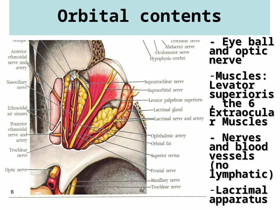
Orbital contents
- Eye ball - Eye ball and optic and optic nervenerve-Muscles: Muscles: Levator Levator superioris, superioris, the 6 the 6 Extraocular Extraocular Muscles Muscles
- Nerves and - Nerves and blood blood vessels (no vessels (no lymphatic)lymphatic)-Lacrimal Lacrimal apparatus apparatus -Orbital fatOrbital fat

1-1-skinskin
2-subcutaneous tissue.2-subcutaneous tissue.
3-orbicularis occuli muscle.3-orbicularis occuli muscle.
4-orbital septum and tarsal plate.4-orbital septum and tarsal plate.
5-conjunctiva.5-conjunctiva.
--Aponeurosis of levator Aponeurosis of levator palpebrae superioris muscle inpalpebrae superioris muscle in
اللور >> عن يفرقه الي اللور >> هذا عن يفرقه الي upper upperهذاeye lideye lid..
--smooth muscle (upper and lower tarsal muscles)smooth muscle (upper and lower tarsal muscles)..
The LidThe Lid

Lid margin:Lid margin:
Cilliary portion.Cilliary portion.
Lacrimal portion. Lacrimal portion. No cillia here No cillia here
Opening of tarsal Opening of tarsal (meibomian) (meibomian) glands.glands.
Gray line: junction Gray line: junction between ant. Part between ant. Part (skin and muscle) (skin and muscle) and post. Part and post. Part (tarsus and (tarsus and conjunctiva).conjunctiva).

• Blood supply: medial palpebral (of ophthalmic) and lateral palpebral (of lacrimal of ophthalmic) arteries.
• Lymphatic drainage: superficial parotid and submandibular nodes.
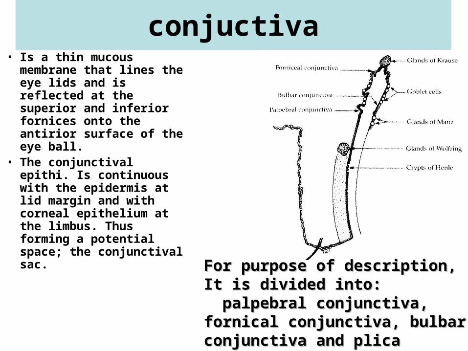
conjuctiva• Is a thin mucous
membrane that lines the eye lids and is reflected at the superior and inferior fornices onto the antirior surface of the eye ball.
• The conjunctival epithi. Is continuous with the epidermis at lid margin and with corneal epithelium at the limbus. Thus forming a potential space; the conjunctival sac. For purpose of description, It is For purpose of description, It is
divided into:divided into: palpebral conjunctiva, fornical palpebral conjunctiva, fornical conjunctiva, bulbar conjunctiva and conjunctiva, bulbar conjunctiva and plica semilunaris.plica semilunaris.


• Palpebral conjunctiva : firmly adherent to the tarsus. Sulcus subtarasalis is a shallow groove on the back of the eye lid 2mm from post. Lid margin.

Histologically, the conjunctiva is formed of 2-5 layers of stratified columnar epith.
Resting on loose connective tissue .
• Goblet cells are scattered along the surface of the conjunctiva, it secretes the mucous component of the tear film.
• Accessory lacrimal glands situated in the connective tissue of the conjunctiva.

• Blood supply of the conjunctiva comes from the palpebral vessels of the eye lid that supply palpebral conjunctiva, fornical conjunctiva (posterior conjunctival arteries), and bulbar conjunctiva reaching for 3mm from the limbus, where they anastomose with the anterior conjunctival arteries which are branches of the anterior cilliary arteries.
• Lymphatic drainage: to superficial parotid and submandibular nodes.

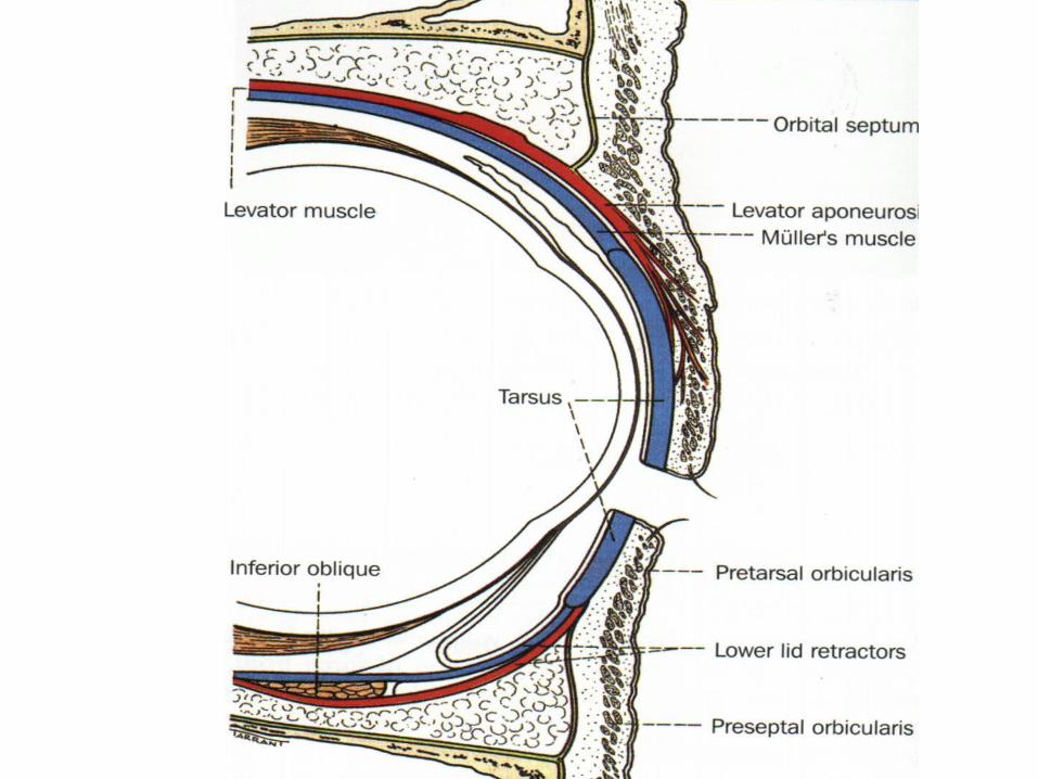
Orbital septum and tarsal plateOrbital septum and tarsal plate
Tarsal glands (meibomian gl.) 20-30 embedded in Tarsal glands (meibomian gl.) 20-30 embedded in the substances of the tarsus in postirior wall . the substances of the tarsus in postirior wall . Opening at lid margin. Opening at lid margin.
Orbital Orbital septum is the septum is the framework of framework of the lid. the lid. Attached to Attached to orbital orbital margin. margin. Separates lid Separates lid from orbital from orbital cavity. cavity. Become Become thickened to thickened to form the form the tarsus.tarsus.

Nerve supply: facial (7Nerve supply: facial (7thth) N enter the muscle from ) N enter the muscle from temporal side.temporal side.
Orbicularis Orbicularis oculioculi::
-close eye -close eye Lid << most Lid << most importanant importanant
-dilates -dilates lacrimal sac lacrimal sac (suction of (suction of tears).tears).

Insertion: -aponeurosis into the upper edge of tarsus Insertion: -aponeurosis into the upper edge of tarsus (smooth muscle of tarsus), ant. surface of tarsus(smooth muscle of tarsus), ant. surface of tarsus ( also ( also skin. -Medial and lateral expansion of aponeurosis into med. and lat. palpeb skin. -Medial and lateral expansion of aponeurosis into med. and lat. palpeb ligaments. –upper conj fornix).ligaments. –upper conj fornix).
Nerve supply by superior branch of oculomotor (3Nerve supply by superior branch of oculomotor (3rdrd)N. )N.
part which is smooth muscle of the tarsus: sympathetic Ns from superior part which is smooth muscle of the tarsus: sympathetic Ns from superior cervical sympathetic ganglion. cervical sympathetic ganglion.
Origin from apex of eye lid
Levator Levator palpebrae palpebrae
superioris :superioris :
only in the upper eye only in the upper eye lid.lid.Function: elevation Function: elevation of the eye lid.of the eye lid.

Consists of Consists of large orbital large orbital part and small part and small palpebral part palpebral part that are that are continuous continuous with each with each other around other around the apenorosis the apenorosis of the levator of the levator Ms.Ms.
lies in the anterior upper temporal part of the orbit
Lacrimal gland

Lacrimal gland
• Ducts from the orbital part and plapebral part open in the superior fornix.
• Blood supply: lacrimal artery of opthalmic artery.
• Lymphatic drainage: with conjunctival drainage to superficial parotid nodes.

Lacrimal drainage system- 2 Lacrimal puncti - 2 Lacrimal puncti on the summit on the summit lacrimal papillea.lacrimal papillea.
- 2 Lacrimal - 2 Lacrimal canaliculi each canaliculi each has vertical and has vertical and horizontal portion.horizontal portion.
- Lacrimal sac: - Lacrimal sac: has fundus , body, has fundus , body, and neck.and neck.
- Nasolacrimal duct: connects sac with inferior meatus - Nasolacrimal duct: connects sac with inferior meatus of the nose. Directed downwards, backwards, and of the nose. Directed downwards, backwards, and laterally. laterally.

The Exrtraocular Muscles
There are 6 extraocular muscles:
4 straight muscles (4 recti)
1- superior rectus. 2- inferior rectus.
3- medial rectus. 4- lateral rectus.
2 0blique muscles
1- superior oblique.
2- inferior oblique.

Origin of extraocular muscles5 arise from 5 arise from the apex of the the apex of the orbit except orbit except the inferior the inferior oblique.oblique.
The 4 recti: The 4 recti: from annulus from annulus of zinn of zinn (common (common tendinous tendinous ring).ring).
The superior oblique: above and medial to The superior oblique: above and medial to the annulus of Zinn.the annulus of Zinn.
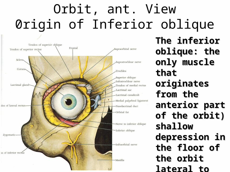
Orbit, ant. View0rigin of Inferior oblique
The inferior The inferior oblique: the only oblique: the only muscle that muscle that originates from originates from the anterior part the anterior part of the orbit) of the orbit) shallow shallow depression in depression in the floor of the the floor of the orbit lateral to orbit lateral to the fossa for the fossa for lacrimal sac.lacrimal sac.

Insertion of extraocular muscles
All are inserted in the sclera.
The 4 recti muscles are inserted into the sclera anterior to the equator close to the limbus.
The oblique muscles are inserted into the sclera, posterior to the equator at the posterior temporal part of the sclera.

Insertion of the 4 Recti (right Eye)In the In the
sclera sclera anterior anterior to the to the equator equator (spiral of (spiral of Tillaux << Tillaux << junction junction bettwen bettwen cornea cornea and and seclera ).seclera ).

Insertion of the oblique musclesin the sclera posterior to the equator (right
Eye).
Superior Superior oblique: oblique: upper post. upper post. Lateral Lateral part.part.
Inferior Inferior oblique: oblique: lower post. lower post. Lateral partLateral part
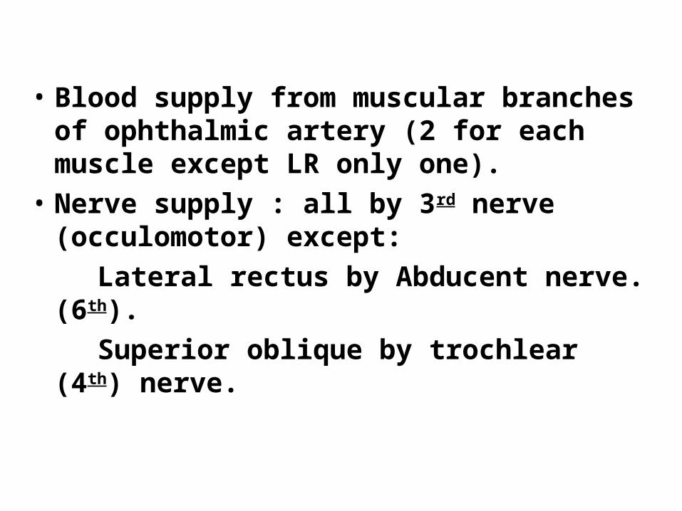
• Blood supply from muscular branches of ophthalmic artery (2 for each muscle except LR only one).
• Nerve supply : all by 3rd nerve (occulomotor) except:
Lateral rectus by Abducent nerve. (6th).
Superior oblique by trochlear (4th) nerve.

Uniocular eye movements
A- elevation B- depression C- adbuction d- adduction E–extortion << away from the nose F-
entortion

The horizontal rectiThe medial rectus:Arise from annulus of zinn, inserts 5.5
mm from the limbus on the medial side of the globe.
Its sole action is adduction.The lateral rectus:Arise from annulus of zinn, inserts 6.5
mm from the limbus on the lateral side of the globe.
Its sole action is abduction.

The vertical recti
The vertical recti run in the same direction of the orbital axis. So they form angle of 23° with the optical axis.

Orbital axis
Optical axis in primary position
Axis of vertical recti
Axis of oblique recti

The superior rectus main The superior rectus main (primary) action in the (primary) action in the primary position is primary position is elevationelevation..Secondary actions are Secondary actions are adduction and intortionadduction and intortion..In position of 23In position of 23°° adbuction the only adbuction the only action is elevation. action is elevation. (optimal position for (optimal position for function testing) function testing) In position of 67In position of 67°°
adduction the only adduction the only action is intortion. (the action is intortion. (the line of pull of the muscle line of pull of the muscle makes 90 degree with makes 90 degree with the optical axis). the optical axis).
Action of right SR

The inferior rectus main The inferior rectus main action in the primary action in the primary position is position is depressiondepression..
Secondary actions areSecondary actions are لو لومع برلل ونوت وورد فور مع كانت برلل ونوت وورد فور كانتثالث لها بول االي حق ثالث االكسس لها بول االي حق االكسس
adduction and adduction andحركات حركات extortionextortion..In position of 23In position of 23°°adbuctionadbuction
االكسس حق برلل تقريبا االكسس ووصار حق برلل تقريبا ووصارحركه لها انقل ومافي بول أي حركه اوف لها انقل ومافي بول أي اوف
the only action is the only action is وحده وحده depression. (optimal position depression. (optimal position for function testing)for function testing)..In position of 67 In position of 67 °° adduction adduction the only action is extortion. the only action is extortion. (the line of pull of the muscle (the line of pull of the muscle makes 90 degree with the makes 90 degree with the optical axis)optical axis)..
Action of right IR
The oblique muscles
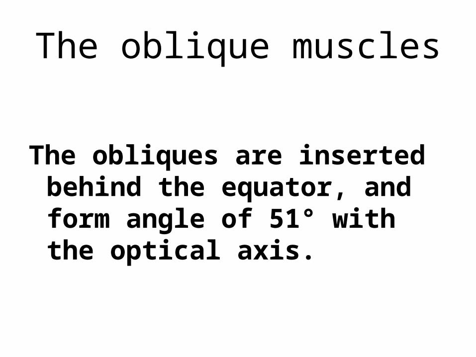
The obliques are inserted behind the equator, and form angle of 51° with the optical axis.

Orbital axis
Optical axis in primary position
Axis of vertical recti
Axis of oblique recti

The superior oblique
Originates above and medial to optic canal, passes through the trochlea between the superior and medial orbital walls, become reflected backwards, to insert in the posterior upper temporal quadrant of the glob.


Insertion of the oblique muscles in the sclera posterior to the
equator (right Eye).Superior Superior oblique: oblique: upper post. upper post. Lateral Lateral part.part.
Inferior Inferior oblique: oblique: lower post. lower post. Lateral partLateral part

Its primary action in the primary position is intortion, its secondary actions are depression and abduction.
In position of 51° adduction, its only action is depression. (0ptimal position for testing of function).
In position of 39° abduction its only action is intortion. (the line of pull of the muscle makes 90° with the optical axis).
Action of SO

The inferior oblique
Originates from small depression just behind the lower orbital margin, lateral to the tear duct, to insert in the posterior lower temporal quadrant of the glob. (close to the macula).
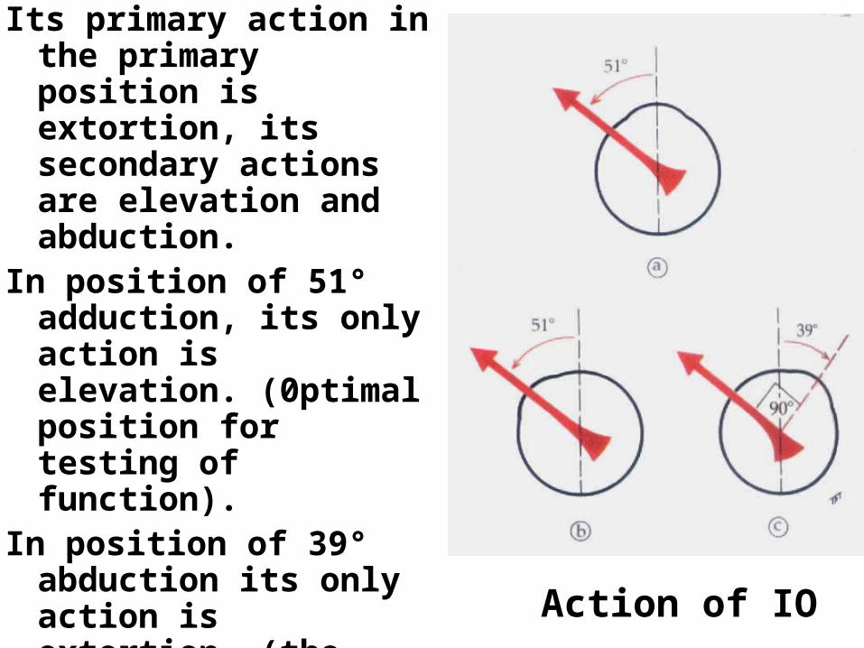
Its primary action in the primary position is extortion, its secondary actions are elevation and abduction.
In position of 51° adduction, its only action is elevation. (0ptimal position for testing of function).
In position of 39° abduction its only action is extortion. (the line of pull of the muscle makes 90° with the optical axis).
Action of IO

Remember,
The 0bliques are abductors and verticals are adductors.
The superiors are intortors, and the inferiors are extortors.

The eye ball
• Surrounded by fascial sheath (tenon capsule)
• Formed of segments of 2 spheres; ant transparent smaller and post opaque larger.
• Ant. Pole is the center of curvature of ant. Segment, post. pole is the center of curvature of post. Segment. The equator lies midway between the two poles.
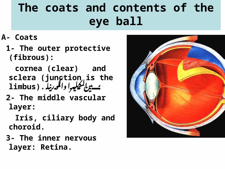
The coats and contents of the eye ball
A- Coats
1- The outer protective (fibrous):
cornea (clear) and sclera (junction is the limbus). بين
والكورنيا السكليرا 2- The middle vascular layer:
Iris, ciliary body and choroid.
3- The inner nervous layer: Retina.

The coats and contents of the eye ball
B- contents:- Anterior chamber between
cornea and iris ( have hol in middel << pupil ) , filled with aqueous.
- Crystalline lens البيوبل ورى- Posterior chamber between iris
and lens, filled with aqueous from cillary body .
-The vitreous cavity filled with the vitreous body

Refractive media of the eye : الريتنا يوصل لين خاللها يمر الضووؤء اليCornea, aqueous, the lens and the vitreous body.

The cornea
• Transparent anterior 1/6 of the outer coat 70% of refrection of eye {{ major part in refraction and it fixed
• Diameter: about 12 mm.• Thickness: central 0.56mm peripheral 1mm.• Refractive power: 42 D (75% of the
refractive power of the eye).

Histologically: formed of 5 layers.1- Epithelium (Stratified nonkeratinized
squamous epith).2- Bowman’s membrane.3- stroma. (( main thikmness ) 4- Descemet’s membrane.5- Endothelium.
The limbus is the corneoscleral junction

The cornea
• Nerve supply: very reach in sensory supply from nasociliary branch of trigeminal nerve (5th) cranial nerve (sensory).
• It is avascular, nutrition through limbal capillaries, aqueous humor, and tear film caver the cornea

Tear film

The cornea: is the part with main refractive power in the eye.
Factors responsible for corneal Transparency:
• Avascular << in ifnlamation will be opay • Regular stromal lamellae << if irregular will be
opay • Endothelial pump: removal of excess fluid by
the endothelium<< any difunction << edema << opay
• Non-myelination of corneal nerves << will be mailenated in liprosy for example

Sclera
• Posterior 5/6 of the outer coat • White in color ,may be bluish in
children• Thickness: 1 mm posterior, 0.6 mm
at equator 0.3 mm at insertion of recti muscles (thinnest area).

Scleral foramina كل الزمالسكليرا حق تروح الستركجر
• Anterior : exit of
anterior ciliary vessels
and nerves
( at the insertion of
the recti)• Equatorial: exit of
vortex veins• Lamina cribrosa: exit
of optic nerve bundles • Posterior: around optic nerve:
exit of posterior ciliary vessels and nerves

Sclera , structure, function
• Episclera.• sclera is separated from choroid by
suprachoroidal space.• Thick collagenous bundles running in
various directions.- Functions:• Protection • Insertion of muscles• Preserve shape of globe

Vascular layer
• Iris
• Ciliary body
• Choroid

The Iris• Pigmented contractile diaphragm with
central opening called the pupil.

-Iris pattern is the presence of fine irregularities on the anterior iris surface due to presence of the collarette (irregular circular line) and the crypts (areas of dark depressions).
-It is divided by the collarets into pupillary and cilliary zones.
-The post. Surface is darkly pigmented.
-The iris is attached to the middle of the antrior surface of the cilliary body.

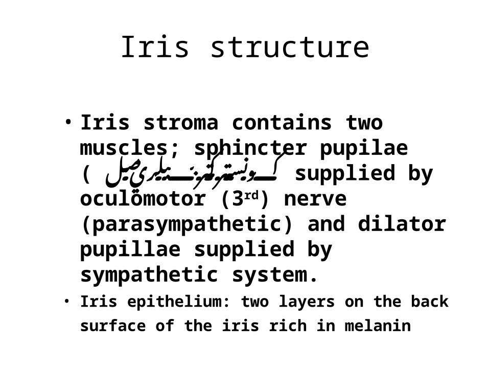
Iris structure
• Iris stroma contains two muscles; sphincter pupilae ( كونستركتر
مصل supplied by ببلريoculomotor (3rd) nerve (parasympathetic) and dilator pupillae supplied by sympathetic system.
• Iris epithelium: two layers on the back surface
of the iris rich in melanin

Iris function
• Control amount of light entering the globe through the action of sphincter and dilator pupillae
• Give the color of the eye through the melanin pigment

The Ciliary bodyIt extends between the iris and choroid.
It is triangular in cross section.
The base of the triangle (anterior surface) is continuous with the iris root.
The apex is directed posteriorly and is continuous with the choroid.

The Ciliary bodyThe anterior surface (base)
is called pars plicata and it contains 60-70 process and gives attachment to the lens zonules and responsible for aqueous formation.
The posterior surface is smooth and is called pars plana (important surgical land mark)
الفترس نوصل عشانانجوري نسوي ما بدون
الريتنا او اللنس It . حقlies against the sclera.

Histologically:Formed of ciliary epithelium, stroma, ciliary muscles.
-ciliary muscles: 3 parts;
1- longitudinal fiber.
2- circular fibers.
3- oblique fibers.
Nerve supply parasympathetic from occulomootr (3rd) nerve.
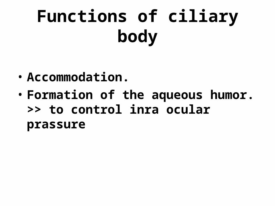
Functions of ciliary body
• Accommodation.
• Formation of the aqueous humor. << to control inra ocular prassure

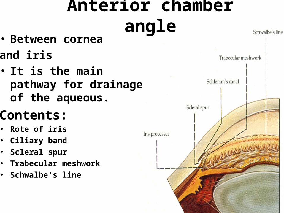
• Between cornea
and iris• It is the main pathway for
drainage of the aqueous.
Contents:• Rote of iris• Ciliary band• Scleral spur• Trabecular meshwork• Schwalbe’s line
Anterior chamber angle

The choroid
• Highly vascualr and pigmented membrane between the sclera and retina
• Formed of large, medium sized and small vessels and choriocapillaris.
• Main function is to nourish the retina
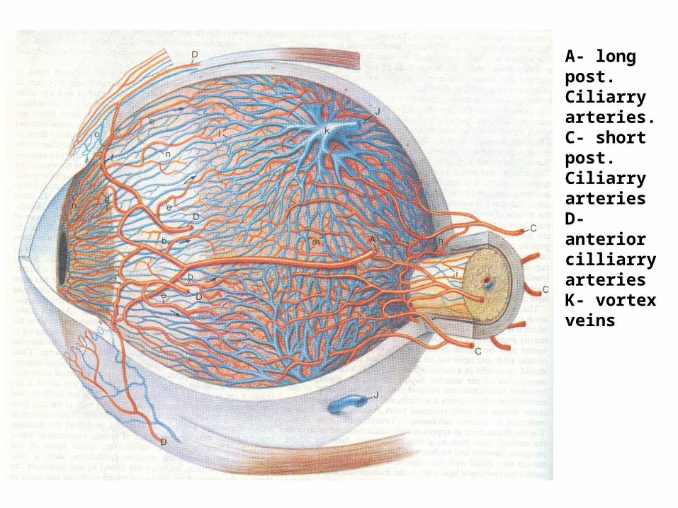
A- long post. Ciliarry arteries.C- short post. Ciliarry arteriesD- anterior cilliarry arteriesK- vortex veins

The crystalline lens• It is a transparent, biconvex
structure lies behind the iris and in front of the vitreous body.
• The convexity of its ant. Surface is less than of its posterior surface.
• 4 mm thick 10 mm diameter• It is kept in place by the
suspensory ligaments.• Center of ant.s called
antirior pole .. And the post. The same بوستيريور اسمه بول
• The priphri of lens called equator

• The lens is formed of:1- lens capsule, elastic clear membrane. {{ caver the lens 2- Subcapsular epithelium, single layer of cells under the anterior capsule and
absent at posterior capsule. At the equator it become transferred to lens fibers.
3- lens fibers. The earliest fibers moves centrally and form the nucleus , and the later fibers form the
cortex.
With advancing of age (about 25 years of age), the nucleus becomes progressively harder in component.النص في كبسول بره
نيوكليس وداخل كورتكسصارت كلما كبرنا كلما اليالى وتؤدي اكثر قاسيه الكتراكتس

• The lens is avascular and nutrition is obtained by diffusion from aqueous.
• Functions of the lens:
1- refractive media (25% of the refractive power of the eye).
2- protects retina from ultraviolet rays.
3- accommodation << main fanction

Accommodation: It is the ability of the lens to increase its
refractive power so that images of near objects can come to focus on the retina. (it is part of the near reflex).
Mechanism: contraction of the circular fibers of ciliary muscle causes Mechanism: contraction of the circular fibers of ciliary muscle causes relaxation of the lens zonules and the lens becomes more spherical relaxation of the lens zonules and the lens becomes more spherical mainly at its anterior surface and its refractive power increases.mainly at its anterior surface and its refractive power increases.

The Vitreous Body• It fills the posterior cavity
of the eye between the lens and retina.
• It is clear transparent semi-fluid, and avascualr.
• Loosely adherent to the retina except around the optic disc, the major retinal vessels (both gets weaker with ageing) and at the extreme periphery ;vitreous base (never gets weaker with ageing). Sport to ball << main function
The anterior surface is The anterior surface is adherent to the lens by the adherent to the lens by the hyaloido-capsular hyaloido-capsular ligament. It becomes ligament. It becomes weaker with aging.weaker with aging.

The Retina• It is the inner
most sensitive (nervous) layer of the eye.
• It extends from the ciliary body to the optic disc.
• transparent showing the red color of the underling chororidal vessels.
Retina, Left Eye

The retina
• Orra serrata: junction of retina to ciliary body
• Macula: central part of the retina, contain xanthophil pigment
• Optic disc: exit of nerve fibers to higher visual centers

the macula lutea:The fovea is a central
depression formed by displacement of layers of the retina leaving the photoreceptors with highest concentration of cones. Its avascualr.
The center of the depression is called the foveola. اهم
الفجن حق منطقهالنها فجن والكلورسل كونز واجد فيها
حييل حساسه فاهي

Structure:formed of 10 layers1-the Retinal pigment
epithelium.2-rods and cones.3-external limiting
membrane.4-outer nuclear layer.5-outer plexiform
layer.6-inner nuclear layer.7-inner plexiform layer.8-Gangelion cells.9-nerve fiber layer.10-internal limiting
membrane.
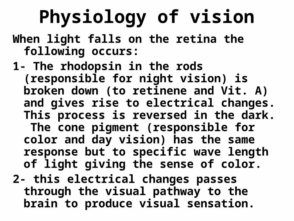
Physiology of visionWhen light falls on the retina the following
occurs:1- The rhodopsin in the rods (responsible for
night vision) is broken down (to retinene and Vit. A) and gives rise to electrical changes. This process is reversed in the dark. The cone pigment (responsible for color and day vision) has the same response but to specific wave length of light giving the sense of color.
2- this electrical changes passes through the visual pathway to the brain to produce visual sensation.
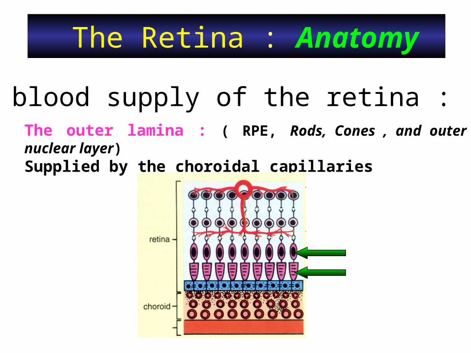
The Retina : Anatomy
The blood supply of the retina :The outer lamina : ( RPE, Rods, Cones , and outer nuclear layer)Supplied by the choroidal capillaries

The Retina : Anatomy
The blood supply of the retina :The inner lamina : (The rest of the retinal layers)Supplied by the central retinal artery&vein .

The Optic Nerve- Is made of all ganglion cells axons that becomes
myelinated behind lamina cribrosa (scleral foramina that gives passage to the nerve).
- It runs through lamina cribrosa leaves the orbit through the optic canal to reach the optic chiasma.
- Its surrounded by dense sheath of dura mater, middle delicate sheath of arachnoid, and inner most vascular pial sheath. الي الفاتحه النقطه
نيرف االوبتك بدايه اهي البيوبل من نشووفهاا

-central physiological cup مار نررف مافيهالوسط .Lamina cribrosa is seen , في
- Central retinal vessels enter and leave the eye from the cup.
the optic disc:the optic disc: -Nasal to the -Nasal to the macula. macula. -1,5 mm in -1,5 mm in diameter.diameter.-Pale pink in color. -Pale pink in color. -rounded in shape, -rounded in shape, Well-defined edges.Well-defined edges.

The visual pathwayMade of:
- optic nerve.- optic chiasma (crossing
of nasal fibers occurs).- optic tracts.- lateral geniculate
bodies.- optic radiations.- visual cortical areas (17-
18)

The visual nerve pathway
• Sensory end organs: Rods and cones.
• The first order neuron: bipolar cells of retina.
• The second order neuron: Ganglion cells of retina.
• Third order neuron: cells of the lateral geniculate body.