Anatomic Variants Mimicking Pathology on Echocardiography ... · Anatomic Variants Mimicking...
10
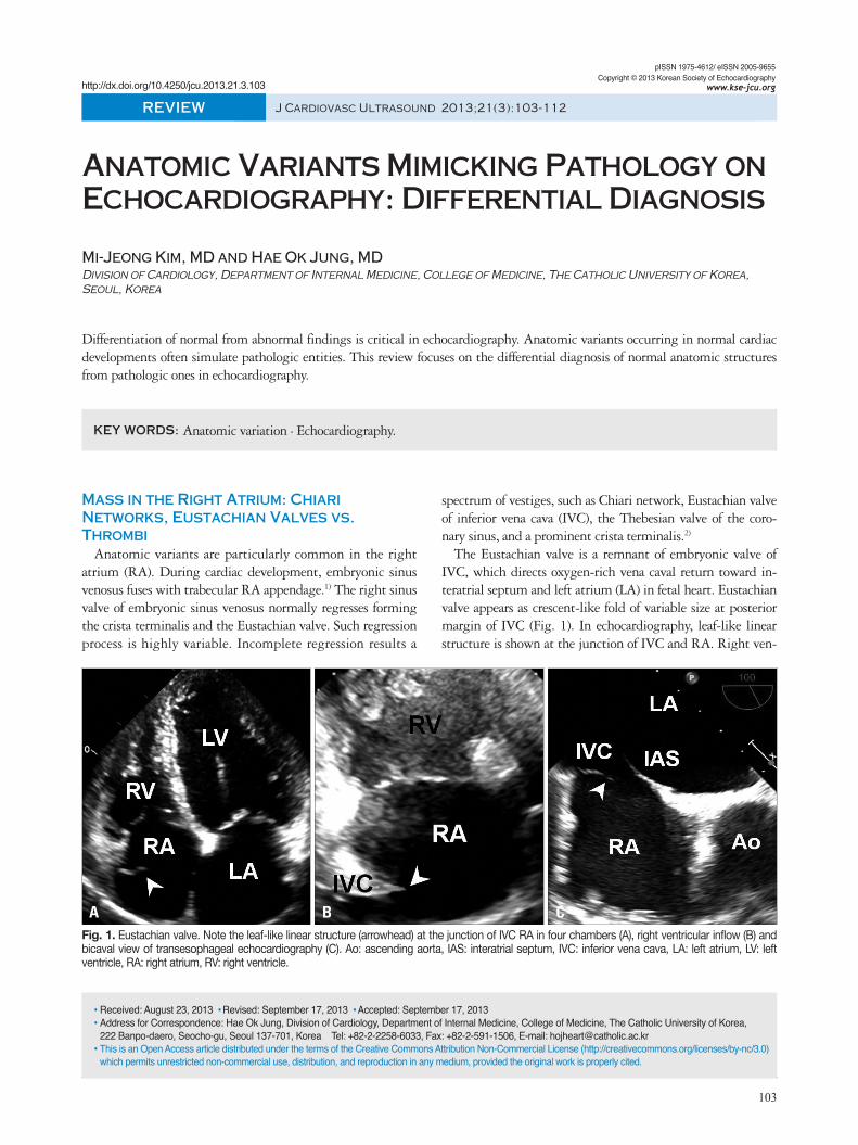
103 Mass in the Right Atrium: Chiari Networks, Eustachian Valves vs. Thrombi Anatomic variants are particularly common in the right atrium (RA). During cardiac development, embryonic sinus venosus fuses with trabecular RA appendage. 1) The right sinus valve of embryonic sinus venosus normally regresses forming the crista terminalis and the Eustachian valve. Such regression process is highly variable. Incomplete regression results a spectrum of vestiges, such as Chiari network, Eustachian valve of inferior vena cava (IVC), the Thebesian valve of the coro- nary sinus, and a prominent crista terminalis. 2) The Eustachian valve is a remnant of embryonic valve of IVC, which directs oxygen-rich vena caval return toward in- teratrial septum and left atrium (LA) in fetal heart. Eustachian valve appears as crescent-like fold of variable size at posterior margin of IVC (Fig. 1). In echocardiography, leaf-like linear structure is shown at the junction of IVC and RA. Right ven- pISSN 1975-4612/ eISSN 2005-9655 Copyright © 2013 Korean Society of Echocardiography www.kse-jcu.org http://dx.doi.org/10.4250/jcu.2013.21.3.103 REVIEW J Cardiovasc Ultrasound 2013;21(3):103-112 Anatomic Variants Mimicking Pathology on Echocardiography: Differential Diagnosis Mi-Jeong Kim, MD and Hae Ok Jung, MD Division of Cardiology, Department of Internal Medicine, College of Medicine, The Catholic University of Korea, Seoul, Korea Differentiation of normal from abnormal findings is critical in echocardiography. Anatomic variants occurring in normal cardiac developments often simulate pathologic entities. This review focuses on the differential diagnosis of normal anatomic structures from pathologic ones in echocardiography. KEY WORDS: Anatomic variation · Echocardiography. • Received: August 23, 2013 • Revised: September 17, 2013 • Accepted: September 17, 2013 • Address for Correspondence: Hae Ok Jung, Division of Cardiology, Department of Internal Medicine, College of Medicine, The Catholic University of Korea, 222 Banpo-daero, Seocho-gu, Seoul 137-701, Korea Tel: +82-2-2258-6033, Fax: +82-2-591-1506, E-mail: [email protected] • This is an Open Access article distributed under the terms of the Creative Commons Attribution Non-Commercial License (http://creativecommons.org/licenses/by-nc/3.0) which permits unrestricted non-commercial use, distribution, and reproduction in any medium, provided the original work is properly cited. Fig. 1. Eustachian valve. Note the leaf-like linear structure (arrowhead) at the junction of IVC RA in four chambers (A), right ventricular inflow (B) and bicaval view of transesophageal echocardiography (C). Ao: ascending aorta, IAS: interatrial septum, IVC: inferior vena cava, LA: left atrium, LV: left ventricle, RA: right atrium, RV: right ventricle. A B C
Transcript of Anatomic Variants Mimicking Pathology on Echocardiography ... · Anatomic Variants Mimicking...
103
Mass in the Right Atrium: Chiari Networks, Eustachian Valves vs. Thrombi
Anatomic variants are particularly common in the right atrium (RA). During cardiac development, embryonic sinus venosus fuses with trabecular RA appendage.1) The right sinus valve of embryonic sinus venosus normally regresses forming the crista terminalis and the Eustachian valve. Such regression process is highly variable. Incomplete regression results a
spectrum of vestiges, such as Chiari network, Eustachian valve of inferior vena cava (IVC), the Thebesian valve of the coro-nary sinus, and a prominent crista terminalis.2)
The Eustachian valve is a remnant of embryonic valve of IVC, which directs oxygen-rich vena caval return toward in-teratrial septum and left atrium (LA) in fetal heart. Eustachian valve appears as crescent-like fold of variable size at posterior margin of IVC (Fig. 1). In echocardiography, leaf-like linear structure is shown at the junction of IVC and RA. Right ven-
pISSN 1975-4612/ eISSN 2005-9655 Copyright © 2013 Korean Society of Echocardiography
www.kse-jcu.orghttp://dx.doi.org/10.4250/jcu.2013.21.3.103
REVIEW J Cardiovasc Ultrasound 2013;21(3):103-112
Anatomic Variants Mimicking Pathology on Echocardiography: Differential Diagnosis
Mi-Jeong Kim, MD and Hae Ok Jung, MDDivision of Cardiology, Department of Internal Medicine, College of Medicine, The Catholic University of Korea, Seoul, Korea
Differentiation of normal from abnormal findings is critical in echocardiography. Anatomic variants occurring in normal cardiac developments often simulate pathologic entities. This review focuses on the differential diagnosis of normal anatomic structures from pathologic ones in echocardiography.
KEY WORDS: Anatomic variation · Echocardiography.
•Received: August 23, 2013 •Revised: September 17, 2013 •Accepted: September 17, 2013 •Address for Correspondence: Hae Ok Jung, Division of Cardiology, Department of Internal Medicine, College of Medicine, The Catholic University of Korea, 222 Banpo-daero, Seocho-gu, Seoul 137-701, Korea Tel: +82-2-2258-6033, Fax: +82-2-591-1506, E-mail: [email protected]•This is an Open Access article distributed under the terms of the Creative Commons Attribution Non-Commercial License (http://creativecommons.org/licenses/by-nc/3.0) which permits unrestricted non-commercial use, distribution, and reproduction in any medium, provided the original work is properly cited.
Fig. 1. Eustachian valve. Note the leaf-like linear structure (arrowhead) at the junction of IVC RA in four chambers (A), right ventricular inflow (B) and bicaval view of transesophageal echocardiography (C). Ao: ascending aorta, IAS: interatrial septum, IVC: inferior vena cava, LA: left atrium, LV: left ventricle, RA: right atrium, RV: right ventricle.
A B C
Journal of Cardiovascular Ultrasound 21 | September 2013
104
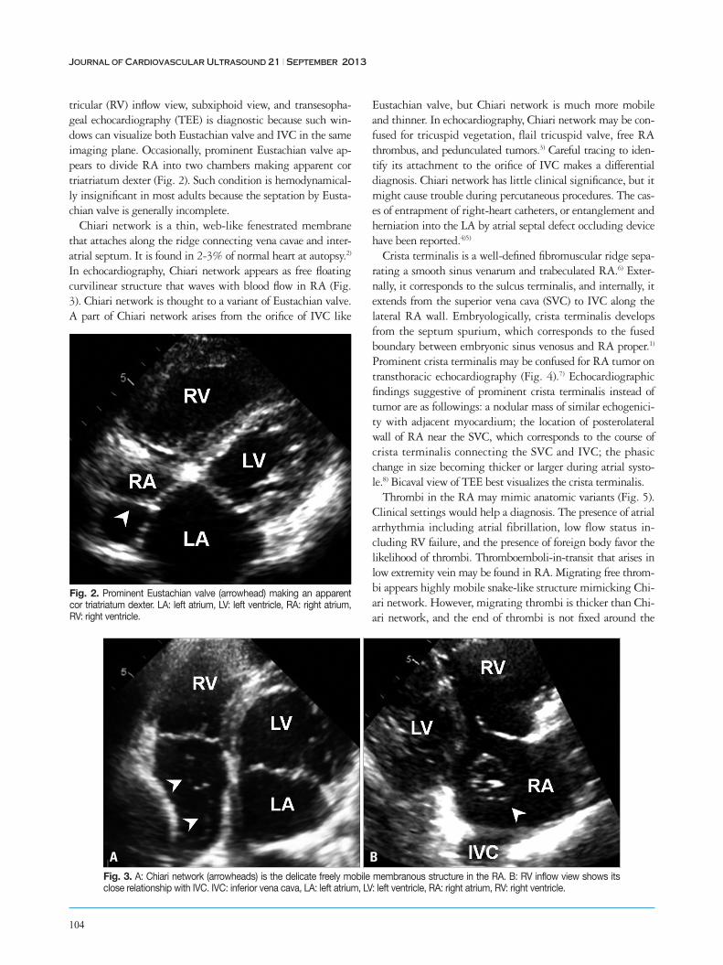
tricular (RV) inflow view, subxiphoid view, and transesopha-geal echocardiography (TEE) is diagnostic because such win-dows can visualize both Eustachian valve and IVC in the same imaging plane. Occasionally, prominent Eustachian valve ap-pears to divide RA into two chambers making apparent cor triatriatum dexter (Fig. 2). Such condition is hemodynamical-ly insignificant in most adults because the septation by Eusta-chian valve is generally incomplete.
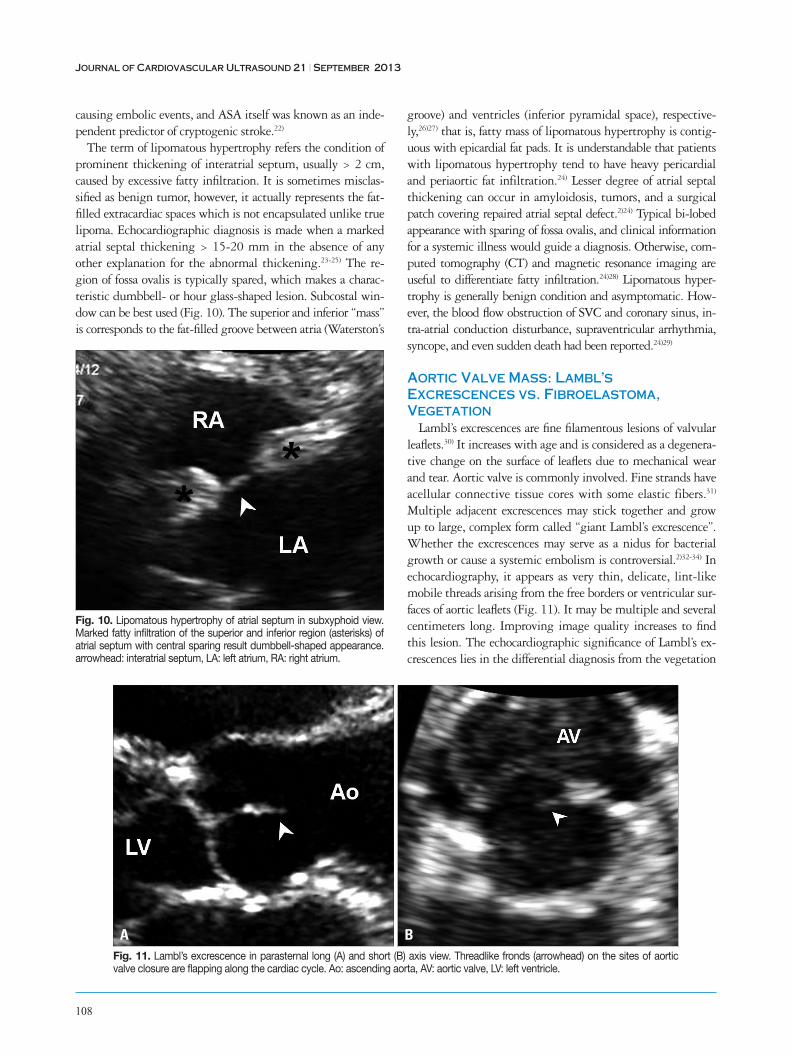
Chiari network is a thin, web-like fenestrated membrane that attaches along the ridge connecting vena cavae and inter-atrial septum. It is found in 2-3% of normal heart at autopsy.2) In echocardiography, Chiari network appears as free floating curvilinear structure that waves with blood flow in RA (Fig. 3). Chiari network is thought to a variant of Eustachian valve. A part of Chiari network arises from the orifice of IVC like
Eustachian valve, but Chiari network is much more mobile and thinner. In echocardiography, Chiari network may be con-fused for tricuspid vegetation, flail tricuspid valve, free RA thrombus, and pedunculated tumors.3) Careful tracing to iden-tify its attachment to the orifice of IVC makes a differential diagnosis. Chiari network has little clinical significance, but it might cause trouble during percutaneous procedures. The cas-es of entrapment of right-heart catheters, or entanglement and herniation into the LA by atrial septal defect occluding device have been reported.4)5)
Crista terminalis is a well-defined fibromuscular ridge sepa-rating a smooth sinus venarum and trabeculated RA.6) Exter-nally, it corresponds to the sulcus terminalis, and internally, it extends from the superior vena cava (SVC) to IVC along the lateral RA wall. Embryologically, crista terminalis develops from the septum spurium, which corresponds to the fused boundary between embryonic sinus venosus and RA proper.1) Prominent crista terminalis may be confused for RA tumor on transthoracic echocardiography (Fig. 4).7) Echocardiographic findings suggestive of prominent crista terminalis instead of tumor are as followings: a nodular mass of similar echogenici-ty with adjacent myocardium; the location of posterolateral wall of RA near the SVC, which corresponds to the course of crista terminalis connecting the SVC and IVC; the phasic change in size becoming thicker or larger during atrial systo-le.8) Bicaval view of TEE best visualizes the crista terminalis.
Thrombi in the RA may mimic anatomic variants (Fig. 5). Clinical settings would help a diagnosis. The presence of atrial arrhythmia including atrial fibrillation, low flow status in-cluding RV failure, and the presence of foreign body favor the likelihood of thrombi. Thromboemboli-in-transit that arises in low extremity vein may be found in RA. Migrating free throm-bi appears highly mobile snake-like structure mimicking Chi-ari network. However, migrating thrombi is thicker than Chi-ari network, and the end of thrombi is not fixed around the
Fig. 2. Prominent Eustachian valve (arrowhead) making an apparent cor triatriatum dexter. LA: left atrium, LV: left ventricle, RA: right atrium, RV: right ventricle.
Fig. 3. A: Chiari network (arrowheads) is the delicate freely mobile membranous structure in the RA. B: RV inflow view shows its close relationship with IVC. IVC: inferior vena cava, LA: left atrium, LV: left ventricle, RA: right atrium, RV: right ventricle.
BA

Differential Diagnosis of Anatomic Variants | Mi-Jeong Kim and Hae Ok Jung
105
IVC orifice.Lastly, RA tumors are needed to be differentiated from ana-
tomic variants. Myxoma is the most common primary tumor occurring in RA. Echocardiographically, myxoma appears as a globular or spherical mass with friable surface and heteroge-nous internal echogenicity. Myxomas typically arise from the interatrial septum around the fossa ovalis.2) Metastatic tumors including hepatoma, renal cell cancer, and sarcoma from pel-vic organs reaching RA through IVC can be seen in echocar-diography. Careful tracing the origin of mass will give a clue for differential diagnosis from benign anatomic variants.
Right Ventricular Mass: Moderator Band vs. Thrombus
Heavy trabeculation, prominent or redundant papillary muscle, and moderator band may be normally seen in the RV.
They need to be differentiated from pathologic entities such as primary and metastatic tumors, thrombi, and vegetations.
Multiple trabeculation is a characteristic of RV. The pattern of trabeculation is highly variable and the exaggeration of nor-mal trabeculation might be confused for cardiomyopathy. True pathologic hypertrabeculation occurring in developmen-tal arrest of RV myocardium is rare, and it is often accompa-nied by the other congenital heart diseases including tricuspid valve anomaly, atrial or ventricular septal defect, and left ven-tricular (LV) non-compaction.
Moderator band is a prominent trabeculation of RV extend-ing from the base of anterior papillary muscle to the interven-tricular septum, which contains right bundle branch. It is present in the majority of normal adults but tremendous indi-vidual variations are observed in thickness and shape. In echo-cardiography, moderator band is a thick echo-dense band-like
Fig. 4. Crista terminalis (arrowhead) appears as the mass arising from the posterior wall of RA in four chamber view (A), but it is well visualized as muscular ridge near SVC in transesophageal echocardiography (B). EV: Eustachian valve, IVC: inferior vena cava, LA: left atrium, LV: left ventricle, RA: right atrium, RV: right ventricle, SVC: superior vena cava.
Fig. 5. A: Thrombi of RA (arrowheads) in parasternal short axis view. B: Computed tomography imaging of the same patient. Ao: ascending aorta, LA: left atrium, LV: left ventricle, RA: right atrium, RV: right ventricle.
B
B
A
A

Journal of Cardiovascular Ultrasound 21 | September 2013
106
structure across the RV cavity and connects the lower inter-ventricular septum and the anterior papillary muscle (Fig. 6).
RV may be involved by true pathologic lesions. Thrombi may be present in RV failure, eosinophilic endocarditis, myo-cardial infarction involving right coronary artery, and rarely RV cardiomyopathy with aneurysm. As thrombi involving RV often occur at distal RV, plenty of trabeculation normally present in the distal RV cavity may prevent to identify a small thrombi. Careful evaluation with multiple off-axis imaging planes is needed for the suspected case with RV dysfunction. RV may be involved a tumorous condition including rhabdo-myoma and metastatic tumors. Tumors may cause RV failure by direct invasion and myocardial replacement with tumor in-filtration, cavity obliteration or limiting tricuspid motion (Fig. 7).9)
Left Ventricular Bands: False Tendon vs. Thrombi
LV bands or false tendons are fibromuscular structures cross-ing the LV cavity. LV bands may pass between papillary mus-cles, from papillary muscle to the ventricular septum, between free walls, or from free wall to interventricular septum, in con-trary to true chordae tendineae connecting papillary muscle and mitral valve leaflets.10)11) False tendon is found up to 55% in normal hearts by autopsy study.12) In echocardiography, LV bands appear as string-like thin bands passing LV cavity (Fig. 8), which may be transverse, longitudinal, or sagittal, and sin-gle or multiple. The location, direction, length and thickness of LV bands may vary depending on their embryonic origin of inner cardiac muscle layer and contents. Muscular bands be-come shorter and thicker in systole, and vice versa in diastole.
Fig. 7. Pathologic lesions involving RV. A: Thrombus (arrowheads) attached to chordae tendineae and the wall of RV with significant dilation and impaired contractility. B: Irregular thickening of RV free wall (arrowheads) due to metastatic lung cancer. Note the pericardial effusion around the RV. LA: left atrium, LV: left ventricle, RA: right atrium, RV: right ventricle.
BA
Fig. 6. Normal variants of RV. A: Moderator band (arrowhead). B: Prominent papillary muscle. C: Hypertrabeculation (asterisk). IVS: interventricular septum, LA: left atrium, LV: left ventricle, RA: right atrium, RV: right ventricle.
A B C

Differential Diagnosis of Anatomic Variants | Mi-Jeong Kim and Hae Ok Jung
107
Fibrous bands become straight and taut in diastole, and vice versa in systole.13) Off-axis images demonstrating the overall length of bands, normal LV structures on both ends, and con-stant motion during cardiac cycle are the key features.11) False tendon located near LV apex may be confused for mural thrombus particularly in images of true LV apex being not completely visualized (Fig. 8B).
Papillary muscles vary in shape, thickness, and the location in LV wall. More than one belly is observed in up to 50% of nor-mal hearts.14) Accessory papillary muscle may be confused for pathologic structures such as LV thrombus or papillary muscle tumors when it arises from an unusual location. The presence of LV band is strongly indicative of the accessory papillary muscle instead of pathologic entity.15) Normally contractile adjacent LV wall help to exclude the mural thrombi. Papillary muscle vari-ants in architecture and location have peculiar clinical signifi-cance in hypertrophic cardiomyopathy. Anterior displacement of hypertrophied papillary muscle is known to accentuate the resting trans-LV outflow tract pressure gradients.16)17)
Atrial Septum Variants: Atrial Septal Aneurysm vs. Lipomatous Hypertrophy
Atrial septal aneurysm (ASA) is found in 1% of adults at autopsy.18) An excursion of > 10 mm beyond the plane of in-teratrial septum is recognized as ASA,19) although such cut-off value is arbitrary. ASA may involve only the region of fossa ovalis, or the entire interatrial septum.20) Frequent association with atrial septal defect, patent foramen ovale (PFO), mitral valve prolapse, Marfan syndrome suggests that ASA is con-genital malformation with genetic background.19) Longstand-ing elevation of atrial pressure may contribute to ASA forma-tion, inducing a septum to bulge toward the lower pressure chamber. In echocardiography, redundant interatrial septum bulges beyond the atrial septal plane (Fig. 9). Phasic oscilla-tion along the cardiac or respiratory cycle is common. Promi-nent ASA may appear as cystic mass in long axis views, but diagnosis is rarely difficult particularly with the aid of TEE. ASA is known to be associated with atrial arrhythmia and ischemic stroke.21)22) ASA is frequently accompanied by PFO
Fig. 8. Normal LV variants. A: Fibrous LV band (arrowhead) pass the LV cavity with transverse direction. B: Muscular LV band (arrowhead). C: Apically located papillary muscle (arrowheads) with chordae tendineae. LA: left atrium, LV: left ventricle, RA: right atrium, RV: right ventricle.
Fig. 9. A: Atrial septal aneurysm (arrowhead) in four chamber view. B: It may show tissue drop mimicking atrial septal defect. C: The degree of excursion (a) from the atrial septal plane (b) is used to quantify the size of atrial septal aneurysm. LA: left atrium, LV: left ventricle, RA: right atrium, RV: right ventricle.
A
A
B
B
C
C
Journal of Cardiovascular Ultrasound 21 | September 2013
108
causing embolic events, and ASA itself was known as an inde-pendent predictor of cryptogenic stroke.22)
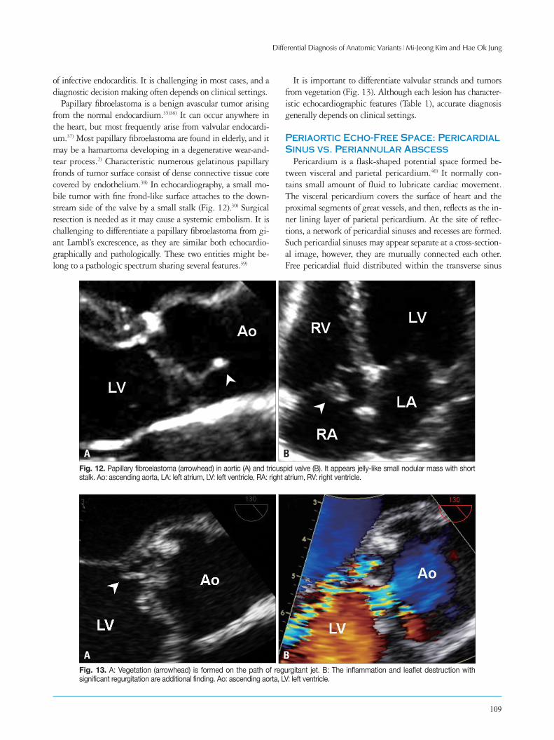
The term of lipomatous hypertrophy refers the condition of prominent thickening of interatrial septum, usually > 2 cm, caused by excessive fatty infiltration. It is sometimes misclas-sified as benign tumor, however, it actually represents the fat-filled extracardiac spaces which is not encapsulated unlike true lipoma. Echocardiographic diagnosis is made when a marked atrial septal thickening > 15-20 mm in the absence of any other explanation for the abnormal thickening.23-25) The re-gion of fossa ovalis is typically spared, which makes a charac-teristic dumbbell- or hour glass-shaped lesion. Subcostal win-dow can be best used (Fig. 10). The superior and inferior “mass” is corresponds to the fat-filled groove between atria (Waterston’s
groove) and ventricles (inferior pyramidal space), respective-ly,26)27) that is, fatty mass of lipomatous hypertrophy is contig-uous with epicardial fat pads. It is understandable that patients with lipomatous hypertrophy tend to have heavy pericardial and periaortic fat infiltration.24) Lesser degree of atrial septal thickening can occur in amyloidosis, tumors, and a surgical patch covering repaired atrial septal defect.2)24) Typical bi-lobed appearance with sparing of fossa ovalis, and clinical information for a systemic illness would guide a diagnosis. Otherwise, com-puted tomography (CT) and magnetic resonance imaging are useful to differentiate fatty infiltration.24)28) Lipomatous hyper-trophy is generally benign condition and asymptomatic. How-ever, the blood flow obstruction of SVC and coronary sinus, in-tra-atrial conduction disturbance, supraventricular arrhythmia, syncope, and even sudden death had been reported.24)29)
Aortic Valve Mass: Lambl’s Excrescences vs. Fibroelastoma, Vegetation
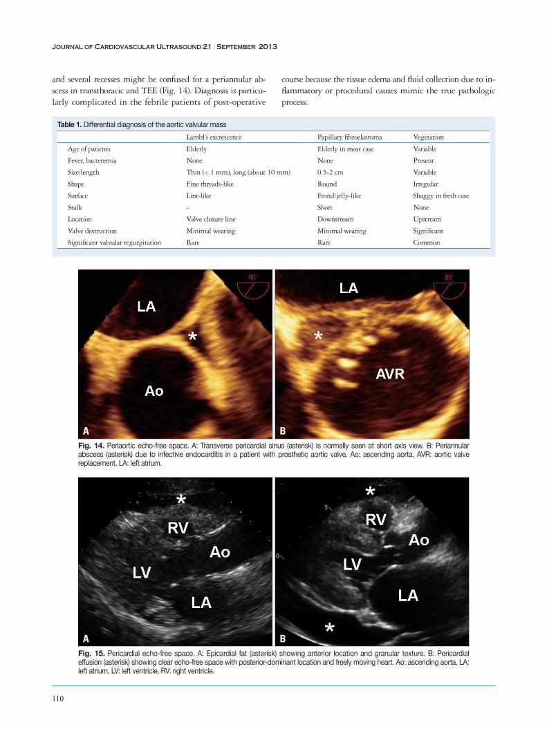
Lambl’s excrescences are fine filamentous lesions of valvular leaflets.30) It increases with age and is considered as a degenera-tive change on the surface of leaflets due to mechanical wear and tear. Aortic valve is commonly involved. Fine strands have acellular connective tissue cores with some elastic fibers.31) Multiple adjacent excrescences may stick together and grow up to large, complex form called “giant Lambl’s excrescence”. Whether the excrescences may serve as a nidus for bacterial growth or cause a systemic embolism is controversial.2)32-34) In echocardiography, it appears as very thin, delicate, lint-like mobile threads arising from the free borders or ventricular sur-faces of aortic leaflets (Fig. 11). It may be multiple and several centimeters long. Improving image quality increases to find this lesion. The echocardiographic significance of Lambl’s ex-crescences lies in the differential diagnosis from the vegetation
Fig. 10. Lipomatous hypertrophy of atrial septum in subxyphoid view. Marked fatty infiltration of the superior and inferior region (asterisks) of atrial septum with central sparing result dumbbell-shaped appearance. arrowhead: interatrial septum, LA: left atrium, RA: right atrium.
Fig. 11. Lambl’s excrescence in parasternal long (A) and short (B) axis view. Threadlike fronds (arrowhead) on the sites of aortic valve closure are flapping along the cardiac cycle. Ao: ascending aorta, AV: aortic valve, LV: left ventricle.
BA
Differential Diagnosis of Anatomic Variants | Mi-Jeong Kim and Hae Ok Jung
109
of infective endocarditis. It is challenging in most cases, and a diagnostic decision making often depends on clinical settings.
Papillary fibroelastoma is a benign avascular tumor arising from the normal endocardium.35)36) It can occur anywhere in the heart, but most frequently arise from valvular endocardi-um.37) Most papillary fibroelastoma are found in elderly, and it may be a hamartoma developing in a degenerative wear-and-tear process.2) Characteristic numerous gelatinous papillary fronds of tumor surface consist of dense connective tissue core covered by endothelium.38) In echocardiography, a small mo-bile tumor with fine frond-like surface attaches to the down-stream side of the valve by a small stalk (Fig. 12).30) Surgical resection is needed as it may cause a systemic embolism. It is challenging to differentiate a papillary fibroelastoma from gi-ant Lambl’s excrescence, as they are similar both echocardio-graphically and pathologically. These two entities might be-long to a pathologic spectrum sharing several features.39)
It is important to differentiate valvular strands and tumors from vegetation (Fig. 13). Although each lesion has character-istic echocardiographic features (Table 1), accurate diagnosis generally depends on clinical settings.
Periaortic Echo-Free Space: Pericardial Sinus vs. Periannular Abscess
Pericardium is a flask-shaped potential space formed be-tween visceral and parietal pericardium.40) It normally con-tains small amount of fluid to lubricate cardiac movement. The visceral pericardium covers the surface of heart and the proximal segments of great vessels, and then, reflects as the in-ner lining layer of parietal pericardium. At the site of reflec-tions, a network of pericardial sinuses and recesses are formed. Such pericardial sinuses may appear separate at a cross-section-al image, however, they are mutually connected each other. Free pericardial fluid distributed within the transverse sinus
Fig. 12. Papillary fibroelastoma (arrowhead) in aortic (A) and tricuspid valve (B). It appears jelly-like small nodular mass with short stalk. Ao: ascending aorta, LA: left atrium, LV: left ventricle, RA: right atrium, RV: right ventricle.
Fig. 13. A: Vegetation (arrowhead) is formed on the path of regurgitant jet. B: The inflammation and leaflet destruction with significant regurgitation are additional finding. Ao: ascending aorta, LV: left ventricle.
B
B
A
A
Journal of Cardiovascular Ultrasound 21 | September 2013
110
and several recesses might be confused for a periannular ab-scess in transthoracic and TEE (Fig. 14). Diagnosis is particu-larly complicated in the febrile patients of post-operative
course because the tissue edema and fluid collection due to in-flammatory or procedural causes mimic the true pathologic process.
Table 1. Differential diagnosis of the aortic valvular mass
Lambl’s excrescence Papillary fibroelastoma Vegetation
Age of patients Elderly Elderly in most case Variable
Fever, bacteremia None None Present
Size/length Thin (< 1 mm), long (about 10 mm) 0.5-2 cm Variable
Shape Fine threads-like Round Irregular
Surface Lint-like Frond/jelly-like Shaggy in fresh case
Stalk - Short None
Location Valve closure line Downstream Upstream
Valve destruction Minimal wearing Minimal wearing Significant
Significant valvular regurgitation Rare Rare Common
Fig. 14. Periaortic echo-free space. A: Transverse pericardial sinus (asterisk) is normally seen at short axis view. B: Periannular abscess (asterisk) due to infective endocarditis in a patient with prosthetic aortic valve. Ao: ascending aorta, AVR: aortic valve replacement, LA: left atrium.
Fig. 15. Pericardial echo-free space. A: Epicardial fat (asterisk) showing anterior location and granular texture. B: Pericardial effusion (asterisk) showing clear echo-free space with posterior-dominant location and freely moving heart. Ao: ascending aorta, LA: left atrium, LV: left ventricle, RV: right ventricle.
B
B
A
A

Differential Diagnosis of Anatomic Variants | Mi-Jeong Kim and Hae Ok Jung
111
Pericardial Echo-Free Space: Epicardial Fat vs. Pericardial Effusion
Epicardial adipose tissue often appears as echo-free space mimicking pericardial effusion in echocardiography. Most adi-pose tissue distributes in atrio-ventricular or interventricular groove. Excessive fatty infiltration tends to occur in old, obese, and diabetic patients, particularly in women.
Echocardiographic differentiation of adipose tissue from flu-id is based on echogenicity, texture, mobility, and location.2) Inflammatory or bloody effusion may be extremely thick to show high and heterogenous echogenicity. With exception of loculated effusion, free pericardial fluid accumulates on depen-dent region, usually posterior of left in supine position. Ante-riorly located echo-free space is likely to be epicardial fat depo-sition rather than pericardial effusion (Fig. 15). Mobility of the adjacent tissue is another clue to differentiation. As adi-pose tissue is less mobile than pericardial fluid, surrounding epicardial and pericardial layer move less freely in case of fatty infiltration. Heart moves like “swinging” in moderate amount of pericardial effusion, but not in fat-filled space. Lastly, adi-pose tissue is more echogenic and lobulated than fluid with homogenous echogenicity. CT imaging can definitely differ-entiate epicardial adipose tissue from fluid collection in com-plicated case.
ConclusionsThe differential diagnosis of “mass” occurring heart is often
challenging although the imaging quality and techniques are quite beautiful in current echocardiography. Knowledge about anatomic variants would guide to proper interpretation of echo-cardiographic findings. Previous images should be reviewed carefully whenever it is available. Short-term repetition of im-aging on the mass is extremely helpful to diagnosis and not to miss the critical change of the pathologic entities.
References1. Schoenwolf GC, Bleyl SB. Larsen’s human embryology, 4th ed. Philadel-
phia: Churchill Livingstone/Elsevier;2009. 2. Weyman AE. Principles and practice of echocardiography, 2nd ed. Phila-
delphia: Lippincott Williams & Wilkins;1994.3. Werner JA, Cheitlin MD, Gross BW, Speck SM, Ivey TD. Echocar-
diographic appearance of the Chiari network: differentiation from right-heart pathology. Circulation 1981;63:1104-9.
4. Cooke JC, Gelman JS, Harper RW. Chiari network entanglement and herniation into the left atrium by an atrial septal defect occluder device. J Am Soc Echocardiogr 1999;12:601-3.
5. Goldschlager A, Goldschlager N, Brewster H, Kaplan J. Catheter entrapment in a Chiari network involving an atrial septal defect. Chest 1972;62:345-6.
6. Loukas M, Tubbs RS, Tongson JM, Polepalli S, Curry B, Jordan R, Wagner T. The clinical anatomy of the crista terminalis, pectinate muscles and the teniae sagittalis. Ann Anat 2008;190:81-7.
7. Mirowitz SA, Gutierrez FR. Fibromuscular elements of the right atrium: pseudomass at MR imaging. Radiology 1992;182:231-3.
8. Salustri A, Bakir S, Sana A, Lange P, Al Mahmeed WA. Prominent crista terminalis mimicking a right atrial mass: case report. Cardiovasc Ul-
trasound 2010;8:47.9. Ports TA, Schiller NB, Strunk BL. Echocardiography of right ventricu-
lar tumors. Circulation 1977;56:439-47.10. Nishimura T, Kondo M, Umadome H, Shimono Y. Echocardiograph-
ic features of the false tendons in the left ventricle. Am J Cardiol 1981;48: 177-83.
11. Keren A, Billingham ME, Popp RL. Echocardiographic recognition and implications of ventricular hypertrophic trabeculations and aberrant bands. Circulation 1984;70:836-42.
12. Luetmer PH, Edwards WD, Seward JB, Tajik AJ. Incidence and dis-tribution of left ventricular false tendons: an autopsy study of 483 normal human hearts. J Am Coll Cardiol 1986;8:179-83.
13. George A, Parameswaran A, Nekkanti R, Lurito K, Movahed A. Normal anatomic variants on transthoracic echocardiogram. Echocardiogra-phy 2009;26:1109-17.
14. Victor S, Nayak VM. Variations in the papillary muscles of the normal mitral valve and their surgical relevance. J Card Surg 1995;10:597-607.
15. Misra S, Koshy T, Pal S. Echo rounds: false tendons and accessory papil-lary muscle in the left ventricle. Anesth Analg 2011;113:1016-8.
16. Kwon DH, Setser RM, Thamilarasan M, Popovic ZV, Smedira NG, Schoenhagen P, Garcia MJ, Lever HM, Desai MY. Abnormal papil-lary muscle morphology is independently associated with increased left ven-tricular outflow tract obstruction in hypertrophic cardiomyopathy. Heart 2008;94:1295-301.
17. Harrigan CJ, Appelbaum E, Maron BJ, Buros JL, Gibson CM, Lesser JR, Udelson JE, Manning WJ, Maron MS. Significance of pap-illary muscle abnormalities identified by cardiovascular magnetic resonance in hypertrophic cardiomyopathy. Am J Cardiol 2008;101:668-73.
18. Silver MD, Dorsey JS. Aneurysms of the septum primum in adults. Arch Pathol Lab Med 1978;102:62-5.
19. Mügge A, Daniel WG, Angermann C, Spes C, Khandheria BK, Kronzon I, Freedberg RS, Keren A, Denning K, Engberding R, et al. Atrial septal aneurysm in adult patients. A multicenter study using transthoracic and transesophageal echocardiography. Circulation 1995;91: 2785-92.
20. Hanley PC, Tajik AJ, Hynes JK, Edwards WD, Reeder GS, Hagler DJ, Seward JB. Diagnosis and classification of atrial septal aneurysm by two-dimensional echocardiography: report of 80 consecutive cases. J Am Coll Cardiol 1985;6:1370-82.
21. Ong LS, Nanda NC, Falkoff MD, Barold SS. Interatrial septal aneu-rysm, systolic click and atrial tachyarrhythmia--a new syndrome? Ultra-sound Med Biol 1982;8:691-3.
22. Mattioli AV, Aquilina M, Oldani A, Longhini C, Mattioli G. Atrial septal aneurysm as a cardioembolic source in adult patients with stroke and normal carotid arteries. A multicentre study. Eur Heart J 2001;22:261-8.
23. Page DL. Lipomatous hypertrophy of the cardiac interatrial septum: its de-velopment and probable clinical significance. Hum Pathol 1970;1:151-63.
24. Heyer CM, Kagel T, Lemburg SP, Bauer TT, Nicolas V. Lipomatous hypertrophy of the interatrial septum: a prospective study of incidence, imag-ing findings, and clinical symptoms. Chest 2003;124:2068-73.
25. Fyke FE 3rd, Tajik AJ, Edwards WD, Seward JB. Diagnosis of lipo-matous hypertrophy of the atrial septum by two-dimensional echocardiogra-phy. J Am Coll Cardiol 1983;1:1352-7.
26. Cunningham KS, Veinot JP, Feindel CM, Butany J. Fatty lesions of the atria and interatrial septum. Hum Pathol 2006;37:1245-51.
27. Shirani J, Roberts WC. Clinical, electrocardiographic and morphologic features of massive fatty deposits (“lipomatous hypertrophy”) in the atrial septum. J Am Coll Cardiol 1993;22:226-38.
28. Stephant E, Barthelet M, Leroux PY, Revel D. Images in cardiovascu-lar medicine. Lipomatous hypertrophy of the interventricular septum: echo-cardiography, cardiac magnetic resonance, and multidetector computerized to-mography imaging. Circulation 2008;118:e71-2.

Journal of Cardiovascular Ultrasound 21 | September 2013
112
29. Riva L, Banfi C, Gaeta R, Viganò M. Lipomatous hypertrophy of the in-teratrial septum. Description of a clinical case and literature review. Miner-va Cardioangiol 2006;54:789-92.
30. Armstrong WF, Ryan T. Feigenbaum’s echocardiography, 7th ed. Phila-delphia: Lippincott Williams & Wilkins;2010.
31. Magarey FR. On the mode of formation of Lambl’s excrescences and their relation to chronic thickening of the mitral valve. J Pathol Bacteriol 1949; 61:203-8, 5 pl.
32. Freedberg RS, Goodkin GM, Perez JL, Tunick PA, Kronzon I. Valve strands are strongly associated with systemic embolization: a transesophageal echocardiographic study. J Am Coll Cardiol 1995;26:1709-12.
33. Roldan CA, Shively BK, Crawford MH. Valve excrescences: prevalence, evolution and risk for cardioembolism. J Am Coll Cardiol 1997;30:1308-14.
34. Aziz F, Baciewicz FA Jr. Lambl’s excrescences: review and recommenda-tions. Tex Heart Inst J 2007;34:366-8.
35. Klarich KW, Enriquez-Sarano M, Gura GM, Edwards WD, Tajik AJ, Seward JB. Papillary fibroelastoma: echocardiographic characteristics
for diagnosis and pathologic correlation. J Am Coll Cardiol 1997;30:784-90.
36. Ngaage DL, Mullany CJ, Daly RC, Dearani JA, Edwards WD, Ta-zelaar HD, McGregor CG, Orszulak TA, Puga FJ, Schaff HV, Sundt TM 3rd, Zehr KJ. Surgical treatment of cardiac papillary fibroelastoma: a single center experience with eighty-eight patients. Ann Thorac Surg 2005;80:1712-8.
37. Gowda RM, Khan IA, Nair CK, Mehta NJ, Vasavada BC, Sacchi TJ. Cardiac papillary fibroelastoma: a comprehensive analysis of 725 cases. Am Heart J 2003;146:404-10.
38. Lammers RJ, Bloor CM. Cancer and the heart. New York: Springer-Verlag;1986.
39. Melduni RM, Klarich KW, Nesbitt GC, Shub C. Lambl’s excrescences: is surgical excision really necessary? Tex Heart Inst J 2008;35:89; author reply 90.
40. D’Avila A, Scanavacca M, Sosa E, Ruskin JN, Reddy VY. Pericardial anatomy for the interventional electrophysiologist. J Cardiovasc Electrophysi-ol 2003;14:422-30.