
ANATOMIC REPORT - neuroanatomia3dsevilla.es · surrounding bones, especially at the sutures, at the...
41
ANATOMIC REPORT MICROSURGICAL ANATOMY OF THE DURAL ARTERIES Carolina Martins, M.D. Department of Neurological Surgery, University of Florida, Gainesville, Florida Alexandre Yasuda, M.D. Department of Neurological Surgery, University of Florida, Gainesville, Florida Alvaro Campero, M.D. Department of Neurological Surgery, University of Florida, Gainesville, Florida Arthur J. Ulm, M.D. Department of Neurological Surgery, University of Florida, Gainesville, Florida Necmettin Tanriover, M.D. Department of Neurological Surgery, University of Florida, Gainesville, Florida Albert Rhoton, Jr., M.D. Department of Neurological Surgery, University of Florida, Gainesville, Florida Reprint requests: Albert Rhoton, Jr., M.D., Department of Neurological Surgery, University of Florida College of Medicine, P.O. Box 100265, Gainesville, FL 32610-0265. Email: [email protected] Received, March 26, 2004. Accepted, August 13, 2004. OBJECTIVE: The objective was to examine the microsurgical anatomy basic to the microsurgical and endovascular management of lesions involving the dural arteries. METHODS: Adult cadaveric heads and skulls were examined using the magnification provided by the surgical microscope to define the origin, course, and distribution of the individual dural arteries. RESULTS: The pattern of arterial supply of the dura covering the cranial base is more complex than over the cerebral convexity. The internal carotid system supplies the midline dura of the anterior and middle fossae and the anterior limit of the posterior fossa; the external carotid system supplies the lateral segment of the three cranial fossae; and the vertebrobasilar system supplies the midline structures of the posterior fossa and the area of the foramen magnum. Dural territories often have overlapping supply from several sources. Areas supplied from several overlapping sources are the parasellar dura, tentorium, and falx. The tentorium and falx also receive a contribution from the cerebral arteries, making these structures an anastomotic pathway between the dural and parenchymal arteries. A reciprocal relationship, in which the territories of one artery expand if the adjacent arteries are small, is common. CONCLUSION: The carotid and vertebrobasilar arterial systems give rise to multiple branches that supply the dura in a complex and overlapping pattern. A knowledge of the microsurgical anatomy of these dural arteries and their assessment on pretreatment evaluations plays a major role in safe and accurate treatment of multiple lesions. KEY WORDS: Cranial base, Cranial nerves, Dura mater, External carotid artery, Internal carotid artery, Intracranial arteries, Microsurgical anatomy, Skull base, Vertebral artery Neurosurgery 56[ONS Suppl 2]:ONS-211–ONS-251, 2005 DOI: 10.1227/01.NEU.0000144823.94402.3D T he dural arteries arise from the internal and external carotid arteries and the ver- tebrobasilar system. These arteries may be the site of formation of saccular aneurysms, pseudoaneurysms, and arteriovenous fistulae and the source of traumatic and spontaneous hemorrhage into the epidural, subdural, and intraparenchymal areas, in addition to their well-known role in the vascularization of me- ningiomas, other tumors, and parenchymal arteriovenous malformations (AVMs). Ad- vances in the microsurgical and endovascular management of lesions involving these arter- ies have created the need for a deeper under- standing of the dural arterial network. This study examined the microsurgical anatomy of this network important in planning the thera- peutic approach not only to lesions invading the cranial base, convexity dura, tentorium, and falx but also to numerous intraparenchy- mal lesions. MATERIALS AND METHODS Five dry skulls and 20 adult cadaveric heads were examined using 3 to 40 mag- nification of the surgical microscope (Carl Zeiss Inc., Göttingen, Germany). The arteries and veins were perfused with colored silicon (Dow Corning, Midland, MI; Crayola, Easton, PA). The dissections were performed to dem- onstrate and photograph the individual dural artery branches and their common areas of supply. GENERAL STRUCTURE: ENDOSTEAL AND MENINGEAL LAYERS The cranial dura mater is a thick, collage- nous sheath that lines the cranial cavity and is continuous with the spinal dura at the fora- men magnum. The dura is adherent to the NEUROSURGERY VOLUME 56 | OPERATIVE NEUROSURGERY 2 | APRIL 2005 | ONS-211
-
Upload
truongquynh -
Category
Documents
-
view
216 -
download
0
Transcript of ANATOMIC REPORT - neuroanatomia3dsevilla.es · surrounding bones, especially at the sutures, at the...

ANATOMIC REPORT
MICROSURGICAL ANATOMY OF THE DURAL ARTERIES
Carolina Martins, M.D.Department of NeurologicalSurgery, University of Florida,Gainesville, Florida
Alexandre Yasuda, M.D.Department of NeurologicalSurgery, University of Florida,Gainesville, Florida
Alvaro Campero, M.D.Department of NeurologicalSurgery, University of Florida,Gainesville, Florida
Arthur J. Ulm, M.D.Department of NeurologicalSurgery, University of Florida,Gainesville, Florida
Necmettin Tanriover, M.D.Department of NeurologicalSurgery, University of Florida,Gainesville, Florida
Albert Rhoton, Jr., M.D.Department of NeurologicalSurgery, University of Florida,Gainesville, Florida
Reprint requests:Albert Rhoton, Jr., M.D.,Department of NeurologicalSurgery, University of FloridaCollege of Medicine,P.O. Box 100265,Gainesville, FL 32610-0265.Email:[email protected]
Received, March 26, 2004.
Accepted, August 13, 2004.
OBJECTIVE: The objective was to examine the microsurgical anatomy basic to themicrosurgical and endovascular management of lesions involving the dural arteries.METHODS: Adult cadaveric heads and skulls were examined using the magnificationprovided by the surgical microscope to define the origin, course, and distribution ofthe individual dural arteries.RESULTS: The pattern of arterial supply of the dura covering the cranial base is morecomplex than over the cerebral convexity. The internal carotid system supplies themidline dura of the anterior and middle fossae and the anterior limit of the posteriorfossa; the external carotid system supplies the lateral segment of the three cranialfossae; and the vertebrobasilar system supplies the midline structures of the posteriorfossa and the area of the foramen magnum. Dural territories often have overlappingsupply from several sources. Areas supplied from several overlapping sources are theparasellar dura, tentorium, and falx. The tentorium and falx also receive a contributionfrom the cerebral arteries, making these structures an anastomotic pathway betweenthe dural and parenchymal arteries. A reciprocal relationship, in which the territoriesof one artery expand if the adjacent arteries are small, is common.CONCLUSION: The carotid and vertebrobasilar arterial systems give rise to multiplebranches that supply the dura in a complex and overlapping pattern. A knowledge ofthe microsurgical anatomy of these dural arteries and their assessment on pretreatmentevaluations plays a major role in safe and accurate treatment of multiple lesions.
KEY WORDS: Cranial base, Cranial nerves, Dura mater, External carotid artery, Internal carotid artery,Intracranial arteries, Microsurgical anatomy, Skull base, Vertebral artery
Neurosurgery 56[ONS Suppl 2]:ONS-211–ONS-251, 2005 DOI: 10.1227/01.NEU.0000144823.94402.3D
The dural arteries arise from the internaland external carotid arteries and the ver-tebrobasilar system. These arteries may
be the site of formation of saccular aneurysms,pseudoaneurysms, and arteriovenous fistulaeand the source of traumatic and spontaneoushemorrhage into the epidural, subdural, andintraparenchymal areas, in addition to theirwell-known role in the vascularization of me-ningiomas, other tumors, and parenchymalarteriovenous malformations (AVMs). Ad-vances in the microsurgical and endovascularmanagement of lesions involving these arter-ies have created the need for a deeper under-standing of the dural arterial network. Thisstudy examined the microsurgical anatomy ofthis network important in planning the thera-peutic approach not only to lesions invadingthe cranial base, convexity dura, tentorium,and falx but also to numerous intraparenchy-mal lesions.
MATERIALS AND METHODS
Five dry skulls and 20 adult cadavericheads were examined using �3 to �40 mag-nification of the surgical microscope (CarlZeiss Inc., Göttingen, Germany). The arteriesand veins were perfused with colored silicon(Dow Corning, Midland, MI; Crayola, Easton,PA). The dissections were performed to dem-onstrate and photograph the individual duralartery branches and their common areas ofsupply.
GENERAL STRUCTURE:ENDOSTEAL AND
MENINGEAL LAYERS
The cranial dura mater is a thick, collage-nous sheath that lines the cranial cavity and iscontinuous with the spinal dura at the fora-men magnum. The dura is adherent to the
NEUROSURGERY VOLUME 56 | OPERATIVE NEUROSURGERY 2 | APRIL 2005 | ONS-211

surrounding bones, especially at the sutures, at the cranialbase, and around the foramen magnum. With increasing age,the dura becomes less pliable and more firmly adherent to theinner surface of the cranium, particularly the calvaria.
The dura is composed of an endosteal layer that faces thebone and a meningeal layer that faces the brain (16). Theselayers are distinguished as separate sheaths at the venoussinuses, foramen magnum, and optic canal. The meningeallayer is continuous with the dural covering of the spinal cordand optic nerves, providing tubular sheaths for the cranialnerves as they pass through the cranial foramina. Thesesheaths fuse with the epineurium as the cranial nerves emergefrom the cranium, except at the optic nerve, where the duralsheath blends into the sclera. At the vascular foramina, themeningeal layer fuses with the adventitia of the vessel. Themeningeal layer folds inward to form the falx cerebri, thetentorium cerebelli, the falx cerebelli, and the diaphragm sel-lae, which partially divide the cranial cavity into freely com-municating spaces. The endosteal layer of dura is continuousthrough the cranial sutures and foramina with the pericra-nium and through the superior orbital fissure and optic canalwith the periorbita (16).
VASCULAR ORGANIZATION OFTHE DURA
The origin of the membranes of the cranium starts when theembryo has a crown-to-hump length of 12 to 20 mm, at whichtime the differentiation of the cranium, dura, arachnoid, andpia begins. The gradual cleavage of the vascular system intoexternal, dural, and cerebral layers also takes place at thisstage, which has been referred to as the third stage of thecerebrovascular development (79). As the membranes cover-ing the brain differentiate, the anastomosing channels thatconnect the deep capillary plexus with the superficial vesselsclose, thus separating the vessels surrounding the brain fromthose belonging to the cranium and its coverings (79, 80). Themajor meningeal arteries originating from this cleavage giverise to a rich anastomotic network that may enlarge aftervarious insults (22) and play a role in the genesis of duralAVMs. This anastomotic network divides progressively intoprimary, secondary, and penetrating vessels.
The primary anastomotic vessels change little in diameter asthey course over the dural surface and anastomose frequentlywith each other. They cross the superior sagittal sinus, con-necting the dura over the paired cerebral hemispheres into asingle vascular unit. Crossing vessels are particularly largewhen one middle meningeal artery is hypoplastic. The pri-mary anastomotic arteries have a straight course and measure100 to 300 �m in diameter, whereas the main meningealfeeders have a diameter of 400 to 800 �m. The primary anas-tomotic arteries give rise to arteries to the cranium, secondaryanastomotic arteries, penetrating dural vessels, and arterio-venous shunts (29).
The secondary anastomotic arteries also lie on the outerdural surface. They measure 20 to 40 �m in caliber, are short,
and their anastomotic pattern forms a regular polygonal net-work (29). Penetrating vessels arise from primary and second-ary anastomotic arteries, leave the dural surface, and extend towithin 5 to 15 �m of the inner and juxta-arachnoid surface ofthe dura, to end in the capillary network. Capillaries, 8 to 12�m in diameter, are present throughout dura, including thefalx and tentorium, and are especially rich parasagittally,where they may form several layers. The capillary bed islocated on the inner or cerebral surface of dura, being sepa-rated from arachnoid by only a few micrometers (29).
The arteries to the cranium originated from the primaryanastomotic vessels. They are clearly seen when the dura isstripped from the cranium and many small arteries are tornout of the diploe, revealing their tiny foramina on the innertable of the cranium. They measure 40 to 80 �m and supplythe metabolic needs of the cranium and diploic contents.These vessels, which are often enlarged in dural AVMs, can bea source of copious bleeding during elevation of the bone flapduring craniotomy.
OVERVIEW OF DURAL SUPPLY
This section provides an overview of the supply of theintracranial dura before discussing the origin, course, andterritory of each of the individual dural arteries. The duracovering the anterior fossa floor draws its supply from theanterior and posterior ethmoidal arteries, the superficial re-current ophthalmic artery, and the middle meningeal artery(Fig. 1) (Tables 1–3). The middle meningeal artery will notcontribute to the supply of the dura lining the floor of theanterior fossa if the artery or its anterior branch arises from theophthalmic arterial system. The territory of the anterior con-vexity and parasagittal area is supplied by both the anteriorbranch of the middle meningeal artery and the anterior men-ingeal branch from the ophthalmic artery (Fig. 2).
The supply to the middle fossa and paracavernous duraderives laterally from the middle meningeal, accessory men-ingeal, and ascending pharyngeal arteries. In an anterior-to-posterior direction, it receives contributions from the recurrentbranches of the ophthalmic and lacrimal arteries as well asfrom the medial tentorial artery (Figs. 1 and 3). Medially, thosearteries anastomose with the intracavernous branches of theinternal carotid artery. The sellar dura has a bilateral supplyfrom the paired capsular, inferior hypophysial, medial clival,and dorsal meningeal arteries that anastomose across the mid-line on the anterior and posterior surfaces of the dorsum sellae(Figs. 1, 3, and 4) (21, 36, 68, 83).
The convexity dura is supplied predominantly by branchesof the middle meningeal arteries (Fig. 2). These branchescourse toward the superior sagittal sinus, where they aredistributed to the sinus walls and give off descendingbranches to the adjacent falx cerebri. The scalp arteries,through the emissary foramina, also send branches to theconvexity dura. The dura over the frontal convexity is sup-plied by the anterior meningeal branch of the anterior eth-moidal artery and branches of the anterior division of the
MARTINS ET AL.
ONS-212 | VOLUME 56 | OPERATIVE NEUROSURGERY 2 | APRIL 2005 www.neurosurgery-online.com

FIGURE 1. Superior view of the cra-nial base showing the area of supply ofthe individual meningeal arteries. Du-ral branches from the internal carotidarterial system are highlighted in shadesof green, external carotid system inshades of blue, and the vertebrobasilarsystem in shades of red. A, internalcarotid system. The dura covering themedial part of the anterior fossa floor issupplied by the anterior and posteriorethmoidal arteries, the superficial recur-rent ophthalmic artery, and olfactorybranches of the anterior cerebral artery.The internal carotid will contribute tothe supply of the dura lining the floor ofthe anterior fossa if the middle menin-geal artery or its anterior branch arisesfrom the ophthalmic arterial system.The internal carotid system, through itsinferolateral trunk and dorsal menin-geal artery, supplies most of the parasel-lar dura and part of the anterior wall ofthe posterior fossa and the sellar durathrough its paired capsular, inferior hy-pophysial, medial clival, and dorsalmeningeal arteries. A branch of the in-ferolateral trunk may join the carotidbranch of the ascending pharyngeal ar-tery to form the recurrent artery of theforamen lacerum. The medial clival ar-tery and dorsal meningeal arteries sup-ply the upper clivus. B, external carotidsystem. The anterior and posterior divi-sions of the middle meningeal arteryand its petrosal branch supply the duracovering the lateral cranial base. Theterritories of the anterior and posteriorbranches of the middle meningeal arteryextend toward the supratentorial andinfratentorial convexity dura and medi-ally over the falx and tentorium. Theaccessory meningeal and the ascendingpharyngeal artery branches contributeto the supply of the area between theinternal carotid and middle meningealterritories on the middle and posteriorfossae. The jugular and hypoglossalbranches of the ascending pharyngealarteries supply the inferior portion of theposterior surface of the petrous bone, thelateral cerebellar dura, the midclivus,and the anterolateral foramen magnum. The mastoid branch of the occipital artery constitutes the main supply to the lateral part of the cerebellar fossae. C,vertebrobasilar system. The anterior and posterior meningeal branches of the vertebral artery supply the foramen magnum dura. The posterior meningeal artery providesthe major supply to the paramedial and medial portions of the dura covering the cerebellar convexity. The subarcuate artery, a branch of the AICA, supplies the duraof the posterior surface of the petrous bone and adjacent part of the internal acoustic meatus, as well as the bone in the region of the superior semicircular canal. D,overview. At the cranial base, the internal carotid system supplies the midline structures of the anterior and middle fossae and the anterior limit of the posterior fossa.The vertebrobasilar system supplies the midline structures of the posterior fossa and the area of the foramen magnum. The external carotid system distributes branchesto the lateral segment of the three cerebral fossae. A., artery; Access., accessory; Ant., anterior; Asc., ascending; Br., branch; Brs., branches; Caps., capsular; Car.,carotid; Cer., cerebral; Cliv., clival; Div., division; Dors., dorsal; Eth., ethmoidal; For., foramen; Hypogl., hypoglossal; Inf., inferior; Jug., jugular; Lac., lacrimal; Lat.,lateral; Med., medial; Men., meningeal; Mid., middle; Occip., occipital; Olf., olfactory; Ophth., ophthalmic; Pharyng., pharyngeal; Pet., petrosal; Post., posterior;Rec., recurrent; Subarc., subarcuate; Tr., trunk.
DURAL ARTERIES
NEUROSURGERY VOLUME 56 | OPERATIVE NEUROSURGERY 2 | APRIL 2005 | ONS-213

TABLE 1. Area of supply of meningeal branchesa
Meningeal branches Area of supply Other nomenclature
Ascending pharyngeal artery1. Carotid branch Periosteal of foramen lacerum and dura of carotid canal, wall of carotid artery, pericarotid
sympathetic plexus, lower edge of trigeminal ganglion.2. Jugular branch Dura of jugular foramen, lateral portion of the wall of inferior petrosal sinus, walls of
jugular bulb and inferior sigmoid sinus, dura of inferior portion of the posterior petrosalsurface, CN IX, X, XI.
3. Hypoglossal branch Dura of foramen magnum (anterolateral segment), inferolateral cerebellar fossa, CN XII.
Occipital artery1. Jugular branch Auxiliary branch for the dura of jugular foramen, lateral portion of the wall of inferior
petrosal sinus, walls of jugular bulb and inferior sigmoid sinus, dura of inferior portion ofthe posterior petrosal surface, and CN IX, X, XI.
Artery of C2 somite
2. Hypoglossal branch Dura of foramen magnum (anterolateral segment), inferolateral cerebellar fossa, and CN XII.3. Mastoid branch Dura over the lateral portion of the posterior petrosal surface including endolymphatic duct
and sac and jugular foramen edge (ascending and descending branches), lateral, andparamedial cerebellar fossa (descending and posteromedial branches).
Transmastoid branch, artery ofmastoid foramen
4. Parietal emissary branch Dura of posterior parietal convexity.
Middle meningeal artery1. Petrosal branch Lateral edge of trigeminal ganglion, V2, V3, and inferior part of the lateral wall of cavernous
sinus (cavernous branch). Dura over the posteromedial floor of middle fossa, medial half ofthe insertion of tentorium along the petrous ridge and superior petrosal sinus. Greatersuperficial petrosal nerve and geniculate ganglion CN VII. Branches to the walls oftympanic cavity.
2. Anterior division Dura over frontal and anterior parietal convexity, including walls of superior sagittal sinusand falx. Dura over the lateral portion of the anterior and middle fossa, including the lateralsegment of the posterior border of the lesser sphenoid wing (medial branch).
3. Posterior division Dura over the posterolateral floor of the middle fossa, lateral half of the insertion oftentorium along the petrous ridge and superior petrosal sinus. Dura around the confluencebetween superior petrosal, transverse, and sigmoid sinus and superior portion of the dura ofthe lateral cerebellar fossa (petrosquamosal branch). Dura of the temporosquamous andparieto-occipital convexity, including walls of superior sagittal sinus and falx (parieto-occipital branch).
Accessory meningeal artery Extracranial territory: eustachian tube, external acoustic meatus, pterygoid muscle, V3
branches.Lesser/small meningeal artery,pterygomeningeal artery
Intracranial territory: gasserian ganglion, middle fossa dura (medial portion), lateral wall ofcavernous sinus (inferior portion), and CN III, IV, V, VI, VII.
Cavernous carotid1. Recurrent artery of
foramen lacerumPeriosteal lining of foramen lacerum and dura of carotid canal, wall of carotid artery,pericarotid sympathetic plexus, lower edge of trigeminal ganglion.
2. Medial tentorial artery Transdural segment of CN III and IV, roof of cavernous sinus, medial third of tentorium, andposterior attachment of falx cerebri.
Marginal tentorial artery,Bernasconi’s artery, artery ofBernasconi and Cassinari
3. Lateral tentorial artery Lateral third of tentorium at its attachment to petrous bone. Basal tentorial artery4. Dorsal meningeal artery CN VI into Dorello’s canal, dura over dorsum sellae and clivus (medial branch). Tentorial
attachment to the petrous bone (lateral branch).Lateral clival artery (35)
5. Inferior hypophyseal artery Pituitary gland. Dura over the posterior sellar floor (hypophyseal arterial circle). Dura ofposterior clinoid and medial wall of cavernous sinus (medial clival artery).
6. Medial clival artery Dura over posterior clinoid, dorsum sellae, and medial wall of cavernous sinus.7. Inferolateral trunk Inferolateral wall of cavernous sinus and adjacent middle fossa. Superior division: transdural
segment of CN III, IV, roof of cavernous sinus, medial third of tentorium, and posteriorattachment of falx cerebri. Anterior division: CN III, IV, VI and cavernous sinus dura aroundthe superior orbital fissure, V2, dura around foramen rotundum. Posterior division: VI, V3,CN VII (petrosal) and dura around gasserian ganglion.
Lateral main stem (73), arteryof the inferior cavernous sinus(64)
8. Capsular arteries Dura of the floor and anterior margin of the roof of sella. McConnell’s arteries
a CN, cranial nerve.
MARTINS ET AL.
ONS-214 | VOLUME 56 | OPERATIVE NEUROSURGERY 2 | APRIL 2005 www.neurosurgery-online.com

middle meningeal artery, which also supplies the dura in theanterior parietal region. The dura over the posterior convexityis supplied by the parieto-occipital and petrosquamosalbranches of the posterior division of the middle meningealartery (36). This area also receives a contribution from theposterior meningeal branch of the vertebral artery when thisvessel extends above the torcula (Fig. 2) (25).
The falx cerebri, falx cerebelli, and tentorium are suppliedby basal and convexity branches of the meningeal arteries andreceive a contribution from the cerebral arteries, making thesestructures an anastomotic pathway between the dural andparenchymal arteries (Figs. 3 and 5). Most of the vascularsupply of the falx cerebri comes through its insertion on thevault, with the anterobasal insertion, the falcotentorial angle,and the free margin receiving independent contributions (54).The dural walls of the superior sagittal sinus, the site ofinsertion of the falx on the dura of the convexity, are suppliedby the middle meningeal arteries, which form two paramedialarcades and are reinforced anteriorly, at the level of the inser-tion of the falx on the crista galli, by the anterior falcinearteries.
Posteriorly, at the falcotentorial junction, the paramedialarcades are reinforced from the posterior meningeal branch ofthe vertebral artery, the medial tentorial branch of the intra-cavernous carotid, and an infrequent branch of the posteriorcerebral artery (Figs. 3 and 5). The posterior meningeal artery,the major contributor, ascends in the falx cerebelli and extendsalong the insertion of the falx cerebri. The medial tentorialartery, which supplies the medial third of the tentorium,reaches the straight sinus and torcula and may ascend in theposterior portion of the falx cerebri (Fig. 5) (36). The perical-losal branches of the anterior cerebral artery may also piercethe falx at or near its free edge.
The tentorium receives supratentorial and infratentorialcontributions (Figs. 3, 5, and 6). The supratentorial sources arethe medial and lateral tentorial branches of the intracavernouscarotid medially and the branches of the middle meningealartery anterolaterally. The infratentorial components are themost superior extensions of the jugular branch of the ascend-ing pharyngeal artery and an occasional tentorial branch of theposterior cerebral artery medially, the occipital artery later-ally, and the posterior meningeal artery posteriorly (Fig. 3).
TABLE 1. Continued
Meningeal branches Area of supply Other nomenclature
Ophthalmic artery1. Anterior ethmoidal artery Dura over anterior convexity (anterior meningeal artery), medial third of the floor of the
anterior fossa, and anterior third of falx (anterior falcine artery).2. Posterior ethmoidal artery Dura over the medial third of the floor of the anterior fossa, extending posteriorly to the
anterior clinoid process and chiasmatic groove.3. Deep recurrent
ophthalmic arteryDura of the walls of the cavernous sinus around the superior orbital fissure.
4. Superficial recurrentophthalmic
Dura over the anterior clinoid and lesser sphenoid wings. Dura over the anteromedialportion of middle fossa.
5. Lacrimal arterya. Meningolacrimal artery Dura over the lateral part of the superior orbital fissure, sphenoid wings and ridge.b. Sphenoid artery Dura over the lateral part of the superior orbital fissure, sphenoid wings and ridge. Recurrent meningeal artery,
orbital branch of middlemeningeal artery (10)
Anterior cerebral artery1. Olfactory branches Auxiliary branch for the dura over the medial third of the floor of the anterior fossa.2. Pericallosal branches Auxiliary branches to the free edge of falx cerebri.
Vertebral artery1. Anterior meningeal artery Anterior spinal dura and odontoid process. Dura of atlanto-occipital space and anterolateral
border of foramen magnum, including dura over occipital condyles.2. Posterior meningeal artery Dura over the posterior atlanto-occipital space, falx cerebelli, medial and paramedial
cerebellar fossa. Dura forming the walls of transverse sinus and torcula. Dura over theoccipital convexity.
Artery of C3 somite (35)
Anteroinferior cerebellar artery1. Subarcuate artery Dura of the posterior surface of petrous temporal bone around acoustic meatus.
Posterior cerebral artery1. Tentorial branch Posterior third of the falx cerebri and adjacent medial part of tentorium. Artery of Davidoff and
Schechter, meningeal branchof posterior cerebral artery
DURAL ARTERIES
NEUROSURGERY VOLUME 56 | OPERATIVE NEUROSURGERY 2 | APRIL 2005 | ONS-215

The lateral two-thirds of the tentorium and its edge along thetransverse sinus derive their supply primarily from the petro-sal and occipital arcades. The petrosal arcade follows thesuperior petrosal sinus and is composed of the lateral tentorialartery, branches from the petrous and petrosquamosal trunkof the middle meningeal artery, and the lateral branch of thedorsal meningeal artery. The occipital arcade is composedabove the tentorium by the petrosquamosal trunk and occip-ital branches of the middle meningeal artery and below thetentorium by the occipital and posterior meningeal arteries(Fig. 6) (36). The medial third of the tentorium is supplied bythe medial tentorial artery. This area may also infrequentlyreceive a contribution from a dural branch of the posteriorcerebral artery (Figs. 3 and 6).
The dura of the posterior fossa has been subdivided intoseveral areas. The clival dura extends from the dorsum sellaeto the anterior border of the foramen magnum and is limitedlaterally by the petroclival fissure. The posterior petrous duraextends from the petroclival fissure to the sigmoid and supe-rior petrosal sinuses. The cerebellar fossa dura is limited lat-erally by the sigmoid sinuses and extends over the cerebellarsurface from the transverse sinus to the foramen magnum. Thedura over the cerebellar convexity has been further subdi-vided into a medial region adjacent to the falx cerebelli, alateral region adjacent to the sigmoid sinus, and a paramedialregion between the two. The dural supply at the level of theforamen magnum arises predominantly from the external ca-rotid and vertebral arteries (Fig. 6) (51, 60). The anterior andposterior meningeal branches of the vertebral artery anasto-mose with the jugular and hypoglossal branches of the as-cending pharyngeal artery and the mastoid branch of theoccipital artery to supply the area. The intracavernous carotidmay also contribute through the clival branches of the dorsalmeningeal arteries (68). The dural branches of the vertebralartery are usually small but may enlarge to supply dura-basedlesions.
The clival area derives its supply from the medial clival anddorsal meningeal branches of the intercavernous carotid, theanterior meningeal branch of the vertebral artery, andbranches of the ascending pharyngeal artery (Figs. 1 and 6).The dura on the posterior surface of the petrous bone issupplied by the dorsal meningeal and subarcuate arteries andbranches of the middle meningeal, occipital, and ascendingpharyngeal arteries. The dura of the lateral portion of thecerebellar fossa receives its supply from the ascending pha-ryngeal, occipital, and vertebral arteries. The posterior men-ingeal artery provides the major supply to the paramedial andmedial portions of the cerebellar dura, but this area alsoreceives contributions from the middle meningeal and occip-ital branches to the region of the torcula (Fig. 7).
Dural territories often have overlapping supply from sev-eral sources. A reciprocal relationship between the territoriesof adjacent arteries is common, so that when the area suppliedfrom one source is small, the territory of another artery en-larges to cover that area. This reinforces the need to see allpossible sources of supply to a lesion before any surgical or
endovascular treatment. Areas supplied from several overlap-ping sources are the tentorium and adjacent falx, the walls ofthe cavernous sinus, and the dura around the gasserian gan-glion (75).
INDIVIDUAL DURAL ARTERIES
The dural branches of the external carotid artery will beconsidered initially, followed by those of the internal carotidand vertebrobasilar systems (Tables 1 and 2).
External Carotid Artery Branches
Three posteriorly directed branches of the external carotidartery, the ascending pharyngeal, occipital, and maxillary ar-teries, give rise to dural branches. The superficial temporaland/or posterior auricular branches of the external carotidartery may occasionally have connections over the convexitythat pass through the emissary foramina to supply the dura.
Ascending Pharyngeal Artery
The ascending pharyngeal artery, the smallest branch of theexternal carotid artery, usually arises from the proximal por-tion of the external carotid artery. It has an ascending verticalcourse, along the posterolateral wall of the pharynx, anteriorto the longus capitis muscle, and medial to the styloglossusand stylopharyngeus muscles (Fig. 8, A and B). This initialsegment of the artery can be seen, in the lateral angiogram, infront of the vertebral column and medial to the main externalcarotid trunk on the anteroposterior view. Although the as-cending pharyngeal artery arises from the external carotidartery in approximately 65% of cases (38), many variations ofits origin have been reported. It can originate from the lingual,occipital, or internal carotid arteries or even from the cervicalarteries, or as a single trunk with the occipital artery (16, 36,38). The ascending pharyngeal artery has a wide craniofacialdistribution that should be examined during angiographicstudies performed for lesions of the cranial base, cerebellopon-tine angle, and sella (38). It may supply dural AVMs, menin-giomas, sarcomas, schwannomas, and glomus tumors of theposterior cranial base and occipital bone (59).
The ascending pharyngeal artery has a variable branchingpattern. In the more complete variant, it divides, near thecranial base, into three divisions: an anterior division, whichgives off the pharyngeal rami; a middle division, which cor-responds to the inferior tympanic artery; and a posterior orneuromeningeal division (Fig. 8, A and B) (36, 38). The supe-rior pharyngeal artery, also called the eustachian ramus, arisesfrom the anterior division, supplies the eustachian tube, andsends a carotid branch that accompanies the internal carotidartery within the carotid canal. The middle division may beabsent or may arise with the anterior or posterior divisions.The posterior division is the site of origin of branches to thejugular foramen and hypoglossal canal and a prevertebralbranch, which ascends in front of the anterior surface of theatlas and axis and may contribute to the odontoid arterial arch
MARTINS ET AL.
ONS-216 | VOLUME 56 | OPERATIVE NEUROSURGERY 2 | APRIL 2005 www.neurosurgery-online.com

(18, 38). The anterior meningeal artery from the vertebralartery, which runs into the spinal canal, must be distinguishedfrom the prevertebral branch of the ascending pharyngealartery (18, 38).
The meningeal contribution of the ascending pharyngealartery is by way of the hypoglossal, jugular, and carotidbranches (Fig. 8). The hypoglossal and jugular branches, themore constant, originate from the posterior division (38), and
TABLE 2. Meningeal branches
External carotid artery1. Ascending pharyngeal arterya
a. Carotid branchb. Jugular branchc. Hypoglossal branchb
2. Occipital arterya. Jugular branchb. Hypoglossal branchc. Mastoid branchc
d. Parietal emissary branch3. Maxillary artery
a. Middle meningeal arteryd
i. Anterior divisione
1. Lateral branch2. Medial branch
a. Meningolacrimal arteryb. Sphenoidal artery
ii. Posterior division1. Petrososquamosal branch2. Parieto-occipital branch
iii. Petrosal branchf
1. Cavernous branch2. Petrosal artery
b. Accessory meningeal arteryg
Internal carotid artery2. Cavernous segment
a. Recurrent artery of foramen lacerumb. Meningohypophyseal trunkh
i. Tentorial trunk1. Media tentorial arteryi
2. Lateral tentorial arteryi
ii. Dorsal meningeal artery1. Medial branch2. Medial clival branch3. Lateral branch
iii. Inferior hypophyseal artery1. Hypophyseal circle2. Medial clival arteryj
c. Inferolateral trunkk
i. Superior division1. Medial tentorial artery
ii. Anterior division1. Medial branch
a. Deep recurrent ophthalmic artery2. Lateral branch
a. Artery of foramen rotundumiii. Posterior division
1. Medial branch2. Lateral branch
d. Capsular arteriesl
i. Inferior capsular arteryii. Anterior capsular artery
TABLE 2. Continued
3. Supraclinoid segmenta. Ophthalmic arterym
i. Anterior ethmoidal artery1. Anterior falcine artery2. Anterior meningeal artery
ii. Posterior ethmoidal arteryiii. Deep recurrent ophthalmic arteryiv. Superficial recurrent ophthalmic arteryv. Lacrimal artery
1. Meningolacrimal artery2. Sphenoidal artery
b. Anterior cerebral arteryi. Olfactory branchesii. Pericallosal branches
Vertebral branches1. Anterior meningeal artery2. Posterior meningeal arterym
a. Artery of falx cerebelli
Anterior inferior cerebellar artery1. Subarcuate arteryo
Posterior cerebral artery1. Tentorial branch
a May arise from lingual, occipital, internal carotid, or ascending cervicalartery. It may arise as a single trunk with the occipital artery.b May arise from vertebral artery.c May arise from ascending pharyngeal artery.d May arise from petrous or cavernous internal carotid, ophthalmic, lacri-mal, or basilar artery.e May arise from ophthalmic artery.f May arise from the posterior division.g May arise from the middle meningeal artery, ophthalmic or lacrimalartery.h All listed branches of the meningohypophyseal trunk can arise separatelyor in different combinations directly from the cavernous carotid artery.i May arise directly from the posterior vertical or horizontal intracavernouscarotid. May arise from the lateral branch of the dorsal meningeal, middlemeningeal, accessory meningeal, ophthalmic and lacrimal arteries, or as abranch of the inferolateral trunk.j May arise directly from the intracavernous carotid.k May arise from the meningohypophyseal trunk.l May arise from the inferior hypophyseal artery.m May arise from the intracavernous carotid or middle meningeal artery.n May arise from occipital artery, hypoglossal branch of ascending pharyn-geal, cervical internal carotid or posterior inferior cerebellar artery.o May originate from labyrinthine artery or as a cerebellosubarcuate artery.
DURAL ARTERIES
NEUROSURGERY VOLUME 56 | OPERATIVE NEUROSURGERY 2 | APRIL 2005 | ONS-217

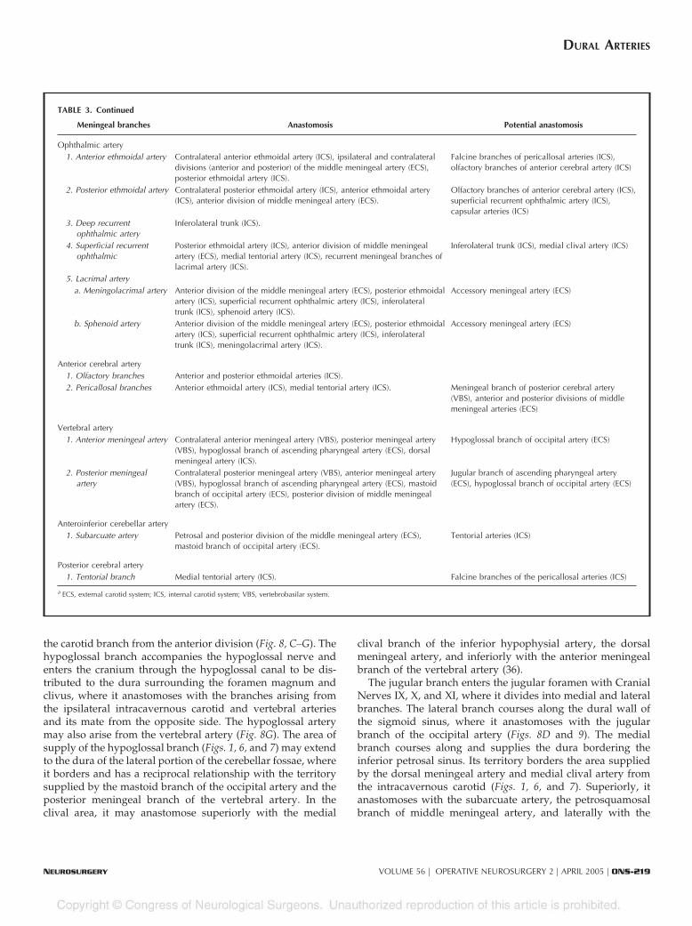
TABLE 3. Potential anastomosis of the meningeal branchesa
Meningeal branches Anastomosis Potential anastomosis
Ascending pharyngeal artery1. Carotid branch Recurrent artery of foramen lacerum (ICS), inferolateral trunk (ICS),
cavernous branch of middle meningeal artery (ECS).Accessory meningeal artery (ECS)
2. Jugular branch Jugular and mastoid branches of occipital artery (ECS), hypoglossal branchof ascending pharyngeal artery (ECS), dorsal meningeal artery (ICS),subarcuate artery (VBS).
Posterior division of middle meningeal artery(ECS), posterior meningeal artery (VBS), medialclival artery (ICS)
3. Hypoglossal branch Hypoglossal and mastoid branches of occipital artery (ECS), jugular branchof ascending pharyngeal artery (ECS), dorsal meningeal artery (ICS),anterior and posterior meningeal arteries (VBS).
Medial clival artery (ICS)
Occipital artery1. Jugular branch Same potential anastomosis as the jugular branch of ascending pharyngeal
artery, with which this artery has a strong reciprocal relationship.2. Hypoglossal branch Same potential anastomosis as the hypoglossal branch of ascending
pharyngeal artery, with which this artery has a strong reciprocalrelationship.
3. Mastoid branch Jugular and hypoglossal branches of ascending pharyngeal and occipitalarteries (ECS), posterior meningeal artery (VBS).
Lateral tentorial artery (ICS), posterior division ofmiddle meningeal artery (ECS), subarcuate artery(VBS)
4. Parietal emissary branch Posterior division of middle meningeal artery (ECS). Posterior meningeal artery (VBS)
Middle meningeal artery1. Petrosal branch Anterior and posterior middle meningeal artery divisions (ECS), accessory
meningeal artery (ECS), carotid branch of ascending pharyngeal artery(ECS), inferolateral trunk (ICS), medial and lateral tentorial arteries (ICS).
Dorsal meningeal artery (ICS), jugular branch ofascending pharyngeal artery (ECS), mastoid branchof occipital artery (ECS), subarcuate artery (VBS)
2. Anterior division Anterior (including the anterior falcine artery) and posterior ethmoidalarteries (ICS), contralateral anterior division of the middle meningeal artery(ECS), ipsilateral and contralateral posterior division of the middle meningealartery (ECS), petrosal branch of middle meningeal artery (ECS), recurrentmeningeal branches of lacrimal artery (ICS), inferolateral trunk (ICS).
Superficial recurrent ophthalmic artery (ICS),accessory meningeal artery (ECS)
3. Posterior division Petrosal branch, anterior and contralateral posterior divisions of themiddle meningeal artery (ECS), posterior meningeal artery (VBS), lateraltentorial artery (ICS), mastoid branch of occipital artery (ECS).
Medial tentorial artery (ICS), dorsal meningealartery (ICS), jugular branch of ascendingpharyngeal artery (ECS), subarcuate artery (VBS)
Accessory meningeal artery Petrosal branch and anterior division of the middle meningeal artery (ECS),inferolateral trunk (ICS), medial tentorial artery (ICS).
Posterior division of middle meningeal artery (ECS),carotid branch of ascending pharyngeal artery (ECS),recurrent meningeal branches of lacrimal artery (ICS)
Cavernous carotid1. Recurrent artery of
foramen lacerumCarotid branch of ascending pharyngeal artery (ECS), inferolateral trunk(ICS).
Accessory meningeal artery (ECS), petrosal branchof middle meningeal artery (ECS)
2. Medial tentorial artery Contralateral medial tentorial artery (ICS), lateral tentorial artery (ICS),petrosal branch and posterior division of middle meningeal artery (ECS),dorsal meningeal artery (ICS), inferolateral trunk (ICS).
Meningeal branch of posterior cerebral artery(VBS), posterior meningeal artery (VBS), superficialrecurrent ophthalmic artery (ICS), medial clivalartery (ICS), accessory meningeal artery (ECS)
3. Lateral tentorial artery Medial tentorial artery (ICS), petrosal branch and posterior division ofmiddle meningeal artery (ECS), posterior meningeal artery (VBS), mastoidbranch of occipital artery (ECS).
Subarcuate artery (VBS), dorsal meningeal artery(ICS), medial clival artery (ICS), inferolateral trunk(ICS)
4. Dorsal meningeal artery Contralateral dorsal meningeal artery (ICS), medial clival artery (ICS),tentorial arteries (ICS), petrosal branch and posterior division of middlemeningeal artery (ECS), jugular and hypoglossal branch of ascendingpharyngeal artery (ECS).
Jugular and hypoglossal branches of occipitalartery (ECS).
5. Inferior hypophysealartery
Contralateral inferior hypophyseal artery (ICS), capsular arteries (ICS). Medial clival artery when arising fromintracavernous carotid (ICS)
6. Medial clival artery Contralateral medial clival artery (ICS), dorsal meningeal artery (ICS),medial tentorial artery (ICS).
Lateral tentorial artery (ICS), jugular andhypoglossal branches of ascending pharyngeal andoccipital arteries (ECS), superficial recurrentophthalmic artery (ICS), capsular arteries (ICS)
7. Inferolateral trunk Middle meningeal artery (ECS), accessory meningeal artery (ECS), carotidbranch of ascending pharyngeal artery (ECS), recurrent branch of foramenlacerum (ICS), medial tentorial artery (ICS).
Superficial recurrent branch of ophthalmic artery(ICS), recurrent meningeal branches of lacrimalartery (ICS)
8. Capsular arteries Contralateral capsular arteries (ICS), inferior hypophyseal artery (ICS). Medial clival artery (ICS), posterior ethmoidalartery (ICS)
MARTINS ET AL.
ONS-218 | VOLUME 56 | OPERATIVE NEUROSURGERY 2 | APRIL 2005 www.neurosurgery-online.com
the carotid branch from the anterior division (Fig. 8, C–G). Thehypoglossal branch accompanies the hypoglossal nerve andenters the cranium through the hypoglossal canal to be dis-tributed to the dura surrounding the foramen magnum andclivus, where it anastomoses with the branches arising fromthe ipsilateral intracavernous carotid and vertebral arteriesand its mate from the opposite side. The hypoglossal arterymay also arise from the vertebral artery (Fig. 8G). The area ofsupply of the hypoglossal branch (Figs. 1, 6, and 7) may extendto the dura of the lateral portion of the cerebellar fossae, whereit borders and has a reciprocal relationship with the territorysupplied by the mastoid branch of the occipital artery and theposterior meningeal branch of the vertebral artery. In theclival area, it may anastomose superiorly with the medial
clival branch of the inferior hypophysial artery, the dorsalmeningeal artery, and inferiorly with the anterior meningealbranch of the vertebral artery (36).
The jugular branch enters the jugular foramen with CranialNerves IX, X, and XI, where it divides into medial and lateralbranches. The lateral branch courses along the dural wall ofthe sigmoid sinus, where it anastomoses with the jugularbranch of the occipital artery (Figs. 8D and 9). The medialbranch courses along and supplies the dura bordering theinferior petrosal sinus. Its territory borders the area suppliedby the dorsal meningeal artery and medial clival artery fromthe intracavernous carotid (Figs. 1, 6, and 7). Superiorly, itanastomoses with the subarcuate artery, the petrosquamosalbranch of middle meningeal artery, and laterally with the
TABLE 3. Continued
Meningeal branches Anastomosis Potential anastomosis
Ophthalmic artery1. Anterior ethmoidal artery Contralateral anterior ethmoidal artery (ICS), ipsilateral and contralateral
divisions (anterior and posterior) of the middle meningeal artery (ECS),posterior ethmoidal artery (ICS).
Falcine branches of pericallosal arteries (ICS),olfactory branches of anterior cerebral artery (ICS)
2. Posterior ethmoidal artery Contralateral posterior ethmoidal artery (ICS), anterior ethmoidal artery(ICS), anterior division of middle meningeal artery (ECS).
Olfactory branches of anterior cerebral artery (ICS),superficial recurrent ophthalmic artery (ICS),capsular arteries (ICS)
3. Deep recurrentophthalmic artery
Inferolateral trunk (ICS).
4. Superficial recurrentophthalmic
Posterior ethmoidal artery (ICS), anterior division of middle meningealartery (ECS), medial tentorial artery (ICS), recurrent meningeal branches oflacrimal artery (ICS).
Inferolateral trunk (ICS), medial clival artery (ICS)
5. Lacrimal arterya. Meningolacrimal artery Anterior division of the middle meningeal artery (ECS), posterior ethmoidal
artery (ICS), superficial recurrent ophthalmic artery (ICS), inferolateraltrunk (ICS), sphenoid artery (ICS).
Accessory meningeal artery (ECS)
b. Sphenoid artery Anterior division of the middle meningeal artery (ECS), posterior ethmoidalartery (ICS), superficial recurrent ophthalmic artery (ICS), inferolateraltrunk (ICS), meningolacrimal artery (ICS).
Accessory meningeal artery (ECS)
Anterior cerebral artery1. Olfactory branches Anterior and posterior ethmoidal arteries (ICS).2. Pericallosal branches Anterior ethmoidal artery (ICS), medial tentorial artery (ICS). Meningeal branch of posterior cerebral artery
(VBS), anterior and posterior divisions of middlemeningeal arteries (ECS)
Vertebral artery1. Anterior meningeal artery Contralateral anterior meningeal artery (VBS), posterior meningeal artery
(VBS), hypoglossal branch of ascending pharyngeal artery (ECS), dorsalmeningeal artery (ICS).
Hypoglossal branch of occipital artery (ECS)
2. Posterior meningealartery
Contralateral posterior meningeal artery (VBS), anterior meningeal artery(VBS), hypoglossal branch of ascending pharyngeal artery (ECS), mastoidbranch of occipital artery (ECS), posterior division of middle meningealartery (ECS).
Jugular branch of ascending pharyngeal artery(ECS), hypoglossal branch of occipital artery (ECS)
Anteroinferior cerebellar artery1. Subarcuate artery Petrosal and posterior division of the middle meningeal artery (ECS),
mastoid branch of occipital artery (ECS).Tentorial arteries (ICS)
Posterior cerebral artery1. Tentorial branch Medial tentorial artery (ICS). Falcine branches of the pericallosal arteries (ICS)
a ECS, external carotid system; ICS, internal carotid system; VBS, vertebrobasilar system.
DURAL ARTERIES
NEUROSURGERY VOLUME 56 | OPERATIVE NEUROSURGERY 2 | APRIL 2005 | ONS-219

mastoid branches of the occipital artery. The jugular branchdistal to the jugular foramen supplies the dura facing theinferior part of the cerebellopontine angle (Fig. 6) (36). Thehypoglossal and jugular branches also supply the adjacentsegments of Cranial Nerves IX through XII (34, 38, 59). Onlateral angiograms, the posterior division of the ascendingpharyngeal artery ascends beside and overlaps the foramenmagnum. On anteroposterior views, the hypoglossal is themost medial of the terminal branches of the posterior division.
The carotid branch originates from the anterior division ofthe ascending pharyngeal artery. It courses in the periosteallining of the carotid canal and anastomoses, at the level of theforamen lacerum, with branches arising from the carotid si-phon to form the recurrent artery of the foramen lacerum (36).This recurrent artery also anastomoses at the lower edge of thetrigeminal ganglion with the posterior branch of the inferolat-eral trunk and the cavernous branch of the middle meningealartery. The recurrent artery of the foramen lacerum may be
FIGURE 2. Superior view of theconvexity showing the area of supplyof the individual meningeal arteries.Dural branches from the internal ca-rotid arterial system are highlightedin shades of green, the external ca-rotid system meningeal branches inshades of blue, and the vertebrobasilarsystem in red. A, internal carotid sys-tem. The anterior ethmoidal artery hasalso been called the anterior menin-geal artery when its territory extendsto the dura of the frontal convexity. Itgives origin to the anterior falcine ar-tery, also called the artery of the falxcerebri, which supplies the anteriorportion of the falx cerebri and adjacentdura covering the frontal pole. B, ex-ternal carotid system. The convexitydura is supplied predominantly bybranches of the middle meningeal ar-teries, which supply the dura of thefrontal, temporal, and parietal convex-ities and the adjacent walls of thetransverse and sigmoid sinus. Thesebranches course toward the superiorsagittal sinus, where they are distrib-uted to the sinus walls and give offdescending branches to the adjacentfalx cerebri. C, vertebrobasilar system.The posterior meningeal artery mayreach the dura of the posterior convex-ity in the area above the torcula. Itextends along the insertion of the falxcerebri into the tentorium after as-cending in the insertion of the falxcerebelli. D, overview. The dura overthe frontal convexity is supplied bythe anterior meningeal branch of theanterior ethmoidal artery andbranches of the anterior division of themiddle meningeal artery that alsoreach the dura in the anterior parietalregion. The parieto-occipital andpetrosquamosal branches of the poste-rior division of the middle meningealartery supply the dura over the poste-rior convexity. This area also receivesa contribution from the posterior meningeal branch of the vertebral artery when this vessel extends above the torcula. A., artery; Access., accessory; Ant., anterior;Div., division; Men., meningeal; Mid., middle; Post., posterior.
MARTINS ET AL.
ONS-220 | VOLUME 56 | OPERATIVE NEUROSURGERY 2 | APRIL 2005 www.neurosurgery-online.com

FIGURE 3. Superior view of the ten-torium showing the area of supply ofthe individual meningeal arteries.Dural branches from the internal ca-rotid arterial system are highlightedin shades of green, the external ca-rotid in shades of blue, and the verte-brobasilar system in shades of red. A,internal carotid system. From medialto lateral, the dorsal meningeal, themedial, and the lateral tentorial arter-ies supply the tentorium at its petrosalattachment. The dorsal meningeal ar-tery (lateral branch) passes aboveMeckel’s cave and accompanies thesuperior petrosal sinus along the pe-trous ridge, thus participating in thebasal arterial arcade of the tentoriumcerebelli. The medial tentorial arteryascends to the roof of the cavernoussinus and along the free edge of thetentorium to contribute to the supplyof the medial third of the tentorium.As it approaches the region of thestraight sinus, it curves laterally,ramifying within the tentorium andanastomosing along the base of thefalx. The lateral tentorial artery entersthe tentorium along its attachment tothe petrous ridge and continues back-ward to supply the tentorial area lat-eral to that supplied by the medialtentorial artery. B, external carotidsystem. The branches of the posteriordivision of the middle meningeal ar-tery contribute to the supply of theanterolateral tentorium and extendsuperiorly to supply the falcotentorialjunction and falx. The posteriorbranch of the middle meningeal arterygives rise to the petrosquamosalbranch at the junction of the cranialbase and convexity and supplies theinsertion of the tentorium along thepetrous ridge and groove for the trans-verse sinus, the dura of the torcula,and the junction of the sigmoid, trans-verse, and superior petrosal sinuses. Itextends to the dura of the posteriorfossa bordering the area supplied bythe other branches of the external carotid artery. The occipital artery (not seen) may also provide tentorial branches along the lateral tentorial attachment. The externalcarotid system may supply most of the tentorium if it gives rise to a medial tentorial branch that may infrequently arise from either of the divisions of the middlemeningeal artery or from the accessory meningeal artery. C, vertebrobasilar system. The posterior meningeal artery forms the infratentorial limb of the arcade supplyingthe falcotentorial junction. The posterior cerebral artery may also contribute to the supply of the medial edge of the tentorium through the artery of Davidoff andSchechter (tentorial branch of the P.C.A.). D, overview. The carotid sources of supply to the tentorium include the medial and lateral tentorial branches of theintracavernous carotid medially and the branches of the middle meningeal artery anterolaterally. The vertebrobasilar component to the medial edge is supplied bythe tentorial branch of the posterior cerebral artery medially and the posterior meningeal artery posteriorly. The lateral two thirds of the tentorium and its edge along thetransverse sinus derive their supply primarily from the petrosal and occipital arcades. The petrosal arcade follows the superior petrosal sinus and is composed ofthe lateral tentorial artery, branches from the petrosal and petrosquamosal trunk of the middle meningeal artery, and the lateral branch of the dorsal meningealartery. The occipital arcade is formed by the petrosquamosal trunk and occipital branches of the middle meningeal artery and the occipital and posteriormeningeal arteries. The medial third of the tentorium is supplied by the medial tentorial branch of the intracavernous carotid and the tentorial branch of theposterior cerebral artery. A., artery; Ant., anterior; Br., branch; Div., division; Dors., dorsal; Lat., lateral; Med., medial; Men., meningeal; Mid., middle; P.C.A.,posterior cerebral artery; Pet., petrosal; Post., posterior; Tent., tentorial.
DURAL ARTERIES
NEUROSURGERY VOLUME 56 | OPERATIVE NEUROSURGERY 2 | APRIL 2005 | ONS-221

involved in the supply of angiomas, lymphoid tumors, angio-fibromas of the nasopharynx, and tumors of the cavernoussinus and caroticocavernous fistulae (36).
Occipital Artery
The occipital artery originates from the posterior surface ofthe external carotid artery, at the level of the angle of themandible, and ascends posteriorly, being crossed superficiallyby the hypoglossal nerve. It passes deep to the posterior bellyof the digastric muscle and lateral to the internal jugular vein,vagus nerve, internal carotid artery, and accessory nerve (Fig.9). At the level of a vertical plane crossing the posterior borderof the external auditory canal, the occipital artery can be foundin a tunnel formed above by the occipital groove, a prominentsulcus on the undersurface of the temporal bone, medially bythe attachment of the superior oblique muscle on the trans-verse process of the atlas, and laterally by the insertion of theposterior belly of the digastric muscle in the digastric groove(Fig. 9, B–F). The presence of the occipital groove is dependenton whether the artery courses superficially or deep to thelongissimus capitis muscle. The groove is present if the arterycourses deep to the longissimus capitis muscle along the lower
surface of the cranial base and is absent if the artery coursesinferior to the cranial base or lateral to the longissimus capitismuscle (Fig. 9, E–G) (68).
The occipital artery at the level of the posterior border of theupper insertion of the longissimus capitis muscle courses inthe upper part of the space between the occipital bone and C1and lateral to the rectus capitis posterior major and semispi-nalis capitis muscle. It is covered by a deeper layer formed bythe splenius capitis muscle and a more superficial layerformed by the sternocleidomastoid muscle. The occipital ar-tery pierces the fascia between the trapezius and sternoclei-domastoid, near the superior nuchal line, and ascends in thesuperficial fascia of the scalp, where it is accompanied by thegreater occipital nerve (Fig. 9, E–G). The occipital artery givesrise to an auricular branch, which anastomoses with the pos-terior auricular artery behind the ear; muscular branches tothe sternocleidomastoid, digastric, stylohyoid, splenius, andlongissimus capitis muscles; and meningeal branches to theposterior fossa that enter the cranium through the jugularforamen and condylar canal and to inconstant branches thatrun through the mastoid emissary foramen (Fig. 9, B, D, andG).
FIGURE 4. Enlarged superior viewshowing the supply of the parasellararea. Dural branches from the inter-nal carotid arterial system have beenhighlighted in shades of green, theexternal carotid system meningealbranches in shades of blue, and thevertebrobasilar system in shades ofred. A, internal carotid system. In ananterior-to-posterior direction, theparasellar dura receives contributionsfrom the recurrent branches of theophthalmic artery and the meningo-lacrimal, medial tentorial, medialclival, and dorsal meningeal arteries.The medial clival and dorsal menin-geal arteries supply the dura over theposterior roof of the cavernous sinusand posterior diaphragma sellae andanastomoses laterally with thebranches of the inferolateral trunk, themain supplier of the lateral wall of thecavernous sinus. B, external carotidsystem. The supply to the parasellarpart of the middle fossa arises from themain divisions of the middle menin-geal artery. The accessory meningeal and ascending pharyngeal arteries may provide an alternative supply of the lateral portion of the parasellar area in their reciprocalrelationship with the branches of the internal carotid artery that supply the same area. C, vertebrobasilar system. There are no branches of the vertebrobasilar systemto the parasellar dura. The anterior meningeal artery from the vertebral artery supplies the anterolateral portion of the posterior fossa and foramen magnum. D,overview. The intracavernous carotid branches provide the major supply to the roof and posterior and lateral walls of the cavernous sinus. These branches border laterallywith the ascending pharyngeal and accessory meningeal branches. The main divisions of the middle meningeal artery supply the middle fossa dura. The branches ofthe internal carotid artery supplying the posterior wall of the cavernous sinus may anastomose with the branches of the ascending pharyngeal and vertebral artery tosupply the clival dura. A., artery; Access., accessory; Ant., anterior; Asc., ascending; Br., branch; Car., carotid; Cliv., clival; Div., division; Dors., dorsal; Eth.,ethmoidal; Inf., inferior; Lac., lacrimal; Lat., lateral; Med., medial; Men., meningeal, meningo; Mid., middle; Ophth., ophthalmic; Pharyng., pharyngeal; Post.,posterior; Rec., recurrent; Tent., tentorial; Tr., trunk.
MARTINS ET AL.
ONS-222 | VOLUME 56 | OPERATIVE NEUROSURGERY 2 | APRIL 2005 www.neurosurgery-online.com

The occipital artery is divided into three portions: 1) ascend-ing cervical, 2) cervico-occipital or horizontal, and 3) ascend-ing occipital (Fig. 9A) (45). The meningeal branches mostfrequently originate from the second and third arterial seg-ments. The first portion gives rise to muscular branches, theauricular branch, and occasionally to the ascending pharyn-geal artery. The second segment of the occipital artery givesrise to three types of branches: meningeal, muscular, and adescending branch. The descending branch, located at the firstcervical interspace, originates at the point at which the arterycrosses behind the superior oblique muscle and divides intosuperficial and deep branches (Fig. 9C). The deep branchanastomoses with the vertebral and deep cervical artery (36,37). These anastomoses are the adult remnants of the embry-onic arterial connections and explain the variants in which theoccipital artery arises from the vertebral, internal carotid, orascending cervical artery (36).
The second segment is frequently the source of the stylo-mastoid artery when it arises from the occipital artery ratherthan from other external carotid branches. The stylomastoidartery ascends along the anterior aspect of the mastoid processto enter the stylomastoid foramen and supplies the facialnerve and middle ear (45). It anastomoses with the petrosalbranch of the middle meningeal artery, which supplies thepetrosal segment of the facial nerve (Fig. 9J) (2). Occlusion ofthe stylomastoid artery during either embolization or surgerymay result in a facial nerve deficit. The stylomastoid artery,when present, on angiograms marks the position of the distalpart of the facial canal.
The mastoid branch, also called the transmastoid branch or theartery of the mastoid foramen, is present in approximately half of allspecimens and originates from the second segment of the occipitalartery at the level of the insertion of the semispinalis capitis muscle,midway between the inferior and superior nuchal lines (Fig. 9, B, D,
FIGURE 5. Lateral view showingthe supply of the tentorium and falx.The dural branches from the internalcarotid arterial system are highlightedin shades of green, the external ca-rotid system in shades of blue, and thevertebrobasilar system in shades ofred. A, internal carotid system. Theanterior falcine artery, the distal con-tinuation of the anterior ethmoidal ar-tery, enters the falx at the cribriformplate and supplies the anterior portionof the falx cerebri and adjacent duracovering the frontal pole. The free bor-der of the falx and the walls of theinferior sagittal sinus receive branchesfrom the pericallosal arteries anteri-orly and the medial tentorial arteryposteriorly. B, external carotid sys-tem. The anterior and posterior divi-sions of the middle meningeal arterysupply the walls of the superior sagit-tal sinus and give rise to descendingbranches that are the main supply tothe falx and the falcotentorial junc-tion. The posterior division of the mid-dle meningeal artery also reaches thewalls of the straight and transversesinuses. C, vertebrobasilar system. The posterior meningeal arteries reach the falcotentorial junction and posterior third of the falx cerebri. The presence of the infrequenttentorial branch of the posterior cerebral artery represents an additional potential supply to the lower edge of the falx and the falcotentorial junction. D, overview. Thefalx cerebri, falx cerebelli, and tentorium are supplied by basal and convexity branches of the meningeal arteries and receive a contribution from the cerebral arteries,making these structures an anastomotic pathway between the dural and parenchymal arteries. Most of the vascular supply of the falx cerebri comes through the middlemeningeal artery at its insertion on the vault, with the anterior basal insertion, the falcotentorial angle, and the free margin receiving contributions from the internalcarotid and vertebrobasilar branches. The dural walls of the superior sagittal sinus, the site of insertion of the falx on the convexity, is supplied by the middle meningealarteries, which form paired paramedial arcades, which are reinforced anteriorly, at the level of the insertion of the falx on the crista galli, by the anterior falcine arteries.Posteriorly, at the falcotentorial junction, the paramedial arcades are reinforced from three sources: the posterior meningeal artery from the vertebral artery, the medialtentorial artery from the intracavernous carotid, and an occasional branch of the posterior cerebral artery. The posterior meningeal artery ascends along the insertionof the falx cerebelli. The medial tentorial artery, which supplies the medial third of the tentorium, reaches the straight sinus and torcula and may ascend in the posteriorportion of the falx cerebri. The pericallosal branches of the anterior cerebral artery may also pierce the falx at or near its free edge to reinforce the arterial network alongthe deep edge of the falx. A., artery; Ant., anterior; Br., branch; Brs., branches; Div., division; Falc., falcine; Lat., lateral; Med., medial; Men., meningeal; Mid., middle;P.C.A., posterior cerebral artery; Perical., pericallosal; Pet., petrosal; Post., posterior; Tent., tentorial.
DURAL ARTERIES
NEUROSURGERY VOLUME 56 | OPERATIVE NEUROSURGERY 2 | APRIL 2005 | ONS-223

and G) (16, 36, 45). From its origin, the mastoid branch coursesbetween the splenius capitis muscle and the junction of the mastoidand occipital bones. It enters the cranial cavity at the level of thesuperior nuchal line by passing through the mastoid foramen. In-tracranially, the superior nuchal line corresponds to the level of thetransverse sinus. The mastoid branch emerges intracranially at theposterior border of the upper end of the sigmoid sinus and dividesinto three groups of branches: descending, ascending, and postero-medial (45). The descending branches are directed toward the jug-ular foramen and border the dural territory supplied by the jugularbranch of the ascending pharyngeal artery (Fig. 8D). The postero-medial branches anastomose with the petrosquamous branch of themiddle meningeal artery and constitute the main supply to thelateral part of the cerebellar fossae, which borders the territory of thehypoglossal branch of the ascending pharyngeal artery and/or theposterior meningeal branch of the vertebral artery. The ascendingbranches, which are directed to the dura covering the superior partof the posterior surface of the temporal bone that faces the cerebel-
lopontine angle, anastomoses with the subarcuate branch of theanteroinferior cerebellar artery (Figs. 1 and 6) and can supply acous-tic neurinomas, meningiomas, and arteriovenous fistulae.
The mastoid branches also supply the endolymphatic duct andsac (15). By selectively filling the branches of the external carotidartery with colored methylmethacrylate, it has been demon-strated that the occipital artery sends branches that pass throughthe mastoid part of the temporal bone and exit through tinyforamina in the groove for the sigmoid sinus to run superiorlyand medially on the posterior surface of the temporal bonetoward the region of the endolymphatic sac. On reaching theendolymphatic duct and sac, these arteries are oriented along thelong axis of the sac, branching in the region of the distal duct. Thedistal branches of the internal carotid and vertebral systems mayreach the tiny dural vessels around the internal auditory meatusand in the wall of the sigmoid sinus but do not reach the en-dolymphatic sac or cross the sigmoid sinus. This finding sup-
FIGURE 6. View of posterior fossaand tentorial dura. The view is di-rected from medially into the left halfof a posterior fossa in which the cere-bellum was removed. The clivus is onthe right and the transverse sinus onthe left. Dural branches from the in-ternal carotid arterial system havebeen highlighted in shades of green,the external carotid system in shadesof blue, and the vertebrobasilar sys-tem in shades of red. A, internal ca-rotid system. The medial tentorial ar-tery supplies the medial third of thetentorium, and the dorsal meningealand the lateral tentorial artery con-tribute to the arcade that supply theattachment of the tentorium to thepetrous ridge. The medial clival anddorsal meningeal arteries supply thedorsum sellae and upper clivus. B,external carotid system. The hypo-glossal and jugular branches of theascending pharyngeal artery and thebranches of the occipital artery supplythe dura of the lateral part of the cer-ebellar fossa and the inferior portion ofthe posterior surface of the petrous temporal bone. The mastoid branch of the occipital artery constitutes the main supply of the lateral part of the cerebellar fossae andhas a role in the supply of the lateral tentorial attachment. C, vertebrobasilar system. The subarcuate artery, a branch of the AICA, supplies the posterior surface ofthe petrous bone above the internal acoustic meatus and surrounding the subarcuate fossa. The anterior and posterior meningeal artery branches of the vertebral arterysupply the foramen magnum dura. The posterior meningeal artery supplies the medial and intermediate portions of the cerebellar fossa dura. The vertebrobasilar systemmay also infrequently supply the medial edge of the tentorium through a branch of the posterior cerebral artery. D, overview. Branches derived from all three arterialsystems supply the dura covering the posterior surface of the petrous bone and clivus. The clival area, from the dorsum sellae to the foramen magnum, derives its supplyfrom the medial clival and dorsal meningeal branches of the internal carotid artery, the jugular and hypoglossal branches of the ascending pharyngeal artery, and theanterior meningeal branch of the vertebral artery. From medial to lateral, the dura over the posterior surface of the petrous bone is supplied by the dorsal meningeal,subarcuate, occipital and ascending pharyngeal arteries, and branches of the middle meningeal artery (not seen). The dura of the lateral portion of the cerebellar fossa,from above to below, receives its supply from the branches of the occipital, ascending pharyngeal, and vertebral arteries. The posterior meningeal artery is the majorsupplier of the paramedial and medial portions of the cerebellar dura, with contributions from the middle meningeal and occipital arteries. A., artery; Ac., acoustic; Asc.,ascending; Ant., anterior; Br., branch; Brs., branches; Cliv., clival; Dors., dorsal; For., foramen; Hypogl., hypoglossal; Int., internal; Jug., jugular; Lat., lateral; Med.,medial; Men., meningeal; Occip., occipital; P.C.A., posterior cerebral artery; Pharyng., pharyngeal; Post., posterior; Sig., sigmoid; Subarc., subarcuate; Tent.,tentorial; Transv., transverse.
MARTINS ET AL.
ONS-224 | VOLUME 56 | OPERATIVE NEUROSURGERY 2 | APRIL 2005 www.neurosurgery-online.com

ports previous animal studies showing that obstruction of theinternal auditory artery resulted in degeneration of the membra-nous labyrinth and cochlea, but with morphological preservationof the endolymphatic sac. On the basis of these findings, it hasbeen postulated that manipulation of the blood flow in the oc-cipital artery can modify the fluid dynamics of the inner earthrough its branches to the endolymphatic sac, opening a possi-ble therapeutic window to Meniere disease (15).
The third or ascending occipital portion gives rise to theterminal branches of the occipital artery, which supply themusculocutaneous structures of the posterior portion of thecranial vault and anastomoses with the branches of the super-ficial temporal artery (Fig. 9H). The parietal foramen (Fig. 9J),which is an inconstant opening located near the sagittal sutureapproximately 3 to 5 cm above the lambda (16), transmits ameningeal branch of the ascending occipital segment and asmall emissary vein (45).
Variations of the stylomastoid artery and the mastoidbranch include their origin from the ascending pharyngealartery or from the posterior auricular artery. Alternatively,other meningeal arteries that commonly have other sites oforigin, such as the posterior meningeal artery and the branchto the falx cerebelli from the posterior meningeal artery, mayalso arise from the occipital artery. When the occipital artery isabsent, its arterial territory is taken by the posterior auricular,the ascending cervical, or the vertebral artery (45). The originof the occipital artery shifts during the embryological period,successively belonging to vertebral, internal carotid, and ex-ternal carotid arteries (45).
Maxillary Artery
The maxillary artery, through its middle meningeal andaccessory meningeal branches (Figs. 10 and 11), provides al-most all of the supply to the dura over the convexity andimportant contributions to the supply of the basal dura (Figs.1–4).
Middle Meningeal Artery. The middle meningeal arterynormally arises from the first or mandibular segment of themaxillary artery, just behind the condylar process of the man-dible, and enters the cranium through the foramen spinosum(Fig. 10, A–H). After passing through the foramen spinosum,the main stem courses laterally, grooving the greater sphenoidwing, where it divides into its anterior and posterior divisions,which supply the dura of the frontal, temporal, and parietalconvexities, the upper surface of the temporal bone, and theadjacent walls of the transverse and sigmoid sinus as well asthe middle fossa dura adjacent to the cavernous sinus (Fig. 10,F–N). In its path between the anterosuperior angle of thegreater sphenoid wing and the sphenoid angle of the parietalbone, the anterior division and sometimes the sphenoparietalsinus can be encased in a bony canal that varies in extensionbetween 1 and more than 30 mm (8). The anterior division isusually single but may be composed of two branches (dupli-cated) in 0.8% or absent in 0.7% of cases, whereas the posteriordivision is duplicated in 8.1% (8). At the level of the superiorsagittal sinus, the middle meningeal artery anastomoses withthe anterior falcine branch of the ophthalmic artery to supplythe dural layers of the falx (Fig. 5).
FIGURE 7. Posterior view of the dura covering the cerebellum andforamen magnum. A suboccipital craniectomy and C2 laminectomy hasbeen performed, with preservation of the posterior arch of C1. Duralbranches from the external carotid system are highlighted in shades ofblue and the vertebrobasilar system in shades of red. No branches ofthe internal carotid system supply the dura covering the posterior cere-bellar surface. A, external carotid system. The mastoid branches of theoccipital artery constitute the main supply to the lateral part of thecerebellar fossae. The posteromedial division of the mastoid branchanastomoses with the petrosquamous branch of the middle meningealartery above and below with the hypoglossal branch of the ascendingpharyngeal artery. B, vertebrobasilar system. The posterior meningeal
artery supplies the medial and paramedial cerebellar fossae between the trans-verse sinus and torcula above and the posterior edge of the foramen magnumbelow. C, overview. The dura of the lateral portion of the cerebellar fossareceives its supply from the middle meningeal, occipital, ascending pharyn-geal, and vertebral arteries. The walls of the falx cerebelli and enclosed occipi-tal sinus are supplied primarily by the branches of the posterior meningealartery. The posterior meningeal artery is also the major supplier of the para-medial and medial portions of the cerebellar dura, with lesser contributionsfrom the middle meningeal and occipital arteries. A., artery; Asc., ascending;Br., branch; Brs., branches; Div., division; Hypogl., hypoglossal; Men.,meningeal; Mid., middle; Occip., occipital; Pharyng., pharyngeal; Post.,posterior.
DURAL ARTERIES
NEUROSURGERY VOLUME 56 | OPERATIVE NEUROSURGERY 2 | APRIL 2005 | ONS-225

The middle meningeal artery and the osseous groove inwhich it courses begin at the foramen spinosum and divideinto anterior and posterior divisions 15 to 30 mm anterolateralto the foramen spinosum (Fig. 10, F and I). The anterior divi-sion and its groove divide behind the lateral part of the greaterwing into a lateral branch, which passes across the pterion toreach the dura of the lateral convexity, and a medial branch,which courses medially along the lower surface of the sphe-
noid ridge, where it anastomoses with the recurrent branch ofthe lacrimal artery. In nine of 10 orbits dissected, Liu andRhoton (46) reported the presence of anastomotic connectionsbetween the recurrent meningeal branch of the lacrimal arteryand the medial branch of the anterior division of the middlemeningeal artery. Occasionally, the recurrent meningealbranch of the lacrimal artery gives rise to the anterior segmentof the middle meningeal artery, or, more rarely, the ophthal-
FIGURE 8. A, lateral view of the leftparapharyngeal space and the ascendingpharyngeal artery origin and course. Theparapharyngeal space lies between thepharyngeal wall medially and the medialpterygoid muscle laterally. This predomi-nantly fat-filled space contains thebranches of the ascending pharyngeal andfacial arteries and branches of the glosso-pharyngeal nerve. The jugular foramenand the inferior opening of the carotidcanal are in the roof of this space. Theascending pharyngeal artery usuallyarises from the external carotid artery andascends vertically between the internal ca-rotid artery and the longus capitis muscle.The artery makes a sharp anterior turn atthe base of the cranium, running down-ward and forward, following the upperborder of the superior pharyngeal con-strictor muscle to supply the pharynx andthe auditory tube. At the uppermost pointof the vertical segment, the ascending pha-ryngeal artery gives off a carotid branch,which follows the internal carotid arteryinto the carotid canal and anastomoseswith the artery of the foramen lacerum,which arises from the internal carotid ar-tery. The ascending pharyngeal artery canoriginate from the lingual, occipital, orinternal carotid artery or even from thecervical arteries, or it may arise as a singletrunk with the occipital artery. B, the lefttemporal bone has been drilled to open thejugular foramen. Some of the branches ofthe external carotid artery have been re-moved while the proximal part of the oc-cipital artery and its jugular branch andthe ascending pharyngeal artery have been preserved. The segment of the internal carotid artery below thecarotid canal has been removed and the stump retracted posteriorly to expose the anterior and posteriordivisions of the ascending pharyngeal artery. The anterior division gives off the pharyngeal rami, and theposterior, or neuromeningeal, division sends branches to the posterior fossa dura. The ascending ramus of theanterior branch, also called the eustachian branch, supplies the eustachian tube and gives off a carotid branch,which accompanies the internal carotid artery within the carotid canal, supplying the periosteum, thesympathetic network around the vessel, and the arterial walls. The jugular branches of the ascendingpharyngeal and occipital arteries send branches to Cranial Nerves IX, X, and XI. C, intracranial view of theleft jugular foramen. The jugular branch descends below the jugular foramen. D, lateral view of a leftmastoidectomy. The mastoid air cells have been removed to expose the superior, posterior, and lateralsemicircular canals, the facial nerve, the sigmoid sinus, and the jugular bulb. The lateral division of thejugular branch of the ascending pharyngeal artery ascends along the anterior edge of the sigmoid sinus andanastomoses medially with the meningeal branches of the internal carotid artery, superiorly with thesubarcuate artery, and laterally with the mastoid branches of the occipital artery.
MARTINS ET AL.
ONS-226 | VOLUME 56 | OPERATIVE NEUROSURGERY 2 | APRIL 2005 www.neurosurgery-online.com

mic artery can give rise to themain stem of the middle menin-geal artery itself. In these cases,with an ophthalmic or lacrimalorigin of the middle meningealartery, the grooves marking thecourse of the main stem of themiddle meningeal artery willoriginate at the lateral edge ofthe superior orbital fissure (72)and the foramen spinosum will be hypoplastic or absent (53).Another, less frequent, site of origin of the middle meningealartery is from the petrous portion of the internal carotid artery,referred to as a stapedial-middle meningeal artery, an anom-aly that results from failure of the embryonic stapedial branchof the internal carotid artery to regress and allow the middlemeningeal artery to become connected to the external carotidartery (53).
Angiographically, in the anterior view, the middle meningealartery is easily recognized by a sharp turn along the floor of the
middle fossa after passingthrough the foramen spino-sum. Its course along the in-ner table is characterized bysmooth curves, in contrastto the sinuous course of theoverlapping superficialtemporal artery. This initialintracranial portion of themiddle meningeal arterycan be elevated andstretched by lesions arisingat the cranial base. Radio-graphically, the grooves forthe meningeal branches canbecome tortuous and the fo-ramen spinosum can en-large in meningiomas andvascular malformations (24,86).
Immediately adjacent tothe foramen spinosum, themiddle meningeal arterygives off a short branch,which divides into thepetrosal artery laterally anda cavernous branch to thetrigeminal ganglion medi-ally (Fig. 10, G and H). Thepetrosal branch runs withthe greater petrosal nerveand penetrates the temporalbone by passing throughthe facial hiatus and sup-plies the facial nerve andwalls of the tympanic cavity(4, 70). Damage to the petro-sal branch occurring as thedura is elevated in a sub-temporal extradural ap-proach to the trigeminalnerve, cavernous sinus, orinternal acoustic meatusmay result in a facial nervedeficit. Bleeding at this siteshould be controlled by a
method other than coagulation to avoid damaging the facialnerve, which may be exposed in the floor of the middle fossa atthe level of the hiatus fallopi (70, 72).
The posterior division of the middle meningeal artery givesrise to a petrosquamosal branch at the junction of the cranialbase and convexity that supplies the insertion of the tentoriumalong the petrous ridge and groove for the transverse sinus,the dura of the torcula, and the junction of the sigmoid,transverse, and superior petrosal sinuses and extends to thedura of the posterior fossa bordering the area supplied by the
FIGURE 8. Continued. E, posterior viewof the left lower cranial nerves. The cere-bellum and accessory nerve have been ele-vated to expose the hypoglossal nerve. Thehypoglossal branch of the ascending pha-ryngeal artery passes through the hypo-glossal canal with the hypoglossal nerveand enters the dura. At the level of theforamen magnum, the territory suppliedby the hypoglossal branch is interposed be-tween the territories of the anterior andposterior meningeal arteries of the verte-bral artery. F, posterior view of a hypoglos-sal canal that has been opened to expose thehypoglossal nerve and the hypoglossalbranch of the ascending pharyngeal artery.The hypoglossal branch supplies the duraof the lateral portion of the foramen mag-num and inferolateral cerebellar fossa. Theposteroinferior cerebellar artery arises fromthe extradural segment of the vertebral ar-tery. G, the right cerebellar tonsil has beenelevated to expose a meningeal artery thatarises from the intradural vertebral arteryand supplies the dura on the lateral edge ofthe lateral foramen magnum. A., artery;Ant., anterior; Asc., ascending; Br.,branch; Cap., capitis; Car., carotid; CN,cranial nerve; Constr., constrictor; Div.,division; Ext., external; For., foramen; Hy-pogl., hypoglossal; Int., internal; Jug.,jugular; Lat., lateral; Long., longus; M.,muscle; Men., meningeal; Occip., occipi-tal; P.I.C.A., posteroinferior cerebellar ar-tery; Palat., palatini; Pharyng., pharyn-geal; Post., posterior; Proc., process;Semicirc., semicircular; Sig., sigmoid; Sup.,superior; Tens., tensor; Vert., vertebral.
DURAL ARTERIES
NEUROSURGERY VOLUME 56 | OPERATIVE NEUROSURGERY 2 | APRIL 2005 | ONS-227

other branches of the external carotid artery (Figs. 7 and 10, Iand J) (57). The petrosquamosal artery may infrequently sup-ply almost all the posterior fossa dura, including the cerebellarfossa and tentorium cerebelli (57). The parieto-occipital branch
of the middle meningeal artery supplies the dura over theposterior convexity (Fig. 10J).
The middle meningeal artery in the middle fossa has anas-tomotic connections with the ophthalmic system and the men-
FIGURE 9. A, lateral view of aleft occipital artery in the areabelow the mastoid process. Theexternal carotid artery has beenretracted anteriorly and the poste-rior belly of the digastric down-ward to expose the occipital artery.The occipital artery originates fromthe posterior surface of the externalcarotid artery, courses posteriorlyand upward, and passes deep tothe posterior belly of the digastricmuscle in the occipital groove ofthe temporal bone. B, posteriorview of the retroauricular area. Thesternocleidomastoid and spleniuscapitis muscle have been reflected.The fascial cuff of the spleniuscapitis muscle has been leftattached to the superior nuchalline. The occipital artery passesbetween the longissimus capitisand semispinalis capitis musclesand gives rise to a mastoid branchthat passes through the mastoidforamen to reach the dura in thearea of the junction of the sigmoidand transverse sinuses. The petros-quamosal branch of the middlemeningeal artery passes toward thedura above the transverse sinus,where it anastomoses with themastoid branch of the occipitalartery. C, the bone has beenremoved around the mastoidbranch of the occipital artery. Adescending branch of the occipitalartery arises as the artery passesabove the superior oblique muscleand gives rise to deep rami thatanastomose with the vertebralartery. D, enlarged view of C. Themastoid branch of the occipitalartery enters the cranium by pass-ing through the mastoid foraminaand anastomoses over the junctionof the sigmoid and transversesinuses with the branches of themiddle meningeal artery. E and F,left (E) and right (F) retromastoidareas. The sternocleidomastoid andsplenius capitis muscles have beenreflected forward to demonstrate the variable relationship of the occipital artery to the lower surface of the occipital bone and longissimus capitis muscle. E,when the course of the occipital artery is low, no occipital groove is present, and the artery passes superficial to the longissimus capitis muscle. F, if theartery courses below the cranial base in an occipital groove, it passes deep to the longissimus capitis muscle.
MARTINS ET AL.
ONS-228 | VOLUME 56 | OPERATIVE NEUROSURGERY 2 | APRIL 2005 www.neurosurgery-online.com

FIGURE 9. Continued. G, posterior view of the left occipitomastoid area. The occipitalgroove is a prominent sulcus on the lower surface of the temporal bone, medial to thedigastric groove in which the occipital artery courses. The mastoid foramen transmits themastoid emissary vein and the mastoid branch of the occipital artery. It is usually locatedat the level of the lateral part of the superior nuchal line and may be composed of multipleforamina. H, the terminal branches of the occipital artery form an anastomotic networkin the scalp with the branches of the superficial temporal artery. A meningeal branch ofthis segment courses toward the midline and passes through the parietal foramen. I,posterior view of the sagittal and lambdoid sutures. The parietal foramen, which trans-mits an emissary vein and a meningeal branch of the terminal segment of the occipitalartery, is located near the midline, 3 to 5 cm above the lambda. J, lateral view of the leftinfrapetrosal area with the posterior belly of the digastric retracted anteriorly. Thestylomastoid artery arises from the posterior surface of the external carotid artery andpasses through the stylomastoid foramen to reach the facial canal, where it supplies themastoid portion of the facial nerve and walls of the tympanic cavity. The stylomastoidartery may also arise from the posterior auricular, occipital, or ascending pharyngealartery. K, the posterior belly of the digastric has been removed to expose the jugularbranch of the occipital artery, which ascends behind the carotid sheath to supply the duraaround the jugular foramen and Cranial Nerves IX, X, and XI. A., artery; Br., branch;Brs., branches; Cap., capitis; Car., carotid; CN, cranial nerve; Desc., descending;Digast., digastric; Ext., external; For., foramen; Gr., greater; Inf., inferior; Int., internal;Jug., jugular; Longiss., longissimus; M., muscle; Men., meningeal; Mid., middle; N.,nerve; Obl., oblique; Occip., occipital; Occipitomast., Occipitomastoid; Par., parietal;Petrosquam., petrosquamosal; Post., posterior; Proc., process; Sag., sagittal; Sup.,superior; Superf., superficial; Temp., temporal; Transv., transverse; V., vein; Vert.,vertebral.
DURAL ARTERIES
NEUROSURGERY VOLUME 56 | OPERATIVE NEUROSURGERY 2 | APRIL 2005 | ONS-229

ingeal branches of the intracavernous carotid (Fig. 1). It maycontribute to the supply of the second and third trigeminaldivisions in addition to the facial nerve and may supply mostof the tentorium if it gives rise to a medial tentorial branch(34). The medial tentorial artery may arise from either themain divisions of the middle meningeal artery or from theaccessory meningeal artery (57), to be described in the nextsection.
The middle meningeal artery anastomoses over the upperclivus and adjacent posterior surface of the temporal bone
with the dorsal meningeal artery and the subarcuate artery.The distal part of the petrosquamosal branch anastomoses, atthe level of the junction of the sigmoid, transverse, and supe-rior petrosal sinuses, with the branches of the occipital artery,which passes through the mastoid foramen and the meningealbranches of the ascending pharyngeal and vertebral arteries.
The middle meningeal artery may also anastomose with abranch of the basilar artery. Various explanations on the originof the basilar connection have been reported (74, 82). In onevariant, the anastomotic branch of the basilar artery arises
FIGURE 10. A, lateral view of theleft mandible and infratemporal area.The parotid gland and masseter havebeen removed, and a portion of thezygoma has been resected to exposethe attachment of the temporal muscleto the coronoid process of mandible.The superficial temporal artery arisesfrom the external carotid artery andcourses behind the condylar process ofthe mandible and the temporomandib-ular joint. B, the coronoid process hasbeen removed and the temporalismuscle retracted upward to expose themaxillary and superficial temporal ar-teries and branches of the external ca-rotid artery. The maxillary artery isdivided into mandibular, pterygoid,and pterygopalatine segments. Themandibular segment courses deep tothe neck of the mandible. The ptery-goid segment courses between thetemporalis and pterygoid muscles andgives rise to the deep temporal andpterygoid arteries. The pterygopala-tine segment passes through thepterygomaxillary fissure to enter thepterygopalatine fossa. The deep tem-poral arteries and nerves enter thedeep surface of the temporalis muscle.C, the temporalis muscle has been re-moved and a craniotomy performed toexpose the anterior and posterior divi-sions of the middle meningeal arteryabove the foramen spinosum. Themiddle meningeal artery enters thecranium below the midpoint of a lineextending from the lateral canthus tothe anterior wall of the external acous-tic meatus. The middle meningealveins accompany the divisions of theartery and communicate above withthe superior sagittal sinus through the venous lacunae. D, the condylar process of the mandible has been divided, and it and the pterygoid muscles have been retractedforward to expose the middle meningeal artery, which arises from the mandibular segment of the maxillary artery and passes upward between the roots of theauriculotemporal nerve and the foramen spinosum to reach the middle fossa dura. The lateral wall of the cavernous sinus and the trigeminal divisions have been exposed.E, enlarged view. The pterygoid venous plexus has been removed to expose the middle meningeal artery arising from the maxillary artery and coursing between theroots of the auriculotemporal nerve. At the foramen spinosum, the middle meningeal artery is anterolateral to the carotid canal, the greater petrosal nerves, and thetensor tympani muscle. F, the dura has been elevated from the floor of the left middle fossa to expose the bifurcation of the middle meningeal artery into anterior andposterior divisions lateral to the foramen spinosum. The medial branch of the anterior division courses near the sphenoid ridge and anastomoses with themeningolacrimal and/or sphenoidal branches of the ophthalmic system. The lateral branch ascends toward the superior sagittal sinus.
MARTINS ET AL.
ONS-230 | VOLUME 56 | OPERATIVE NEUROSURGERY 2 | APRIL 2005 www.neurosurgery-online.com

between the anteroinferior cerebellar and the posteroinferiorcerebellar arteries, enters the internal acoustic meatus, piercesthe superior wall of the internal acoustic meatus, and proceedslaterally between the bone and dura to supply the territory ofthe middle meningeal artery. In this case, the connection rep-resents the persistence of a presegmental artery, which accom-panies the acoustic-facial nerve and connects to the superiorbranch of the former stapedial artery rather than the internalcarotid artery, as would be the case with a persistent primitive
otic artery. Another variant of basilar-middle meningeal ar-tery connection occurs when a primitive trigeminal arteryanastomoses with the middle meningeal artery. This occurswhen the portion of the primitive trigeminal artery from theinternal carotid artery to the trigeminal ganglion regresses,whereas the connection from the basilar artery to the trigem-inal ganglion persists and anastomoses with the middle men-ingeal artery. Another variant includes a prominent lateralpontine branch of the basilar artery that leaves the basilar
FIGURE 10. Continued. G, the duracovering the lateral wall of the leftcavernous sinus and adjacent middlefossa has been removed. The middlemeningeal artery gives rise to cavern-ous and petrous branches before split-ting into anterior and posterior divi-sions just anterior and lateral to theforamen spinosum. The medial brancharising from the anterior divisioncourses along the lower surface of thesphenoid ridge and anastomoses withthe recurrent branch of the lacrimalartery. H, enlarged view of G. Imme-diately after entering the cranial cav-ity, the middle meningeal artery givesoff a short vessel, which divides intothe petrosal artery laterally and a cav-ernous branch to the trigeminal gan-glion medially. The petrosal branchruns with the greater petrosal nerve,passes through the facial hiatus andsupplies the facial nerve and walls ofthe tympanic cavity. Damage to thisbranch occurring as the dura is ele-vated in a subtemporal extradural ap-proach may result in a facial nervedeficit. Bleeding at this site should becontrolled by a method other than co-agulation to avoid damaging the facialnerve, which may be exposed in thefloor of the middle fossa. The cavern-ous branch anastomoses with the pos-terior branch of the inferolateraltrunk. The petrosquamosal brancharises from the posterior trunk at thejunction of the cranial base and con-vexity; supplies the insertion of thetentorium to the petrous ridge, the dura of the torcula, and the junction of the sigmoid, transverse, and superior petrosal sinuses; and extends to the dura of the posteriorfossa bordering the area supplied by the external carotid branches. I, lateral view of the anterior division of the left middle meningeal artery in another specimen. Theanterior division, usually the larger, crosses the greater sphenoid wing to reach the groove in the sphenoidal angle of the parietal bone, where it divides into severalbranches. A branch of the anterior division ascends, grooving the parietal bone approximately 1.5 cm behind the coronal suture. In its path, between the anterosuperiorangle of the greater sphenoid wing and the sphenoid angle of the parietal bone, the anterior branch and sometimes the sphenoparietal sinus can be encased in a bonycanal that varies between 1 and more than 30 mm in length. In this case, the segment encased in a bony canal was removed in elevating the bone. The petrosquamosalbranch, described in H, arises from the middle meningeal artery at the junction of the cranial base and convexity. J, posterolateral view of the dura over the righttransverse sinus and torcula. The petrosquamosal branch of the middle meningeal artery supplies the insertion of the tentorium, the dura of the torcula, and the junctionof the sigmoid, transverse, and superior petrosal sinuses and extends to the dura of the posterior fossa bordering the area supplied by the external carotid branches. Thedistal part of the petrosquamous branch anastomoses, at the level of the junction of the sigmoid, transverse, and superior petrosal sinuses, with the occipital branch thatpasses through the mastoid foramen and the meningeal branches of the ascending pharyngeal and vertebral arteries. The petrosquamosal artery may infrequently supplyalmost all of the posterior fossa dura, including the cerebellar fossa and tentorium cerebelli.
DURAL ARTERIES
NEUROSURGERY VOLUME 56 | OPERATIVE NEUROSURGERY 2 | APRIL 2005 | ONS-231

artery above the anteroinferior cerebellar artery 5 to 6 mmproximal to the superior cerebellar artery and sends branchesalong the trigeminal nerve (trigeminal artery of Stephens andStilwell) that anastomose with middle meningeal branches(74).
Accessory Meningeal Artery. The accessory meningealartery, also called the lesser or small meningeal artery (2, 12)(Table 1), may arise from either the maxillary or middle men-ingeal artery, depending on the relationship of the maxillaryartery to the pterygoid muscles (Fig. 11) (2, 39). It arises fromthe maxillary artery if the maxillary artery courses deep to thepterygoid muscles and from the middle meningeal artery ifthe maxillary artery passes superficial to the pterygoid mus-cle. In the cases in which the middle meningeal artery arisesfrom the ophthalmic, internal carotid, or basilar artery, theaccessory meningeal artery will arise directly from the trunkof the maxillary artery (36). The caliber of the accessory mid-dle meningeal artery is approximately one-third to one-half ofthe middle meningeal artery, and in 30 to 45% of the cases, it
is formed of multiple small arteries, especially if it arises fromthe maxillary artery (Fig. 11) (2, 12).
From its origin, the accessory meningeal artery courses to-ward the angle between the posterosuperior edge of the lateralpterygoid plate and the infratemporal surface of the sphenoidbone. It usually passes posterior to the inferior alveolar andlingual nerves (2). In 78% of the cases, the accessory meningealartery enters the cranium through the foramen ovale. In theremaining 22%, it passes through the sphenoidal emissaryforamen (foramen of Versalius), an opening occasionallyfound 2 to 3 mm medial to the anterior edge of the foramenovale, which also transmits an emissary vein linking the ptery-goid plexus and the cavernous sinus (Fig. 11C) (2, 39).
The extracranial segment of the accessory meningeal arteryhas anastomoses with the ascending pharyngeal artery andpterygopalatine arteries (Fig. 11, A and F) (36, 39). It suppliesthe membranous portion of the Eustachian tube and externalacoustic meatus, the lateral pharyngeal wall and medial ptery-goid muscle, the mandibular nerve below the foramen ovale,
FIGURE 10. Continued. K, superiorview of the convexity dura. The middlemeningeal arteries give rise to a rich anas-tomotic layer of vessels referred to as theprimary anastomotic arteries. These arter-ies change little in diameter as they courseand anastomose over the dural surface.They cross the superior sagittal sinus,connecting the dura over the paired cere-bral hemispheres into a single vascularunit. Crossing vessels are particularlylarge when one middle meningeal artery ishypoplastic. The primary anastomotic ar-teries have a straight course and measure100 to 300 �m in diameter. The primaryanastomotic arteries give rise to the arter-ies to the cranium, secondary anastomoticarteries, penetrating dural vessels, and ar-teriovenous shunts. Secondary anasto-motic arteries also lie on the outer duralsurface. They measure 20 to 40 �m incaliber, they are short, and their anasto-motic pattern forms a polygonal network.L, enlarged view of the area of the superiorsagittal sinus. Each middle meningeal ar-tery forms a paramedian arcade just lat-eral to the superior sagittal sinus. The arcades anastomose across the midline, connecting the dural arterialnetwork in a single vascular unit. M, enlarged view of the sagittal sinus. The middle meningeal branches reachand participate in the supply of the walls of the superior sagittal sinus, where they give off descending branchesto the adjacent falx cerebri and anastomose with the other falcine arteries. N, the superior sagittal sinus hasbeen opened and its walls are held laterally with pins to expose the branching pattern of the middle meningealarteries along the sinus walls. A., artery; Alv., alveolar; Ant., anterior; Auriculotemp., auriculotemporal; Br.,branch; Car., carotid; Clin., clinoid; CN, cranial nerve; Div., division; Ext., external; Fiss., fissure; For.,foramen; Gr., greater; Inf., inferior; Int., internal; Lat., lateral; M., muscle; Mandib., mandibular; Med.,medial; Men., meningeal; Mid., middle; N., nerve; Parieto-Occip., parieto-occipital; Palat., palatini; Pet.,petrosal; Petrosquam., petrosquamosal; Plex., plexus; Post., posterior; Proc., process; Pteryg., pterygoid;Pterygomax., pterygomaxillary; Sag., sagittal; Sup., superior; Superf., superficial; Temp., temporal, tem-poralis; Tens., tensor; Transv., transverse; Tymp., tympani; V., vein; Ven., venous; Zygo., zygomatic.
MARTINS ET AL.
ONS-232 | VOLUME 56 | OPERATIVE NEUROSURGERY 2 | APRIL 2005 www.neurosurgery-online.com

and the sphenoid periosteum. It has been suggested that it becalled the pterygomeningeal artery, because the extracranial
structures receive the predominance of its flow, whereas theintracranial branch receives only 10% (39).
FIGURE 11. A, inferolateral view of theright foramina ovale and spinosum andthe middle and accessory meningeal arter-ies passing through the cranial base. Thepterygoid muscles have been removed.The internal carotid artery has been dis-placed posteriorly to expose the ascendingpharyngeal artery between the internalcarotid artery and the lateral pharyngealwall. The middle meningeal artery arisesfrom the maxillary artery, ascends be-tween the roots of the auriculotemporalnerve, and passes through the foramenspinosum. The accessory middle menin-geal artery arises from the maxillary ar-tery and passes through the foramen ovalein this case. It may occasionally passthrough the sphenoidal emissary foramen,an opening located 2 to 3 mm medial tothe anterior edge of the foramen ovale thattransmits an emissary vein linking thepterygoid plexus and the cavernous sinus.B, anterior view of the right foramen ovaleexposing the sharp lateral curve of themiddle meningeal artery above the fora-men spinosum. The diameter of the acces-sory meningeal artery, which passesthrough the foramen ovale in this case, isapproximately one-third of the diameter ofthe middle meningeal artery. The intra-cranial territory of the accessory menin-geal artery includes the gasserian gan-glion and adjacent middle fossa dura,where it anastomoses with the meningealbranches from the ophthalmic and middlemeningeal arteries and the carotid siphon.C, endocranial surface of the sella andmiddle fossa. The sphenoidal emissary fo-ramen is present in approximately 40% ofthe skulls. It is located medial to the fora-men ovale. D, the infratemporal fossa hasbeen exposed by removing the anteriorpart of the mandibular ramus, the zygo-matic arch, and the lower part of the tem-poralis muscle. The deep temporal nervesand arteries pierce the deep surface of thetemporalis muscle. The middle meningealartery arises from the mandibular seg-ment of the maxillary artery and gives riseto the accessory meningeal artery. E, en-larged view of D after removing the pterygoid segment of the maxillary artery. The middle meningeal artery courses between the roots of the auriculotemporal nerveand passes through the foramen spinosum to enter the middle fossa. The accessory meningeal artery is similar in caliber to the middle meningeal artery, from whichit arises. In this specimen, the accessory meningeal artery ascends superficial to the lingual and inferior alveolar nerves. F, the maxillary artery has been removed andthe cervical internal carotid exposed. The upper segment of the ascending pharyngeal artery makes a sharp anterior turn superficial to the constrictor pharynx muscleand gives rise to a well-developed carotid branch that follows the carotid artery into the carotid canal. A., artery; Access., accessory; Alv., alveolar; Ant., anterior; Asc.,ascending; Auriculotemp., auriculotemporal; Br., branch; Cap., capitis; Car., carotid; Clin., clinoid; CN, cranial nerve; Emiss., emissary; For., foramen; Inf., inferior;Int., internal; Jug., jugular; Long., longus; M., muscle; Men., meningeal; Mid., middle; N., nerve; Occip., occipital; Pharyng., pharyngeal; Post., posterior; Sphen.,sphenoidal; Temp., temporal; Superf., superficial; V., vein.
DURAL ARTERIES
NEUROSURGERY VOLUME 56 | OPERATIVE NEUROSURGERY 2 | APRIL 2005 | ONS-233

The intracranial territory of the accessory meningeal arteryincludes the gasserian ganglion and adjacent middle fossadura, where it anastomoses with the meningeal branches fromthe ophthalmic and middle meningeal arteries and the carotidsiphon. The accessory meningeal artery has a reciprocal rela-tionship with the inferolateral trunk of the intracavernouscarotid in the supply of the mandibular nerve and the duraadjacent to the cavernous sinus. It has prominent anastomoseswith the posterolateral branch of the inferolateral trunk (Figs.1 and 4). Lasjaunias and Theron (39) found that the accessorymeningeal artery was small in comparison to the inferolateraltrunk in 25% of the cases, had a size similar to the inferolateraltrunk in 59%, and was the only supply to the part of thecavernous sinus area normally supplied by the inferolateraltrunk in 16%. In the latter case, the caliber of this accessorymeningeal artery approaches that of the middle meningealartery (36). Occlusion of this artery during endovascular pro-cedures may result in cranial nerve deficits because of itssupply to the oculomotor, trochlear, trigeminal, abducens, andfacial nerves (36).
Dilenge and Geraud (12) found that the artery could beidentified in lateral angiograms throughout its extracranialcourse in 60% of 100 selective angiographies, but it was rec-ognizable intracranially in only 6 cases because of its smallsize. It was more easily identified when it was part of ananastomotic network between the carotid siphon and the in-ternal maxillary artery. On the anteroposterior angiographicview, the accessory meningeal artery slants medially, abovethe cranial base, toward the cavernous sinus at the point ofarborization of the inferolateral trunk. It may contribute to thevascular pedicle of meningiomas and schwannomas of thegasserian ganglion (12) and can be involved in paracavernousAVMs.
Internal Carotid Artery Branches
Intracavernous Segment
The intracavernous carotid gives rise to branches that sup-ply the walls and enclosed structures of sella, cavernous sinus,and tentorium (Figs. 3 and 4). The branches can be divided onthe basis of the direction of their course into a medial groupthat includes the inferior hypophysial, medial clival, and cap-sular arteries; a lateral group that includes the inferolateraltrunk, also called the artery of the inferior cavernous sinus,and its branches and the lateral tentorial artery; and a poste-rior group that includes the dorsal meningeal artery and me-dial tentorial artery (Fig. 12, A–L). The inferior hypophysialand medial clival arteries derive from the primitive maxillaryartery, and the dorsal meningeal artery derives from the prim-itive trigeminal artery. When these two embryological arteriesoriginate in a single trunk, the branches will arise from a singlesource, referred to as the meningohypophysial trunk (36).
The meningohypophysial trunk and the inferolateral trunkare the most consistent branches of the intracavernous carotid.They arise from a single trunk in 6% of the cavernous sinus(26). They anastomose with their mates of the opposite side
and with the meningeal branches of the external carotid, oph-thalmic, and vertebral arteries (Figs. 1 and 4) (83). The com-munication between the external carotid and internal carotidthrough the cavernous branches is of significance in the man-agement of carotid cavernous fistulae, which must be based onevaluation of all these communicating channels. An increasein opacification of the cavernous carotid branches may occurwith alteration of cerebral dynamics associated with increasedintracranial pressure or a distant intracranial lesion and incerebrovascular disease in which the cavernous branches actas the rete mirabili (83).
Meningohypophysial Trunk. The meningohypophysialtrunk is the largest branch of the intracavernous carotid (52,64, 73). It arises lateral to the dorsum sellae at or just proximalto the apex of the first curve of the intracavernous carotid (Fig.12). It is approximately the same size as the ophthalmic artery(64). In its most complete form, it gives rise to the tentorial,inferior hypophysial, and dorsal meningeal arteries (Fig. 12,B–G and J). However, these branches can arise separately fromthe internal carotid artery or in different combinations (26, 36),and the origin of some secondary arteries directly from themeningohypophysial trunk can give the appearance of morethan the usual number of branches (Fig. 12, H and I). Theposterior bend of the internal carotid artery and the origin ofthe meningohypophysial trunk can be exposed through Par-kinson’s triangle, located in the lateral view between the troch-lear and ophthalmic nerves, except when the carotid is elon-gated and tortuous, causing the posterior bend to rise abovethe trochlear nerve (Fig. 12, D–F) (21, 68). The oculomotor andtrochlear nerves enter the dural roof of the cavernous sinusjust above or slightly behind the trifurcation of the meningo-hypophysial trunk. According to Harris and Rhoton (21), themeningohypophysial trunk provided a branch to the tento-rium in 100% of 50 cavernous sinuses examined, making thetentorial artery the most constant branch leaving this trunk.
Tentorial Arteries. The tentorium is supplied by the me-dial and lateral tentorial arteries (Figs. 3, 5, and 6). The medialtentorial artery, also referred to as the marginal tentorial ar-tery, usually arises from the meningohypophysial trunk (Fig.12, E, F, K, and Q). It ascends to the roof of the cavernous sinusand then posterolaterally, along the free edge of the tentorium,to contribute to the supply of the transdural segment of theoculomotor and trochlear nerves, the walls of the cavernoussinus, and the medial third of the tentorium (31, 36, 68, 76). Itdeparts from the cavernous sinus just beneath the entrance ofthe trochlear nerve and initially courses posteriorly approxi-mately 5 mm from the free margin of the tentorium (Fig. 12, Dand E). As it approaches the region of the straight sinus, itcurves laterally, ramifying within the tentorium and anasto-mosing along the base of the falx with branches from its matefrom the opposite side (Figs. 3 and 5) (64, 68, 76). Althoughusually described as a branch of the tentorial division of themeningohypophysial trunk (26), the medial tentorial arterymay also arise directly from the posterior vertical or from thehorizontal segment of the cavernous carotid, the inferolateral
MARTINS ET AL.
ONS-234 | VOLUME 56 | OPERATIVE NEUROSURGERY 2 | APRIL 2005 www.neurosurgery-online.com

trunk, the accessory meningeal, the intraorbital ophthalmic,the lacrimal, or the middle meningeal arteries (35, 75).
The term “Bernasconi’s artery” is used as a synonym for themedial tentorial artery (21, 35, 68) (Table 1). Bernasconi and
FIGURE 12. A, superior view of thesella and roof of the cavernous sinus. Theright anterior clinoid has been removed.The oculomotor nerve enters the roof ofthe cavernous sinus lateral to the posteriorclinoid process. The medial clival artery,usually a branch of the inferior hypophy-sial artery and less commonly of the cav-ernous carotid artery, runs in the dura ofthe sinus roof and is distributed to thedura over the posterior clinoid and upperdorsum. B, superolateral view of the leftcavernous sinus. The roof has been openedand the venous contents removed to ex-pose the branches of the meningohypophy-sial trunk. The meningohypophysialtrunk gives origin to the dorsal menin-geal, medial clival, and tentorial arteries.C, superolateral view after opening thelateral sinus wall. The first division of thetrigeminal nerve has been retracted later-ally to expose the inferolateral trunk,which arises from the lateral side of themidportion of the horizontal segment ofthe cavernous carotid, passes above theabducens nerve, and deep to the first tri-geminal segment, supplies the dura of theinferolateral wall of the cavernous sinusand adjacent middle fossa and anastomo-ses with the recurrent artery of the fora-men lacerum. The dorsal meningeal ar-tery passes posteriorly with the abducensnerve and is distributed to the dura overthe dorsum sellae and clivus and anasto-moses with its mate from the opposite side.Its territory has a reciprocal relationshipwith that of the medial clival artery. Themedial clival artery arises from the menin-gohypophysial trunk in this specimen. Itsinitial course is anterior to the posteriorclinoid, but it also reaches to the dura overthe posterior surface of the dorsum sellae.The tentorial arteries pass laterally toreach the tentorium. D, lateral view ofanother left cavernous sinus after removalof the lateral dural wall to expose the or-igin of the meningohypophysial trunk.The abducens nerve was exposed by de-pressing the first trigeminal division. Themedial edge of the petrolingual ligament marks the beginning of the intracavernous segment of the carotid. E, enlarged view of D. The meningohypophysial trunk arisesnear the apex of the posterior bend of the intracavernous carotid on the medial side of the trochlear nerve. The tentorial artery arises as a branch of themeningohypophysial trunk and divides into the medial and lateral tentorial arteries at the level of the petrous ridge. The medial tentorial artery supplies the medial edgeand medial one-third of the tentorium, reaching the area around the straight sinus and the posterior attachment of falx. The lateral tentorial artery supplies the lateraltwo-thirds of the tentorium and the attachment of the tentorium to the petrous ridge and anastomoses with the petrosal and petrosquamosal branches of the middlemeningeal artery, the lateral branch of the dorsal meningeal artery, and the mastoid branch of the occipital artery. The dorsal meningeal artery runs posteriorly andpasses through Dorello’s canal, located below the petrosphenoidal ligament. F, the posterior bend of the intracavernous carotid artery has been retracted laterally toexpose the inferior hypophysial artery passing medially across the cavernous sinus to reach the lateral surface of the posterior lobe and capsule of the pituitary gland.
DURAL ARTERIES
NEUROSURGERY VOLUME 56 | OPERATIVE NEUROSURGERY 2 | APRIL 2005 | ONS-235

FIGURE 12. Continued. G, lateral view of the posterior part of a right cav-ernous sinus. A portion of the dorsum sellae and upper clivus has beenremoved to expose the posterior lobe of the pituitary gland and the posteriorbend of the intracavernous carotid. The tentorial, inferior hypophysial, anddorsal meningeal arteries arise from the meningohypophysial trunk. The ten-torial artery has been divided near its origin and the tentorium removed. Thepetrosphenoidal ligament has been excised to expose the passage of the dorsalmeningeal artery to the clival dura. The inferior hypophysial artery passes tothe posterior lobe of the pituitary gland and sellar floor. H, posterior viewafter removal of the dorsum sellae in another specimen. The tentorial, inferiorhypophysial, and dorsal meningeal arteries arise directly from the cavernouscarotid artery. The paired inferior hypophysial arteries anastomose on the pos-terior surface of the posterior lobe to form an arterial circle that reaches the
dura over the floor and posterior wall of the sellae. Dorello’s canal has beenunroofed on the right. The dorsal meningeal artery divides into medial andlateral branches. The lateral branch supplies the abducens nerve and the duraaround Dorello’s canal, and the medial branch supplies the dura over the dor-sum and upper clivus. The territory supplied by the medial branch of the dor-sal meningeal artery has a reciprocal relationship with the territory of themedial clival artery. I, enlarged view of the right cavernous sinus shown inH. The tentorial, dorsal meningeal, and inferior hypophysial arteries ariseseparately from the posterior bend of the cavernous carotid artery. J, lateralview of the left cavernous sinus. The tentorial and the meningohypophysialarteries arise from the posterior bend of the carotid. The meningohypophysialtrunk gives rise to the inferior hypophysial, medial clival, and dorsal menin-geal arteries.
MARTINS ET AL.
ONS-236 | VOLUME 56 | OPERATIVE NEUROSURGERY 2 | APRIL 2005 www.neurosurgery-online.com

Cassinari (3) were the first to describe an arterial vessel in-volved in the supply of the tentorium and its lesions. At thattime, they thought the vessel originated from the externalcarotid artery, but its true origin from the internal carotidartery later became apparent (20, 73, 76, 78).
When visible during normal angiography, the medial tentorialartery ranges in length from 5 to 35 mm. A pathological lesionhas been considered a possibility if the tentorial artery can befollowed, in the angiogram, for a distance longer than 40 mm (68,73). Other aspects, such as increased caliber, undulating course,
and multiple branching, also suggest the presence of a lesion (83).Its presence on angiograms is not diagnostic of a tentorial me-ningioma as first suggested, because it can be seen in AVMs,gliomas with tentorial invasion, trigeminal neuromas, and evenin the normal patient (20, 30, 68, 76).
In this study, the lateral tentorial artery, also called the basaltentorial artery, commonly arose as a single trunk with themedial tentorial artery (Fig. 12E). From its origin, it passedbackward, upward, and slightly laterally to enter the tento-rium along its attachment to the petrous ridge and continued
FIGURE 12. Continued. K, lateralview of the right cavernous sinusarea in another specimen, in whichthe medial tentorial artery arisesfrom the inferolateral trunk, asoccurs in 40% of cavernoussinuses. The medial tentorial arteryruns parallel to the trochlear nervein the upper portion of Parkinson’striangle located between the troch-lear nerve and the first trigeminaldivision. The anterolateral branchof the inferolateral trunk coursesbetween V1 and V2 and toward theforamen rotundum. L, superiorview of the specimen shown in K.The inferolateral trunk arises fromthe lateral side of the midportion ofthe horizontal segment of the intra-cavernous carotid and passesbetween the abducens nerve andthe first trigeminal division to sup-ply the dura over the inferolateralwall of the cavernous sinus andadjacent middle fossa. The anteriordivision of the inferolateral trunkgives rise to anterolateral andanteromedial branches. The antero-medial branch passes forward andsupplies the oculomotor, trochlear,and abducens nerves and entersthe orbit through the superiororbital fissure. The medial tentorialartery has been removed. M, supe-rior view of the right cavernoussinus. The roof has been opened,and the oculomotor, trochlear, andophthalmic nerves have beenretracted laterally to expose thedorsal ophthalmic artery, the seg-ment of the deep recurrent ophthal-mic artery that courses inside thecavernous sinus. The deep recurrent ophthalmic artery arises from the initial intra-orbital part of the ophthalmic artery and courses backward through theannulus of Zinn and the medial portion of the superior orbital fissure to cross the anterior venous space of the cavernous sinus. The deep recurrent oph-thalmic artery anastomoses with the anterolateral branch of the inferolateral trunk. N, anterior view of the sphenoid sinus and sellar region. The osseouswalls of the sella have been removed. The optic canals, containing the optic nerves and ophthalmic arteries, course above the cavernous carotid. The ante-rior intercavernous sinus is usually the largest intrasellar connection between the cavernous sinuses. A right capsular artery arises from the horizontalsegment of the intracavernous carotid and runs medially to supply the dura over the floor of the sella. It anastomoses with the branches of the left inferiorhypophysial artery, which originates from the posterior bend of the intracavernous carotid.
DURAL ARTERIES
NEUROSURGERY VOLUME 56 | OPERATIVE NEUROSURGERY 2 | APRIL 2005 | ONS-237

backward to supply the tentorial area lateral to that suppliedby the medial tentorial artery (30, 35, 86). The lateral tentorialartery anastomoses with the petrosal and petrosquamosalbranches of the middle meningeal artery and the lateralbranch of the dorsal meningeal artery (Figs. 3 and 6).
Dorsal Meningeal Artery. The dorsal meningeal artery,also called the lateral clival artery (31), arises from the menin-gohypophysial trunk in most cases and passes posteriorlythrough the cavernous sinus to supply the dura of the dorsumsellae and clivus and anastomose with its mate from theopposite side across the midline (Fig. 12, B–E and G–J). It arosefrom the meningohypophysial trunk in 90% of the 50 cavern-ous sinus studied by Harris and Rhoton (21). In 6% of cases, itarises directly from the lateral surface of the posterior ascend-ing portion of the intracavernous carotid, just below the originof the meningohypophysial trunk (21, 68, 69).
The dorsal meningeal artery divides into medial and lateralbranches (Fig. 12, H and I). The medial branch passes belowthe petrosphenoid ligament, which roofs Dorello’s canal, toaccompany and supply the abducens nerve into the canal andanastomoses with the clival ramus of the jugular branch of theascending pharyngeal artery (Figs. 1 and 6) (36, 64). The me-dial branch has a reciprocal relationship with the medial clivalartery, which arises as a branch of the intracavernous carotidor from the inferior hypophysial artery. The medial clivalarteries’ initial course is anterior to the posterior clinoid pro-cess, but it also reaches to the dura over the posterior surfaceof dorsum sellae, where it anastomoses, across the midline,with its counterpart from the opposite side and also with themedial branch of the dorsal meningeal artery (Fig. 12, A–C). Ifno medial clival artery was found in this study, a branch arosedirectly from the dorsal meningeal artery or its medial branch
FIGURE 12. Continued. O, lat-eral view of the right cavernoussinus after removal of the duralwalls. The inferolateral trunkarises medial to the first trigeminaldivision, but its branches can beseen between the trigeminal divi-sions. The anterolateral branch ofthe anterior division coursestoward and gives a branch to theforamen rotundum. The posteriordivision is exposed between thesecond and third trigeminal divi-sions. P, posterosuperior view ofthe right cavernous sinus with thetrigeminal nerve reflected forward.The posterior division of the infero-lateral trunk courses above themotor root of the trigeminal gan-glion and supplies the gasserianganglion and adjacent dura. It hasa reciprocal relationship with theterritory supplied by the cavernousbranch of the middle meningealartery and may reach the hiatusfallopi to supply the facial nerve.Q, the trigeminal nerve has beenremoved to expose the inferolateral trunk and its divisions. In this specimen, the supe-rior division of the inferolateral trunk gives rise to the medial tentorial artery, whichsupplies the medial third of the tentorium and posterior attachment of the falx cerebri.The anterior division supplies the segment of the oculomotor, trochlear, and abducensnerves near the superior orbital fissure. The posterior division reaches the gasserianganglion, mandibular nerve, and adjacent dura and anastomoses with the recurrentartery of the foramen lacerum. The dorsal meningeal artery arises from the posteriorcarotid bend and supplies the abducens nerve in the region of Dorello’s canal. A.,artery; Ant., anterior; Br., branch; Caps., capsular; Car., carotid; Cav., cavernous;Clin., clinoid, clinoidal; Cliv., clival; CN, cranial nerve; Diaph., diaphragma; Div.,division; Dors., dorsal; For., foramen; Gr., greater; Hyp., hypophysial; Inf., inferior,infero; Int., internal; Lat., lateral; Lig., ligament; Med., medial; Men., meningeal;Meningohyp., meningohypophysial; N., nerve; Ophth., ophthalmic; P.C.A., posteriorcerebral artery; Pet., petrosal, petrous; Petroling., petrolingual; Petrosphen., petros-phenoid; Pit., pituitary; Post., posterior; Rec., recurrent; Seg., segment; Sup., superior;Tent., tentorial; Tr., trunk; V, vein.
MARTINS ET AL.
ONS-238 | VOLUME 56 | OPERATIVE NEUROSURGERY 2 | APRIL 2005 www.neurosurgery-online.com
and coursed medially and superiorly on the posterior aspectto the posterior clinoid process and dorsum sellae to supplythe territory of the medial clival artery (Fig. 12, H and I).
The lateral branch of the dorsal meningeal artery passes abovethe trigeminal cistern (Meckel’s cave) and accompanies the su-perior petrosal sinus along the petrous ridge, thus participatingin the basal arterial arcade of the tentorium cerebelli (Fig. 3). Thisbranch anastomoses, lateral to the trigeminal ganglion, with thetentorial arteries and branches of the middle meningeal arteryrunning over the superior surface of the temporal bone.
The dorsal meningeal artery is the adult remnant of thetrigeminal artery. Persistence of parts of the primitive trigem-inal artery may explain anomalous connections between themiddle meningeal artery and the basilar artery as well as theorigin of the cerebellar arteries from the carotid siphon (36).
Inferior Hypophysial Artery. The inferior hypophysial ar-tery arises most frequently from the meningohypophysial trunkor directly from the medial surface of the posterior ascendingsegment of the cavernous carotid artery (Fig. 12, F–J and N) (21,36). It passes medially across the cavernous sinus to reach thelateral surface of the posterior lobe and capsule of pituitarygland. The artery divides into superior and inferior branches thatanastomose with their mates of the opposite side, forming anarterial circle anterior to the dorsum sellae (Fig. 12H). The inferiorbranch of this arterial circle, along with the more anteriorlylocated capsular arteries, may supply the dura on the sellar floor(21, 52, 69, 83). The capsular arteries may also be branches of theinferior hypophysial artery (Fig. 12N) (64). The dura of the pos-terior clinoid and cavernous sinus can also be supplied by theinferior hypophysial branch, through the medial clival artery,which can also arise directly from the intracavernous carotid(Table 2) (36). In the lateral angiogram, the inferior hypophysialartery is superimposed on the carotid siphon and is thereforeimpossible to identify even after subtraction studies (36).
Inferolateral Trunk. The inferolateral trunk, also called thelateral main stem artery (73) or the artery of the inferior cavern-ous sinus (64), arises from the lateral side of the midportion of thehorizontal segment of the intracavernous carotid, approximately5 to 8 mm distal to the origin of the meningohypophysial trunk(Fig. 12, K, L, and O) (21, 69). It arises directly from the carotidartery in 84% of the cavernous sinuses and from the meningo-hypophysial trunk in another 6% (21).
The inferolateral trunk passes above (96%) or below (4%)the abducens nerve (27), descends through or lateral to thefirst trigeminal division, and supplies the dura of the infero-lateral wall of the cavernous sinus and adjacent middle fossaup to the gasserian ganglion (Figs. 1, 4, and 12, K, L, and O).The branches of the inferolateral trunk anastomose with themiddle and accessory meningeal arteries (69). The branches tothe gasserian ganglion may run in the dura lateral to theganglion or pass superior to the motor root to reach the duraon the medial side of the ganglion (Fig. 12P).
In its most complete form, the inferolateral trunk gives rise tosuperior, anterior, and posterior branches (Fig. 12Q) (36, 43). Thesuperior branch supplies the roof of the cavernous sinus. It givesrise to a medial tentorial branch in approximately 40% of cases
(Fig. 12, K and Q) (43). The anterior and the posterior divisionsdivide into a medial and a lateral branch. The medial branch ofthe anterior division passes forward; supplies the oculomotor,trochlear, and abducens nerves; enters the orbit through thesuperior orbital fissure; and terminates as the deep recurrentophthalmic artery (Fig. 12M). The anterolateral branch passestoward the foramen rotundum and supplies the dura of theadjacent temporal fossa and maxillary nerve (Fig. 12, K, O, andQ). The medial branch of the posterior division is distributed tothe abducens nerve, the medial third of the gasserian ganglion,and the mandibular nerve (Fig. 12P). The lateral branch of theposterior division supplies the middle and lateral thirds of thegasserian ganglion and adjacent dura (Fig. 12Q) (43). Because ofits reciprocal relationship with the cavernous branch of the mid-dle meningeal artery, the posterior division may also reach thehiatus fallopi to supply the facial nerve (4, 34, 36). The posteriordivision of the inferolateral trunk anastomoses with the recurrentartery of foramen lacerum (Figs. 1 and 4).
McConnell’s Capsular Arteries. The term “McConnell’scapsular arteries” refers to the anterior and inferior capsulararteries, tiny branches that arise distal to the origin of the infero-lateral trunk. The inferior capsular artery is the more proximal ofthe capsular arteries. It arises from the inferomedial surface of thehorizontal segment of the intracavernous carotid, distal to theorigin of the inferolateral trunk, or as a secondary branch of theinferior hypophysial artery (Fig. 12N). It runs medially in thedural covering of the inferior surface of the anterior lobe, givingbranches to the dura of the floor of the sella turcica. The anteriorcapsular artery arises from the medial aspect of the internalcarotid artery just before it pierces the roof of the cavernous sinusand runs medially in the dura of the anterior margin and roof ofthe sella turcica, anastomosing with its mate of the opposite side.
McConnell’s capsular arteries are frequently absent, beingfound in 8 to 50% of cavernous sinuses (21, 26, 52, 64, 69). Thisvariability may be because of difficulty in injecting these ar-teries or its origin as a branch of the inferior hypophysialartery (52, 70). The capsular arteries have been visualizedangiographically in patients with sphenoid sinus carcinoma,craniopharyngioma, and parasellar meningiomas (83).
Recurrent Artery of the Foramen Lacerum. This tiny ar-tery originates from the posterior ascending portion of thecarotid siphon and descends into the foramen lacerum, sup-plying the pericarotid autonomic nerve plexus and the arterialwall (Fig. 12C) (36). This artery cannot be seen angiographi-cally after internal carotid artery injections because of thedensity of the parent carotid but is visible after ascendingpharyngeal injection because of its anastomoses with the ca-rotid branch of the ascending pharyngeal artery. The recurrentartery of the foramen lacerum also anastomoses along theinferior surface of the trigeminal ganglion with the posteriorbranch of the inferolateral trunk (Figs. 1 and 4).
Supraclinoid Internal Carotid Branches
The ophthalmic and anterior cerebral branches of the su-praclinoid carotid may supply the dura.
DURAL ARTERIES
NEUROSURGERY VOLUME 56 | OPERATIVE NEUROSURGERY 2 | APRIL 2005 | ONS-239

Ophthalmic ArteryContributions from the ophthalmic artery to the dura derive
primarily from its ethmoidal, recurrent ophthalmic, and lacri-mal branches (Figs. 1, 4, and 13–15)
Ethmoidal Arteries. The anterior and posterior ethmoidalarteries arise from the ophthalmic artery in the medial third ofthe orbit (Fig. 13, A and B) and range in diameter between 0.5and 1 mm in diameter (81). These arteries enter the anterior
FIGURE 13. Superior view. A, the leftorbit has been unroofed and the levatorpalpebrae and superior rectus muscleshave been reflected posteriorly to exposethe ophthalmic artery. The anterior andposterior ethmoidal arteries arise fromthe ophthalmic artery, and the anteriorand posterior ethmoidal nerves arisefrom the nasociliary nerves; both thearteries and nerves course medially,passing above the optic nerve and be-tween the superior oblique and medialrectus muscles to enter the ethmoidalcanals. The orbital opening of the eth-moidal canals is located at the junctionof the roof and medial wall of the orbit.B, the left orbit has been unroofed, andthe levator and superior rectus muscleshave been removed to expose the eth-moidal arteries, which emerge from theethmoidal canals intracranially at thelateral edge of the cribriform plate. Thelacrimal artery arises from the initialsegment of the ophthalmic artery,courses laterally, and anastomosesthrough its recurrent meningeal or themeningolacrimal branch with the mid-dle meningeal artery. C, superior viewof the dura around the cribriform plateafter removal of the olfactory bulbs. Theanterior ethmoidal arteries emerge fromthe ethmoidal canal at the lateral edge ofthe cribriform plate. The anterior eth-moidal arteries run anteriorly and me-dially to reach and ascend in the falx,where they continue as the anterior fal-cine arteries. The anterior falcine arteryprovides the major supply to the ante-rior third of falx. D, superior view. Theorbital plate of the frontal bone has beenremoved, and the superior oblique mus-cle has been retracted to expose the en-trance of the anterior ethmoidal arteryand nerve into the anterior ethmoidalcanal. The anterior ethmoidal arteryreaches the anterior fossa at the antero-lateral edge of the cribriform plate andthe inferior attachment of the falx to thecrista galli. E, superior view of the samespecimen. The left frontal lobe has beenretracted laterally to expose the falx. Theanterior falcine artery ascends withinthe falx and anastomoses with the middle meningeal branches that reach the sagittal sinus and descend on the falx and with the falcine branches of pericallosal branchof the anterior cerebral artery. A., artery; Ant., anterior; CN, cranial nerve; Crib., cribriform; Eth., ethmoidal; Front., frontal; Lac., lacrimal; Med., medial; M., muscle;N., nerve; Nasocil., nasociliary; Obl., oblique; Olf., olfactory; Ophth., ophthalmic; Post., posterior; Sup., superior.
MARTINS ET AL.
ONS-240 | VOLUME 56 | OPERATIVE NEUROSURGERY 2 | APRIL 2005 www.neurosurgery-online.com

and posterior ethmoidal canals with their corresponding eth-moidal nerves and leave the canals to enter the anterior cranialfossa at the anterior and posterior ends of the lateral edge ofthe cribriform plate (Fig. 13, A–D). The orbital opening of theethmoidal canals are located at the junction of the roof andmedial wall of the orbit, along the frontoethmoid suture (Fig.14F). Intracranially, the ethmoidal canals open on the suture
between the orbital part of the frontal bone and the cribriformplate. Before reaching the intracranial cavity, the ethmoidalarteries send branches to the ethmoid sinuses and nasal cavityand septum. The ethmoidal arteries are prominently enlargedin vascular tumors or dural AVMs of the anterior fossa.
Intracranially, the anterior ethmoidal artery has also beencalled the anterior meningeal artery, especially when its terri-
FIGURE 14. A, osseous relationships. Anterior view of the right orbit.The superior orbital fissure is located in the orbital apex and opens inferi-orly into the inferior orbital fissure. The recurrent meningeal (sphenoidal)branch of the lacrimal artery courses through the lateral portion of thesuperior orbital fissure to anastomose with branches of the middle menin-geal arteries. In as many as 50% of dissected specimens, an additionalvessel links the lacrimal and middle meningeal artery. This accessoryanastomotic branch between the lacrimal and middle meningeal artery,called the meningolacrimal artery, courses through the lacrimal foramenlocated just below the lesser sphenoidal wing lateral to the superior orbitalfissure. B, enlarged view of the right orbit. The lacrimal foramen occupiesa variable position relative to the superior orbital fissure. It can be locatedlateral to the superior orbital fissure, confluent with its lateral end, oroccupy any intermediate position between these extremes. The lacrimalforamen is composed of multiple openings in 5 to 15% of the cases. C,intracranial view of the superior orbital fissure. The surface of the anteriorclinoid process exhibits the opening of a tiny bony tunnel that startsinside the optic canal and gives passage to the superficial recurrent oph-thalmic artery, which supplies the roof of the cavernous sinus and maycontinue posteriorly along the tentorium as the medial tentorial artery.The lateral portion of the superior orbital fissure is enlarged (arrow) forthe passage of the recurrent meningeal (sphenoidal) artery, which anasto-
moses with the anterior branch of the middle meningeal artery. The lacri-mal foramen is located lateral to the superior orbital fissure. It is presentalong 30 to 50% of sphenoid wings and gives passage to the meningolac-rimal artery, another anastomotic channel between the ophthalmic andmiddle meningeal arteries. D, intracranial view of the right superiororbital fissure and sphenoid ridge. The recurrent meningeal branch of theophthalmic artery anastomoses with the medial branch of the anteriordivision of the middle meningeal artery, which courses along the spheno-parietal groove with the sphenoparietal sinus. E, superior view of theright sphenoid ridge. The anterior division of the middle meningeal arterymay be encased in a 1- to 30-mm canal, like that shown, in its coursealong the sphenoidal ridge. After reaching the upper or distal end of thecanal, the branches of the artery ascend in bony grooves on the innertable of the cranium. F, view of the medial wall of the orbit. The eth-moidal arteries and nerves course through the ethmoidal canals, located inthe suture between the orbital plates of the frontal and ethmoid bones. A.,artery; Ant., anterior; Br., branch; Clin., clinoid; Eth., ethmoidal; Fiss.,fissure; For., foramen; Front., frontal; Gr., greater; Inf., inferior;Infraorb., infraorbital; Lac., lacrimal; Less., lesser; Max., maxillary;Med., medial; Men., meningeal; Mid., middle; Ophth., ophthalmic;Orb., orbital; Post., posterior; Rec., recurrent; Sphen., sphenoidal; Sup.,superior; Supraorb., supraorbital.
DURAL ARTERIES
NEUROSURGERY VOLUME 56 | OPERATIVE NEUROSURGERY 2 | APRIL 2005 | ONS-241

tory extends to the dura of the frontal convexity (Fig. 2) (32). Itgives origin to the anterior falcine artery, also called the arteryof the falx cerebri, which enters the falx at the cribriform plateand supplies the anterior portion of the falx cerebri and adja-cent dura covering the frontal pole that borders with the duralterritory of the middle meningeal artery (Fig. 13, C–E) (19, 32,65). The anterior falcine artery may be present on both sides,but either the right or left may predominate (65). On normalcarotid angiograms, the artery is frequently seen ascending inthe falx near its attachment to the convexity dura (65). It mayenlarge in falx meningiomas and occlusive cerebrovasculardiseases (19). The anterior meningeal branches of the eth-moidal arteries often supply meningiomas from the olfactorygroove and may be seen on angiography to arch along thesurface of the tumor (86). The anterior falcine artery is notshifted laterally by intracranial masses because it courseswithin the rigid falx (65).
The posterior ethmoidal artery passes through the posteriorethmoidal canal and enters the dura at the posterior margin ofthe cribriform plate and supplies the dura of the medial thirdof the floor of the anterior cranial fossa, including the planumsphenoidale, anterior clinoid process, and chiasmatic groove(Fig. 1). It anastomoses posteriorly with the branches of theinternal carotid artery, laterally with branches of the middlemeningeal artery, and anteriorly with the meningeal branchesof the anterior ethmoidal artery (Figs. 1, 2, and 4).
Recurrent Ophthalmic Arteries. The ophthalmic arteriesmay give rise to two recurrent ophthalmic arteries, one super-
ficial and one deep, that supply the dura. The superficialrecurrent ophthalmic artery generally arises at a sharp anglefrom the proximal portion of the ophthalmic artery in the opticcanal and passes backward through the canal to reach thedura over the anterior clinoid, the adjacent lesser sphenoidwing, and the anterior and medial parts of the middle fossaand anastomoses with branches of the middle meningeal ar-tery and posterior ethmoidal artery (Figs. 1, 4, and 14C) (32). Itsupplies the dural roof of the cavernous sinus and may infre-quently continue as the medial tentorial artery (Tables 1 and 2).In the lateral angiographic view, it projects above the C4portion of the carotid siphon crossing the C3 portion under oron the anterior clinoid process somewhat more cephalad thanthe deep recurrent ophthalmic artery (40).
The deep recurrent ophthalmic artery arises from the initialintraorbital part of the ophthalmic artery and courses laterally,taking a recurrent course through the annulus of Zinn and themedial portion of the superior orbital fissure, crossing theanterior venous space of the cavernous sinus to supply thedura of the sinus wall bordering the territory of the inferolat-eral trunk (Fig. 12M). The presence of the deep recurrentophthalmic artery is closely related to the embryological pro-cess that results in the adult form of the ophthalmic artery.Initially, the primitive ophthalmic artery arises from twosources, the anterior cerebral artery and the intracavernouscarotid artery. The ophthalmic artery arising from the anteriorcerebral artery undergoes a process of migration to arise fromthe paraclinoid internal carotid. The ophthalmic artery arising
FIGURE 15. Superolateral view.A, part of the roof and lateral wallof the left orbit have been removedand the intraorbital structuresexposed to demonstrate the anasto-motic pathways between the lacri-mal and middle meningeal arteries.The anterior division of the middlemeningeal artery gives off a medialbranch, which runs medially alongthe sphenoid ridge and anastomo-ses with the lacrimal branch of theophthalmic system. In this speci-men, there is a dual connectionbetween the middle meningeal andlacrimal arteries. The most lateralartery is the meningolacrimalbranch, a recurrent meningeal branch that pierces the sphenoid wing by passing through thelacrimal foramen. Another vessel, called the recurrent meningeal artery or sphenoid artery(shown in B), courses through the superior orbital fissure to create a second anastomosisbetween the anterior division of the middle meningeal and the ophthalmic system. B, enlargedview of A. The meningolacrimal artery has been depressed to expose the tortuous course of therecurrent meningeal artery, also called the sphenoidal artery, which courses through the lateraledge of the superior orbital fissure to reach the dura of the middle fossa and parasellar area. C,lateral view of the left frontal dura. A frontal branch that arises from the ophthalmic arterypasses through the orbital roof to supply the frontal dura, reaching forward to the dura thatcovers the frontal pole. A., artery; Ant., anterior; Br., branch; CN, cranial nerve; Div., divi-sion; Front., frontal; Lac., lacrimal; Lat., lateral; M., muscle; Men., meningeal, meningo;Mid., middle; Ophth., ophthalmic; Orb., orbital; Post., posterior; Sphen., sphenoidal.
MARTINS ET AL.
ONS-242 | VOLUME 56 | OPERATIVE NEUROSURGERY 2 | APRIL 2005 www.neurosurgery-online.com

from the intracavernous carotid also undergoes regression tobecome the deep recurrent ophthalmic artery (40). A cavern-ous origin of the ophthalmic artery, either with or without anophthalmic artery arising in the usual intradural location, hasbeen reported in 6 to 8% of the cases (21), a finding explainedby the persistence of a part of the primitive ophthalmic artery.When there are two ophthalmic arteries, one passing throughthe optic canal and one through the superior orbital fissure,either may be dominant.
Lacrimal Branch. The most important collateral bloodsupply to the orbit is the middle meningeal artery and, in areverse manner, the ophthalmic arterial system can provideflow to the territory of the middle meningeal artery and itsbranches through the anastomoses between the anteriorbranch of the middle meningeal artery and the lacrimal branchof the ophthalmic artery (Fig. 15) (11). The presence of arterialconnections between the ophthalmic system and the middlemeningeal artery has its origin in the embryonic developmentof the stapedial artery (62) and involves the persistence ofanastomoses that are normal at one stage of the developmentbut later regress (46, 48). In the 20-mm embryo, the primitivestapedial artery divides into maxillomandibular and supraor-bital divisions. The maxillomandibular division penetrates theforamen spinosum and is annexed by the external carotidartery to form the maxillary artery and extracranial segment ofmiddle meningeal artery. The supraorbital division is respon-sible for the formation of the intracranial segment of themiddle meningeal artery and the ophthalmic artery (14, 48).The supraorbital division also gives off a branch near thesuperior orbital fissure that courses medially along the poste-rior edge of the lesser wing of the sphenoid to be distributedto the anterior clinoid process and roof of the cavernous sinus,participating in the supply of the oculomotor and trochlearnerves and sometimes coursing posteriorly as the medial ten-torial artery. This arterial branch provides the link between theintraorbital vessels (lacrimal or ophthalmic) and the branchesof the carotid siphon (36). In the adult, this artery probablycorresponds to the superficial recurrent ophthalmic artery.
Partial or complete persistence of the branches of the prim-itive supraorbital artery, one of which forms the ophthalmicartery and the other the middle meningeal artery, explainsboth the dependence of the orbital vascularization on themiddle meningeal artery and the variable participation of theophthalmic artery in vascularization of the convexity dura(Fig. 15C) (42, 48). If the proximal portion of the ophthalmicartery regresses, the adult ophthalmic artery originates notfrom the internal carotid artery but rather from the middlemeningeal artery. A unilateral middle meningeal artery originof the ophthalmic artery was seen in 2 of 170 anatomic spec-imens and 3 of 3500 cerebral angiograms (11, 46). Demonstra-tion of this anomaly bilaterally is extremely rare; only fourcases have been reported (84). An ophthalmic origin of themiddle meningeal artery can be detected in skulls by theabsence or reduced size of the foramen spinosum and/or theabsence, attenuation, or interruption of the osseous sulcus forthe middle meningeal artery along the floor of the middle
fossa and has been found in 10% of specimens (11, 58, 78).Elevation of the dura from the greater and lesser wings of thesphenoid, removal of the sphenoid ridge, or embolizationprocedures involving the external carotid artery risks blind-ness in patients with a middle meningeal origin of the oph-thalmic artery (46, 48, 84). If the primitive supraorbital branchthat forms the ophthalmic artery persists but its anastomoseswith the branch that forms the middle meningeal artery re-gresses, the resultant anomaly includes an ophthalmic arterythat supplies only the globe and remains separated from in-traorbital extraocular branches (muscular and lacrimalbranches), which are then supplied by the middle meningealartery.
The ophthalmic artery complex can supply the dura of theconvexity and related lesions by three different anomalousmeningeal vessels. The most common, found in 0.5% of an-giograms, is the middle meningeal artery that originates fromthe ophthalmic artery. The ophthalmic artery may also supplydural lesions through an anterior branch of the middle men-ingeal artery or an accessory meningeal artery that arises fromthe ophthalmic artery. The anatomicoradiological features ofanomalous meningeal branches arising from the ophthalmicartery are typical. These vessels usually arise at the point atwhich the ophthalmic artery passes above the intraorbitaloptic nerve near the origin of the posterior ethmoidal arteryand pass upward through the superior orbital fissure to reachthe cranial dura. Meningeal vessels of ophthalmic origin andrelated lesions are opacified exclusively by internal carotidartery injection, whereas external carotid artery injection failsto visualize them. The anterior branch of the middle menin-geal artery and the accessory meningeal artery of ophthalmicorigin may be distinguished on angiograms from the anteriorfalcine artery because the latter branch courses near the mid-line in the anteroposterior view and a few millimeters inside thefrontal convexity in the lateral view, whereas the anterior branchof the middle meningeal artery and the accessory meningealartery of ophthalmic origin have a more lateral course, awayfrom the midline in the anteroposterior view and posterior to thefrontal convexity in the lateral view (48). The anastomotic ramusbetween the lacrimal and the middle meningeal arteries usuallyenters the orbit through the superior orbital fissure; however, inas many as 50% of dissected specimens, an additional foramencan be seen in the greater wing of the sphenoid (Fig. 14, A–C) (36,58). This foramen, called the lacrimal foramen, has also beencalled the Hyrtl, meningo-orbital, cranio-orbital, sinus canal, orsphenofrontal foramen (11, 58).
The lacrimal foramen is composed of multiple openings in 5 to15% of the cases and occupies a variable position relative to thesuperior orbital fissure (11, 58). The lacrimal foramen can belocated lateral to the superior orbital fissure or confluent with itslateral end (Fig. 14, A–C) (11). Two middle meningeal branchescan coexist: one penetrating the orbit through the superior orbitalfissure and the other, the lacrimal foramen. The branch thatpasses through the lacrimal foramen is referred to as the menin-golacrimal artery, and the one entering the orbit through thesuperior orbital fissure is called the recurrent meningeal or sphe-
DURAL ARTERIES
NEUROSURGERY VOLUME 56 | OPERATIVE NEUROSURGERY 2 | APRIL 2005 | ONS-243

noidal artery or the orbital branch of the middle meningeal artery(Fig. 15, A and B). Sometimes the meningolacrimal artery is intactdistally but fails to anastomose proximally with the middle men-ingeal artery and instead breaks up into a fine anastomoticplexus within the dura (11). The recurrent meningeal artery(sphenoidal artery) runs in the sphenoparietal sulcus, on thelower edge of the sphenoid ridge, with the sphenoparietal sinus.This artery is long and tortuous, whereas the meningolacrimalhas a short, straight path to the orbit and to its anastomosis withthe lacrimal artery (Fig. 15, A and B). The recurrent meningealartery (sphenoidal artery) may be associated with a laterallyexpanded superior orbital fissure (Fig. 14C). The meningealbranches of the ophthalmic artery, because of this variable dis-tribution, should be carefully studied in sphenoid ridge, fronto-basal, and anterior falcine tumors. A common angiographic find-ing in these lesions is enlargement of the ophthalmic artery (32).
Anterior Cerebral Artery. Dural branches can arise at twolevels along the anterior cerebral artery (36). The olfactorybranches arise more proximally on the anterior cerebral artery,course on the olfactory bulb, and may anastomose with theolfactory branches from the ethmoidal arteries in the region ofthe cribriform plate (Fig. 1). Farther distally, the pericallosalartery can send branches to the free margin of the falx, whichanastomose anteriorly with the anterior falcine branch of theophthalmic artery and posteriorly with the dural branchesfrom the posterior cerebral artery (Fig. 5) (36).
Vertebrobasilar System
The vertebral, anteroinferior cerebellar, or posterior cere-bral arteries may send branches to the dura.
Vertebral Artery Branches
The anterior and posterior meningeal arteries arise from theextracranial segment of the vertebral artery and supply aportion of the posterior fossa dura (Figs. 1–7).
Anterior Meningeal Artery. The anterior meningeal ar-tery arises from the vertebral artery at the level of the C2,passes medially through the C2–C3 intervertebral foramen infront of the C3 root, and courses upward near the midline,sending several twigs to the anterior dura along its course (Fig.16A). These paired arteries join to form an arch in the dura atthe level of the apex of the dens, which gives off multiple finerami to the dura in the atlanto-occipital space (10, 17, 51).Intracranially, the anterior meningeal artery anastomoses withthe hypoglossal branch of the ascending pharyngeal artery(Figs. 1 and 6) (17, 18, 44, 51).
The anterior meningeal artery can be identified in approx-imately 50% of subtraction angiograms (58). Its small sizeresults in only the first 1 to 1.5 cm of its course being seenangiographically (17, 60). In the frontal angiogram, it is seen toarise from the medial aspect of the vertebral artery andcourses upward toward the foramen magnum. In the lateralangiogram, its initial segment is projected behind the vertebralartery, but it is subsequently seen in the anterior portion of the
spinal canal, immediately posterior to the vertebral bodies andanterior to the anterior spinal artery (17, 51, 60).
Posterior Meningeal Artery. The posterior meningeal ar-tery usually arises from the third segment of the vertebralartery, which runs in the groove for the vertebral artery on theupper edge of the posterior arch of the atlas (Fig. 16, B–D). Itsorigin is usually closer to the dural entrance than to thetransverse foramen of the atlas. Its initial course is along theupper posterior aspect of the extradural vertebral artery to-ward the posterolateral edge of the foramen magnum, whereit enters the intracranial dura. It ascends posterosuperiorly,nearly parallel to the internal occipital crest, to reach theattachment of the cerebellar falx. Around the level of theexternal occipital protuberance, the artery bifurcates and anas-tomoses with the meningeal branches of the occipital andmiddle meningeal arteries (Figs. 5, 7, and 16, D and E) (80).
The posterior meningeal artery can be divided into an ex-tracranial and an intracranial portion. The extracranial portionis tortuous, probably as a response to the motility of the neck(51), and extends from the origin to the atlanto-occipital space(Fig. 16, C and D). The intracranial portion shows a relativelystraight configuration (Fig. 16, D and E) (60). This pattern onangiography facilitates differentiation of the posterior menin-geal artery from the branches of the posteroinferior cerebellarartery. The posterior meningeal artery, seen on 30 to 40% ofthe angiograms, is easier to identify on lateral than on antero-posterior films (51, 60).
The posterior meningeal artery may also originate from theoccipital artery, the hypoglossal branch of the ascending pharyn-geal artery, the cervical internal carotid artery, and the postero-inferior cerebellar artery (Fig. 16F) (33, 80). Its origin from anartery supplying the brain parenchyma can result from the per-sistence of the preexisting anastomotic channels between theprimitive cerebral and meningeal vessels and regression of theproximal stem of the posterior meningeal artery.
Subarcuate Branch of the Anteroinferior Cerebellar Artery
The subarcuate artery usually originates medial to the porus ofthe internal acoustic meatus from the lateral pontine segment ofthe anteroinferior cerebellar artery (AICA) and penetrates thedura covering the subarcuate fossa to enter the subarcuate canal(Fig. 16, G and H). It may also arise as a branch of the labyrinthineartery, which also arises from the AICA, or from a cortical branchof the AICA, in which case it is called a cerebellosubarcuateartery. In the few cases in which the artery arises inside theinternal acoustic canal, it reaches the subarcuate canal after ashort recurrent course through the porus of the canal or bypiercing the meatal roof (67, 68). The subarcuate artery anasto-moses with the branches of the stylomastoid artery within thepetrous bone, the branches of the middle meningeal artery run-ning over the superior surface of the petrous bone, and themastoid branches of the occipital artery. It supplies the duraalong the superolateral edge of the internal acoustic meatus andadjacent posterior surface of the petrous bone and ends near thecenter of the superior semicircular canals (Figs. 1 and 6). Al-
MARTINS ET AL.
ONS-244 | VOLUME 56 | OPERATIVE NEUROSURGERY 2 | APRIL 2005 www.neurosurgery-online.com

though not seen angiographically, the subarcuate artery is in-volved in the formation of the leptomeningeal collateral circula-tion that develops after AICA occlusion (42).
Posterior Cerebral Artery
The posterior part of the falx cerebri and the adjacent medial partof the tentorium may be supplied in part by a meningeal branch ofthe posterior cerebral artery. The posterior cerebral branch has also
been referred to as the artery of Davidoff and Schechter (Figs. 3 and5) (85). This artery originates from the peduncular or ambient seg-ment of the posterior cerebral artery, courses around the brainstemto the midline, and makes a sharp angulated upward turn to piercethe tentorium and supply the tentorium and adjacent falx cerebri.An enlarged meningeal branch of the posterior cerebral artery hasbeen identified angiographically in vascular tumors and AVMsinvolving the falcotentorial junction (85). A meningeal branch of the
FIGURE 16. A, posterior view. Anupper cervical laminectomy and sub-occipital craniotomy have been com-pleted, and the spinal cord has beenretracted to expose the anterior men-ingeal artery. The anterior meningealartery arises from the anteromedialsurface of the extracranial vertebralartery, between the C2 and C3 trans-verse processes, and ascends to anas-tomose with its mate from the oppositeside, forming an arc at the level of theapex of the dens that gives off multiplefine rami to the dura of the atlanto-occipital space. The anterior menin-geal artery anastomoses with the hy-poglossal and jugular branches of theascending pharyngeal artery to sup-ply the dura of the lateral portion offoramen magnum. The second, third,and fourth segments of the vertebralartery are labeled. B, posterior view ofthe craniocervical junction in anotherspecimen. The right superior obliquemuscle has been resected and the rec-tus capitis posterior major reflecteddownward to expose the posteriormeningeal artery arising from thethird segment of the vertebral artery,which courses in a bony sulcus on theupper edge of C1. C, enlarged view ofB after a suboccipital craniotomy. Thethird segment of the vertebral artery islocated between the transverse processof the atlas and the dural entrance andgives rise to the posterior meningealartery near the dura entrance, thenthe transverse process of C1. The ex-tracranial course of the posterior men-ingeal artery is tortuous to compen-sate for the mobility of this cervicalsegment. A lateral branch of the pos-terior meningeal artery runs toward
the occipital condyle. The posterior condylar vein passes through the condylar canal. D, the posterior meningealartery ascends, nearly parallel to the internal occipital crest, to reach the dura over the medial cerebellar fossaeand falx cerebelli and above the torcula to reach the dura of the falx cerebri. E, posterior view of the torcula area.The posterior meningeal artery anastomoses with the meningeal branches of the ascending pharyngeal artery andmastoid branch of the occipital artery at the level of the foramen magnum and over the cerebellar fossae. Abovethe torcula, the posterior meningeal artery anastomoses with the petrosquamosal and parieto-occipital branchesof the middle meningeal arteries.
DURAL ARTERIES
NEUROSURGERY VOLUME 56 | OPERATIVE NEUROSURGERY 2 | APRIL 2005 | ONS-245

posterior cerebral artery could not be identified in this study or in aprevious study from this laboratory (89). However, the senior au-thor (ALR) has noted the presence of this variation in studies relatedto other areas.
CLINICAL CONSIDERATIONS
Pial AVMs
The knowledge of a meningeal contribution to an AVM isimportant in planning their surgical and endovascular treat-ment. AVMs, although fed predominantly by parenchymalarteries, may also be fed by meningeal arteries. The contribu-tion of the meningeal branches from internal carotid or verte-bral arteries in subtentorial AVMs has been well documented,but their supply to supratentorial pial malformations is lesscommon than that from branches of the external carotid artery(56). However, the contribution from the internal carotid andvertebral arteries may be significant in some pial malforma-tions, with important therapeutic implications (56, 77). Theinvolvement of the dural arteries in pial AVMs occurs becauseof an error in the embryological stratification of the vascularsystem into superficial, dural, and pial vessels (62, 79). In thesecases, collateral pathways develop to perfuse the ischemic
hemisphere surrounding the AVM during growth of the le-sion or after partial treatment (62).
Fifty percent of the pial AVMs receive a contribution frommeningeal branches of the external carotid artery (56). The fre-quency of meningeal contribution is significantly higher in tem-poral, parietal, and occipital lesions and in larger ones displayinggreater shunting and steal phenomena on angiography. In dif-fuse, superficial malformations, rates of meningeal supply havevaried from 42 to 65% (56, 77), involving primarily branches ofthe middle meningeal and accessory meningeal arteries. Occipi-tal and parietal malformations often received a contribution fromthe occipital, the middle meningeal, and occasionally the poste-rior meningeal arteries. The incidence of transdural blood supplyincreased as shunt volume and age increased, and it can evendevelop after treatment. In patients who had no meningeal con-tributions to pial lesions before embolization, this collateral duralsupply developed during a mean time of 12 months in as muchas 25% of cases (77). Ignoring the initial presence or subsequentdevelopment of dural supply to a pial malformation may resultin part of a nidus being untreated (77).
Dural AVMs
Dural AVMs, consisting of a nidus of arteriovenous shunt-ing within the dura, compose 6 to 15% of all intracranial
FIGURE 16. Continued. F, posteriorview. The left posterior meningeal ar-tery has an anomalous origin from pos-teroinferior cerebellar artery. At thelevel of the cisterna magna, the caudalloop of the posteroinferior cerebellar ar-tery gives rise to a meningeal branch,which pierces the arachnoid to supplythe territory of the posterior meningealartery. G, posterior view of the rightcerebellopontine angle. The AICA givesoff the subarcuate and labyrinthine ar-teries. The subarcuate supplies the duracovering the superolateral portion of theposterior petrous temporal bone. H, pos-terior view of the right petrous boneadjacent to the subarcuate fossa. Thedura lateral to the petroclival fissure hasbeen removed to expose the subarcuateartery entering the subarcuate fossa. A.,artery; A.I.C.A., AICA; Ant., anterior;Asc., ascending; Atl., atlas, atlantal;Br., branch; C1, first cervical nerve; C2,second cervical nerve; C3, third cervicalnerve; CN, cranial nerve; Cap., capitis;Cond., condylar; Dors., dorsal; En-dolymph., endolymphatic; Flocc., floc-culus; For., foramen; Gang., ganglion;Int., internal; Intermed., intermedius;Jug., jugular; Labyr., labyrinthine;Lat., lateralis; M., muscle; Maj., major; Men., meningeal; Mid., middle; Min., minor; N., nerve, nervous; Occip., occipital; Pet., petrosal; Pharyng., pharyngeal; P.I.C.A.,posteroinferior cerebellar artery; Post., posterior; Subarc., subarcuate; Suboccip., suboccipital; Sup., superior; Transv., transverse; V., vein; V.A.2, vertebral artery secondsegment; V.A.3, vertebral artery third segment; V.A.4, vertebral artery fourth segment.
MARTINS ET AL.
ONS-246 | VOLUME 56 | OPERATIVE NEUROSURGERY 2 | APRIL 2005 www.neurosurgery-online.com

AVMs (1, 9, 24, 25, 41, 58), 6% of supratentorial, and 35% ofinfratentorial malformations (6, 49, 87). They are thought to bethe result of enlargement of normal arteriovenous anastomo-ses within the dura (1, 22, 29, 58) after venous thrombosis andother injuries, including surgery, that may trigger a process ofinflammation, neovascularization, and angiogenesis (36, 58).They can also be acquired after trauma or rupture of ananeurysm (22, 58, 63) that produces a communication betweenthe dural arteries and veins and emphasizes the role of venoushypertension in the origin of these lesions (23). The arterial-ization of the venous flow causes turbulence and changes inthe venous wall that make thrombus formation more likely. Inthis setting, diffusely elevated intracranial pressure or hydro-cephalus is attributable not only to the fistula but also to aprogressive thrombosis of the venous pathways (41). The vol-ume of the arteriovenous shunt in a dural malformation isconsidered insufficient to produce significant cerebral arterialischemia caused by steal, as seen with parenchymal AVMs (24,41). However, significant retrograde drainage with increasedpressure in adjacent cortical veins caused by extensive sinusthrombosis may result in seizures, focal ischemic deficits, orintracranial hemorrhage.
Frequent sites of dural AVMs include the transverse andsigmoid sinuses, followed by the cavernous sinus, anteriorcranial base, and tentorium (6, 9). Lesions located in the ten-torium, torcular area, anterior cranial base, and superior sag-ittal sinuses are especially prone to produce seizures, deficit,or hemorrhage (1, 9), whereas those at the transverse sigmoidand cavernous sinuses tend to have a more benign course,although no location is immune to producing neurologicaldysfunction (1).
Dural AVMs are most frequently supplied by the followingexternal carotid branches: the occipital artery (54%), the mid-dle meningeal artery (32%), and the ascending pharyngealartery (21%) (6, 9). The most frequent internal carotid contri-bution is from the meningohypophysial trunk (25%). The mostcommonly involved vertebral branch is the posterior menin-geal artery (18%). Dural AVMs involving the cavernous, su-perior sagittal, and straight and transverse sinuses and thetorcular area often receive an arterial contribution from thecontralateral side (6).
Generalized central nervous symptoms, such as increasedintracranial pressure, communicating hydrocephalus, second-ary macrocephaly or papilledema, and secondary visual atro-phy in the setting of a dural AVM, are considered to be theresult of venous hypertension and stasis, whereas focal defi-cits, such as seizures, aphasia, motor weakness, and transientischemic attacks and intracerebral hemorrhage, are consideredto be a result of the pattern of venous drainage (sinusal versusleptomeningeal drainage) (1, 9, 25, 41, 49, 87). Cranial nervedeficits may be a result of steal, because the tiny meningealarteries that supply the cranial nerves are directly involved inthe AVM. The location of a dural AVM can help explain someof its clinical features. Tinnitus is perceived whenever thedrainage involves a dural sinus in direct contact with thepetrous pyramid, and exophthalmos is seen primarily with
cavernous lesions. Lesions situated in the anterior fossa andtentorium have a higher risk of focal hemorrhagic complica-tions, because the venous drainage in these areas usuallyinvolves cortical veins.
The natural history of dural AVMs is highly variable (1, 9).Patients may remain asymptomatic or have minimal symptomfor many years (1, 47), but a small group will present withprogressive deficits. Intradural hemorrhage may result in sud-den deterioration (1, 6). Classifications based on angiographicfindings have been used to predict the types of malformationsand associated complications and to direct the therapeuticapproach (5, 9). In a group of patients with dural AVMsfollowed up for a mean duration of 6.6 years, the risk ofhemorrhage reached 1.8% per year, with most hemorrhagesoccurring within 3 years of diagnosis (6). The risk of hemor-rhage from dural AVMs is nearly the same as that of anintraparenchymal malformation and is greater in patients har-boring a venous varix. Sinus or venous outflow occlusion,which may occur during or after cerebral angiography, mayinfrequently result in improvement (6, 9, 24, 41, 49). Tinnitus,which was the most common presenting symptom (39%),improved spontaneously in half of the patients over the years(6).
Nonoperative monitoring of dural AVMs may be appropri-ate in selected cases, because spontaneous regression, thoughtto be related to thrombosis, may occur (41, 47). Bleeding withhematoma formation and associated edema or vasospasmmay also promote thrombosis by reducing blood flow. Arte-riosclerosis has also been suggested as a factor influencingspontaneous regression (47).
Dural AVMs are treated by arterial embolization, trans-venous occlusion, surgical excision, sinus skeletonization, orradiation therapy, often used in combination (1, 9, 36, 49, 58).Embolization or occlusion of the venous components of thelesion results in regression of the arterial feeders. This is incontrast to AVMs of the brain, in which venous occlusioncarries a high risk of nidal rupture and disastrous bleeding(58). Angiographically defining the dural contributors, whichare often from several sources, is important because transar-terial embolization may be used to treat or palliate symptoms,such as tinnitus, in malformations that cannot be embolizedtransvenously (24, 58). However, these lesions are infre-quently cured by transarterial embolization because of therecurrence or persisting formation of tiny dural collaterals.Surgical therapy, with or without preparatory embolization,aimed at resecting the dural leaflets and sinus harboring thelesion and disconnecting the leptomeningeal vascular path-ways, which are the source of serious neurological sequelae,remains an effective therapeutic option (1, 24, 49).
Dural Aneurysms and Arteriovenous Fistulae
Damage to the middle meningeal artery with cranial frac-tures may cause epidural hematomas, traumatic aneurysms,and arteriovenous fistulae (27). Arteriovenous fistulae involv-ing the dural branches of the external carotid artery account
DURAL ARTERIES
NEUROSURGERY VOLUME 56 | OPERATIVE NEUROSURGERY 2 | APRIL 2005 | ONS-247

for 11% of all fistulae involving the carotid artery and 7% ofthe arteriovenous fistulae of the head and neck (66). Theselesions have a tendency to undergo spontaneous resolution(13, 71).
Traumatic aneurysms occur in 0.09 to 0.4% of head injuries,of which 27% are false aneurysms of the middle meningealartery (13). A fracture line is found in all of these cases, andalmost all of the fractures (92%) cross the groove of the middlemeningeal artery (7, 13, 23). Of the traumatic aneurysms, 85%are located in the temporal region, 10% in the occipital region,and 5% in the frontal region.
The intracranial part of the middle meningeal artery hasthinned areas and medial defects. Trauma resulting in a tear ofthese areas may be followed by hematoma formation confinedwithin the dural layers, thus limiting the bleeding. As thehematoma resolves and the wall fibroses, a small pseudoan-eurysmal sac may develop, with subsequent rupture. Theaneurysm may be located in the weakest part of the arterialwall but not necessarily adjacent to or beneath the fracture line(23). The most frequent presentation of a traumatic pseudo-aneurysm, occurring in 70% of the cases, is with an acute ordelayed epidural hematoma. False aneurysms of the middlemeningeal artery can also be associated with subdural, sub-arachnoid, or intracerebral hemorrhage or with a mixed typeof bleeding. Injury to the meningeal vessel may also producea fistulous communication between the artery and the accom-panying diploic vein (7, 13). The interval between trauma andsecondary pseudoaneurysm rupture ranges from 1 to 30 days(7, 13, 71).
Histological examination of this lesion reveals fibroconnec-tive tissue, with loss of the normal arterial layer, surroundingthe hole in the arterial wall (13, 23). On angiography, menin-geal pseudoaneurysms have specific characteristics. They areperipherally located, lack a neck, and have an irregular slowlyfilling and emptying sac usually visible only in the late stagesof selective external carotid artery injections (7, 13). Contrastmedium may settle in the aneurysm and the preaneurysmaland postaneurysmal segments of the middle meningeal arterymay not opacify at the same time. Documented angiographicenlargement suggests that rupture is imminent (13). The highmortality rate, reaching as high as 13 to 20%, partially reflectsthe severity of the initial trauma and makes surgical extirpa-tion reasonable (7, 13).
True saccular aneurysms of the middle meningeal artery areextremely rare and are associated with pathological conditionssuch as Paget’s disease, arterial hypertension, dural AVM,meningiomas, cavernous hemangiomas, moyamoya disease,occlusion of a cerebral artery, or abnormal site of origin of ameningeal artery (28) that chronically increases blood flowand hemodynamic stress on the wall. Histologically, theselesions are similar to their subarachnoid counterparts (90).
Subarachnoid hemorrhage has occurred secondary to rup-ture of a saccular aneurysm of a dural branch of the ascendingpharyngeal artery (59) and in patients harboring aneurysms ofthe middle meningeal and accessory meningeal arteries (90).These cases have been cited as documenting the need to assess
the dural blood supply angiographically if standard four-vessel cerebral angiography does not reveal the cause of thesubarachnoid hemorrhage.
Moyamoya Disease
The development of a meningeal contribution to brain flowin moyamoya disease is believed to occur in response to thegradual cerebral ischemia caused by progressive occlusion ofthe carotid siphons (77). These transdural anastomoses de-velop more frequently around the base of the brain, near thesite of carotid occlusion. It is important to evaluate the degreeof transdural anastomoses if a surgical procedure is beingconsidered, because the operation may result in loss of thesecompensatory anastomoses.
Revascularization Procedures
Yasargil (88) proposed the use of the middle meningealartery for revascularization if the superficial temporal or theoccipital arteries are unsuitable for anastomosis (61). The mid-dle meningeal artery is easily exposed and large enough for adirect anastomosis to the middle cerebral artery (61). It isthinner and more like the cortical vessels, and its proximity tothe cortical vessels facilitates the exposure. The use of themiddle meningeal artery avoids the risk of postoperative skinnecrosis seen after mobilization of the superficial temporalartery and facilitates the bone closure. However, the artery issusceptible to damage during the craniotomy, and the duracontaining the middle meningeal artery is not as mobile as thesuperficial temporal artery, thus limiting the extent and num-ber of cortical branches that might be considered foranastomoses.
Recurrent Chronic Subdural Hematomas
Changes in the dural arteries may play a significant role inthe development of a recurrent subdural hematoma. A recur-rence rate of approximately 10% has been reported (50). His-tological study of the vascular structures between the duraand the outer membrane of the hematoma reveals arteriesoriginating from branches of the middle meningeal artery thatentered the outer membrane, possibly developing as a resultof the inflammation that follows hemorrhage.
Reports on the angiographic findings of chronic subduralhematomas are rare, but they point to dilation of the middlemeningeal artery and the presence of scattered abnormal vas-cular networks, which were considered to be macrocapillariesin the outer membrane (50). On the basis of these findings,multirecurrent subdural collections have been treated success-fully with embolization of the middle meningeal artery. Afterembolization, no enlargement of the hematoma was seen, andcomplete resolution of the hematoma was gradually obtained,suggesting that embolization of the middle meningeal arterycan be used as a therapeutic adjunct in recurrent chronicsubdural hematomas (50).
MARTINS ET AL.
ONS-248 | VOLUME 56 | OPERATIVE NEUROSURGERY 2 | APRIL 2005 www.neurosurgery-online.com

Migraine Headache
Migraine headaches have been postulated to arise fromdural arteries, including the middle meningeal, with disturbedcontrol mechanisms. It is often assumed that dural bloodsupply is dependent only on humoral or efferent neuronalmechanisms and that it is not autoregulated, because the duraseems to be a tissue without major metabolic demand. How-ever, dural arteries are innervated by sympathetic, parasym-pathetic, and sensory neurons, all of which have the potentialto alter caliber and flow within the arteries. It has been dem-onstrated that the middle meningeal artery possesses the abil-ity to autoregulate its blood flow through intrinsic, non-neuralmechanisms, and in this respect, it resembles the cerebralrather than the extracerebral circulation (55).
Neoplasms and Chronic Meningitis
Neoplasms involving the meninges are frequently associatedwith enlargement of the local meningeal branches. Although me-ningiomas are the most notorious among those lesions, almost anytumor, including lymphomas, metastases, and gliomas that invadethe dura, may cause such alterations (65). Hypervascularity, as seenin pachymeningitis, is also a prominent morphological and angio-graphic feature involving the meningeal vessels.
CONCLUSION
The meningeal arteries form a complex and variable anas-tomotic network involved in numerous neurological and neu-rosurgical pathological conditions. A knowledge of their mi-crosurgical anatomy and their assessment on pretreatmentevaluations plays a major role in safe and accurate treatment.
REFERENCES
1. Awad IA, Little JR, Akrawi WP, Ahl J: Intracranial dural arteriovenousmalformations: Factors predisposing to an aggressive neurological course.J Neurosurg 72:839–850, 1990.
2. Baumel JJ, Beard DY: The accessory meningeal artery of man. J Anat95:386–402, 1961.
3. Bernasconi V, Cassinari V: Angiographical characteristics of meningiomasof tentorium [in Italian]. Radiol Med (Torino) 43:1015–1026, 1957.
4. Blunt MJ: The blood supply of the facial nerve. J Anat 88:520–526, 1954.5. Borden JA, Wu JK, Shucart WA: A proposed classification for spinal and
cranial dural arteriovenous malformations and implications for treatment.J Neurosurg 82:166–179, 1995.
6. Brown RD Jr, Wiebers DO, Nichols DA: Intracranial dural arteriovenousfistulae: Angiographic predictors of intracranial hemorrhage and clinicaloutcome in nonsurgical patients. J Neurosurg 81:531–538, 1994.
7. Bruneau M, Gustin T, Zekhnini K, Gilliard C: Traumatic false aneurysm ofthe middle meningeal artery causing an intracerebral hemorrhage: Casereport and literature review. Surg Neurol 57:174–178, 2000.
8. Chandler SB, Derezinki CF: The variations of the middle meningeal arterywithin the middle cranial fossa. Anat Record 62:309–316, 1935.
9. Cognard C, Gobin YP, Pierot L, Bailly A, Houdart E, Casasco A, Chiras J,Merland J: Cerebral dural arteriovenous fistulas: Clinical and angiographiccorrelation with a revised classification of venous drainage. Radiology194:671–680, 1995.
10. de Oliveira EP, Rhoton AL Jr, Peace D: Microsurgical anatomy of the regionof the foramen magnum. Surg Neurol 24:293–352, 1985.
11. Diamond MK: Homologies of the meningeal-orbital arteries of humans: Areappraisal. J Anat 178:223–241, 1991.
12. Dilenge D, Geraud G: Accessory meningeal artery. Acta Radiol Suppl347:63–69, 1976.
13. Flores JS, Vaquero J, Garcia Sola R, Rossi E, Martinez R, Martinez P, SantosH, Bravo G: Traumatic false aneurysms of the middle meningeal artery.Neurosurgery 18:200–203, 1986.
14. Gabriele OF, Bell D: Ophthalmic origin of the middle meningeal artery.Radiology 89:841–844, 1967.
15. Gadre AK, Fayad JN, O’Leary MJ, Zakhary R, Linthicum FH: Arterialsupply of the human endolymphatic duct and sac. Otolaryngol Head NeckSurg 108:141–148, 1993.
16. Gray H: Anatomy of the Human Body. New York, Churchill Livingstone, 1973,ed 35.
17. Greitz T, Lauren T: Anterior meningeal branch of the vertebral artery. ActaRadiol 7:219–224, 1968.
18. Haffajee MR: A contribution by the ascending pharyngeal artery to thearterial supply of the odontoid process of the axis vertebra. Clin Anat10:14–18, 1997.
19. Handa J, Shimizu Y: Dural arteriovenous anomaly supplied by the anteriorfalcine artery. Neuroradiology 6:212–214, 1973.
20. Handa H, Handa J, Tazumi M: Tentorial branch of the internal carotid artery(arteria tentorii). AJR Am J Roentgenol 98:595–598, 1966.
21. Harris FS, Rhoton AL Jr: Anatomy of the cavernous sinus: A microsurgicalanatomy study. J Neurosurg 45:169–180, 1976.
22. Herman JM, Spetzler RF, Bederson JB, Kurbat JM, Zabramski JM: Genesis of adural arteriovenous malformation in a rat model. J Neurosurg 83:539–545, 1995.
23. Holland HW, Thomson JL: Aneurysm of the middle meningeal artery. ClinRadiol 16:334–338, 1965.
24. Houser OW, Baker HL Jr, Rhoton AL Jr, Okazaki H: Intracranial duralarteriovenous malformations. Radiology 105:55–64, 1972.
25. Houser OW, Campbell JK, Campbell RJ, Sundt TM Jr: Arteriovenous mal-formation affecting the transverse dural venous sinus-an acquired lesion.Mayo Clin Proc 54:651–661, 1979.
26. Inoue T, Rhoton AL Jr, Theele D, Barry ME: Surgical approaches to thecavernous sinus: A microsurgical study. Neurosurgery 26:903–932, 1990.
27. Ishii R, Ueki K, Ito J: Traumatic fistula between a lacerated middle menin-geal artery and a diploic vein: Case report. J Neurosurg 44:241–244, 1976.
28. Kahara VJ: Middle meningeal artery aneurysm: Case illustration.J Neurosurg 91:518, 1999.
29. Kerber CW, Newton TH: The macro and microvasculature of the duramater. Neuroradiology 6:175–179, 1973.
30. Kramer R, Newton T: Tentorial branches of the internal carotid artery. AJRAm J Roentgenol 95:826–830, 1965.
31. Krisht A, Barnett DW, Barrow D, Bonner G: The blood supply of the intracavernouscranial nerves: An anatomical study. Neurosurgery 34:275–279, 1994.
32. Kuru Y: Meningeal branches of ophthalmic artery. Acta Radiol 6:241–251,1967.
33. Kwak S, Nagashima T, Kobayashi S: Anomalous origin of the posterior meningealartery from the internal carotid artery. Neuroradiology 19:103–104, 1980.
34. Lapresle J, Lasjaunias P: Cranial nerve ischaemic arterial syndromes. Brain109:207–215, 1986.
35. Lasjaunias P: Anatomy of the tentorial arteries. J Neurosurg 61:1159–1160,1984 (letter).
36. Lasjaunias P, Berenstein A: Surgical Neuroangiography. New York, Springer-Verlag, 1987, vol 1.
37. Lasjaunias P, Manelfe C: Arterial supply of the upper cervical nerves andthe cervicocarotid anastomotic channels. Neuroradiology 18:125–131, 1979.
38. Lasjaunias P, Moret J: The ascending pharyngeal artery: Normal and patho-logic radioanatomy. Neuroradiology 11:77–82, 1976.
39. Lasjaunias P, Theron J: Radiographic anatomy of the accessory meningealartery. Radiology 121:99–104, 1976.
40. Lasjaunias P, Brismar J, Moret J, Theron J: Recurrent cavernous branches ofthe ophthalmic artery. Acta Radiol Diagn (Stockh) 19:553–560, 1978.
41. Lasjaunias P, Chiu M, ter Brugge K, Tolia A, Hurth M, Bernstein M:Neurological manifestations of intracranial dural arteriovenous malforma-tions. J Neurosurg 64:724–730, 1986.
DURAL ARTERIES
NEUROSURGERY VOLUME 56 | OPERATIVE NEUROSURGERY 2 | APRIL 2005 | ONS-249

42. Lasjaunias P, Moret J, Manelfe C, Theron J, Hasso T, Seeger J: Arterialanomalies at the base of the skull. Neuroradiology 13:267–272, 1977.
43. Lasjaunias P, Moret J, Mink J: The anatomy of the inferolateral trunk (ILT)of the internal carotid artery. Neuroradiology 13:215–220, 1977.
44. Lasjaunias P, Moret J, Theron J: The so-called anterior meningeal artery ofthe cervical vertebral artery. Neuroradiology 17:51–55, 1978.
45. Lasjaunias P, Theron J, Moret J: The occipital artery: Anatomy, normalarteriographic aspects and embryological significance. Neuroradiology 15:31–37, 1978.
46. Liu Q, Rhoton AL Jr: Middle meningeal origin of the ophthalmic artery.Neurosurgery 49:401–407, 2001.
47. Magidson MA, Weinberg PE: Spontaneous closure of a dural arteriovenousmalformation. Surg Neurol 6:107–110, 1976.
48. Maiuri F, Donzelli R, de Divitiis O, Fusco M, Briganti F: Anomalous men-ingeal branches of the ophthalmic artery feeding meningiomas of the brainconvexity. Surg Radiol Anat 20:279–284, 1998.
49. Malik GM, Pearce JE, Ausman JI, Mehta B: Dural arteriovenous malforma-tions and intracranial hemorrhage. Neurosurgery 15:332–339, 1984.
50. Mandai S, Sakurai M, Matsumoto Y: Middle meningeal artery embolization forrefractory chronic subdural hematoma: Case report. J Neurosurg 93:686–688, 2000.
51. Manelfe C, Roulleau J, Combelles R, Grisoli F: The vascularization of the duramater of the occipital foramen [in French]. Neurochirurgie 17:469–480, 1971.
52. McConnell EM: The arterial blood supply of the human hypophysis cerebri.Anat Rec 115:175–201, 1953.
53. McLennan JE, Rosembaum AE, Haughton VM: Internal carotid origins ofthe middle meningeal artery. Neuroradiology 7:265–275, 1974.
54. Merland JJ, Bories J, Djindjian R: The blood supply of the falx cerebri, thefalx cerebelli and the tentorium cerebelli. J Neuroradiol 4:175–202, 1977.
55. Michalicek J, Gordon V, Lambert G: Autoregulation in the middle menin-geal artery. J Cereb Blood Flow Metab 16:507–516, 1996.
56. Miyachi S, Negoro M, Handa T, Sugita K: Contribution of meningeal arteriesto cerebral arteriovenous malformations. Neuroradiology 35:205–209, 1993.
57. Moret J, Lasjaunias P, Vignaud J, Doyon D: The middle meningeal bloodsupply to the posterior fossa. Neuroradiology 16:306–307, 1978.
58. Morris P: Practical Neuroangiography. Baltimore, Williams & Wilkins, 1997.59. Mujica PH, Rosemblat AM, Luessenhop AJ: Subarachnoid hemorrhage sec-
ondary to an aneurysm of the ascending pharyngeal artery: Case report.J Neurosurg 54:818–820, 1981.
60. Newton TH: The anterior and posterior meningeal branches of the vertebralartery. Radiology 91:271–279, 1968.
61. Nishikawa M, Hashi K, Shiguma M: Middle meningeal-middle cerebralartery anastomosis for cerebral ischemia. Surg Neurol 12:205–208, 1979.
62. Padget DH: The development of the cranial arteries in the human embryo.Contrib Embryol 32:205–262, 1948.
63. Pakarinen S: Arteriovenous fistula between the middle meningeal arteryand the sphenoparietal sinus: A case report. J Neurosurg 23:438–439, 1965.
64. Parkinson D: Collateral circulation of the cavernous carotid artery: Anat-omy. Can J Surg 115:251–268, 1964.
65. Pollock JA, Newton TH: The anterior falx artery: Normal and pathologicanatomy. Radiology 91:1089–1095, 1968.
66. Pritz MB, Pribram HF: Intracerebral hemorrhage from a middle meningeal arte-riovenous fistula with a giant venous varix. Surg Neurol 37:460–463, 1992.
67. Rhoton AL Jr: Microsurgery of the Temporal Bone and of Acoustic Neuromas.Tryon, Paul Bucy and Associates, 1977, pp 1–29.
68. Rhoton AL Jr: The posterior cranial fossa: Microsurgical anatomy and sur-gical approaches. Neurosurgery 47[Suppl 3]:S195–S210, 2000.
69. Rhoton AL Jr: The supratentorial cranial space: Microsurgical anatomy andsurgical approaches. Neurosurgery 51[Suppl 4]:S1–S410, 2002.
70. Rhoton AL Jr, Pulec JL, Hall GM, Boyd AS Jr: Absence of bone over thegeniculate ganglion. J Neurosurg 28:48–53, 1968.
71. Roski R, Owen M, White RJ, Takaoka Y, Bellon EM: Middle meningealartery trauma. Surg Neurol 17:200–203, 1982.
72. Royle G, Motson R: An anomalous origin of the middle meningeal artery.J Neurol Neurosurg Psychiatry 36:874–876, 1973.
73. Schnurer L, Stattin S: Vascular supply of intracranial dura from internalcarotid artery with special reference to its angiographic significance. ActaRadiol Diagn (Stockh) 1:441–450, 1963.
74. Seeger JF, Hemmer JF: Persistent basilar/middle meningeal artery anasto-mosis. Radiology 118:367–370, 1976.
75. Silvela J, Zamarron MA: Tentorial arteries arising from the external carotidartery. Neuroradiology 14:267–269, 1978.
76. Smith DR, Ferry DJ, Kempe LG: The tentorial artery: Its diagnostic signifi-cance. Acta Neurochir (Wien) 21:57–69, 1969.
77. Soderman M, Rodesch G, Lasjaunias P: Transdural blood supply to cerebralarteriovenous malformations adjacent to the dura mater. AJNR Am JNeuroradiol 23:1295–1300, 2002.
78. Stattin S: Meningeal vessels of the internal carotid artery and theirangiographic significance. Acta Radiol 55:329–336, 1961.
79. Streeter GL: The developmental alterations in the vascular system of thebrain of the human embryo. Contrib Embryol 24:5–38, 1918.
80. Tanohata K, Maehara T, Noda M, Katoh H, Sugiyama S, Okazaki A: Anomalousorigin of the posterior meningeal artery from the lateral medullary segment of theposterior inferior cerebellar artery. Neuroradiology 29:89–92, 1987.
81. Vignaud J, Hasso AN, Lasjaunias P, Clay C: Orbital vascular anatomy andembryology. Radiology 111:617–626, 1974.
82. Waga S, Okada M, Yamamoto Y: Basilar-middle meningeal arterial anasto-mosis: Case report. J Neurosurg 49:450–452, 1978.
83. Wallace S, Goldberg H, Leeds N, Mishkin M: The cavernous branches of theinternal carotid artery. Am J Roentgenol Radium Ther Nucl Med 101:34–46, 1967.
84. Watanabe A, Hirano K, Ishii R: Dural caroticocavernous fistula with bothophthalmic arteries arising from middle meningeal arteries. Neuroradiol-ogy 38:806–808, 1996.
85. Weinstein M, Stein R, Pollock J, Stucker TB, Newton TH: Meningeal branchof the posterior cerebral artery. Neuroradiology 7:129–131, 1974.
86. Wickbom I, Stattin S: Roentgen examination of intracranial meningiomas.Acta Radiol 50:175–186, 1958.
87. Willinsky R, ter Brugge K, Lasjaunias P: The variable presentations of thecraniocervical and cervical dural arteriovenous malformations. Surg Neurol34:118–123, 1990.
88. Yasargil MG: Suturing techniques, in Yasargil MG (ed): Microsurgery Appliedto Neurosurgery. Stuttgart, Georg Thieme, 1969, pp 51–58.
89. Zeal AA, Rhoton AL Jr: Microsurgical anatomy of the posterior cerebralartery. J Neurosurg 48:534–559, 1978.
90. Zubkov YN, Matsko DE, Pak VA: Saccular aneurysms of meningeal artery:Case report. Neurosurgery 42:664–666, 1998.
AcknowledgmentsWe are grateful to Ronald Smith, Director of the Microsurgical International
Laboratory, for his technical advice; Margaret Barry and David Peace, MedicalIllustrators, for their diligent work with anatomic images; and Laura Dickinsonfor her invaluable help with the manuscript.
COMMENTS
Truly seminal contributions to the body of knowledge, in anydiscipline, are rare. Throughout the history of medicine, such
contributions have often included canons that synthesize previ-ous knowledge and present it in new light, altering forever howscholars study, teach, interpret, and apply a given subject. Suchcontributions of Galen, Avicenna, the anatomists of Padua, Wil-lis, Virchow, and others are clear and compelling, as are thosewho have more recently cast ahead of us nets of cellular, molec-ular, psychosocial, or computational dimensions of medicine.Professor Rhoton’s work, and in particular this extraordinarycompendium of knowledge by Martins et al. regarding the anat-omy of pachymeningeal arteries, achieves comparable legitimacyand impact. No student of neurosurgery will consider the anat-omy of dural vessels the same way after perusing this work norforget the simple but brilliant illustrations. No scholar will study,teach, or perform research in this field in the future without
MARTINS ET AL.
ONS-250 | VOLUME 56 | OPERATIVE NEUROSURGERY 2 | APRIL 2005 www.neurosurgery-online.com

reference to this work. In this article, the authors clarify, organize,synthesize, and add novel knowledge, and they use originaldissections, digital images, and their mature artistic style to en-hance their message. They present the simple and more esotericaspects of the subject with equal grace, as in implications for flapviability in everyday neurosurgery or the variants of dural sup-ply at the falx or tentorial incisura. They never neglect clinicalapplications to key diseases (e.g., blood supply to dural-basedtumors and clinical manifestations of dural arteriovenous mal-formations) or optimization of surgical approaches to these andother pathological entities. It is an honor to review this articleand a privilege to have selfishly enjoyed it before others.
Issam A. AwadEvanston, Illinois
In this, probably the most detailed anatomic account of thedural arterial supply in the literature, the authors complement
the exquisite work of Professor Lasjaunias in describing theintracranial dural branches of the carotid and vertebral vessels.The clinical implication of the detailed understanding of thesevessels lies in the assessment and treatment of two main entities:dural arteriovenous malformations/fistulae and cranial base tu-mors (primarily meningiomas). In the latter pathological settings,some of the described dural vessels can enlarge significantly indiameter and can recruit contralateral dural supply distinctlydifferent from the described normal anatomic patterns. We com-mend the senior author, Professor Rhoton, for his continuedcontributions to the anatomic basis of microsurgical techniques.
M. Gazi YasargilLittle Rock, ArkansasSaleem I. AbdulraufSt. Louis, Missouri
The authors present an elegant and comprehensive micro-anatomic review of the arterial supply to the intracranial
dura. The value of this study is obvious: the safety and efficacyof surgical treatment of dural-based neoplasms and vascularlesions depends on knowledge of their vascular supply. Theallure of dural vascular anatomy often suffers in comparisonto cerebral vascular anatomy, but this article helps to bringfresh attention and corrective action to this deficit.
Robert M. FriedlanderBoston, Massachusetts
Martins et al. have provided us with a superb study on themicrosurgical anatomy of the dural arteries, which
surely will become a landmark paper on this topic. As pointedout by the authors, when dealing with cerebrovascular dis-ease, one should have a deep understanding of this anatomy.In endovascular neurosurgery, this knowledge is fundamentalnot only to obtain better results, but also, and more impor-tantly, to prevent complications. When performing emboliza-tion for tumors, arteriovenous malformations, and arterio-venous fistulas, anastomoses between dural arteries and brainvessels, which are sometimes not visualized on angiographicimages, can lead to very serious complications. Awareness ofsuch an occurrence is the best, and sometimes the only, way toavoid a potential adverse event. Once again, the work ofRhoton’s group—in this case, a collaboration of Brazilian neu-rosurgeons under the tutelage of Dr. Rhoton—helps us tobetter understand the anatomy of such a complex vascularsystem.
Ricardo A. HanelL. Nelson HopkinsBuffalo, New York
DURAL ARTERIES
NEUROSURGERY VOLUME 56 | OPERATIVE NEUROSURGERY 2 | APRIL 2005 | ONS-251


![Journal of Neurological Sciences [Turkish] ... · The mass was an immobile, lobulated mass displacing and disturbing the normal anatomic structures. Split cranial sutures and prominent](https://static.fdocuments.net/doc/165x107/5c68150309d3f2bb148cb8ae/journal-of-neurological-sciences-turkish-the-mass-was-an-immobile-lobulated.jpg)