Anatoma comparada de los senos frontales en el f©lido
14
Introduction The cranial sinuses are air-filled spaces located within the skull of Vertebrates, highly variable in shape and development among the different groups. The study of these structures is very inter- esting for the analysis of cranial morphology and functional anatomy, but their functionality has been widely debated and several hypotheses have been proposed (Blanton & Biggs, 1968; Blaney, 1990; Comparative anatomy of the frontal sinuses in the primitive sabre-toothed felid Promegantereon ogygia (Felidae, Machairodontinae) and similarly sized extant felines Anatomía comparada de los senos frontales en el félido dientes de sable primitivo Promegantereon ogygia (Felidae, Machairodontinae) y felinos actuales de tamaño similar G. Siliceo 1 , M.J. Salesa 1 , M. Antón 1 , J.F. Pastor 2 , J. Morales 1 ABSTRACT In the present work, the frontal sinuses of the sabre-toothed felid Promegantereon ogygia are analysed, in comparison to those of the extant felines Acinonyx jubatus, Puma conocolor and Panthera pardus, of similar body weight. The study was carried out using 3D virtual models obtained from CT Scan images, a non-destructive technique that has revealed as a powerful tool for accessing to all kind of intracranial information. Our study shows that the frontal sinuses of P. ogygia were more similar to those of P. concolor, both in the presence of several struts reinforcing the dorsal part, and in the devel- opment of a remarkable caudal expansion. This caudal expansion would act as a thermal insulator of the brain, and would indicate a more open environment than previously supposed for this species, whereas the struts would be related to biomechanical stresses produced during the “canine shear-bite”, the killing method of the machairodontines. Keywords: frontal sinus, pneumatisation, skull, Felidae, Felinae, Machairodontinae. RESUMEN En el presente trabajo, se analizan los senos frontales del félido dientes de sable Promegantereon ogy- gia, en comparación con los de los felinos actuales Acinonyx jubatus, Puma conocolor y Panthera pardus, de similar peso corporal. El estudio se llevó a cabo utilizando modelos virtuales 3D obtenidos por tomogra- fía axial computerizada, una técnica no destructiva que se ha revelado como una poderosa herramienta para acceder a todo tipo de información intracraneal. Nuestro estudio muestra que los senos frontales de P. ogygia eran más similares a los de P. concolor, tanto en la presencia de varios puntales óseos de refuerzo de la parte dorsal, y en el desarrollo de una notable expansión caudal. Esta expansión caudal actuaría como un aislante térmico del cerebro, y podría indicar un entorno más abierto de lo que se supo- ne para esta especie, mientras que los puntales óseos se relacionarían con tensiones biomecánicas pro- ducidas durante el mordisco típico de los macairodontinos, el método de ataque de los machairodontinos. Palabras clave: seno frontal, pneumatización, cráneo, Felidae, Felinae, Machairodontinae. 1 Departamento de Paleobiología, Museo Nacional de Ciencias Naturales - CSIC, C/ José Gutiérrez Abascal 2, 28006. Madrid, Spain. Email: [email protected], [email protected], [email protected], [email protected] 2 Departamento de Anatomía, Facultad de Medicina, Universidad de Valladolid, C/ Ramón y Cajal 7, 47005 Valladolid, Spain. Email: [email protected] Estudios Geológicos, 67(2) julio-diciembre 2011, 277-290 ISSN: 0367-0449 doi:10.3989/egeol.40605.189
Transcript of Anatoma comparada de los senos frontales en el f©lido

Introduction
The cranial sinuses are air-filled spaces locatedwithin the skull of Vertebrates, highly variable inshape and development among the different
groups. The study of these structures is very inter-esting for the analysis of cranial morphology andfunctional anatomy, but their functionality has beenwidely debated and several hypotheses have beenproposed (Blanton & Biggs, 1968; Blaney, 1990;
Comparative anatomy of the frontal sinuses in the primitivesabre-toothed felid Promegantereon ogygia (Felidae,Machairodontinae) and similarly sized extant felinesAnatomía comparada de los senos frontales en el félido dientes desable primitivo Promegantereon ogygia (Felidae, Machairodontinae)y felinos actuales de tamaño similar
G. Siliceo1, M.J. Salesa1, M. Antón1, J.F. Pastor2, J. Morales1
ABSTRACT
In the present work, the frontal sinuses of the sabre-toothed felid Promegantereon ogygia areanalysed, in comparison to those of the extant felines Acinonyx jubatus, Puma conocolor and Pantherapardus, of similar body weight. The study was carried out using 3D virtual models obtained from CTScan images, a non-destructive technique that has revealed as a powerful tool for accessing to all kindof intracranial information. Our study shows that the frontal sinuses of P. ogygia were more similar tothose of P. concolor, both in the presence of several struts reinforcing the dorsal part, and in the devel-opment of a remarkable caudal expansion. This caudal expansion would act as a thermal insulator of thebrain, and would indicate a more open environment than previously supposed for this species, whereasthe struts would be related to biomechanical stresses produced during the “canine shear-bite”, the killingmethod of the machairodontines.
Keywords: frontal sinus, pneumatisation, skull, Felidae, Felinae, Machairodontinae.
RESUMEN
En el presente trabajo, se analizan los senos frontales del félido dientes de sable Promegantereon ogy-gia, en comparación con los de los felinos actuales Acinonyx jubatus, Puma conocolor y Panthera pardus,de similar peso corporal. El estudio se llevó a cabo utilizando modelos virtuales 3D obtenidos por tomogra-fía axial computerizada, una técnica no destructiva que se ha revelado como una poderosa herramientapara acceder a todo tipo de información intracraneal. Nuestro estudio muestra que los senos frontales deP. ogygia eran más similares a los de P. concolor, tanto en la presencia de varios puntales óseos derefuerzo de la parte dorsal, y en el desarrollo de una notable expansión caudal. Esta expansión caudalactuaría como un aislante térmico del cerebro, y podría indicar un entorno más abierto de lo que se supo-ne para esta especie, mientras que los puntales óseos se relacionarían con tensiones biomecánicas pro-ducidas durante el mordisco típico de los macairodontinos, el método de ataque de los machairodontinos.
Palabras clave: seno frontal, pneumatización, cráneo, Felidae, Felinae, Machairodontinae.
1 Departamento de Paleobiología, Museo Nacional de Ciencias Naturales - CSIC, C/ José Gutiérrez Abascal 2, 28006. Madrid,Spain. Email: [email protected], [email protected], [email protected], [email protected] Departamento de Anatomía, Facultad de Medicina, Universidad de Valladolid, C/ Ramón y Cajal 7, 47005 Valladolid, Spain.Email: [email protected]
Estudios Geológicos, 67(2)julio-diciembre 2011, 277-290
ISSN: 0367-0449doi:10.3989/egeol.40605.189
e390-11 Siliceo.qxd 30/1/12 14:20 Página 277

Witmer, 1997). Frontal sinuses are included in acomplex system of cranial cavities (pneumatisa-tions) called paranasal sinuses, which include,besides the frontal sinuses, all the sinuses locatedwithin the ethmoidal, maxilar and sphenoidal bones(Barone, 2010; Evans, 1993; Joeckel, 1998;Edinger, 1950). These cavities may extend to adja-cent bones, such as the temporal bone (Sherwood,1999). In Mammals, the paranasal sinuses derivefrom diverticula of the nasal cavity formed bymeans of a pneumatisation process. Thus, thesesinuses are connected with the nasal cavity, andcovered with a thin epithelial tissue, which is acontinuation of the nasal mucosa. The maxillarysinus derives from the respiratory portion of thenasal cavity, whilst the ethmoidal, frontal and sphe-noidal sinuses, derive from the olfactory portion ofthe nasal cavity (Witmer, 1997, 1999). The mostwidely accepted hypothesis for the origin of thecranial pneumatisation states that this process isproduced when the mucous epithelial tissue fromthe nasopharyngeal cavity expands into differentcranial bones, which are close to the nasal cavity;this mucosa is very rich in osteoclasts, which areresponsible for the resorption of the cranial bonytissue (Witmer, 1997; Smith et al., 2005). Thisprocess of bone remodelling is restricted by severalstructural and biomechanical constrains, which aredifferent depending on the bone in which the cavityis being developed. Thus, in the case of frontalsinuses, they grow as separate left and right entitiesfrom the middle nasal meatus, as their developmentis confined laterally by the orbits, and rostro-cau-dally by the inner and outer tables of the frontalbone (Zollikofer & Weissmann, 2008).
Besides, this process of pneumatisation requirescertain equilibrium between bone resorption anddeposition, in order to keep the sinus biomechani-cally stable (Sherwood, 1999; Witmer, 1997). Thisequilibrium results in a sinus structure with cham-bers and bony struts, the latter supporting the cavity,as they are located in areas of high biomechanicaldemands (Witmer, 1997).
Several hypotheses have been proposed toexplain the function of these cranial pneumatisa-tion, most of them related to physiology or architec-ture, development and biomechanics of the skull,such as: imparting resonance to the voice (Cleland,1862; Bignon, 1889; Leakey & Walker, 1997; Dyceet al., 2002), protection of the brain from shocks(Rui et al., 1960; Schaffer & Reed, 1972; Davis etal., 1996), reducing cranial weight (Cleland, 1862;
Paulli, 1900; Nemours, 1931; Shea, 1936; Buhler,1972; Davis et al., 1996), increasing surface area ofolfactory mucosa (Braune & Clasen, 1877; Negus,1957, 1958), humidifying and warming the inspiredair (Proetz, 1922, 1938; O’Malley, 1924; Gannon etal., 1997), thermoregulation of the brain (Bignon,1889; Bremer, 1940; Proetz, 1953; Verheyen, 1953;Dyce et al., 2002), or producing nitric oxide gas(Lundberg et al., 1994). Nevertheless, no one ofthese hypotheses is completely satisfactory or wide-ly applicable. For example, a function related to res-piratory physiology (humidifying and warming theinspired air) has been refuted based on the evidencethat the sinus epithelium is almost devoid of glan-dular tissue and the sinus ostium (the opening thatconnects a sinus to the nasal cavity itself) is situatedout of the path of the respiratory currents (Proetz,1953; Blanton & Biggs, 1968; Witmer, 1997). Thehumidification and warming of the inspired air isprovided by the epithelium of the maxilloturbinates,which is placed in the line of the respiratory current(Hillenius, 1992). Besides this, for other authors theparanasal sinuses are simply functionless structuresresulting from a process of unnecessary bone tissueremoval, followed by deposition of necessary boneto maintain a strong structure that supports biome-chanical stress (Weidenreich, 1941; Edinger, 1950;Witmer, 1997).
Anyway, in order to assess the functionality ofthe paranasal pneumatisation, it is necessary tostudy the different groups of sinuses as separatedentities, with similar formation processes, but withdifferent structural constrains and physiological fea-tures. Thus, although primarily paranasal sinusescould have been formed as a result of a boneremodeling process, they have probably developeda set of secondary functions, different in each groupof vertebrates.
General morphology of the frontal sinuses inmammals
The frontal sinuses are located between the exter-nal and internal tables of the frontal bone. Thesetwo tables contact by means of several struts devel-oped along the sinus, forming an irregular trans-verse partition; rostrally and medially, the naso-frontal opening connects the sinus with the nasalcavity (Barone, 2010; Evans, 1993). The sinuses aresubdivided into a series of chambers by a variablenumber of bony struts, which can be from more or
278 G. Siliceo, M.J. Salesa, M. Antón, J.F. Pastor, J. Morales
Estudios Geológicos, 67(2), 277-290, julio-diciembre 2011. ISSN: 0367-0449. doi:10.3989/egeol.40605.189
e390-11 Siliceo.qxd 30/1/12 14:20 Página 278
less cylindrical structures to bony sheets. Somespecies have relatively simple frontal sinuses, witha low number of struts, such as felids; others devel-op complex frontal sinuses, such as some bovids,whose sinuses have a great number of struts and acomplicated system of interconnected chambers(Farke, 2010). There are also intermediate mor-phologies, such as that seen in canids, which showfrontal sinuses divided in three chambers (rostral,medial and lateral) and a relatively high number ofstruts (Barone, 2010; Evans, 1993).
The frontal sinuses are invaded by prolongationsof the ethmoturbinate bones. The turbinate bonesare complex bony scrolls developed within thenasal cavity, derived from the ossification of theethmoid bone. There are three groups of turbinates,defined by the name of the bones where they arefixed: ethmoturbinates, nasoturbinates and maxillo-turbinates (Hillenius, 1992; Kardong, 2002).Besides, ethmoturbinates can be divided in endo-turbinates and ectoturbinates, based upon how farthey extend medially toward the nasal septum; themaxilloturbinates are covered by respiratory epithe-lium, and are located in the rostro-ventral portion ofthe nasal cavity, in the path of the respiratory air-flow; on the contrary, nasoturbinates and ethmo-turbinates are primarily, but not entirely covered byolfactory epithelium, and are situated in the dorso-caudal part of the nasal cavity (Negus, 1958;Moore, 1981; Hillenius, 1992). Maxilloturbinates,nasoturbinates and ethmoturbinates serve toincrease the surface of both olfactory and respirato-ry tissues and are partially separated by a thin sheetof bone (Negus, 1958; Moore, 1981; Hillenius,1992). The ethmoturbinates occupy the rostral andmedial parts of the frontal sinus, the caudal partbeing almost empty; a slight prolongation connectsthis caudal portion with the nasal cavity. Theepithelium covering the frontal sinuses is lined witha thin layer of ephithelium of the ethmoturbinates(Evans, 1993).
Previous studies on the frontal sinuses of fossilvertebrates
Classically, the study of frontal sinuses in fossilvertebrates, and in general, of any intracranial cavi-ty, was based on destructive methods (i. e.: cuttingthe skulls) or X-ray techniques (Negus, 1958;Paulli, 1900; Edinger, 1950). Nevertheless, thesemethods do not provide complete information on
the 3D morphology of these cavities, mostlybecause these structures usually have complexshapes, with prolongations and connections to othercavities. Also, the very nature of the destructivetechniques drastically reduces the available fossilskulls, so they were used only when relatively largesamples were available.
The development of non-invasive and non-destructive methods like computerized axial tomog-raphy (CT Scan, CAT) and 3D virtual reconstruc-tions generated from CT data, has facilitated theaccess to new information on the endocranial struc-tures, as they provide with precise morphologicalinformation without damaging the skulls. Due tothis, the use of these techniques in anatomical stud-ies of cranial internal morphology has greatlyincreased in the last two decades (Brochu, 2000;Colbert et al., 2005; García et al., 2007; Jin et al.,2007; Dong, 2008; Silcox et al., 2009).
In recent years, most studies on frontal orparanasal sinuses have been focused on extant andfossil primates, including hominids, and many ofthese on maxillary sinus (Rae & Koppe, 2003,2004; Márquez et al., 2008). More recently, thefrontal sinus of extant Bovidae (Farke, 2007, 2010)or the cranial sinuses of theropod and ceratopsiandinosaurs (Witmer & Ridgely, 2008; Farke, 2010)have been described in detail. In carnivoran mam-mals, most of the studies have been focused on thefrontal sinuses of fossils and extant Hyaenidae, dueto their exceptional development. In fact, this groupshows unique frontal sinuses, caudally elongated,and skulls with domed-forehead, whose functionhas been related to the dissipation of stresses pro-duced when cracking bones (Joeckel, 1998; Tanneret al., 2008; Tseng et al., 2011). There are othergroups of carnivorans such as ursids, percrocutids,and borophagine canids, and members of the Cre-odonta, whose frontal sinuses have been recentlydescribed (Joeckel et al., 1997; García et al., 2007;Tseng, 2009; Tseng & Wang, 2010).
Frontal sinuses in Felidae
The available information on the morphology ofthe frontal sinuses in Felidae is quite scarce. Classi-cal anatomical studies use to include short descrip-tions of the frontal sinuses of the domestic cat (Feliscatus), although never in detail (Reighard & Jen-nings, 1901; Negus, 1954; Barone, 2010). Thefrontal sinus of F. catus (fig. 1) is described as a
Estudios Geológicos, 67(2), 277-290, julio-diciembre 2011. ISSN: 0367-0449. doi:10.3989/egeol.40605.189
Comparative anatomy of the frontal sinuses in the primitive sabre-toothed felid Promegantereon ogygia 279
e390-11 Siliceo.qxd 30/1/12 14:20 Página 279

simple cavity, with a small rostral portion, and acaudal portion equivalent to the lateral and medialfrontal sinuses of canids (Barone, 2010). The rostralportion is invaded by the first ethmoturbinates,whereas the caudal portion is almost empty, butshowing a slight prolongation of the ethmoturbinatesin its rostral portion (fig. 1) (Barone, 2010; Negus,1954). Nevertheless, we have to consider that this isa domestic form, and might not reflect the generalmorphology of wild small felines.
Concerning fossil felids, the classical work byMerriam & Stock (1932) briefly describes thefrontal sinuses of the sabre-toothed felid Smilodonfatalis indicating the existence of a large caudalportion, and a rostral portion containing the upperscrolls (ectoturbinates) of the ethmoturbinate(fig. 2); these authors also describe the frontalsinuses of the feline Panthera atrox, but just indi-cating that “the frontal sinus is of large size” inrelation to that of S. fatalis. Joeckel & Stavas(1996) described the frontal sinuses of the bar-bourofelid (a felid-related family of carnivorans)Barbourofelis fricki as being relatively larger andmore caudally extended than those of extant felids,this latter feature also present in S. fatalis; for theseauthors, this caudal displacement of the sinuswould be caused by the hyper-development of theupper canines in both B. fricki and S. fatalis, whichalso would be indicating certain degree of paral-lelism in the development of this structure in thesespecies. Also, Joeckel & Stavas (1996) describe thefrontal sinus of the sabre-toothed felid Machairo-dus as extending into the sagittal crest, but they donot provide any reference supporting this. Recent-ly, Christiansen & Mazák (2008) have inferred rel-
atively large frontal sinuses in the primitive chee-tah Acinonyx kurteni, from China, although basedon the external anatomy of the frontal bone, whichis greatly inflated.
All of these fossils felids are relatively derivedspecies within their lineages, and the morphologyof the frontal sinus in primitive forms remainsundescribed. In the case of the primitive sabre-toothed felid Promegantereon ogygia, an animal ofsimilar body weight to those of the extant P. concol-or or P. pardus, the only available data came fromvery fragmentary fossils. The discovery in 1991 ofthe Batallones-1 fossil site in central Spain(Morales et al., 2000, 2004) changed this situation,as cranial and post-cranial fossils of many individu-als of P. ogygia have been recovered in the excava-tions of 1991-2008, which has allowed several stud-ies on the functional anatomy, palaeoecology andsystematics of this species (Salesa et al., 2005,2006, 2010a, 2010b). Some other aspects of itspalaeobiology, such as the development, function orphysiology of the intracranial cavities remainedcompletely unknown, in spite of the excellent col-lection of skulls of this species recovered fromBatallones-1. Thus, in the present paper we carryout the first study of the frontal sinuses of this prim-itive member of the Smilodontini, providing valu-able data for future comparisons with other sabre-toothed felids, in order to understand the evolutionof these cavities in Felidae.
Material and methods
Material
The skull of Promegantereon ogygia (BAT-1’06F6-57) analysed in this study belongs to the excep-tional collection from the Batallones-1 fossil site(Late Miocene, Vallesian, MN 10, Madrid) housedat the collections of the Museo Nacional de Cien-cias Naturales-CSIC (Madrid, Spain). This skull isone of the most complete and less deformed withinthe sample from Batallones-1, and was discoveredduring the excavations of 2006. Although CT Scanof other skulls of P. ogygia from Batallones-1 wereperformed, the fossils were severely flattened, thefrontal sinuses being so collapsed that the speci-mens were not suitable for the present study.
For comparison, skulls of the following extantspecies of Felinae were used: one male Panthera par-dus (MAV-4882), one male Puma concolor (MAV-
280 G. Siliceo, M.J. Salesa, M. Antón, J.F. Pastor, J. Morales
Estudios Geológicos, 67(2), 277-290, julio-diciembre 2011. ISSN: 0367-0449. doi:10.3989/egeol.40605.189
Fig. 1.—Sagittal section of the skull of Felis catus showing someof the intracranial structures discussed in the text (modified fromBarone, 2010): cp, caudal portion of the frontal sinus; ect, ecto-turbinates filling the rostral portion of the frontal sinus; end, endo-turbinates; nt, nasoturbinates.
e390-11 Siliceo.qxd 30/1/12 14:20 Página 280

3686), and one Acinonyx jubatus (MNCN-3438) ofunknown sex. These specimens belong to the collec-tions of the Museo Anatómico de la Universidad deValladolid (Valladolid, Spain) (with the acronymMAV) and Museo Nacional de Ciencias Naturales-CSIC (Madrid, Spain) (with the acronym MNCN),and were chosen due to their similarity in size with P.ogygia, which eliminates any allometric effect in ourstudy. The skull of P. pardus was scanned with its softtissue, as it was also used for dissection.
Acquisition and processing of data
The skulls of P. ogygia, P. concolor and A. juba-tus were scanned in coronal orientation on a PhilipsBrilliance 64 CT Scan at the Hospital Nuestra Seño-ra de América (Madrid, Spain), with the followingparameters: a slice thickness of 0.67 mm, and aninter-slice spacing of 0.33 mm, which generated amatrix size of 768 x 768 pixels. Scanner energy was120 kV and 101mA for extant skulls, and 250 mAfor the skull of P. ogygia. The head of P. pardus,with soft tissue, was scanned on a General Electric64 CT at the Hospital Clínico Universitario (Val-ladolid) with the following parameters: slice thick-ness and inter-slice spacing of 0.625 mm, the matrixsize was 512 x 512 pixels and scanner energy was120 kV and 127.80 mA.
The acquired CT Scan data consist in a series ofslices in coronal view, the number of which variesin each specimen depending on the size of the skull.These slices were obtained in DICOM format andwere used for reconstructing and creating a 3-dimensional virtual model.
Processing of data
The slices obtained were imported into Mimics 9(Materialise N. V.) software package, in which eachslice is processed individually by a process ofthresholding and a combination of manual and auto-matic segmentation. With this method, any internalstructure of the skull can be clearly observed, and a3D reconstruction of the frontal sinuses can be cre-ated through a virtual filling of this cavity.
Comparative description of frontal sinusesof Puma concolor, Panthera pardus andAcinonyx jubatus
The frontal sinus of P. concolor, P. pardus and A.jubatus, as that of other felids, is located betweenthe external and internal tables of the frontal bone,developed following the shape of this bone, extend-ing from the nasal process of the frontal bone to its
Estudios Geológicos, 67(2), 277-290, julio-diciembre 2011. ISSN: 0367-0449. doi:10.3989/egeol.40605.189
Comparative anatomy of the frontal sinuses in the primitive sabre-toothed felid Promegantereon ogygia 281
Fig. 2.—Sagittal section of the skull of Smilodon fatalis from Rancho La Brea (Los Angeles, USA) showing the development of thefrontal sinus (modified from Merriam & Stock, 1932): cp, caudal portion of the frontal sinus; mp, medial portion of the frontal sinus.
e390-11 Siliceo.qxd 30/1/12 14:20 Página 281
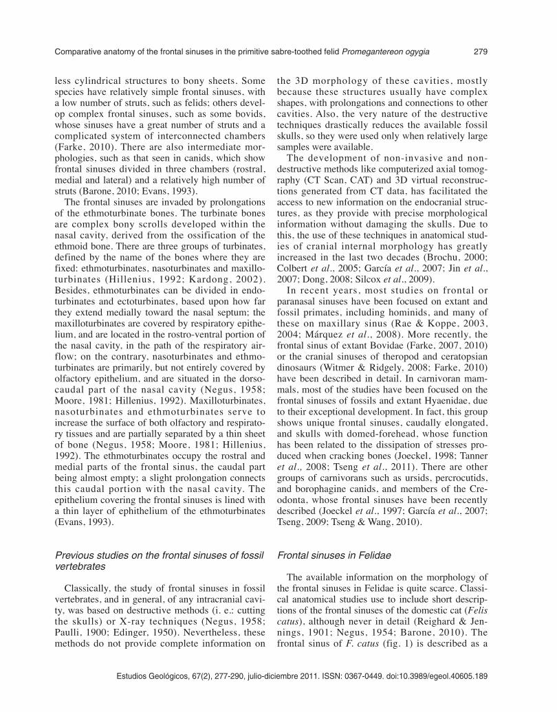
fronto-parietal suture (fig. 3). It shows a little devel-oped rostral portion, although relatively larger thanthat of F. catus, and a caudal widening that followsthe supraorbital margin, penetrating into the zygo-matic process of the frontal bone (fig. 3). Thedevelopment of the frontal sinus is similar in bothsides, separated by means of the septum sinuumfrontalium, which coincides with the interfrontalsuture. The extension of the sinuses over the braincavity is similar in three species, covering only itsrostral portion. However, in A. jubatus and P. con-color the sinus extends more caudally than in P.pardus (fig. 3)
The internal table of the medial portion of thefrontal sinus is irregular and discontinuous. The eth-moturbinates invade this region, developing severalprolongations, being difficult to distinguish the ven-tral boundary of the sinus in this part. So, these eth-moturbinates prolongations constitute the ventral
border of the sinus, separating it from the nasal cav-ity.
The rostral portion of the sinus is small and digi-tiform; it extends rostro-caudally, being filled withethmoturbinates. Both A. jubatus and P. concolorshow a small rostral extension, absent in P. pardus,which reaches the level of the frontal process of thenasal bone. This extension should not be strictlyconsidered part of the frontal sinus, but just aninternal concavity of the nasal bone, not derivedfrom a pneumatisation process, named “recess”(Rossie, 2006; Farke, 2010). Therefore, we considerthat in A. jubatus, P. pardus and P. concolor the ros-tral portion of the frontal sinus reaches the level ofthe fronto-nasal suture, and not beyond this (fig. 3).
Caudally to the rostral part of the frontal sinus,there is a reduced medial portion, hardly distin-guishable from the rostral portion. Nevertheless,this portion is also occupied with ethmoturbinate
282 G. Siliceo, M.J. Salesa, M. Antón, J.F. Pastor, J. Morales
Estudios Geológicos, 67(2), 277-290, julio-diciembre 2011. ISSN: 0367-0449. doi:10.3989/egeol.40605.189
Fig. 3.—Digital reconstructions in dorsal view of the skulls of Acinonyx jubatus (A), Puma concolor (B), Panthera pardus (C) andPromegantereon ogygia (D), showing the virtual reconstruction of the volume occupied by the frontal sinus.
e390-11 Siliceo.qxd 30/1/12 14:20 Página 282
prolongations, even to a higher degree than in therostral part in the three species. In P. pardus, thismedial portion is more differentiated from the ros-tral one than in A. jubatus and P. concolor, havingless ethmoturbinate prolongations inside (fig. 4B).These two later species show a nasal cavity totallyoccupied by dense packets of scrolling turbinates(ethmoturbinates, nasoturbinates and maxillo-turbinates). Although in these three species, prolon-gations of the ethmoturbinates penetrate in themedial portion of the frontal sinus, in A. jubatus andP. concolor they show a greater development thanin P. pardus, almost occupying the whole cavity ofthis portion (figs 4-5). There are few and smallstruts in the rostral and medial portions of thefrontal sinus.
The caudal portion of the frontal sinus is thelargest of the 3 portions, and it is well separated
from the others by means of a sheet-like caudalstrut. Unlike the rostral and medial portions, theethmoturbinates do not invade massively the cau-dal portion, this cavity being almost empty, and itsventral boundary is clearly defined, formed by theventral table of the frontal sinus (figs 4-5). Thiscaudal portion is communicated with the nasalcavity through a slight prolongation of one of theethmoturbinates. Both A. jubatus and P. concolorhave relatively larger frontal sinus than P. pardus,with a caudal portion longer caudally (figs 3, 6).The squama frontalis (in the frontal bone) of theselatter species has a vaulted shape, especially in A.jubatus, and is in that part of the frontal where thecaudal portion of the frontal sinus is located(fig. 6).
Although A. jubatus, P. pardus and P. concolorhave relatively simple frontal sinuses, with fewbony struts, A. jubatus and P. concolor show a
Estudios Geológicos, 67(2), 277-290, julio-diciembre 2011. ISSN: 0367-0449. doi:10.3989/egeol.40605.189
Comparative anatomy of the frontal sinuses in the primitive sabre-toothed felid Promegantereon ogygia 283
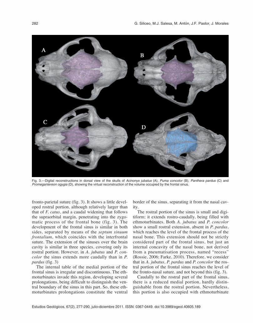
Fig. 4.—Sagittal sections of the virtual reconstructions of theskulls of Puma concolor (A) and Panthera pardus (B), showingthe main structures and cavities within the frontal sinus: rostralportion (rp); medial portion (mp); caudal portion (cp); nasal cavity(nc) filled with the turbinates and the ethmoturbinates (eth); cau-dal strut (cst) separating the caudal portion from the rest of thesinus; dorsal chambers (dch); struts in the caudal end of thefrontal sinus (st).
Fig. 5.—Two sagittal sections at different levels of the virtualreconstruction of the skull of Acinonyx jubatus. A1, dorsal cham-bers (dch) developed in the dorso-medial part of the sinus; A2,large caudal portion (cp) of the frontal sinus communicated withthe dorsal chambers.
e390-11 Siliceo.qxd 30/1/12 14:20 Página 283

great number of struts than P. pardus. Most of thestruts are placed in the rostral and caudal extremesof the caudal portion of the sinus, at the level of thezygomatic process of the frontal bone, and in thecaudal end of the sinus (figs 4-5). Also, both A.jubatus and P. concolor have a set of small cham-bers in the dorso-medial part of the sinus, and inthe dorso-rostral part of the caudal portion of thesinus, resulted from the interconnection of thesmall struts situated in those parts (figs 4A-5). InA. jubatus these chambers are connected to the cau-dal portion of the sinus, whilst in P. concolor thechambers are isolated due to the more caudal situa-tion of the caudal strut (figs 4A-5); also, in this lat-ter species, the strut is curved, with a concave ros-tral face and a convex caudal one, producing anincrease in the volume of the medial portion atexpenses of the caudal one.
Finally, in dorsal view, the different developmentof the frontal sinus can be observed, with P. pardusshowing a clearly shorter sinus than A. jubatus andP. concolor. Besides this, whereas in P. pardus andA. jubatus the sinus gradually becomes narrowercaudally, in P. concolor the sinus shows a markedpost-orbitary constriction (fig. 3).
Morphology of the frontal sinus inPromegantereon ogygia
The studied skull of P. ogygia from Batallones-1shows a good state of preservation, although unfor-tunately, as in most of the fossil skulls of mammals,the ethmoturbinates are not preserved. Its internalcavities keep the three-dimensional structure exceptfor a small collapsed area near the naso-frontalsuture, which prevents the description of the rostralportion of the sinus. In spite of this, and althoughno 3D reconstruction can be made of this portion,the fronto-nasal suture, the predictable rostral limitof this portion, is visible.
The extension of the frontal sinus in P. ogygia isdirectly correlated with the extension of the frontalbone, as in the studied felines; it is limited rostrally bythe naso-frontal suture, and caudally by the fronto-parietal suture. In dorsal view, the frontal sinus, whichhas a very reduced rostral portion, follows the mor-phology of the frontal bone, extending caudally andpenetrating into the zygomatic process of the frontal;then, the sinus narrows following the shape of thepost-orbitary constriction of the frontal, and finishesat the level of the fronto-parietal suture (fig. 3D).
The internal table of the frontal bone in the cau-dal portion of the sinus is well preserved, but in themedial and rostral portions this table is so fragmen-tary that it cannot be described. The caudal portionof the frontal sinus is the best preserved of the threeportions. The septum sinuum frontalium, whichdivides both left and right sides, is well developed,and coincides with the interfrontal suture, as inother felids. In the caudal end of the sinus, severalstruts are seen.
The general shape of the sinus of P. ogygia issimilar to that of P. concolor. In dorsal view itextends more caudally, and it is wider than that of P.pardus, although it is caudally shorter and narrowerthan that of A. jubatus, in accordance with the mor-phology of the frontal bone (fig. 3). However, thecaudal end of the sinus in P. ogygia does not showany trace of constriction and posterior widening,and the caudal strut, which separates the caudal por-tion from the rest of the sinus, is more or lessstraight, unlike the curved strut seen in P. concolor.
In the medial portion of the sinus there are sever-al struts within the sediment filling the cavity; someof them seem to keep their original position, whilstothers just maintain their connection to the dorsalwall of the sinus. These struts are similarly locatedas in the compared species, but they are relativelymore abundant than in P. pardus, resembling tomorphology seen in P. concolor and A. jubatus.Nevertheless, nothing more can be said on theirdegree of complexity or on the possible existence ofchambers.
Discussion
As in other groups of mammals, the size andmorphology of the frontal sinuses of felids areclosely correlated to those of the frontal bone. Thisintracranial cavity is relatively simple in felids, anddoes not show great variability in both extensionand development. Nevertheless, within the studiedspecies, some differences in relative size and num-ber of struts are found, with A. jubatus showing therelatively largest frontal sinuses. Also, this speciesshows a typical dome-shaped skull, linked to largerfrontal sinuses than those of other felids, and char-acterised by an inflated caudal portion with a highernumber of small struts in its dorso-medial part,which interconnect forming several small chambers(figs 4A-5). This latter feature is shared by P. con-color, which also shows a vaulted frontal bone,
284 G. Siliceo, M.J. Salesa, M. Antón, J.F. Pastor, J. Morales
Estudios Geológicos, 67(2), 277-290, julio-diciembre 2011. ISSN: 0367-0449. doi:10.3989/egeol.40605.189
e390-11 Siliceo.qxd 30/1/12 14:20 Página 284

although lacking the dome-shaped skull observed inA. jubatus. On the other hand, the frontal sinuses ofP. pardus are rostro-caudally shorter, with a lessdeveloped caudal portion, and lacking the cham-bered dorso-medial portion. In these features, thesinus of P. pardus resembles that of Felis catus,probably reflecting the primitive condition for Feli-dae. At this respect, it is remarkable that the frontalsinus of P. ogygia shares the caudal elongation seenin P. concolor and A. jubatus, and even the sabre-toothed felid could have had the chambers observedin the dorso-medial part in these two species, or atleast a higher number of struts than P. pardus. Thissimilarity between P. ogygia, P. concolor and A.jubatus cannot be easily explained, as there is noconsensus on the function of the frontal sinuses.Anyway, the caudal development and the cham-bered region could have derived independently inboth groups, as they are not closely related.
As mentioned before, the function of the frontalsinus in felids, or mammals in general, has not beensatisfactorily explained. Negus (1957, 1958) pro-posed an olfactory function for the frontal sinusesof Carnivora, as the surface of the olfactory epithe-lium is increased by the complex system of scrolledethmoturbinates, which are housed in the sinus cav-ity; following this, the presence of empty frontalsinuses would be explained by a reduction in the
complexity of the ethmoturbinates. For otherauthors (Rui et al., 1960; Witmer, 1997) frontalsinuses would be primarily empty cavities, andmacrosmatic mammals (those with well developedolfactory sense) would have experimented a sec-ondary expansion process of the ethmoturbinatesinto the sinus cavities. Nevertheless, as shown byour analysis, both A. jubatus and P. concolor havewell developed ethmoturbinates, but they onlyoccupy the medial portion of the sinus, the inflatedcaudal portion almost lacking ethmoturbinates. Thiswould clearly disagree with this “olfactory hypothe-sis”, as would the fact that these two species do notshow differences in their sense of smell in relationto other felids.
Other proposed functions for the large frontalsinus of A. jubatus are the warming and humidifica-tion of the inhaled air that penetrates in the respira-tory system (Krausman & Morales, 2005; Lecastre& Flamarion, 2010) and the thermoregulation of thebrain and related sense organs (Bignon, 1889; Bre-mer, 1940; Proetz, 1953; Verheyen, 1953; Dyce etal., 1987). The former hypothesis has been convinc-ingly refuted (Proetz, 1953; Blanton & Biggs, 1968;Shea, 1977; Witmer, 1997), whilst the other could bemore plausible at least for some groups (Bignon,1889; Bremer, 1940; Proetz, 1953; Verheyen, 1953;Dyce et al., 1987). In this latter hypothesis, the air
Estudios Geológicos, 67(2), 277-290, julio-diciembre 2011. ISSN: 0367-0449. doi:10.3989/egeol.40605.189
Comparative anatomy of the frontal sinuses in the primitive sabre-toothed felid Promegantereon ogygia 285
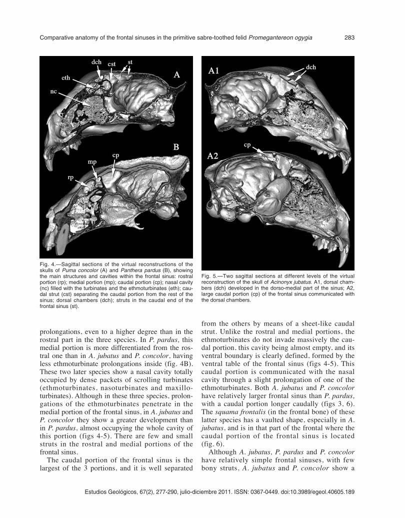
Fig. 6.—Digital reconstructions in lateral (left) view of the skulls of Acinonyx jubatus (A), Puma concolor (B), Panthera pardus (C) andPromegantereon ogygia (D), showing the virtual reconstruction of the volume occupied by the frontal sinus.
e390-11 Siliceo.qxd 30/1/12 14:20 Página 285
chamber formed by the sinus would act as thermalinsulator of the central nervous system, and thus theobserved differences in relative size and caudalexpansion in the frontal sinuses of P. concolor, A.jubatus, P. ogygia and P. pardus would imply differ-ences in the capacity for brain thermal insulation,with P. pardus having the shortest sinus, and thusthis function relatively reduced. Acinonyx jubatusare found in low-structured habitats, such as savan-nas or grasslands with some shrub coverage (Alder-ton, 1998; Bothma & Walker, 1999; Nowak, 2005),whereas P. pardus and P. concolor can occupy arange of very different habitats, from arid savannasto dense tropical forests (Currier, 1983; Johnson etal., 1993; Alderton, 1998; Bothma & Walker, 1999;Nowak, 2005). Nevertheless, recent studies on mole-cular phylogeny show A. jubatus and P. concolor asclosely related taxa (Mattern & McLennan, 2000; Yu& Zhang, 2005; Johnson et al., 2006), and theirsharing of a long frontal sinus, although havingstrong physiological implications, would be reflect-ing their inheritance from a common open habitat-dweller ancestor (Van Valkenburgh et al., 1990;Hemmer et al., 2004) with high capacity for brainthermal regulation. The morphology observed in P.ogygia, more similar to those of P. concolor an A.jubatus than that of P. pardus, poses an interestingquestion on its palaeoecology, as the inferred habitatfor this machairodontine felid is a more or lessclosed habitat (Salesa et al., 2006). A possible expla-nation may be associated with the progressive aridi-fication process that occurred during the Vallesianand Turolian of Europe (Fortelius et al., 2002),which led to the predominance of savannas overwooded habitats. The caudally expanded frontalsinus of P. ogygia would be reflecting an adaptationto these new climatic conditions, where the insola-tion was higher than in wooded habitats. Neverthe-less, the frontal sinus of P. ogygia, and those of allthe analysed felids, extends as far as the level of thefronto-parietal suture, this is, the sinus expands cau-dally following the development of the frontal bone.According to this, any environmental explanationfor this expansion should be established with cau-tion, even if we consider that the derived Smilodonfatalis, the last Smilodontini, associated to relativelyopen environments (Gonyea, 1976; Kurtén &Werdelin, 1990; Stock & Harris, 1992; Coltrain etal., 2004), shows this caudal expansion in the frontalsinus as much developed as P. ogygia (fig. 2).
The other observed difference in the frontalsinus of the analysed felids, this is, the presence in
A. jubatus and P. concolor of several struts in thedorso-medial part of the frontal sinus, absent in P.pardus, could be related to the necessity of a rein-forcement in the larger cavities of the two formerspecies, due to the greater tensions and biomechan-ical demands that a more inflated sinus requires(Witmer, 1997). This dorso-medial part of thefrontal sinus is damaged in the analysed specimenof P. ogygia, but it could have contained severalstruts, as indicated in the descriptions above. Nev-ertheless, these struts would not be reinforcing alarge dorso-medial part of the sinus, as this is notinflated, but they could be necessary if great ten-sions occurred in this part when the animal killedits prey. The killing technique employed by thesabre-toothed felids, the so-called “canine shear-bite”, based on a strong flexion movement of thehead (Emerson & Radinsky, 1980; Akersten, 1985;Turner & Antón, 1997; Antón & Galobart, 1999)could imply an increase in the stress experimentedby the frontal bone, and thus the need for somekind of reinforcement in this area. Several cranialand dental features support the development of thiskilling technique in P. ogygia (Salesa et al., 2005),and thus, it could be possible for these struts tohave played a role in resisting the tensions generat-ed during the machairodont bite. The results of thepresent study on the size and shape of the frontalsinus in P. ogygia are not enough to support thisinterpretation, and the use of different techniquesfor assessing the resistance of the skull of thisspecies would be highly valuable. One of thesemethods, the Finite Element Analysis has beenrecently used to quantitatively assess the biome-chanical performance of the skull of S. fatalis dur-ing the bite, showing that a moderate stress is pro-duced in the frontal bone during this action(McHenry et al., 2007: fig. 4C). Future studies onthe skulls of P. ogygia from batallones-1, using thismethodology, will help to elucidate the functionalimplications of the observed morphology of frontalsinuses and related structures in this primitivesabre-toothed felid.
Conclusions
The morphology of the frontal sinuses of P. ogy-gia has revealed as being more derived than couldbe expected considering that this species is one ofthe most primitive of the machairodontines. Thecaudal expansion of the sinus could be reflecting a
286 G. Siliceo, M.J. Salesa, M. Antón, J.F. Pastor, J. Morales
Estudios Geológicos, 67(2), 277-290, julio-diciembre 2011. ISSN: 0367-0449. doi:10.3989/egeol.40605.189
e390-11 Siliceo.qxd 30/1/12 14:20 Página 286
more open habitat than previously inferred for thisspecies, whereas the presence of several struts inthe dorso-medial part of the sinus would indicate areinforcement of the frontal bone in relation to thecanine shear-bite. Nevertheless, the present workmust be considered as a first approach to theseaspects of the palaeobiology of this primitive sabre-toothed felid, and further studies are need in orderto complete our results.
ACKNOWLEDGEMENTS
We dedicate this work to the memory of Professeur LeonardGinsburg.
We would like to thank the Hospital Nuestra Señora deAmérica (Madrid), and especially María Jesús Siliceo, for per-forming the CT Scans of A. jubatus, P. concolor and P. ogygia,and the Hospital Clínico Universitario de Valladolid, especiallyJosé Manuel Montes, for the CT Scan of P. pardus. We alsothank Dr. Josefina Barreiro (curator, MNCN-CSIC) and theMuseo Anatómico de Valladolid, for access to the specimensanalysed in this work. This study is part of the research projectsCGL2008-00034/BTE and CGL2008-05813-C02-01/BTE(Dirección General de Investigación, Ministerio de Ciencia eInnovación, Spain), and the Research Group CAM-UCM910607. M. J. Salesa is a contracted researcher within the“Ramón y Cajal” program (Ministerio de Ciencia e Innovación,reference RYC2007-00128) and G. Siliceo is a predoctoral FPIfellowship within the project CGL2008-00034/BTE.
References
Adams, D.R. (2004). Canine anatomy: a systemic study.Iowa State Press, Ames, 491 pp.
Akersten, W.A. (1985). Canine function in Smilodon(Mammalia; Felidae; Machairodontinae). Contribu-tions in Science, 356: 1-22.
Alderton, D. (1998). Wild cats of the world. Blandford,London, 192 pp.
Antón, M. & Galobart, A. (1999). Neck function and preda-tory behaviour in the scimitar-toothed cat Homotheriumlatidens (Owen). Journal of Vertebrate Paleontology, 19(4): 771-784. doi:10.1080/02724634.1999.10011190
Barone, R. (2010). Anatomie Comparée des mammifèresdomestiques - Tome 1. Ostéologie. Vigot Freres, Edi-teurs, Paris, 761 pp.
Bignon, F. (1889). Contribution a l’étude de la pneumati-cité chez les oiseaux. Les cellules aëriennes cervico-céphalique des oiseaux et leurs rapports avec les os dela tête. Mémoires de la Société Zoolologique de Fran-ce, 2: 260-320.
Blaney, S.P.A. (1990). Why paranasal sinuses?. TheJournal of Laryngology and Otology, 104: 690-693.doi:10.1017/S0022215100113635
Blanton, P.L. & Biggs, N.L. (1968). Eighteen HundredYears of Controversy: The Paranasal Sinuses. Ameri-
can Journal of Anatomy, 124: 135-148. doi:10.1002/aja.1001240202
Bothma, J. & Walker, C. (1999). Larger carnivores ofthe African savannas. Springer-Verlag, Berlin, 274pp.
Braune, W. & Clasen, F.E. (1877). Die Nebenhöhlen dermenschlichen Nase in ihre Bedeutung für den Mecha-nismus des Rieches. Zeitschrift für Tierzüchtung undZüchtungsbiologie, 2: 1-28.
Bremer, J.L. (1940). The pneumatization of the head ofthe common fowl. Journal of Morphology, 67: 143-157. doi:10.1002/jmor.1050670107
Brochu, C.A. (2000). A digitally-rendered endocast forTyrannosaurus rex. Journal of Vertebrate Paleonto-logy, 20 (1): 1-6. doi:10.1671/0272-4634(2000)020[0001:ADREFT]2.0.CO;2
Buhler, P. (1972). Sandwich structures in the skull cap-sules of various birds: the principle of lightweightstructures in organisms. Mitterilungen aus dem Institutfür leichte Flächentragwerke, 4: 39-50.
Christiansen, P. & Mazák, J.H. (2008). A primitive LatePliocene cheetah, and evolution of the cheetah lineage.PNAS, 106 (2): 515-515.
Cleland, J. (1862). On the relations of the vomer, eth-moid, and intermaxillary bones. Philosophical Tran-sactions, 62: 289-321. doi:10.1098/rstl.1862.0019
Colbert, M.W.; Racicot, R. & Rowe, T. (2005). Anatomyof the Cranial Endocast of the Bottlenose Dolphin,Tursiops truncatus, Based in HRXCT. Journal ofMammalian Evolution, 12: 195-207. doi:10.1007/s10914-005-4861-0
Coltrain, J.B.; Harris, J.M.; Cerling, T.E.; Ehleringer,J.R.; Dearing, M.D.; Ward, J. & Allen, J. (2004).Rancho La Brea stable isotope biogeochemistry andits implications for the palaeoecology of late Pleisto-cene, coastal southern California. Palaeogeography,Palaeoclimatology, Palaeoecology, 205: 199-219.doi:10.1016/j.palaeo.2003.12.008
Currier, M.J.P. (1983). Felis concolor. Mammalian Spe-cies, 200: 1-7. doi:10.2307/3503951
Davis, W.E.; Templer, J. & Parsons, D.S. (1996). Ana-tomy of the paranasal sinuses. Otolaryngologic Clinicsof North America, 29: 57-74.
Dong, W. (2008). Virtual cranial endocast of the oldestgiant panda (Ailuropoda microta) reveals great simila-rity to that of its extant relative. Naturwissenschaften,95: 1079-1083. doi:10.1007/s00114-008-0419-3
Dyce, K.M.; Sack, W.O. & Wensing, C.J.G. (2002).Textbook of Veterinary Anatomy. W. B. SaundersCompany, Philadelphia, 864 pp.
Edinger, T. (1950). Frontal sinus evolution (particularyin the Equidae). Bulletin of the Museum of Comparati-ve Zoology at Harvard collage, 103: 411-496.
Emerson, S.B. & Radinsky, L. (1980). Functional analy-sis of sabertooth cranial morphology. Paleobiology, 6(3): 295-312.
Evans, H.E. (1993). Miller’s Anatomy of the dog. 3rd Edi-tion. Saunders. 1113 pp.
Farke, A.A. (2007). Morphology, constraits, and scalingof frontal sinuses in the hartebeest, Alcelaphus buse-
Estudios Geológicos, 67(2), 277-290, julio-diciembre 2011. ISSN: 0367-0449. doi:10.3989/egeol.40605.189
Comparative anatomy of the frontal sinuses in the primitive sabre-toothed felid Promegantereon ogygia 287
e390-11 Siliceo.qxd 30/1/12 14:20 Página 287

laphus (Mammalia: Artiodactyla, Bovidae). Journal ofMorphology, 268: 243-253. doi:10.1002/jmor.10511
Farke, A.A. (2010). Evolution and functional morpho-logy of the frontal sinuses in Bovidae (Mammalia:Artiodactyla), and implications for the evolution ofcranial pneumaticity. Zoological Journal of the Linne-an Society, 159: 988-1014. doi:10.1111/j.1096-3642.2009.00586.x
Fortelius, M.; Eronen, J.; Jernvall, J.; Liu, L.; Pushinka,D.; Rinne, J.; Tesakov, A.; Vislobokova, I., Zhang, Z.& Zhou, L. (2002). Fossil mammals resolve regionalpatterns of Eurasian climate change over 20 millionyears. Evolutionary Ecology Research, 4: 1005-1016.
Gannon, P.J.; Doyle, W.J.; Ganjian, E.; Márquez, S.;Gnoy, A.; Gabrielle, H.S. & Lawson, W. (1997).Maxillary sinus mucosal blood flow during nasal vstracheal respiration. Archives of Otolaryngology-Head& Neck Surgery, 123 (12): 1336-1340.
García, N.; Santos, E.; Arsuaga, J.L. & Carretero, J.M.(2007). Endocranial morphology of the Ursus denin-geri von Reichenau 1904 from the Sima de los huesos(Sierra de Atapuerca) middle Pleistocene site. Jour-nal of vertebrate paleontology, 27 (4): 1007-1017.doi :10.1671/0272-4634(2007)27[1007:EMO-TUD]2.0.CO;2
Gonyea, W.J. (1976). Behavioral Implications of Saber-Toothed Felid Morphology. Paleobiology, 2 (4): 332-342.
Hemmer, H.; Kahlke, R.D. & Vekua, A.K. (2004). TheOld World puma - Puma pardoides (Owen, 1846)(Carnivora: Felidae) - in the Lower Villafranchian(Upper Pliocene) of Kvabebi (East Georgia, Transcau-casia) and its evolutionary and biogeographical signifi-cance. Neues Jahrbuch für Geologie und Paläontolo-gie, 233: 197-231.
Hillenius, W.J. (1992). The evolution of nasal turbinatesand mammalian endothermy, Paleobiology, 18: 17-29.
Jin, C.; Ciochon, R.L.; Dong, W.; Hunt Jr., R.M.; Liu, J.;Jaeger, M. & Zhu, Q. (2007). The first skull of the ear-liest giant panda. PNAS, 104 (26): 10932-10937.doi:10.1073/pnas.0704198104
Joeckel, R.M. (1998). Unique frontal sinuses in fossil andliving Hyaenidae (Mammalia, Carnivora): descriptionand interpretation. Journal of Vertebrate Paleontology,18 (3): 627-639. doi:10.1080/02724634.1998.10011089
Joeckel, R.M., Bond, H.W. & Kabalka, G.W. (1997).Internal anatomy of the snout and paranasal sinusesof Hyaenodon (Mammalia, Creodonta). Journal ofVertebrate Paleontology. 17 (2): 440-446. doi:10.1080/02724634.1997.10010989
Joeckel, R.M. & Stavas, J.M. (1996). New insights intothe cranial anatomy of Barbourofelis fricki (Mamma-lia, Carnivora). Journal of Vertebrate Paleontology. 16(3): 585-591. doi:10.1080/02724634.1996.10011344
Johnson, K.G.; Wei, W.; Reid, D.G. & Jinchu, H. (1993).Food habits of Asiatic leopards (Panthera pardusfusea) in Wolong Reserve, Sichuan, China. Journal ofMammalogy, 74: 646-650. doi:10.2307/1382285
Johnson, W.E.; Eizirik, E.; Pecon-Slattery, J.; Murphy,W.J.; Antunes, A.; Teeling, E. & O’Brien, S.J.
(2006). The Late Miocene Radiation of Modern Feli-dae: A Genetic Assessment. Science, 311: 73-77.doi:10.1126/science.1122277
Kardong, K.V. (2002). Vertebrates: Comparative Ana-tomy, Function, Evolution. McGraw-Hill, New York,762 pp.
Kurtén, B. & Werdelin, L. (1990). Relationships betweenNorth and South American Smilodon. Journal of Ver-tebrate Paleontology, 10 (2): 158-169. doi:10.1080/02724634.1990.10011804
Leakey, M. & Walker, A. (1997). Afropithecus functionand phylogeny. In: Function, Phylogeny, and Fossils:Miocene Hominoid Evolution and Adaptations (Begun,D.R., Ward, C.V. & Rose, M.D., eds). Plenum Press,New York, 225-239.
Lundberg, J.O.; Rinder, J.; Weitzberg, E.; Lundberg,J.M. & Alving, K. (1994). Nasally exhaled nitric oxidein humans originates mainly in the paranasal sinuses.Acta Physiologica Scandinavica, 152: 431-432.doi:10.1111/j.1748-1716.1994.tb09826.x
Márquez, S., Tessema, B., Clement, P.A. & Schaefer,S.D. (2008). Development of the Ethmoid Sinus andExtramural Migration: The Anatomical Basis of thisParanasal Sinus. The Anatomical Record, 291: 1535-1553. doi:10.1002/ar.20775
Mattern, M.Y. & McLennan, D.A. (2000). Phylogenyand Speciation of Felids. Cladistics, 16: 232-253.doi:10.1111/j.1096-0031.2000.tb00354.x
McHenry, C.R., Wroe, S., Clausen, P.D., Moreno, K. &Cunningham, E. (2007). Supermodeled sabercat, pre-datory behaviour in Smilodon fatalis revealed by high-resolution 3D computer simulation. PNAS, 104 (41):16010-16015. doi:10.1073/pnas.0706086104
Merriam, J.C. & Stock, C. (1932). The Felidae of Ran-cho La Brea. Carnegie Institute of Washington Publi-cations, 442: 1-231.
Moore, W.J. (1981). The Mammalian Skull. CambridgeUniversity Press, Cambridge, 369 pp.
Morales, J., Alcalá, L., Amezua, L. et al. (2000) El yaci-miento del Cerro de los Batallones. In: PatrimonioPaleontológico de la Comunidad de Madrid (edsMorales J, Nieto M, Amezua L, et al.), pp. 179–190,Madrid: Servicio de Publicaciones de la Comunidad deMadrid.
Morales, J., Alcalá, L., Alvárez-Sierra, M.A., et al.(2004). Paleontología del sistema de yacimientos demamíferos miocenos del Cerro de los Batallones,Cuenca de Madrid. Geogaceta, 35: 139-142.
Negus, V. (1957). The function of the paranasal sinuses.A.M.A. Archives of Otolaryngology, 66 (4): 430-442.
Negus, V. (1958). The comparative anatomy and physio-logy of the nose and paranasal sinuses. E&S Livings-tone Ltd, Edinburgh and London, 402 pp.
Nemours, P.R. (1931). A comparison of the accessorynasal sinuses of man with those of lower vertebrates.Transactions of the American Laryngological, Rhino-logical, and Otological Society, 1931: 195–199.
Nowak, R.M. (2005). Walker’s Carnivores of the World.Baltimore: The Johns Hopkins University Press, Balti-more and London, 313 pp.
288 G. Siliceo, M.J. Salesa, M. Antón, J.F. Pastor, J. Morales
Estudios Geológicos, 67(2), 277-290, julio-diciembre 2011. ISSN: 0367-0449. doi:10.3989/egeol.40605.189
e390-11 Siliceo.qxd 30/1/12 14:20 Página 288

O’Malley, J.F. (1924). Evolution of the nasal cavitiesand sinuses in relation to function. Journal of Laryngo-logy and Otology, 39: 57-64.
Paulli, S. (1900). Über die Pneumaticität des Schädelsbei den Säugethieren. III. Über die Morphologie desSiebbeins und Pneumaticität bei den Insectivoren,Hyracoideen, Chiropteren, Carnivoren, Pinnipedien,Edentaten, Rodentiern, Prosimien und Primaten.Gegenbaurs morphologisches Jahrbuch, 28: 483-564.
Proetz, A.W. (1922). Observations upon the formationand function of the accessory nasal sinuses and themastoid cells. Annals of Otology, Rhinology & Laryn-gology, 31: 1083-1096.
Proetz, A.W. (1938). Nasal physiology and its relation tothe surgery of the accessory nasal sinuses. Proceedingsof the Royal Society of Medicine, 31: 1408-1416.
Proetz, A.W. (1953). Applied physiology of the nose. 2d.ed. Annals Plublishing Company, Saint Luis, 395 pp.
Rae, T.C. & Koppe, T. (2003). The Term “LateralRecess” and Craniofacial Pneumatization in Old WorldMonkeys (Mammalia, Primates, Cercopithecoidea).Journal of Morphology, 258: 193-199. doi:10.1002/jmor.10144
Rae, T.C. & Koppe, T. (2004). Holes in the Head: Evolu-tionary Interpretations of the Paranasal Sinuses inCatarrhines. Evolutionary Anthropology, 13: 211-223.doi:10.1002/evan.20036
Reighard, J. & Jennings, H.S. (1901). Anatomy of thecat. Henry Holt and Company, New York, 498 pp.
Rossie, J.B. (2006). Ontogeny and homology of the para-nasal sinuses in Platyrrhini (Mammalia: Primates). Jour-nal of Morphology, 267: 1-40. doi:10.1002/jmor.10263
Rui, R., Den, L. & Gourlaouen, L. (1960). Contribution ál’étude du role des sinus paranassaux. Revue de Laryn-gologie et Oto-Rhinologie, 81: 796-839.
Salesa, M.J., Antón, M., Turner, A. & Morales, J. (2005).Aspects of the functional morphology in the cranialand cervical skeleton of the sabre-toothed cat Parama-chairodus ogygia (Kaup, 1832) (Felidae, Machairo-dontinae) from the Late Miocene of Spain: implica-tions for the origins of machairodont killing bite. Zoo-logical Journal of the Linnean Society, 144: 363–377.doi:10.1111/j.1096-3642.2005.00174.x
Salesa, M.J., Antón, M., Turner, A. & Morales, J. (2006)Inferred behaviour and ecology of the primitive sabre-toothed cat Paramachairodus ogygia (Felidae,Machairodontinae) from the Late Miocene of Spain.Journal de Zoology, 268: 243-254. doi:10.1111/j.1469-7998.2005.00032.x
Salesa, M.J., Antón, M., Turner, A., Alcalá, L., Montoya,P., Morales, J. (2010a) Systematic revision of the LateMiocene sabre-toothed felid Paramachaerodus in Spain.Palaeontology, 53 (6): 1369-1391. doi:10.1111/j.1475-4983.2010.01013.x
Salesa, M. J., Antón, M., Turner, A. & Morales, J. (2010b).Functional anatomy of the forelimb in the primitive felidPromegantereon ogygia (Machairodontinae, Smilodonti-ni) from the Late Miocene of Spain and the origins of thesaber-toothed felid model. Journal of Anatomy, 216:381-396. doi:10.1111/j.1469-7580.2009.01178.x
Schaffer, W.M. & Reed, C.A. (1972). The co-evolutionof social behavior and cranial morphology in sheepand goats (Bovidae, Caprini). Fieldiana Zoology, 61:1-88.
Shea, B. (1977). Eskimo craniofacial morphology,cold stress and the maxillary sinus. American Jour-na l o f Phys ica l An thropology , 47 : 289-300 .doi:10.1002/ajpa.1330470209
Shea, J.J. (1936). Morphologic characteristics of thesinuses. Archives of Otolaryngology, 23: 484-487.
Sherwood, R.J. (1999). Pneumatic processes in the tempo-ral bone of chimpanzee (Pan troglodytes) and gorilla(Gorilla gorilla). Journal of Morphology, 241: 127-137.doi:10.1002/(SICI)1097-4687(199908)241:2<127::AID-JMOR3>3.0.CO;2-P
Silcox, M.T., Dalmyn, C.K. & Bloch, J.I. (2009). Virtualendocast of Ignacius graybullianus (Paromomyidae,Primates) and brain evolution in early primates. PNAS,106 (27): 10987-10992. doi:10.1073/pnas.0812140106
Smith, T.D.; Rossie, J.B.; Cooper, G.M.; Mooney,M.P. & Siegel, M.I. (2005). Secondary Pneumatiza-tion of the Maxillary Sinus in Callitrichid Primates:Insights From Immunohistochemistry and Bone CellDistribution. The Anatomical Record, Part A, 285A:677-689.
Stock, C. & Harris, J.M. (1992). Rancho La Brea: arecord of Pleistocene life in California. Natural His-tory Museum of Los Angeles County Museum, LosAngeles, 81 pp.
Tanner, J.B.; Dumont, E.R.; Sakai, S.T.; Lundrigan, B.L.& Holekamp, K.E. (2008). Of arcs and vaults: the bio-mechanics of bone-cracking in spotted hyenas (Crocu-ta crocuta). Biological Journal of the Linnean Society,95: 246-255. doi:10.1111/j.1095-8312.2008.01052.x
Tseng, Z.J. (2009). Cranial function in a late MioceneDinocrocuta gigantea (Mammalia: Carnivora) revea-led by comparative finite element analysis. Biologi-cal Journal of the Linnean Society, 96: 51-67.doi:10.1111/j.1095-8312.2008.01095.x
Tseng, Z.J. & Wang, X. (2010). Cranial FunctionalMorphology of Fossil Dogs and Adaptation for Durop-hagy in Borophagus and Epicyon (Carnivora, Mamma-lia). Journal of Morphology , 271: 1386-1398.doi:10.1002/jmor.10881
Tseng, Z.J.; Antón, M. & Salesa, M.J. (2011). Theevolution of the bone-cracking model in carnivo-rans: Cranial functional morphology of the Pliocenecursorial hyaenid Chasmaporthetes lunensis (Mam-malia: Carnivora). Paleobiology , 37: 140-156.doi:10.1666/09045.1
Turner, A. & Antón, M. (1997). The Big Cats and theirfossil relatives. Columbia University Press, New York,234 pp.
Van Valkenburgh, B.; Grady, F. & Kurtén, B. (1990). ThePlio-Pleistocene Cheetah-like Cat Miracinonyx inexpecta-tus of North America. Journal of Vertebrate Palaeonto-logy, 10: 434-454. doi:10.1080/02724634.1990.10011827
Verheyen, R. (1953). Contribution à l’étude de la structu-re pneumatique du crâne chez les oiseaux. InstitutRoyal des Sciences Naturelles de Belgique, 29: 1-24.
Estudios Geológicos, 67(2), 277-290, julio-diciembre 2011. ISSN: 0367-0449. doi:10.3989/egeol.40605.189
Comparative anatomy of the frontal sinuses in the primitive sabre-toothed felid Promegantereon ogygia 289
e390-11 Siliceo.qxd 30/1/12 14:20 Página 289

Weidenreich, F. (1941). The extremity bones of Sinanth-ropus pekinensis. Palaeontologia Sinica, 110: 1-150.
Witmer, L.M. (1997). The evolution of the antorbitalcavity of Archosaurs: A study in soft-tissue reconstruc-tion in the fossil record with an analysis of the functionof pneumaticity. Journal of Vertebrate Paleontology,17: 1-76. doi:10.1080/02724634.1997.10011027
Witmer, LM. (1999). The phylogenetic history of para-nasal air sinuses. In: The paranasal sinuses of higherprimates (Koppe, T., Nagai, H. & Alt, K.W., eds.).Quintessence, Chicago, 21-34.
Witmer, L.M. & Ridgely, R.C. (2008). New Insights Intothe Brain, Braincase, and Ear Region of Tyrannosaurs(Dinosauria, Theropoda), with Implications for Sen-
sory Organization and Behavior. The AnatomicalRecord, 292: 1266-1296. doi:10.1002/ar.20983
Yu, L. & Zhang, Y. (2005). Phylogenetic studies of pant-herine cats (Felidae) based on multiple genes, withnovel application of nuclear b-fibrinogen intron 7 tocarnivores. Molecular Phylogenetics and Evolution,35: 483-495. doi:10.1016/j.ympev.2005.01.017
Zollikofer, C.P.E. & Weissmann, J.D. (2008). A morpho-genetic model of cranial pneumatization based on theinvasive tissue hypothesis. The Anatomical Record,291: 1446-1454. doi:10.1002/ar.20784
Recibido el 3 de marzo de 2011Aceptado el 29 de agosto de 2011
290 G. Siliceo, M.J. Salesa, M. Antón, J.F. Pastor, J. Morales
Estudios Geológicos, 67(2), 277-290, julio-diciembre 2011. ISSN: 0367-0449. doi:10.3989/egeol.40605.189
e390-11 Siliceo.qxd 30/1/12 14:20 Página 290