Analysis of two minimally invasive procedures for ...
13
SYSTEMATIC REVIEW Open Access Analysis of two minimally invasive procedures for osteoporotic vertebral compression fractures with intravertebral cleft: a systematic review and meta-analysis Hongyu Wei 1* , Chunke Dong 1,2 , Yuting Zhu 3 and Haoning Ma 1 Abstract Background: A systematic review and meta-analysis to assess the pros and cons of percutaneous vertebroplasty (PVP) versus kyphoplasty (PKP) for osteoporotic vertebral compression fractures (OVCFs) with intravertebral cleft (IVC) including all available evidence from controlled trials. Methods: Databases including Pubmed, Embase, Cochrane Library, China National Knowledge Infrastructure (CNKI), and Wanfang Data were searched to identify relevant studies comparing PVP and PKP for OVCFs with IVC. The outcomes mainly included visual analog scale (VAS), Oswestry Disability Index (ODI), local kyphotic angle (LKA), rate of vertebral height (VH%), and adverse events. Results: Nine studies enrolling 688 patients were eligible for meta-analysis. The results indicated no significant differences between the two groups in the short-and long-term VAS, ODI, LKA, or VH% (P > 0.05). Compared with PVP, PKP was associated with significantly longer operation time (P < 0.05), higher cost (P > 0.05), and more injected cement volume (P < 0.05). In terms of adverse events, PKP has a lower risk of cement leakage (P < 0.05), while with no significant difference in adjacent-level fracture rates (P > 0.05). Conclusion: The two procedures have similar short- and long-term pain relief, functional recovery, local kyphosis correction, and vertebral height maintenance in OVCFs with IVC. PKP is superior to PVP for the injected cement volume, and lower cement leakage rate, however, with longer operation time, more fluoroscopy times, and higher cost. Further randomized controlled trials (RCTs) should be conducted to confirm these results. Keywords: Osteoporotic vertebral compression fractures, Intravertebral cleft, Vertebroplasty, Kyphoplasty, Meta- analysis © The Author(s). 2020 Open Access This article is licensed under a Creative Commons Attribution 4.0 International License, which permits use, sharing, adaptation, distribution and reproduction in any medium or format, as long as you give appropriate credit to the original author(s) and the source, provide a link to the Creative Commons licence, and indicate if changes were made. The images or other third party material in this article are included in the article's Creative Commons licence, unless indicated otherwise in a credit line to the material. If material is not included in the article's Creative Commons licence and your intended use is not permitted by statutory regulation or exceeds the permitted use, you will need to obtain permission directly from the copyright holder. To view a copy of this licence, visit http://creativecommons.org/licenses/by/4.0/. The Creative Commons Public Domain Dedication waiver (http://creativecommons.org/publicdomain/zero/1.0/) applies to the data made available in this article, unless otherwise stated in a credit line to the data. * Correspondence: [email protected] 1 Department of Orthopaedic Surgery, China-Japan Friendship Hospital, 2 Yinghuadong Road, Chaoyang District, Beijing 100029, China Full list of author information is available at the end of the article Wei et al. Journal of Orthopaedic Surgery and Research (2020) 15:401 https://doi.org/10.1186/s13018-020-01938-6
Transcript of Analysis of two minimally invasive procedures for ...
SYSTEMATIC REVIEW Open Access
Analysis of two minimally invasiveprocedures for osteoporotic vertebralcompression fractures with intravertebralcleft: a systematic review and meta-analysisHongyu Wei1* , Chunke Dong1,2, Yuting Zhu3 and Haoning Ma1
Abstract
Background: A systematic review and meta-analysis to assess the pros and cons of percutaneous vertebroplasty(PVP) versus kyphoplasty (PKP) for osteoporotic vertebral compression fractures (OVCFs) with intravertebral cleft(IVC) including all available evidence from controlled trials.
Methods: Databases including Pubmed, Embase, Cochrane Library, China National Knowledge Infrastructure (CNKI),and Wanfang Data were searched to identify relevant studies comparing PVP and PKP for OVCFs with IVC. Theoutcomes mainly included visual analog scale (VAS), Oswestry Disability Index (ODI), local kyphotic angle (LKA), rateof vertebral height (VH%), and adverse events.
Results: Nine studies enrolling 688 patients were eligible for meta-analysis. The results indicated no significantdifferences between the two groups in the short-and long-term VAS, ODI, LKA, or VH% (P > 0.05). Compared withPVP, PKP was associated with significantly longer operation time (P < 0.05), higher cost (P > 0.05), and moreinjected cement volume (P < 0.05). In terms of adverse events, PKP has a lower risk of cement leakage (P < 0.05),while with no significant difference in adjacent-level fracture rates (P > 0.05).
Conclusion: The two procedures have similar short- and long-term pain relief, functional recovery, local kyphosiscorrection, and vertebral height maintenance in OVCFs with IVC. PKP is superior to PVP for the injected cementvolume, and lower cement leakage rate, however, with longer operation time, more fluoroscopy times, and highercost. Further randomized controlled trials (RCTs) should be conducted to confirm these results.
Keywords: Osteoporotic vertebral compression fractures, Intravertebral cleft, Vertebroplasty, Kyphoplasty, Meta-analysis
© The Author(s). 2020 Open Access This article is licensed under a Creative Commons Attribution 4.0 International License,which permits use, sharing, adaptation, distribution and reproduction in any medium or format, as long as you giveappropriate credit to the original author(s) and the source, provide a link to the Creative Commons licence, and indicate ifchanges were made. The images or other third party material in this article are included in the article's Creative Commonslicence, unless indicated otherwise in a credit line to the material. If material is not included in the article's Creative Commonslicence and your intended use is not permitted by statutory regulation or exceeds the permitted use, you will need to obtainpermission directly from the copyright holder. To view a copy of this licence, visit http://creativecommons.org/licenses/by/4.0/.The Creative Commons Public Domain Dedication waiver (http://creativecommons.org/publicdomain/zero/1.0/) applies to thedata made available in this article, unless otherwise stated in a credit line to the data.
* Correspondence: [email protected] of Orthopaedic Surgery, China-Japan Friendship Hospital, 2Yinghuadong Road, Chaoyang District, Beijing 100029, ChinaFull list of author information is available at the end of the article
Wei et al. Journal of Orthopaedic Surgery and Research (2020) 15:401 https://doi.org/10.1186/s13018-020-01938-6
BackgroundThe intravertebral cleft (IVC), which was first describedby Maldague in 1978 [1], has long been considered as theresult of ischemic necrosis in osteoporotic vertebral com-pression fractures (OVCFs) [2, 3]. Initial reports dealt ex-clusively with air-filled IVC, presenting with a transverse,linear, or semilunar radiolucent shadow on conventionalradiographs [3, 4]. Then, several reports have also ob-served variable appearance of IVC in MRI, which dependson whether they are filled with gas or fluid at the time theyare imaged [5]. Some scholars propose that this alternat-ing air or fluid phenomenon can be considered indicativeof microinstability at the cleft site, which ultimately formsa painful progressive kyphosis due to delayed vertebralcollapse, also known as Kummell’s disease [6–8].Owing to the progression of kyphosis with vertebral
collapse and intravertebral instability at the cleft site,fractures with IVC are more susceptible to no responseto conservative treatment; thus, surgical interventionmay be a better choice [9, 10]. Open surgery has beendescribed to treat IVC, but it is mainly indicated for pa-tients with neurological deficits [11, 12]. Besides, it is in-appropriate for most patients with serious comorbiditiesand severe osteoporosis [13]. Invasive procedures, suchas PKP and PVP, widely and successfully used in OVCFs,are better choices for patients with IVC.Previous studies suggested that PKP displayed better
performance in long-term VAS, ODI, LKA, and VH %compared with PVP for OVCFs without IVC [14–17].During the past two decades, some scholars have triedto use PKP and PVP to treat IVC, but the results are notconsistent [18–21]. The pros and cons of the two inter-ventions in treating OVCFs with IVC still need to be in-vestigated. Therefore, we conduct a systematic reviewand meta-analysis of the available literature to calculatea pooled estimate of the advantages and disadvantages ofPKP compared to PVP for single-level OVCFs with IVC.
MethodsOur meta-analysis was carried out according to the Pre-ferred Reporting Items for Systematic Reviews andMeta-analyses (PRISMA) [22] statement.
Search strategyElectronic databases Pubmed, Embase, Cochrane Li-brary, CNKI, and Wanfang Data, were searched for allrelevant studies until June 2020. The keywords for thestudy object included “intravertebral cleft,” “intraverteb-ral vacuum cleft (sign),” “intravertebral pseudarthrosis,”“avascular necrosis,” “vertebral osteonecrosis,” “intraoss-eous vacuum phenomena,” or “Kummell's disease.” Thekeywords for the intervention strategy were “vertebro-plasty” and “kyphoplasty.” Reference lists of all eligibleoriginal and review articles were also screened manually
to identify any initially omitted studies. There is no re-striction on publication language.
Inclusion criteria
1. Interventional studies (RCTs) and observationalstudies (cohort or case-control studies).
2. Studies reported the comparisons between PKP andPVP for patients with single-level OVCFs compli-cated IVC.
3. Studies reported at least one of the followingoutcomes: VAS, ODI, LKA, VH%, operation time,injected cement volume, the incidence of cementleakage, and adjacent vertebral fracture.
4. All patients have at least 12-months follow-upperiod.
Exclusion criteria
1. Patients suffered from multi-level OVCFs with orwithout IVC.
2. Pathological fracture due to primary or metastatictumors, infection, or tuberculosis.
3. Patients complicated with nerve disorder, long-termuse of steroidal or nonsteroidal anti-inflammatorydrugs, or previous surgery at the diseased vertebra.
4. Non-original articles (case reports, reviews, letters,meta-analyses, and editorials), animal studies,in vitro biomechanical studies, or computationalmodeling studies.
Study selectionTwo authors (HY.W. and CK.D.) independently screened alltitles and abstracts related to the eligibility criteria describedabove. The full-text of the literature was reviewed thoroughlyfor the final inclusion. Disagreements were resolved byreaching a consensus with the third author (YT.Z.).
Data extraction and quality assessmentBaseline data (first author and year of publication, lan-guage, study design, sample size, age, gender, and durationof follow-up), intervention, and outcomes were independ-ently extracted in duplicate using a standardized form bytwo authors (HY.W. and CK.D.). Data in other forms (i.e.,median, interquartile range, and mean ± 95% confidenceinterval (CI)) were converted to mean ± standard devi-ation (SD) according to the Cochrane Handbook [23]. Weextracted data by manual measurements from figures ifthey were not reported in numbers.The Cochrane Collaboration’s tool for assessing risk of
bias [24] was used to evaluate RCTs’ methodologicalquality. Each study was judged according to six items:random sequence generation, allocation concealment,blinding of participants and personnel, incomplete
Wei et al. Journal of Orthopaedic Surgery and Research (2020) 15:401 Page 2 of 13

outcome data, selective reporting, and other bias. Eachpiece was classified into three levels: high, unclear, andlow risk. The Newcastle-Ottawa-Scale (NOS) [25] wasused to assess the methodological quality of cohort orcase-control studies on three dimensions: selection (0–4points), comparability (0–2 points), and the determin-ation of either the exposure or the outcome of interest(0–3 points). The studies with 7–9 points were consid-ered high quality, 5–6 points as moderate quality, and0–4 as poor quality. The data extraction and quality as-sessment were independently performed by two authors(HY.W. and CK.D.). The third author (HN.M.) was theadjudicator when no consensus could be achieved.
Statistical analysisOur meta-analysis was performed through RevMan v5.3software (Cochrane Collaboration, Oxford, UK). Continu-ous data, such as VAS, ODI, LKA, VH%, operation time,and injected cement volume, were expressed as mean ±
SD and summarized using the mean difference (MD) orstandardized mean difference (SMD) with 95% confidenceinterval (CI). Risk ratio (RR) with 95% CI was calculatedfor binary outcome data like cement leakage and adjacent-level fractures. Heterogeneity was tested using the chi-square test and quantified by calculating the I2 statistics. P< 0.1 and I2 > 50% was considered statistical heterogen-eity. A random-effects model was used for heterogeneousstatistical data. Otherwise, a fixed-effects model was per-formed. Sensitive analysis or subgroup analysis was usedto investigate the source of heterogeneity. Meta-analysesresults were also assessed using forest plots, and P < 0.05was considered statistically significant.
ResultsLiterature search and study characteristicsIn total, 556 citations were identified after an initial sys-tematic search, of which 187 excluded for duplication,and 343 were excluded scrutiny of the titles and
Fig. 1 Summary of study selection and exclusion process
Wei et al. Journal of Orthopaedic Surgery and Research (2020) 15:401 Page 3 of 13
Table
1Basiccharacteristicsof
includ
edstud
iesandNOSscores
fortheno
n-RC
Ts
Stud
yLang
uage
Stud
ydesign
Sample
size
Age(yea
rs)
Gen
der
(M/F)
BMD(T-sco
re)
Cou
rseof
disea
se(m
onths)
Follo
w-up(m
onths)
NOS
scores
PVP
PKP
PVP
PKP
PVP
PKP
PVP
PKP
PVP
PKP
Chang
etal.[26]
English
RCT
2828
75.0±5.8
75.1±5.7
6/22
8/20
−4.35
±0.91
−4.47
±0.89
8.7±3.0
8.4±2.9
33.5forPVP,35.2forPKP
NR
Fang
andYu
-Liang
[29]
Chine
seRC
S20
2072.02±4.96
72.43±5.64
5/15
7/13
−4.7~
−2.6
>3
10~18
∗∗∗∗
∗∗∗
Jiang
andZh
ang[27]
Chine
seRC
S27
2474.92±4.97
75.08±4.99
8/19
6/18
−2.58
±0.57
−2.61
±0.63
NR
12∗∗∗∗
∗∗∗
Kong
etal.[34]
English
RCS
2429
70.5±6.4
71.9±7.0
8/16
7/22
NR
4.4±3.5
4.1±3.7
12∗∗∗∗
∗∗∗
Liet
al.[33]
Chine
seRC
S151
113
66.2±5.63
68.7±6.49
69/82
41/72
NR
0.93
±0.16
1.0±0.22
>12
∗∗∗∗
∗∗
Yuet
al.[31]
Chine
seRC
S48
2074.6(63~85)
75.9(65~87)
10/38
4/16
−4.34
±0.94
−4.35
±0.74
0.83
0.79
24∗∗∗∗
∗∗∗
etal.[30]
Chine
seRC
S20
2867.05±4.03
67.21±4.57
2/18
3/25
NR
NR
>12
∗∗∗∗
∗∗∗
Zhanget
al.[32]
English
RCS
3835
75.58±4.97
73.74±4.35
10/28
9/26
NR
3.76
±1.68
4.05
±2.01
17.7forPVP,17.9forPKP
∗∗∗∗
∗∗∗
Zhanget
al.[28]
English
RCS
2213
72.82±6.99
74.38±5.66
7/15
5/8
−3.27
±0.49
−3.49
±0.62
7.86
±4.37
10.15±6.30
22.7forPVP,19.3forPKP
∗∗∗∗
∗∗∗
BMDbo
nemineral
density
,NOSNew
castle-Ottaw
a-Scale,
PVPpe
rcutan
eous
verteb
roplasty,P
KPpe
rcutan
eous
kyph
oplasty,RC
Trand
omized
controlledtrial,RC
Sretrospe
ctivecoho
rtstud
y,M
male,
Ffemale,
NR
notrepo
rted
Wei et al. Journal of Orthopaedic Surgery and Research (2020) 15:401 Page 4 of 13
abstracts. After reviewing the full text of the remaining26 studies, we excluded 17 additional articles. Finally, 9studies [26–34] eventually satisfied the eligibility criteriafor meta-analysis, which included one RCT study [26]and eight retrospective cohort studies [27–34]. The re-sults of literature search and study selection process aresummarized in Fig. 1. There were a total of 688 patientsand 688 vertebral bodies; 378 patients underwent thePVP and the remaining 310 received the PKP. Individualstudy sample sizes ranged from 35 to 264 patients. Thedemographic characteristics of the included studies aresummarized in Table 1
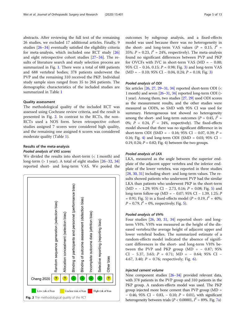
Quality assessmentThe methodological quality of the included RCT wasassessed using Cochrane review criteria, and the result ispresented in Fig. 2. In contrast to the RCTs, the non-RCTs used a NOS form. Seven retrospective cohortstudies assigned 7 scores were considered high quality,and the remaining one assigned 6 scores was consideredmoderate quality (Table 1).
Results of the meta-analysisPooled analysis of VAS scoresWe divided the results into short-term (< 1month) andlong-term (> 1 year). A total of eight studies [26–32, 34]reported short- and long-term VAS. We pooled the
outcomes by subgroup analysis, and a fixed-effectsmodel was used because there was no heterogeneity inthe short- and long-term VAS values (P = 0.15, I2 =35%; P = 0.23, I2 = 24%, respectively). The meta-analysisfound no significant differences between PVP and PKPfor OVCFs with IVC in short-term VAS (MD = − 0.00;95% CI − 0.16, 0.15; P = 0.98; Fig. 3) and long-term VAS(MD = − 0.10; 95% CI − 0.04, 0.24; P = 0.18; Fig. 3)
Pooled analysis of ODISix articles [26, 27, 29–31, 34] reported short-term ODI (<1month) and seven [26–31, 34] reported long-term ODI (>1 year). Among them, two studies [27, 29] used ODI scoresas the measurement results, and the other studies weremeasured as ODI%, so SMD with 95% CI was used forsummary. Heterogeneous test showed no heterogeneityamong the short- and long-term outcomes (P = 0.45, I2 =0%; P = 0.24, I2 = 24%, respectively). The fixed-effectsmodel showed that there was no significant difference in inshort-term ODI (SMD = − 0.16; 95% CI − 0.07, 0.39; P =0.16; Fig. 4) and long-term ODI (SMD = 0.03; 95% CI −0.19, 0.24; P = 0.82; Fig. 4) between the two groups.
Pooled analysis of LKALKA, measured as the angle between the superior end-plate of the adjacent upper vertebra and the inferior end-plate of the lower vertebra, was reported in three studies[28, 30, 31] including short- and long-term values. The re-sults showed patients who underwent PVP had the similarLKA than patients who underwent PKP in the short-term(MD = − 1.29; 95% CI − 2.73, 0.16; P = 0.08; Fig. 5) andlong-term follow-up (MD = − 0.07; 95% CI − 1.39, 1.25; P= 0.91; Fig. 5) in a fixed-effects model (P = 0.19, I2 = 40%;P = 0.79, I2 = 0%, respectively; Fig. 5).
Pooled analysis of VH%Four studies [26, 30, 31, 34] reported short- and long-term VH%. VH% was measured as the height of the dis-eased vertebra/the average height of adjacent upper andlower vertebral bodies. The summarized estimate of arandom-effects model indicated the absence of signifi-cant differences in the short- and long-term VH% be-tween the PVP and PKP group (MD = − 0.87; 95%CI − 5.37, 3.63; P = 0.71; MD = − 0.64; 95% CI −4.67, 3.40; P = 0.76; respectively; Fig. 6).
Injected cement volumeNine component studies [26–34] provided relevant data,with 378 patients in the PVP group and 310 patients in thePKP group. A random-effects model was used. The PKPgroup injected more bone cement than PVP group (MD =− 0.46; 95% CI − 0.83, − 0.10; P = 0.01), with significantheterogeneity between trials (P < 0.00001, I2 = 89%. Fig. 7a)
Fig. 2 The methodological quality of the RCT
Wei et al. Journal of Orthopaedic Surgery and Research (2020) 15:401 Page 5 of 13

Operation timeThis outcome was described in 5 papers [26–30, 32–34]. The PKP group used more operation time thanPVP group (MD = − 10.81; 95% CI − 12.38, − 9.24; P< 0.00001. Fig. 7b), The I2 value attributed 89% vari-ation to heterogeneity; therefore, a random-effectsmodel was used.
Operation costThree studies [28, 29, 34] reported operation cost, andwe ruled out one trial [34] because the premature inclu-sion of patients may impact the results. The meta-analysis of eventually including studies showed that theoperation cost in the PKP group was higher than that inthe PVP group (MD = − 2.34; 95% CI − 2.45, − 2.23; P <0.00001. Fig. 7c)
Fluoroscopy timesThe fluoroscopy times was reported in two studies [26,27]. A fixed-effects model indicated that the PKP group’sfluoroscopy times was more than the PVP group (MD =− 5.03; 95% CI − 6.87, − 3.18; P < 0.00001. Fig. 7d)
IVC located in the thoracolumbar (T11–L2)Five studies [26, 29, 31, 32, 34] described the segmentaldistribution of lesions in detail. A fixed-effects modelshowed that the two groups had similar IVC segmentaldistribution in the thoracolumbar (RR = 0.94; 95% CI0.83, 1.06; P = 0.31. Fig. 7e)
Pooled analysis of cement leakageRelevant data on cement leakage was provided in all stud-ies [26–34], with a total of 688 patients (378 in the PVP
Fig. 3 Forest plot of the mean difference (MD) in pre-operation, short-term, and long-term VAS between the PVP and PKP groups
Wei et al. Journal of Orthopaedic Surgery and Research (2020) 15:401 Page 6 of 13

group and 310 in the PKP group). The meta-analysis ofthe 9 studies showed that the PVP group had a signifi-cantly higher risk of cement leakage than the PVP group(RR = 1.88; 95% CI 1.29, 2.75; P = 0.001; Fig. 8a) in the ab-sence of statistical heterogeneity (P = 0.69, I2 = 0%).
Pooled analysis of adjacent-level fracturesFour studies [26, 27, 32, 34] provided data about the riskratio for adjacent-level fractures. The pooled analysis ofa fixed-effects model showed that there was no signifi-cant difference between the two interventions in the in-cidence of adjacent-level fractures (RR = 1.21; 95% CI0.53, 2.74; P = 0.65; Fig. 8b).
Publication biasThe review of the funnel plots could not rule out the po-tential publication bias for long-term VAS (Fig. 9a) andcement leakage (Fig. 9b). The Egger’s and Begg’s testsshowed no evidence of publication bias for long-termVAS (P = 0.711 and 0.160, respectively; Fig. 9c, d),
and cement leakage (P = 0.350 and 0.186, respect-ively; Fig. 9e, f).
Sensitivity analysisThe sensitivity analysis which was performed by omit-ting 1 study in each turn investigated the influence of asingle study on the overall outcome. The long-term VASin the PVP group was not significantly different fromthat in the PKP group when omitting any of the studiesexcept Jiang et al. [27]. The injected cement volume inthe PVP group was similar to that in the PKP groupwhen removing one study conducted by Feng et al. [29],Jiang et al. [27], Kong et al. [34], or Li et al. [33]. Theother outcomes of sensitivity analysis were not materi-ally differentiated from those of the original analysis.
DiscussionWith the development of imaging technology, IVC, ini-tially, as a rare phenomenon of OVCFs [35], is consid-ered to be a commonly recognized condition at present[36, 37]. Although a consistent pathomechanism has not
Fig. 4 Forest plot of the standardized mean difference (SMD) in pre-operation, short-term, and long-term ODI between the PVP and PKP groups
Wei et al. Journal of Orthopaedic Surgery and Research (2020) 15:401 Page 7 of 13

Fig. 5 Forest plot of the mean difference (MD) in pre-operation, short-term, and long-term LKA between the PVP and PKP groups
Fig. 6 Forest plot of the mean difference (MD) in pre-operation, short-term, and long-term VH% between the PVP and PKP groups
Wei et al. Journal of Orthopaedic Surgery and Research (2020) 15:401 Page 8 of 13

yet been reached, IVC highly suggests a sign of ischemicposttraumatic vertebral necrosis, known as Kummell’sdisease [38]. Instability at the cleft site often leads topseudarthrosis, which eventually results in severe pain,kyphosis, and nerve injury. Due to insufficient know-ledge of the condition and the paucity of literature, spe-cific treatment protocols are limited [39]. Early reportsfocused on conservative treatment, while more recentpreferred surgical interventions for earlier patient ambu-lation and correction of kyphosis [40]. The two minim-ally invasive procedures, PVP and PKP, are indicated toeliminate the motion at the cleft site, to maintain the an-terior height of the vertebral body, and to relieve painand are widely used in the treatment of OVCFs withIVC. However, their results were inconsistent, and it is
not clear which one could provide better outcomes.Therefore, an evidence base is essential for surgeonsto develop a more appropriate scheme. To the best ofour knowledge, this is the first comprehensive meta-analysis to evaluate the therapeutic efficacy differencecomparing PVP and PKP treatments for single-levelOVCFs with IVC.The dynamic motion within the IVC, expressed by the
changes in vertebral body height according to extensionand flexion lateral radiographs [41, 42], has been corre-lated with a high probability of severe pain [43]. VASand ODI were used to evaluate the pain relief and func-tional recovery after the operation, respectively. Com-pared with previous meta-analysis results that PKP wasmore effective on the VAS and ODI assessments than
Fig. 7 a Forest plot of injected cement volume. b Forest plot of operation time. c Forest plot of operation cost. d Forest plot of fluoroscopytimes. e Forest plot of IVC located in the thoracolumbar
Wei et al. Journal of Orthopaedic Surgery and Research (2020) 15:401 Page 9 of 13

the PVP in non-IVC OVCFs [44], the pooled results inour article showed no statistical difference between thetwo groups in short- and long-term follow-up, which in-dicated they both are effective methods for OVCFs withIVC. Taylor et al. [45] found some evidence of moderatecorrelations between the change in VAS pain with thechange in vertebral height (r = 0.62, P = 0.184) andchange in kyphotic angle (r = – 0.68, P = 0.09). Sev-eral meta-analysis results showed that for non-IVCOVCFs, PKP, via balloon expansion, had a better po-tential to restore vertebral height and correct kyphoticdeformity compared to PVP, which resulted in a bet-ter painful and functional improvement in PKP [14,15, 17, 44]. As compared to our pooled results, theLKA and VH% in the IVC patients have no statisticaldifference between the two groups during the follow-up period. Five studies [26, 28, 30–32] in our analysisfound that patients with IVC could achieve a spon-taneous reduction in the hyperextension positionwithout further balloon expansion, which lead to aconsiderable reduction effect in the two procedures.These studies also revealed that the LKA and AH% inthe two groups gradually lost significantly with time,which was consistent with previous findings [18, 46].However, there was no significant difference in the
vertebral recollapse between the two groups due tothe similar bone cement distribution in the low pres-sure IVC region [26, 28].Need more complex procedures, such as the repeated
establishment of expander channel on the pedicle, thePKP calls for more operation time and fluoroscopytimes, which is consistent with our statistic findings.Our pooled analysis also demonstrated that the aver-age cost in the PKP group was significantly higherthan the PVP group because of the use of a balloonduring operation. From the outcomes of our prelimin-ary meta-analysis, the clinical effects had no signifi-cant difference; therefore, the cost gap of the twoapproaches cannot be ignored. In this study, 75.9% ofthe IVCs were located in the thoracolumbar segmentwith no significant difference between the two groups,which suggests that the occurrence of IVC may be at-tributed to the repeated stress activity and high mo-bility in the thoracolumbar segment [26]. Althoughthe analysis of injected cement volume in the PKPgroup was more than in the PVP group (MD = −0.46; 95% CI − 0.83, − 0.10; P = 0.01), six studiesshowed there was no significant difference in theiroutcomes (P < 0.05). The difference may be related toIVC varied locations in the vertebral body [47].
Fig. 8 a Forest plot of cement leakage. b Forest plot of adjacent-level fractures
Wei et al. Journal of Orthopaedic Surgery and Research (2020) 15:401 Page 10 of 13

The rate of cement leakage has drawn considerable at-tention in OVCFs with IVC. The difference in leakagerate between the two procedures was confirmed in ourmeta-analysis, in which the incidence of cement leakageis significantly higher in the PVP group, at 18.3 % thanin the PKP group, at 10.3 %. Possible reasons for the lessfrequent cement leakage that may be related to balloonexpansion could squeeze the surrounding cancellousbone and create a cavity that allows for a more viscouscement to be injected under lower pressure [48, 49].Concerning the incidence of adjacent-level fractures,there was no significant difference between the PVP andPKP groups (8.9% vs.9.8%).Although not mentioned in our included studies, the
refracture in previously cemented vertebrae after PVP orPKP with the incidence of 0.56–2% is a complicationthat should not be ignored in patients with IVC [50, 51].Yu et al. [52] had confirmed that IVC might be the most
important predisposing factor for recollapse of the aug-mented vertebrae in OVCFs. Solid lump cement distri-bution due to the presence of IVC may interceptmechanical interlock with surrounding cancellous bones.Thus, the recollapse in no cemented area is easily devel-oped during daily activity [47]. Once augmented verte-bral refracture occurs, it is difficult for surgeons todetermine the appropriate treatment. Several studiesfound that repeated percutaneous vertebroplasty may bea suitable choice considering the risk of complicationsfor elder patients [50, 53].
LimitationsSeveral limitations involved in this study should be con-sidered: (1) Our analysis included a number of cohortstudies that might result in selective and performancebias due to the absence of random allocation, allocationconcealment, and blinding. (2) Heterogeneity may have
Fig. 9 a, b Funnel plots of long-term VAS and cement leakage. c, d Egger’s publication bias plot and Begg’s funnel plots of long-term VAS. e, fEgger’s publication bias plot and Begg’s funnel plots of cement leakage. MD, mean difference; SE, standard error; RR, risk ratio
Wei et al. Journal of Orthopaedic Surgery and Research (2020) 15:401 Page 11 of 13
been caused by poor non-RCT study design, which in-duced by the unilateral or bilateral surgical technologiesused, varying spinal vertebral bodies, bone mineral dens-ity, age, gender, follow-up time, and course of diseasedifferences. (3) Publication bias comes from significantconclusions being more easily published, and only onecountry publications included in this study may aggra-vate the bias. (4) Finally, given the limited number of theincluded studies in the analysis, the findings should beconfirmed in future research with more relevant RCTsto obtain more reliable and conclusive data.
ConclusionAlthough there is controversy, this systematic reviewcomparing PVP and PKP for treating OVCFs with IVCdemonstrates that the two minimally invasive proce-dures are both safe and efficacious for similar short- andlong-term pain relief, functional recovery, local kyphosiscorrection, and vertebral height maintenance. PKP is su-perior to PVP for the injected cement volume, and lowercement leakage rate, however, with longer operationtime, more fluoroscopy times, and higher cost. FurtherRCTs should be conducted to confirm these results.
AbbreviationsPVP: Percutaneous vertebroplasty; PKP: Percutaneous kyphoplasty;OVCFs: Osteoporotic vertebral compression fractures; IVC: Intravertebral cleft;CNKI: China National Knowledge Infrastructure; VAS: Visual analog scale;ODI: Oswestry Disability Index; LKA: Local kyphotic angle; VH%: Rate ofvertebral height; RCTs: Randomized controlled trials; MD: Mean difference;SMD: Standardized mean difference; CI: Confidence interval; RR: Risk ratio
AcknowledgementsNone
Authors’ contributionsAll authors (HY, CD, YZ, and HM) participated in concept development, datacollection, quality control of the data, data analysis, and interpretation. Thefirst draft of the manuscript was written by HY, and all authors commentedon previous versions of the manuscript. All authors read and approved thefinal manuscript.
FundingNone
Availability of data and materialsAll data generated or analyzed during this study are included in thispublished article
Ethics approval and consent to participateNot applicable
Consent for publicationNot applicable
Competing interestsThe authors declare that they have no competing interests
Author details1Department of Orthopaedic Surgery, China-Japan Friendship Hospital, 2Yinghuadong Road, Chaoyang District, Beijing 100029, China. 2BeijingUniversity of Chinese Medicine, 11 North Third Ring Road East, ChaoyangDistrict, Beijing 100029, China. 3Beijing Tongzhou Integrative MedicineHospital, 89 Chezhan Road, Tongzhou District, Beijing 101100, China.
Received: 30 June 2020 Accepted: 1 September 2020
References1. Maldague BE, Noel HM, Malghem JJ. The intravertebral vacuum cleft: a sign
of ischemic vertebral collapse. Radiology. 1978;129(1):23–9.2. McKiernan F, Faciszewski T. Intravertebral clefts in osteoporotic vertebral
compression fractures. Arthritis Rheum. 2003;48(5):1414–9.3. Lane JI, Maus TP, Wald JT, Thielen KR, Bobra S, Luetmer PH. Intravertebral
Clefts Opacified during Vertebroplasty: Pathogenesis, Technical Implications,and Prognostic Significance. Am J Neuroradiol. 2002;23(10):1642.
4. Theodorou DJ. The intravertebral vacuum cleft sign. Radiology. 2001;221(3):787–8.
5. Linn J, Birkenmaier C, Hoffmann RT, Reiser M, Baur-Melnyk A. Theintravertebral cleft in acute osteoporotic fractures: fluid in magneticresonance imaging-vacuum in computed tomography? Spine (Phila Pa1976). 2009;34(2):E88–93.
6. D’Oria S, Delvecchio C, Dibenedetto M, Zizza F, Somma C. Case report ofKummell’s disease with delayed onset myelopathy and the literature review.Eur J Orthop Surg Traumatol. 2018;28(2):309–16.
7. Swartz K, Fee D. Kummell's disease: a case report and literature review.Spine (Phila Pa 1976). 2008;33(5):E152–5.
8. Kim DY, Lee SH, Jang JS, Chung SK, Lee HY. Intravertebral vacuumphenomenon in osteoporotic compression fracture: report of 67 cases withquantitative evaluation of intravertebral instability. J Neurosurg. 2004;100(1Suppl Spine):24–31.
9. Wang W, Liu Q, Liu WJ, Li QB, Cai L, Wang ZK. Different performance ofintravertebral vacuum clefts in Kümmell’s disease and relevant treatmentstrategies. Orthop Surg. 2020;12(1):199–209.
10. Nakamae T, Fujimoto Y, Yamada K, Takata H, Shimbo T, Tsuchida Y.Percutaneous vertebroplasty for osteoporotic vertebral compression fracturewith intravertebral cleft associated with delayed neurologic deficit. EurSpine J. 2013;22(7):1624–32.
11. Wei H, Dong C, Zhu Y. Posterior fixation combined with vertebroplasty orvertebral column resection for the treatment of osteoporotic vertebralcompression fractures with intravertebral cleft complicated by neurologicaldeficits. Biomed Res Int. 2019;2019:1–10.
12. Zhang X, Hu W, Yu J, Wang Z, Wang Y. An effective treatment option forKümmell disease with neurological deficits. Spine. 2016;41(15):E923–30.
13. Lee K, Adsul N, Kim H, Pee Y, Lee KL, Jang I. Percutaneous pedicle screwfixation with bone cement augmentation under epidural anesthesia fortreatment of Kümmell disease in extremely old age. World Neurosurg. 2018;119:506–10.
14. Zhao G, Liu X, Li F. Balloon kyphoplasty versus percutaneous vertebroplastyfor treatment of osteoporotic vertebral compression fractures (OVCFs).Osteoporos Int. 2016;27(9):2823–34.
15. Wang H, Sribastav SS, Ye F, Yang C, Wang J, Liu H, Zheng Z. Comparison ofpercutaneous vertebroplasty and balloon kyphoplasty for the treatment ofsingle level vertebral compression fractures: a meta-analysis of the literature.Pain Physician. 2015;18(3):209–22.
16. Chang X, Lv Y, Chen B, Li H, Han X, Yang K, Zhang W, Zhou Y, Li C.Vertebroplasty versus kyphoplasty in osteoporotic vertebral compressionfracture: a meta-analysis of prospective comparative studies. Int Orthop.2015;39(3):491–500.
17. Ma X, Xing D, Ma J, Xu W, Wang J, Chen Y. Balloon kyphoplasty versuspercutaneous vertebroplasty in treating osteoporotic vertebral compressionfracture: grading the evidence through a systematic review and meta-analysis. Eur Spine J. 2012;21(9):1844–59.
18. Kim P, Kim SW. Balloon kyphoplasty: an effective treatment for Kummelldisease? Korean J Spine. 2016;13(3):102.
19. Wang G, Yang H, Chen K. Osteoporotic vertebral compression fractures withan intravertebral cleft treated by percutaneous balloon kyphoplasty. J BoneJoint Surg Br. 2010;92(11):1553–7.
20. Heo DH, Chin DK, Yoon YS, Kuh SU. Recollapse of previous vertebralcompression fracture after percutaneous vertebroplasty. Osteoporos Int.2009;20(3):473–80.
21. Chen L, Lai P, Chen W. Unipedicle percutaneous vertebroplasty for spinalintraosseous vacuum cleft. Clin Orthop Relat R. 2005;435:148–53.
22. Moher D, Liberati A, Tetzlaff J, Altman DG. Preferred reporting items forsystematic reviews and meta-analyses: the PRISMA statement. PLoS Med.2009;6(7):e1000097.
Wei et al. Journal of Orthopaedic Surgery and Research (2020) 15:401 Page 12 of 13

23. Higgins J, Green S. Cochrane handbook for systematic reviews ofinterventions version 5.1.0. The Cochrane Collaboration; 2013.
24. Higgins JP, Altman DG, Gotzsche PC, Juni P, Moher D, Oxman AD, Savovic J,Schulz KF, Weeks L, Sterne JA. The Cochrane Collaboration’s tool forassessing risk of bias in randomised trials. BMJ. 2011;343:d5928.
25. Stang A. Critical evaluation of the Newcastle-Ottawa scale for theassessment of the quality of nonrandomized studies in meta-analyses. Eur JEpidemiol. 2010;25(9):603–5.
26. Chang J, Bei M, Shu D, Sun C, Chen J, Xiao Y. Comparison of the clinicaloutcomes of percutaneous vertebroplasty vs. kyphoplasty for the treatmentof osteoporotic Kümmell’s disease: a prospective cohort study. BMCMusculoskelet Disord. 2020;21(1):238.
27. Jiang J, Zhang Y. Unipedicular percutaneous vertebroplasty versuspercutaneous kyphoplasty bone cement for treating Kummell disease.Chinese J Tissue Eng Res. 2019;23(22):3481–7.
28. Zhang J, Fan Y, He X, Meng Y, Huang Y, Jia S, Du J, Wu Q, Hao D. Ispercutaneous kyphoplasty the better choice for minimally invasivetreatment of neurologically intact osteoporotic Kümmell’s disease? Acomparison of two minimally invasive procedures. Int Orthop. 2018;42(6):1321–6.
29. Fang F, Yu-Liang S. A comparison of clinical efficacy of percutaneousvertebroplasty and percutaneous kyphoplasty in the treatment of Kümmelldisease. Chinese J Bone Joint. 2018;7(03):225–9.
30. Xing Y, Ting W, Jizhou Y, Lianyong B, Yongdong Y, Fengxian W, Yi Q, Ziyi Z,Dingyan Z. A retrospective trial of percutaneous vertebroplasty versuspercutaneous kyphoplasty for treatment of Kümmell’s diseases. J TraditChinese Orthopedics Traumatol. 2018;30(6):23–9 33.
31. Yu W, Liang D, Jiang X, Ye L, Yao Z. Comparison of effectiveness betweenpercutaneous vertebroplasty and percutaneous kyphoplasty for treatmentof osteoporotic vertebral compression fracture with intravertebral vacuumcleft. Zhongguo Xiu Fu Chong Jian Wai Ke Za Zhi. 2016;30(9):1104–10.
32. Zhang G, Gao Y, Chen S, Ding S, Gao K, Wang H. Comparison ofpercutaneous vertebroplasty and percutaneous kyphoplasty for themanagement of Kümmell’s disease: A retrospective study. Indian J Orthop.2015;49(6):577.
33. Nan LI, Da HE, Gui-Lin Z, Yong-Gang X, Yu-Qing S, Sai MA, Bo L, Wei T,Hospital BJ. Contrastive study between PVP and PKP for the treatment ofOVCFs with intravertebral cleft. Shandong Med J. 2015;55(04):1–3.
34. Kong L, Wang P, Wang L, Shen Y, Shang Z, Meng L. Comparison ofvertebroplasty and kyphoplasty in the treatment of osteoporotic vertebralcompression fractures with intravertebral clefts. Eur J Orthop SurgTraumatol. 2014;24(S1):201–8.
35. Bickel W, Horn HJ. The intravertebral vacuum phenomenon. A rare findingin vertebral compressions. Aktuelle Radiol. 1993;3(3):199.
36. Matzaroglou C, Georgiou CS, Wilke HJ, Assimakopoulos K, Karageorgos A,Konstantinou D, Velissaris D, Panagiotopoulos E, Kafchitsas K. Kummell’sdisease: is ischemic necrosis or vertebral “microcracking” the first step in thesequence? Med Hypotheses. 2013;80(4):505.
37. Libicher M, Appelt A, Berger I, Baier M, Meeder P, Grafe I, DaFonseca K,Nöldge G, Kasperk C. The intravertebral vacuum phenomen as specific signof osteonecrosis in vertebral compression fractures: results from aradiological and histological study. Eur Radiol. 2007;17(9):2248–52.
38. Kim YM, Kim YMP, Ha KMP. Pathomechanism of intravertebral clefts inosteoporotic compression fractures of the spine. Spine J. 2014;14(4):659–66.
39. Matzaroglou C, Georgiou CS, Panagopoulos A, Assimakopoulos K, Wilke HJ,Habermann B, Panos G, Kafchitsas K. Kummell’s disease: clarifying themechanisms and patients’ inclusion criteria. Open Orthop J. 2014;8:288–97.
40. Ma R, Chow R, Shen FH. Kummell’s disease: delayed post-traumaticosteonecrosis of the vertebral body. Eur Spine J. 2010;19(7):1065–70.
41. McKiernan F, Jensen R, Faciszewski T. The dynamic mobility of vertebralcompression fractures. J Bone Miner Res. 2003;18(1):24–9.
42. Malghem J, Maldague B, Labaisse MA, Dooms G, Duprez T, Devogelaer JP,Berg BV. Intravertebral vacuum cleft: changes in content after supinepositioning. Radiology. 1993;187(2):483–7.
43. Mirovsky Y, Anekstein Y, Shalmon E, Peer A. Vacuum clefts of the vertebralbodies. Am J Neuroradiol. 2005;26(7):1634.
44. Liang L, Chen X, Jiang W, Li X, Chen J, Wu L, Zhu Y. Balloon kyphoplasty orpercutaneous vertebroplasty for osteoporotic vertebral compressionfracture? An updated systematic review and meta-analysis. Ann Saudi Med.2016;36(3):165–74.
45. Taylor RS, Fritzell P, Taylor RJ. Balloon kyphoplasty in the management ofvertebral compression fractures: an updated systematic review and meta-analysis. Eur Spine J. 2007;16(8):1085–100.
46. Wu AM, Lin ZK, Ni WF, Chi YL, Xu HZ, Wang XY, Huang QS. The existence ofintravertebral cleft impact on outcomes of nonacute osteoporotic vertebralcompression fractures patients treated by percutaneous kyphoplasty: acomparative study. J Spinal Disord Tech. 2014;27(3):E88–93.
47. Yu W, Jiang X, Liang YZ, Qiu T, Ye L, Zhang S, Jin D. Intravertebral vacuumcleft and its varied locations within osteoporotic vertebral compressionfractures: effect on therapeutic efficacy. Pain Physician. 2017;20(6):E979–86.
48. Weisskopf M, Weikopf M, Ohnsorge JAK, Niethard FU. Intravertebral pressureduring vertebroplasty and balloon kyphoplasty: an in vitro study. Spine(Phila Pa 1976). 2008;33(2):178–82.
49. Phillips FM, Todd Wetzel F, Lieberman I, Campbell-Hupp M. An in vivocomparison of the potential for extravertebral cement leak aftervertebroplasty and kyphoplasty. Spine (Phila Pa 1976). 2002;27(19):2173–8.
50. Chen L, Hsieh M, Liao J, Lai P, Niu C, Fu T, Tsai T, Chen W. Repeatedpercutaneous vertebroplasty for refracture of cemented vertebrae. ArchOrthop Traum Su. 2011;131(7):927–33.
51. Gaughen JRJ, Jensen ME, Schweickert PA, Marx WF, Kallmes DF. Thetherapeutic benefit of repeat percutaneous vertebroplasty at previouslytreated vertebral levels. Am J Neuroradiol. 2002;23(10):1657.
52. Yu WB, Jiang XB, Liang D, Xu WX, Ye LQ, Wang J. Risk factors and score forrecollapse of the augmented vertebrae after percutaneous vertebroplasty inosteoporotic vertebral compression fractures. Osteoporos Int. 2019;30(2):423–30.
53. Liu H, Zhang J, Chen W, Chen AC, Ji W, Meng B, Yang H, Liu T. Reperfusionrevision surgery for augmented vertebral nonunion with movable cement.World Neurosurg. 2019;132:429–33.
Publisher’s NoteSpringer Nature remains neutral with regard to jurisdictional claims inpublished maps and institutional affiliations.
Wei et al. Journal of Orthopaedic Surgery and Research (2020) 15:401 Page 13 of 13