Analysis of Surgeons Muscle Activity During the Use of a ... of Surgeons Muscle Activity.pdf ·...
13
Analysis of Surgeons’ Muscle Activity During the Use of a Handheld Robotic Instrument in Laparoendoscopic Single-Site Surgery Francisco M. Sánchez-Margallo and Juan A. Sánchez-Margallo Abstract The objective of this study is to assess the surgeon’s performance and ergonomics during the use of a robotic-driven handheld laparoscopic instrument in intracorporeal suturing tasks as well as in digestive and urological laparoscopic procedures performed through single-site surgery, and comparing it with the use of conventional instruments. Seven right-handed experienced surgeons took part in this study. Four surgeons performed nine urethrovesical anastomoses on an ex vivo porcine model and three surgeons a partial nephrectomy and a sigmoidectomy on an in vivo animal model. Surgeons used both conventional laparoscopic instruments and the robotic instrument. Execution times, leakage pressure for the anastomosis, surgical complications and surgeons’ muscle activity were measured. Similar results in surgical performance and ergonomics were obtained using conventional laparo- scopic instruments and the robotic instrument. Muscle activity of the biceps was significantly higher using the robotic instrument during both surgical procedures. Keywords Laparoendoscopic single-site surgery Á Handheld robotic instrument Á Ergonomics Á Muscle activity 1 Introduction Laparoscopic surgery has experienced rapid development in recent years, providing multiple advantages for the patient such as the reduction in postoperative pain, tissue trauma and infection rate, better aesthetic results, and shortened recovery period [1–3]. In this sense, laparoendoscopic single-site surgery (LESS) is being F.M. Sánchez-Margallo (&) Jesús Usón Minimally Invasive Surgical Centre, Cáceres, Spain e-mail: [email protected] J.A. Sánchez-Margallo Department of Computer Systems and Telematics Engineering, University of Extremadura, Badajoz, Spain e-mail: [email protected] © Springer International Publishing Switzerland 2017 V.G. Duffy and N. Lightner (eds.), Advances in Human Factors and Ergonomics in Healthcare, Advances in Intelligent Systems and Computing 482, DOI 10.1007/978-3-319-41652-6_1 3
Transcript of Analysis of Surgeons Muscle Activity During the Use of a ... of Surgeons Muscle Activity.pdf ·...

Analysis of Surgeons’ Muscle ActivityDuring the Use of a Handheld RoboticInstrument in LaparoendoscopicSingle-Site Surgery
Francisco M. Sánchez-Margallo and Juan A. Sánchez-Margallo
Abstract The objective of this study is to assess the surgeon’s performance andergonomics during the use of a robotic-driven handheld laparoscopic instrument inintracorporeal suturing tasks as well as in digestive and urological laparoscopicprocedures performed through single-site surgery, and comparing it with the use ofconventional instruments. Seven right-handed experienced surgeons took part in thisstudy. Four surgeons performed nine urethrovesical anastomoses on an ex vivoporcine model and three surgeons a partial nephrectomy and a sigmoidectomy on anin vivo animal model. Surgeons used both conventional laparoscopic instrumentsand the robotic instrument. Execution times, leakage pressure for the anastomosis,surgical complications and surgeons’muscle activity were measured. Similar resultsin surgical performance and ergonomics were obtained using conventional laparo-scopic instruments and the robotic instrument. Muscle activity of the biceps wassignificantly higher using the robotic instrument during both surgical procedures.
Keywords Laparoendoscopic single-site surgery � Handheld robotic instrument �Ergonomics � Muscle activity
1 Introduction
Laparoscopic surgery has experienced rapid development in recent years, providingmultiple advantages for the patient such as the reduction in postoperative pain,tissue trauma and infection rate, better aesthetic results, and shortened recoveryperiod [1–3]. In this sense, laparoendoscopic single-site surgery (LESS) is being
F.M. Sánchez-Margallo (&)Jesús Usón Minimally Invasive Surgical Centre, Cáceres, Spaine-mail: [email protected]
J.A. Sánchez-MargalloDepartment of Computer Systems and Telematics Engineering,University of Extremadura, Badajoz, Spaine-mail: [email protected]
© Springer International Publishing Switzerland 2017V.G. Duffy and N. Lightner (eds.), Advances in Human Factorsand Ergonomics in Healthcare, Advances in Intelligent Systemsand Computing 482, DOI 10.1007/978-3-319-41652-6_1
3

consolidated as a real alternative to conventional laparoscopic surgery which furtherreduces incision related complications and leads to better cosmetics results.Numerous studies sustaining its feasibility, advantage in pain and recovery timewith respect to conventional surgery, and therapeutic safety [4–6]. In this surgicalapproach, a multichannel surgical port is used to have access to the abdominalcavity of the patient where articulated or prebent instruments are introduced.
LESS surgery as a new evolving surgical technique still represents a challengefor surgeons, which requires surgical expertise [7]. This surgical approach presentssome technical difficulties such as the closer proximity of instruments and loss ofinstruments triangulation, leading to clashing and crossing of the instruments bothinside and outside the patient [8]. These technical constraints lead to a restriction ofmovements for the surgical instruments, which makes surgeons to adopt staticpostures of head and torso and awkward body postures for long periods of time.This could lead to deficient ergonomic conditions during surgery [9, 10], increasingthe possibilities of muscle fatigue and musculoskeletal injuries [11–13].
In order to overcome some of these technical difficulties in LESS, training isnecessary to become proficient in this new surgical approach as well as using itsspecifically designed instruments. In addition, new handheld robotic systems havebeen developed for laparoscopic surgery and single-site surgery [14–16]. Theyprovide precision-driven and articulating instrument tips, which increase the tri-angulation, and therefore improve the performance of some surgical maneuvers.One example of these systems is Kymerax™ (Terumo Europe NV, Leuven,Belgium), which offers interchangeable articulating instruments controlled by itshandle interface.
Apart from dealing with some of the technical limitations of LESS, the use ofthese handheld robotic systems could improve the ergonomic conditions as com-pared to conventional instruments, reducing the risk of musculoskeletal injuries,since they do not require adopting forced postures to perform certain maneuverswithin the abdominal cavity. The objective of this study is to assess the surgeon’sperformance and muscle activity during the use of a robotic-driven, handheldarticulating laparoscopic instrument in intracorporeal suturing tasks as well as indigestive and urological LESS procedures, and comparing it with the use of con-ventional instruments.
2 Materials and Methods
2.1 Participants
Seven right-handed surgeons took part in this study. Four experienced surgeons inlaparoscopy (>100 laparoscopic procedures) and with different experience in LESSparticipated in the study with the training environment. Three experienced surgeonsin laparoscopy and LESS (>20 LESS procedures) and with experience using the
4 F.M. Sánchez-Margallo and J.A. Sánchez-Margallo

robotic instrument participated in the study with the experimental animal model.Participants used both conventional laparoscopic instruments (Conv) and thehandheld robotic instrument (Rob). The type of instrument (conventional orrobotic) to start the task or surgical procedure was randomly assigned to eachsurgeon. All trials were performed at our centre’s experimental surgical theatres.Participants gave informed consent and voluntarily agreed to participate in thestudies.
2.2 Handheld Robotic Instrument
The Kymerax™ system (Terumo Europe NV) is a handheld laparoscopic instru-ment with articulating and interchangeable instruments (scissors, dissector, needleholder and L-hook), which are driven by robotic technology. Surgeons control themovements of the instrument tip through the manipulation of the handle interface.The shaft diameter of its instruments is 8.8 mm.
2.3 Training Environment
The training environment consisted of a validated laparoscopic simulator(SIMULAP®; JUMISC, Cáceres, Spain), with a 10-mm, 30° rigid laparoscope (KarlStorz GmbH & Co. KG, Tuttlingen, Germany) as vision system, and theGelPOINT® Advanced Access Platform (Applied Medical, Rancho SantaMargarita, CA, USA) as surgical access port. The laparoscope was fixed to preventmovements and changes in the instruments. Surgeons hold an angled inlinelaparoscopic dissector (Epix®; Applied Medical) on the left hand. On the righthand, they hold a straight laparoscopic needle holder (Karl Storz GmbH & Co. KG)or the robotic instrument in its needle holder configuration for the conventional androbotic groups, respectively (Fig. 1). Participants were asked to performed nineurethrovesical anastomoses on an ex vivo porcine model in a period of two monthsusing both types of laparoscopic instruments (Fig. 2). The anastomosis was per-formed on an ex vivo porcine bladder using 8 simple interrupted sutures.
During the first (T1) and last (T9) repetitions, execution time, leakage pressureand surgeons’ muscular activity were assessed. The leakage test was performed atthe end of the task to test the integrity of the anastomosis. This test consisted ofintroducing a silicone tube connected to an insufflator (Karl Storz GmbH & Co.KG) through the end of the bladder. While the bladder was immersed in water, thepressure at which air leaked from the anastomosis was recorded. The maximumpressure was set at 30 mmHg.
Analysis of Surgeons’ Muscle Activity … 5

2.4 Experimental Animal Model
Participants performed a partial nephrectomy and a sigmoidectomy on an experi-mental porcine model through LESS approach. For the partial nephrectomy, anartificial pseudotumor was previously created on the upper renal pole of the leftkidney. A mixture of alginate and saline was percutaneously injected to reproducethe tumor. This study was reviewed and approved by the Institutional ReviewBoard of the Jesús Usón Minimally Invasive Surgery Centre.
Suturing tasks were analyzed during both surgical procedures. Specifically,measurements were obtained during the hemostasis in the case of partialnephrectomy and during the anastomosis between the descending colon and rectum
Fig. 2 Use of the roboticinstrument during theurethrovesical anastomosis
Fig. 1 Setup for the study in the training environment using (left) a conventional laparoscopicneedle holder and (right) the robotic instrument
6 F.M. Sánchez-Margallo and J.A. Sánchez-Margallo

in the sigmoidectomy procedure. The GelPOINT® Advanced Access Platform(Applied Medical) was used as surgical access port. In all cases, surgeons hold anarticulated laparoscopic dissector (Dissect SILS®; Covidien, Mansfield, MA, USA)on the left hand. On the right hand, they hold a straight laparoscopic needle holder(Karl Storz GmbH & Co. KG) or the robotic instrument with the needle holderend-effector for the conventional and robotic groups, respectively. For each pro-cedure, the surgery time, surgical complications and the surgeon’s muscular activitywere measure (Fig. 3).
2.5 Surface Electromyography Protocol
For the electromyography (EMG) analysis, we used the MP150 System (BiopacSystems, Inc., Goleta, CA, USA) connected to a laptop (VAIO®; VAIOCorporation, Nagano, Japan) equipped with the AcqKnowledge 3.7 acquisitionsoftware (Biopac Systems, Inc.).
EMG signals were obtained from right biceps brachii, right triceps brachii, rightforearm flexors and extensors, and right trapezius muscles, through triple-surface
Fig. 3 Setup for the study with the animal model. The surgeon is using the robotic instrumentwith the needle holder end-effector during the surgical procedure. Surface electromyography isused to record the surgeon’s muscular activity
Analysis of Surgeons’ Muscle Activity … 7

electrodes. The electrodes were placed according to the SENIAM recommendationsfor each muscle [17]. Before its placement, the skin was cleaned with alcohol toeliminate dirt remnants, grease, and dead skin cells that could impair the acquisitionof EMG signals. To prevent movement of the electrodes, they were fixed using anelastic band. Cables were also attached to the surgeon’s clothes to reduce potentialartifacts. The sample rate was established at 1000 Hz.
Once the electrodes were adequately positioned, the measurement of the max-imal voluntary contraction (MVC) of each muscle was recorded for amplitudenormalization. MVC was recorded separately for each muscle group by asking thesubject to perform specific 8-s tractions against a fixed resistance. This was used asa reference to normalize every EMG recording as a percentage of the MVC, whichallows for comparison between different subjects.
After the EMG data of each group of muscles was recorded for each activity, thesignal was visually inspected and filtered to remove possible artifacts. The rootmean square value of the signal was calculated for each muscle, expressing the finalresults as a percentage of the corresponding MVC.
2.6 Statistical Analysis
For statistical analysis, the Wilcoxon signed rank test was used to compare mea-surements of both study groups. All statistical analyses were carried out using Rversion 3.2.2 (R Foundation for Statistical Computing, Vienna, Austria). The resultsare shown as mean and standard deviation or notched box and whisker plots. Forthe latter, the boxes whose notches do not overlap their medians are significantlydifferent with 95 % confidence. For all tests, p < 0.05 was considered statisticallysignificant.
3 Results
3.1 Training Environment
The average time required to perform an intracorporeal suture during theurethrovesical anastomosis was similar using both instruments during T1 (Conv:5.652 ± 3.744 min; Rob: 5.909 ± 2.384 min). However, during T9, the averagetime was significantly less using the conventional needle holder than the roboticinstrument (Conv: 3.570 ± 1.334 min; Rob: 4.174 ± 1.356 min; p = 0.015).A reduction in the execution time was shown between T1 and T9 for both studygroups.
8 F.M. Sánchez-Margallo and J.A. Sánchez-Margallo

Muscle activity of the analyzed muscles was similar between the use of bothlaparoscopic instruments for the urethrovesical anastomosis during T1 and T9(Fig. 4). Muscle activity of biceps (T1: 22.448 ± 6.845 %MVC; T9: 7.867 ±
1.743 %MVC) and flexor (T1: 31.804 ± 5.630 %MVC; T9: 9.202 ± 6.074 %MVC) muscles decreased significantly from the first (T1) to the last (T9) repetitionusing the robotic instrument.
The leakage pressure for the anastomosis was similar during T1 and T9 for bothgroups of laparoscopic instruments (Fig. 5). The pressure supported by the anas-tomosis performed by the conventional laparoscopic needle holder increased sig-nificantly from the first (T1) to the last (T9) repetition.
3.2 Experimental Animal Model
All surgical procedures were successfully performed and no complications wereregistered (Fig. 6). Surgery time of both procedures was similar using the con-ventional and the robotic laparoscopic instruments.
Fig. 4 Results of the leakage test during T1 and T9. The leakage pressure was measured at theend of the urethrovesical anastomosis performed by the conventional laparoscopic needle holder(Conv) and the robotic instrument (Rob)
Analysis of Surgeons’ Muscle Activity … 9

Fig. 5 Muscle activity (%MVC) of each analyzed muscle using conventional instruments (Conv)and the robotic instrument (Rob) during the first (T1) and last (T9) repetitions of the urethrovesicalanastomosis task
10 F.M. Sánchez-Margallo and J.A. Sánchez-Margallo
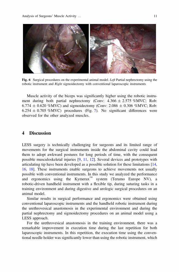
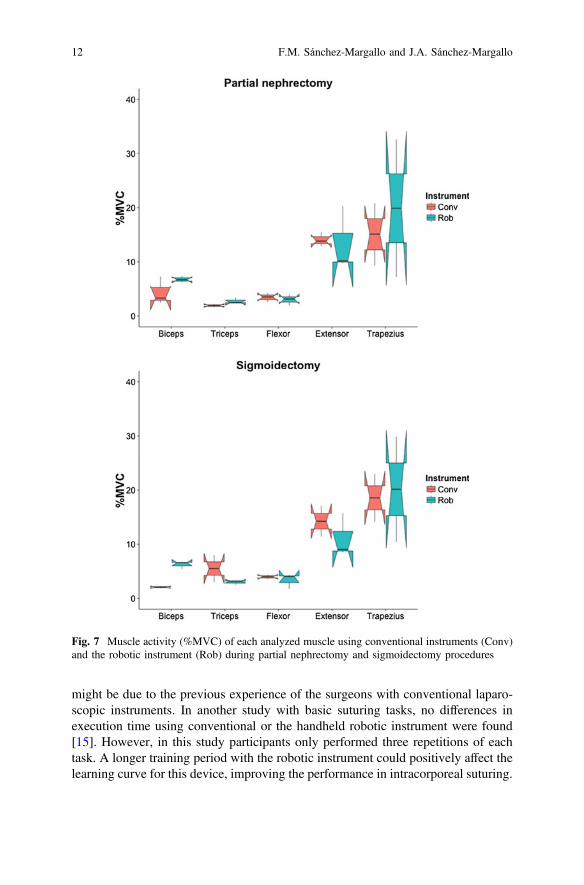
Muscle activity of the biceps was significantly higher using the robotic instru-ment during both partial nephrectomy (Conv: 4.366 ± 2.575 %MVC; Rob:6.774 ± 0.620 %MVC) and sigmoidectomy (Conv: 2.086 ± 0.306 %MVC; Rob:6.254 ± 0.705 %MVC) procedures (Fig. 7). No significant differences wereobserved for the other analyzed muscles.
4 Discussion
LESS surgery is technically challenging for surgeons and its limited range ofmovements for the surgical instruments inside the abdominal cavity could leadthem to adopt awkward postures for long periods of time, with the consequentpossible musculoskeletal injuries [9, 11, 12]. Several devices and prototypes witharticulating tip have been developed as a possible solution for these limitations [14,16, 18]. These instruments enable surgeons to achieve movements not usuallypossible with conventional instruments. In this study we analyzed the performanceand ergonomics using the Kymerax™ system (Terumo Europe NV), arobotic-driven handheld instrument with a flexible tip, during suturing tasks in atraining environment and during digestive and urologic surgical procedures on ananimal model.
Similar results in surgical performance and ergonomics were obtained usingconventional laparoscopic instruments and the handheld robotic instrument duringthe urethrovesical anastomosis in the experimental environment and during thepartial nephrectomy and sigmoidectomy procedures on an animal model using aLESS approach.
For the urethrovesical anastomosis in the training environment, there was aremarkable improvement in execution time during the last repetition for bothlaparoscopic instruments. In this repetition, the execution time using the conven-tional needle holder was significantly lower than using the robotic instrument, which
Fig. 6 Surgical procedures on the experimental animal model. Left Partial nephrectomy using therobotic instrument and Right sigmoidectomy with conventional laparoscopic instruments
Analysis of Surgeons’ Muscle Activity … 11
might be due to the previous experience of the surgeons with conventional laparo-scopic instruments. In another study with basic suturing tasks, no differences inexecution time using conventional or the handheld robotic instrument were found[15]. However, in this study participants only performed three repetitions of eachtask. A longer training period with the robotic instrument could positively affect thelearning curve for this device, improving the performance in intracorporeal suturing.
Fig. 7 Muscle activity (%MVC) of each analyzed muscle using conventional instruments (Conv)and the robotic instrument (Rob) during partial nephrectomy and sigmoidectomy procedures
12 F.M. Sánchez-Margallo and J.A. Sánchez-Margallo

Muscular activity of both biceps and flexor muscles was reduced from the first tothe last repetition during the performance of the urethrovesical anastomosis usingthe robotic instrument. It seems that training improves the ergonomics of surgeonsusing the robotic instrument. However, during the surgical procedures, thisinstrument demanded higher muscular activity for the biceps. This might bebecause the robotic instrument is bigger and heavier than the conventionallaparoscopic instrument, leading to higher workload of the biceps muscle.
As was reported by Pérez-Lanzac et al. [19], the surgeons stated that the use ofthe robotic instrument reduced the technical difficulty of the urethrovesical anas-tomosis performed through LESS approach. Participants in the study with thelaparoscopic simulator considered that it should be necessary a previous training tobe familiarized with the controls on the device.
The use of this robotic instrument has been also proved to be feasible in otherlaparoscopic procedures such as total laparoscopic hysterectomy and radicalprostatectomy [18, 20]. A study with sixty patients who underwent a robot-assistedradical prostatectomy reported no differences between using conventional laparo-scopic and robot-assisted procedures with regard to postoperative pain, blood lossand length of recovery [20].
The main limitation of the presented study is the small sample size. Furtherstudies should be done including other handheld robotic laparoscopic instruments tosupport the results obtained. Understanding how operation conditions, workplacelayout and surgical instruments influence surgeons’ ergonomic condition couldprovide inspiration for new instrument designs, as well as more targeted trainingmethods.
In conclusion, results indicated a positive learning curve in performance andergonomics using the handheld robotic instrument for LESS urethrovesical anas-tomosis. Besides, results showed that partial nephrectomy and sigmoidectomyprocedures performed through LESS approach and using the robotic instrument arefeasible and safe. There were no differences in surgery time and surgeon’s muscleactivity during both surgical procedures, except for the biceps muscle. We considerthat a period of adaptation should be required for this new technology.
Acknowledgments This work has been partially funded by the Government of Extremadura,Spain, and the European Social Fund (PO14034).
References
1. Xourafas, D., Tavakkoli, A., Clancy, T.E., Ashley, S.W.: Distal pancreatic resection forneuroendocrine tumors: is laparoscopic really better than open? J. Gastrointest. Surg. 19,831–840 (2015)
2. Medeiros, L.R., Stein, A.T., Fachel, J., Garry, R., Furness, S.: Laparoscopy versus laparotomyfor benign ovarian tumor: a systematic review and meta-analysis. Int. J. Gynecol. Cancer 18,387–399 (2008)
Analysis of Surgeons’ Muscle Activity … 13

3. Delaney, C.P., Chang, E., Senagore, A.J., Broder, M.: Clinical outcomes and resourceutilization associated with laparoscopic and open colectomy using a large national database.Ann. Surg. 247, 819–824 (2008)
4. Marks, J.H., Montenegro, G.A., Shields, M.V., Frenkel, J.L., Marks, G.J.: Single-portlaparoscopic colorectal surgery shows equivalent or better outcomes to standard laparoscopicsurgery: results of a 190-patient, 7-criterion case-match study. Surg. Endosc. 29, 1492–1499(2014)
5. Fan, X., Lin, T., Xu, K., Yin, Z., Huang, H., Dong, W., et al.: Laparoendoscopic single-sitenephrectomy compared with conventional laparoscopic nephrectomy: a systematic review andmeta-analysis of comparative studies. Eur. Urol. 62, 601–6012 (2012)
6. Kaouk, J.H., Autorino, R., Kim, F.J., Han, D.H., Lee, S.W., Yinghao, S., et al.:Laparoendoscopic single-site surgery in urology: Worldwide multi-institutional analysis of1076 cases. Eur. Urol. 60, 998–1005 (2011)
7. Rais-Bahrami, S., Moreira, D.M., Hillelsohn, J.H., George, A.K., Rane, A., Gross, A.J., et al.:Contemporary perspectives on laparoendoscopic single-site surgery in urologic training andpractice. J. Endourol. 27, 727–731 (2013)
8. Islam, A., Castellvi, A.O., Tesfay, S.T., Castellvi, A.D., Wright, A.S., Scott, D.J.: Earlysurgeon impressions and technical difficulty associated with laparoendoscopic single-sitesurgery: a Society of American gastrointestinal and endoscopic surgeons learning center study.Surg. Endosc. 25, 2597–2603 (2011)
9. Rassweiler, J.J.: Is LESS/NOTES really more? Eur. Urol. 59, 46–8; discussion 48–50 (2011)10. Berguer, R., Forkey, D.L., Smith, W.D.: Ergonomic problems associated with laparoscopic
surgery. Surg. Endosc. 13, 466–468 (1999)11. Morandeira-Rivas, A., Millán-Casas, L., Moreno-Sanz, C., Herrero-Bogajo, M.L.,
Tenías-Burillo, J.M., Giménez-Salillas, L.: Ergonomics in laparoendoscopic single-sitesurgery: survey results. J. Gastrointest. Surg. 16, 2151–2159 (2012)
12. Pérez-Duarte, F.J., Lucas-Hernández, M., Matos-Azevedo, A., Sánchez-Margallo, J.A.,Díaz-Güemes, I., Sánchez-Margallo, F.M.: Objective analysis of surgeons’ ergonomy duringlaparoendoscopic single-site surgery through the use of surface electromyography and amotion capture data glove. Surg. Endosc. 28, 1314–1320 (2014)
13. Pérez-Duarte, F.J., Sánchez-Margallo, F.M., Martín-Portugués, I.D.-G., Sánchez-Hurtado, M.A., Lucas-Hernández, M., Sánchez-Margallo, J.A., et al.: Ergonomic analysis of muscleactivity in the forearm and back muscles during laparoscopic surgery. Surg. Laparosc. Endosc.Percutan. Tech. 23, 203–207 (2013)
14. Bensignor, T., Morel, G., Reversat, D., Fuks, D., Gayet, B.: Evaluation of the effect of alaparoscopic robotized needle holder on ergonomics and skills. Surg. Endosc. 30, 446–454(2016)
15. Zapardiel, I., Hernandez, A., De Santiago, J.: The efficacy of robotic driven handheldinstruments for the acquisition of basic laparoscopic suturing skills. Eur. J. Obstet. Gynecol.Reprod. Biol. 186, 106–109 (2015)
16. Sánchez-Margallo, F.M., Sánchez-Margallo, J.A.: Use of a novel robotic laparoscopicinstrument with ergonomic design for urethrovesical anastomosis: analysis of muscularactivity and posture. International Congress of the European Association for EndoscopicSurgery, Bucharest, Romania (2015)
17. Hermens, H.J., Freriks, B., Merletti, R., Stegeman, D., Blok, J., Rau, G., et al.: SENIAM 8:European recommendation for surface electromyography. Roessingh Research andDevelopment, Enschede (1999)
18. Iacoponi, S., Terán, M., De Santiago, J., Zapardiel, I.: Laparoscopic hysterectomy with ahandheld robotic device in a case of uterine sarcoma. Taiwan. J. Obstet. Gynecol. 54, 84–85(2015)
14 F.M. Sánchez-Margallo and J.A. Sánchez-Margallo

19. Pérez-Lanzac, A., Rosety, J., Okhunov, Z., Soto, J., Garcia-Baquero, R., Ledo, M.J. et al.:Robot-assisted laparoendoscopic hybrid single-site radical prostatectomy: a novel techniqueusing Kymerax. J. Endourol. 27, Part B, Videourology (2013)
20. Nikolov, M., Mekoula, S.T., Hauser, S., Planz, B.: First results comparing between hand-heldrobotic assisted prostatectomy and conventional radical endoscopic extraperitonealprostatectomy (Kymerax, Terumo). 25th World Congress on Videourology. Sofia, Bulgaria(2014)
Analysis of Surgeons’ Muscle Activity … 15