
Analysis of mineral composition & bacterial flora in … · Analysis of mineral composition &...
13
ISSN: 2250-0359 Volume 3 Issue 4 2013 Analysis of mineral composition & bacterial flora in 4 tonsillolith stones. Sudhir M Naik 1 , Ravishankara S 2 , Mohan Appaji 3 , Goutham MK 4 , N Pinky Devi 5 , Sarika S Naik 6 1 Fellow, Department of Cosmetic and Aesthetic surgery, Cosmetic surgery institute of India, Mumbai. 3 Associate Professor , Department of ENT, Head and Neck surgery, KVG Medical College, Sullia, Karnataka. 2 Professor, Department of ENT ,Head and Neck surgery, KVG Medical College, Sullia, Karnataka. 4 Assistant Professor , Department of ENT, Head and Neck surgery, KVG Medical College, Sullia, Karnataka. 5 Junior resident, Department of ENT, Head and Neck surgery, KVG Medical College, Sullia, Karnataka. 6 Senior resident, Department of Anaesthesia and Critical care, Narayana Hrudayalaya, Bangalore. Keyword 1: tonsillolith. Keyword 2: tonsillectomy. Keyword 3: unilateral tonsil enlargement. Abstract : Background/ objectives; Large tonsillolith is a rare entity although small calcifications in the tonsillar tissue are a common clinical finding in adults. These
Transcript of Analysis of mineral composition & bacterial flora in … · Analysis of mineral composition &...

ISSN: 2250-0359 Volume 3 Issue 4 2013
Analysis of mineral composition &
bacterial flora in 4 tonsillolith stones.
Sudhir M Naik1, Ravishankara S2, Mohan Appaji3, Goutham MK4, N Pinky Devi5, Sarika S Naik6 1 Fellow, Department of Cosmetic and Aesthetic surgery, Cosmetic surgery institute of India, Mumbai. 3 Associate Professor , Department of ENT, Head and Neck surgery, KVG Medical College, Sullia, Karnataka. 2 Professor, Department of ENT ,Head and Neck surgery, KVG Medical College, Sullia, Karnataka. 4 Assistant Professor , Department of ENT, Head and Neck surgery, KVG Medical College, Sullia, Karnataka. 5Junior resident, Department of ENT, Head and Neck surgery, KVG Medical College, Sullia, Karnataka. 6 Senior resident, Department of Anaesthesia and Critical care, Narayana Hrudayalaya, Bangalore.
Keyword 1: tonsillolith. Keyword 2: tonsillectomy. Keyword 3: unilateral tonsil enlargement.
Abstract :
Background/ objectives; Large tonsillolith is a rare entity although small
calcifications in the tonsillar tissue are a common clinical finding in adults. These

patients usually present with bad breath odor, pain during swallowing or foreign body
sensation in the throat.
Design: K V G Medical College, Department of ENT, Head & Neck Surgery.
Intervention: Tonsillectomy was done in all the cases & tonsillar specimen sent for
histopathological analysis & tonsillolith for mineral & bacterial flora analysis.
Result: Normal tonsillar lymphoid tissue was seen on histopathology in all the 4 cases.
Calcium carbonate & phosphate was seen in all with magnesium phosphate in one.
Fusobacterium an oral anaerobic commensal were isolated from 2 stones.
Conclusion: Tonsilloliths are sometimes incidental findings in X-ray neck &
nasopharynx. Treatment includes surgical removal for superficial stones &
tonsillectomy for larger & deeper stones.
Introduction:
Tonsillolith are calcifications that form in the crypts of the palatine tonsils.1 These
calculi are usually small but larger stones are also reported. 1
Gross examination & sectioning of tonsillectomy specimens during
histopathological study reveals that small calcifications are not uncommon, but larger
stones are rare. 1 Lang in 1560 was the earliest to describe the tonsillolith2. Most of the
calculi are composed primarily of calcium carbonate and calcium phosphate but other
mineral such as magnesium, sodium, silica, potassium, copper, aluminium, iron,
ammonia radicals have also been reported. 3,4,5

The mechanism of tonsillolith formation in the tonsillar crypts is still debated. 6
The hypothesis widely accepted is that tonsilloliths are formed from retained caseous
secretions in the tonsillar crypts in conjunction with filaments of leptothrix buccalis – a
common oral saprophyte, some times in association with chronic purulent tonsillitis. 6
Tonsillolithiasis can occur at any age but is more frequent in 10-77 year age group
with a mean age of about 50 years with equal sex distribution. 7
The symptoms are usually nonspecific such are sore throat, referred otalgia or
pain in the throat. 6 A foreign body sensation and halitosis may also be presenting
symptoms. 6 It is not unusual for tonsilloliths to be diagnosed on routine radiological
studies. 6
Large calculi are clinically seen as a hard mass in the tonsil. 6
Treatment consists of surgical removal of the stone if it is protruding on the surface. 6
Tonsillectomy is indicated if the calculi are embedded in the tonsillar tissue or when
associated with chronic tonsillitis. 6
Materials & methods :
This is a retrospective of study conducted in KVG Medical College Sullia between
April 2007 to September 2010. 4 cases of tonsillolith clinically palpable were confirmed
on post tonsillectomy specimens. The incidence was 4 in 427 tonsillectomies done in
our department under local as well as general anaesthesia.
All the patients were in the age group 38- 52 years. The youngest patient was 38
year old female & oldest 52 year old female. 3 were females & 1 male. All the 3
females presented with features of chronic tonsillitis & the male patient presented

with h/o mild right sided discomfort in the throat with occasional mild pain in the right
ear while swallowing.
All the 3 females had a significant past history with recurrent throat infections.
Oral examination revealed unilateral tonsillar enlargement & signs of chronic
tonsillitis in all the 3 cases. Also cheesy material was seen over the tonsillolith
prominence and appeared like a large grayish white mass with a pitted rough surface
embedded in the tonsillar tissue. A clinical diagnosis of a tonsillolith was made. X-ray
lateral view of the neck in the 3 cases revealed a radiolucent to radio-opaque shadow
in the tonsillar region.
In the male patient the left tonsil was found to be normal but the right tonsil was
enlarged & the tonsillar surface was congested & normal. Residual congestion of the
right anterior pillar was present. On palpation of the right tonsil stony hard mass was
felt which was not tender. Provisional diagnosis of elongated styloid process was
made, but x-ray lateral view of the neck showed normal styloid process. A small radio-
opaque shadow seen in the right tonsillar region. X-ray submento-vertical view
confirmed the right tonsillolith.(fig 1)
The rest of the ear, nose and throat examination did not reveal any abnormality
in all the 4 cases. An ultra sound examination of submandibular salivary glands, gall
bladder and kidneys did not reveal any evidence of stones. No attempt to dislodge the
stone from the tonsil was made. All the patients were posted for tonsillectomy under
general anaesthesia. Tonsillectomy was performed & the tonsillar tissue along with
the tonsillolith stone was sent for histopathological examination. (fig 2)
The tonsillolith was removed from the tonsil specimen. All the 4 stones were
white, brownish to black in color with a pitted rough surface and measured 1x1cm,

1.5x1 cm, 1.5x1.2 cm & 1.6x 2cm respectively. Histopathology of all the 4 tonsil
specimen showed normal tonsillar lymphoid tissue suggesting chronic tonsillitis.
Calcium carbonate & calcium phosphate were seen in all 4 stones & magnesium
phosphate in one stone only. The post operative period was uneventful.(fig 3,4)
Discussion:
Tonsilloliths or calculi of the tonsil are aggregates of varying size and
consistency which develop in the crypts of the palatine tonsil or around it. 7
As the etio-pathogenesis of tonsilloliths are still uncertain two postulates have
been advocated. According to one postulate chronic tonsillitis associated with
repeated episodes of inflammation produce fibrosis of the openings of the tonsillar
crypts followed by accumulation of bacterial and epithelial debris in these crypts
leading to formation of retention cysts.8 This epithelial debris forms the ideal media
for the growth of bacterial, actinomyces 9 & fungi such as Leptothrix buccalis 6.
Finally dystrophic calcification occurs as a result of deposition of inorganic salts
from the saliva secreted in the mouth by major and minor salivary glands.
10Calcification occurs subsequent to the deposition of inorganic salts following which
gradual enlargement of the stone takes place. 8
The tonsilloliths derive their phosphates and carbonates of calcium and
magnesia from saliva secreted by the major salivary glands. 3 However, this hypothesis
cannot explain the existence of calculi in the peri tonsillar zones and also in cases
where prior tonsillectomy has been done3. For this, some authors have suggested that
tonsilloliths results from stasis of saliva in the efferent ducts of the minor salivary
glands secondary to mechanical obstruction arising from post-tonsillectomy scars or
chronic inflammation. 3

This hypothesis is supported by the histological examination of the tissue
excised from around the tonsillolith which revealed salivary gland lobuli with efferent
ducts surrounded by lymphoid tissue in 2 cases3. Calcification of the peritonsillar
abscess and existence of ectopic tonsillar tissue are other mechanisms by which
peritonsillar stones can form. 3,11,12 The association of tonsillolith with kidney stone,
gall stones and wharton’s duct stones in 3% of the cases suggest that tonsillolithiasis
could be a part of the lithogenic systemic disease. 3,11,12
Large tonsilloliths are relatively uncommon though small calcifications are a
common finding in tonsillectomy specimens. 7
Tonsilloliths can occur at any age but are more frequent in the adults than children.
They are frequently reported in the age group 10-77 yrs with a mean age of 50 yrs. 7
69.7% of the tonsilloliths are located in the tonsillar tissue, 21.2% in the tonsillar
fossa while in 9% in the soft palate. 7 2 cases of large stones in the peritonsillar region
associated with peritonsilar abscess known as peritonsilloliths are reported in
literature. 13 A rare case of a calculus of the lingual tonsil has also been described in
literature. 14 The size of a tonsillolith ranges from a few mm to several cms with largest
described being 41x21x19mm. 15
The commonest symptom is pain in throat followed by swelling in the tonsillar
fossa, dysphagia, odynophagia, otalgia, peritonsillar abscess, swelling in the sub
maxillary triangle and halitosis2. In some young adults tonsillar calcifications may be
associated with presence of whitish expressible foul smelling and foul tasting cheesy
materials from the tonsil. 2

Tonsilloliths which are located deeply in the tonsil may present with unilateral
tonsillar enlargement. 16 Multiple tonsilloliths have also been described. 16 In 9% of
patients the tonsilloliths are asymptomatic with most of the asymptomatic lesions less
than 21mm in the largest dimension. 7 Right sided lesions (60%) are more common
than the left sided. They are usually single & unilateral, but 3 cases of bilateral
tonsilloliths have been reported. 17
Clinically the tonsillolith appears as a white or yellowish hard mass within the
tonsillar crypt. 7 Tonsilloliths may be single or multiple and of variable shapes like
round, oval, cylindrical, pyramidal or lobular. 7 The color also varies from the
commonest grayish yellow to dark gray, black or red brown. 7 Tonsilloliths simulating
peritonsillar abscess15 or malignancy18 have been described.
The diagnosis can usually be easily made on clinical examination including
palpation of the tonsil and for confirmation a lateral x-ray of the upper neck will show
the radio-opaque shadow. 19 Tonsilloliths have been diagnosed on routine x-rays of the
mandible. 1,19
But in the absence of clinical signs & symptoms, these x-rays may be misleading
and give a false impression of an interosseous radio-opaque lesion such as a foreign
body, odontoma, sclerosing osteitis, garre’s osteomyelitis, fibrous dysplasia, idiopathic
osteosclerosis, osteoma or a displaced tooth because of the super imposition of the
tonsillolith on the mandibular ramus. 19
Tonsilloliths should also be differentiated from radio-opaque structures and
lesions that occur in the soft tissues close to the jaws such a sialolith, a phlebolith,
cysticercosis, calcified lymph node , carotid artery arteriosclerosis, Eagle’s syndrome
and dystrophic calcifications in an acne scar, ectopic bone or cartilage, a large
maxillary tuberosity or prominent hamulus of pterygoid. 1,19

Cases of pseudo-bilateral tonsilloliths has also been described in which a
unilateral tonsillolith gave a false impression of a bilateral tonsillolith on lateral
radiograph of the neck because of super imposition of a lesion involving one side of
the jaw which created a ghost or a pseudo image on the contra lateral side. 20 CT scans
may reveal non specific calcified image in the tonsillar zone and is not helpful in
reaching a differential diagnosis. 6
A mineralogical analysis of the tonsillolith reveals primarily carbonates and/ or
phosphates of calcium but, other minerals like magnesium, sodium, silica, potassium,
ammonium radicals, copper, alluminium, iron have also been reported. 7 The
composition of the bacterial flora in tonsilloliths using culture dependent molecular
method and scanning electron microscopy have been described. 21 Anaerobic bacteria
detected in tonsilloliths belong to the genera eubacterium, fusobacterium
meghasphera, porphyromonas, prevotella, selenomonas and tannerella . 21 All these
anaerobes are associated with production of volatile sulphur compounds and this
supports the tonsillolith as a cause of halitosis. 21
Superficial & smaller tonsilloliths can be removed by enucleation or curettage
under local anaesthesia. Tonsillectomy is indicated in larger stones & smaller stones
with chronic tonsillitis. 7 We did tonsillectomy in all the 4 cases as the 3 patients were
having associated chronic tonsillitis & in the other patient the stone was embedded
deep in the tonsillar tissue. Tonsilloliths more than 2cms in largest dimension were
usually associated with significant symptoms & lesser than 2 cm lesser were the
symptoms. 7
In our patients all 4 had stones were lesser than 2cm in greatest dimension. 2 had
tonsilloliths superficially situated & 2 deep in the tonsillar tissue. Stones deeper

resulted in more symptoms than superficially situated stones. So the symptoms of
tonsilloliths depend on the size as well as location of the stones in the tonsillar tissue.
Conclusion:
Tonsilloliths are sometimes incidental findings in X-ray neck & nasopharynx.
Treatment includes surgical removal for superficial stones & tonsillectomy for larger &
deeper stones. No recurrence are reported in literature after enucleation .Calcium
carbonate & calcium phosphate was seen in all the 4 stones & magnesium phosphate
was seen in one stone only. Anaerobic bacterial culture yielded fusobacterium in two
cases.
Fig 1: X-ray lateral view of neck and submento-vertical view showing tonsillolith in
patient 3.

Fig 2: post tonsillectomy fossa after tonsillolith removal.
Fig 3: post-tonsillectomy specimen with tonsillolith in patient 1.
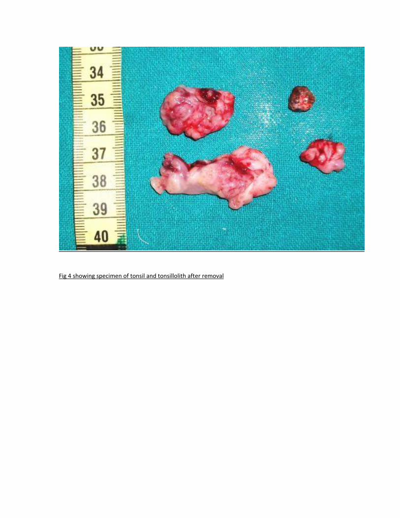
Fig 4 showing specimen of tonsil and tonsillolith after removal

References:
1. Rodrigo CM, Marcus V, Andrea M: Bilateral tonsilloliths in a 77-year old white
man with edentulous jaws: a case report.
2. Pruet CHW, Duplan DA: Tonsil concretions and tonsilloliths. Otolaryngol Clin
North Am 1987, 20(2):305-9.
3. Cooper MM, Steinberg JJ, Lastra M & Antopol S (1993) Tonsillar calculi. Report
of a case & review of the literature. Oral Surg Oral Med Oral pathol 55:239-
245.
4. Heppt W, Schmidt ST, Amstutz GC, Maier H(1989) Tonsillolith : clinical picture &
mineralogic analysis. HNO 37:438-439.
5. Castellano M, Marcolli G(1996) Giant calculi of the tonsil, simulating a
neoplasm. Minerva Med 57:1686-1688.
6. Silvestre-Donat FJ, Pla-Mocholi A, Estelles-Ferriol E, Martinez-Mihi V: Giant
tonsillolith: Report of a case. Med Oral Patol Oral Cir Bucal 2005, 10(3):239-42.
7. Mesolella M, Cimmino M, Martino DM et al (2004) Tonsillolith- Case report &
review of literature Acta Otorhinolaryngol Ital 24:302-307.
8. Paparella MM, Schumrick DA, (1991) Otolaryngology (Vol 3 head & neck), 3rd
edition , Philadelphia. W>B.Saunders p. 2141.
9. Cogolludo Perez FJ, Martin del Guayo G, Olalla Tabar A, Poch Broto J: Report of
a case: large tonsillolith in palatine tonsil. Acta Otorrinolaringol Esp 2002,
53(3):207-10.
10. Jagdeep ST, Ravinder SM, Anamika T, Dev RS& Narinder KM : Giant tonsillolith
causing odynophagia in a child: a rare case report: Cases Journal 2008, 1:50
doi:10.1186/1757-1626-1-50.
11. Neshat K, Penna KJ, Shah DH (2001) Tonsillolith : a case report . J Oral
Maxillofac Surg 59:692-693.
12. Swain HI(1920) Recurrent Calculus of the Tonsil- Report of a case. Ann Otol
Rhinol Laryngol 29:73-78

13. Kimura H, Ohashi N, Nakagawa H, Asai M, Koizumi F: Large tonsillolith
mimicking peritonsillar abscess: a case report. Auris Nasus Larynx 1993,
20(1):73-8.
14. Bando H , Uno T ,N inF, Tei K, Shinomiya T & Has Y (2003) A Case of calculus of
the Lingual Tonsil. Practica oto-rhino-laryngologica 97(7).
15. Modrzynski M, Wrobel B, Zawisza E(2001) Giant tonsillolith simulating
peritonsillar abscess. Pol Merkuriusz Lek H (65):432-433.
16. Dogru H, Tuz M, Aycicek A (2001) Unusual multiple Giant tonsillolith. Oto-
Rhino-Laryngologia Nova H: 319-321.
17. Mosca RC, Cabral MVG, Mantesso A(2006) Bilateral tonsilloliths in a 77 year
old white man with edentulous jaws. A case report. Oral Radiology 22: 34-36.
18. Padmanabhan TK, Chandra Dutt GS, Vasudevan DM, Vijaykumar(1984) Giant
tonsillolith , simulating tumor of the tonsil- a case report. Indian J Cancer21:90-
91.
19. Sezer B, Tugsel Z, Bilgen C(2003) An unusual tonsillolith. Oral surg Oral med
Oral Pathol. Oral radiol Endol 95(4): 471-473.
20. Ram S, Siar CH, Ismail SM & Prepageran N (2004) Pseudo- bilateral tonsilloliths:
a case report & review of literature. Oral Surgery, Oral Medicine, Oral
Pathology, Oral Radilogy & Endodontics 98(1): 110-114.
21. Tusuneishi M, Yamamoto T, Kokeguchi S,Tamaki N, Fukvi K, Watanabe T(2006)
Composition of bacterial flora in tonsillolithiasis. Microbes Infect 8(9-10):
2384-2389.


