Analysis of endocytosis at eisosomes
112
Analysis of endocytosis at eisosomes Dissertation zur Erlangung des Doktorgrades der Fakultät für Biologie der Ludwig-Maximillians-Universität München vorgelegt von Michael Rehman München 2011
Transcript of Analysis of endocytosis at eisosomes

Analysis of endocytosis
at eisosomes
Dissertation zur Erlangung des Doktorgrades
der Fakultät für Biologie
der Ludwig-Maximillians-Universität
München
vorgelegt von
Michael Rehman
München 2011

Erstgutachter: Prof. Dr. Stefan Jentsch
Zweitgutachter: Prof. Dr. Charles David
Tag der mündlichen Prüfung: 19 .09.2011

Table of Contents
1.List of publications .................................................................................................................................... 1
2.Abbreviations .............................................................................................................................................. 2
3.Abstract ......................................................................................................................................................... 3
4.Introduction ................................................................................................................................................. 4
4.1.Plasma membrane organization .................................................................................................. 4
4.1.1.Plasma membrane structure ................................................................................................. 4
4.1.2.Differences between yeast and higher eukaryotes plasma membrane
organization .......................................................................................................................................... 7
4.2.Endocytosis ....................................................................................................................................... 11
4.2.1.Actin patch dependent endocytosis in yeast .............................................................. 13
4.2.2.Eisosomes organize yeast plasma membrane and are static sites of
endocytosis ......................................................................................................................................... 17
4.3.Intracellular trafficking from plasma membrane to vacuoles ........................................ 20
4.3.1.Sorting from the plasma membrane to vacuole ........................................................ 20
4.3.2.AAA-ATPases for disassembly of complexes ............................................................... 20
4.4. Aims ................................................................................................................................................... 25
5.Materials and methods ......................................................................................................................... 26
5.1.Computational analyses .............................................................................................................. 26
5.2.Microbiological and genetic techniques ............................................................................... 26
5.2.1.Bacteria techniques ............................................................................................................... 26
5.2.2.Yeast techniques ..................................................................................................................... 27
5.2.2.1.Gene tagging and deletion............................................................................................. 27
5.2.2.2.Yeast transformation ......................................................................................................... 28
5.2.2.3.Yeast growth assays ........................................................................................................... 28
5.2.2.4.Yeast mating and sporulation ........................................................................................ 29
5.2.2.5.Yeast genomic DNA isolation ........................................................................................ 29
5.2.2.6.Yeast lysate preparation ................................................................................................... 30

5.2.2.6.Yeast lysate preparation for colony PCR .................................................................... 30
5.3. Microscopy ...................................................................................................................................... 31
5.3.1.Imaging...................................................................................................................................... 31
5.3.2. Image processing ................................................................................................................. 31
5.4. Assays ................................................................................................................................................ 32
5.4.1.FM4-64 uptake assay............................................................................................................ 32
5.4.2.Actin staining by phalloidin ............................................................................................... 32
5.4.3.CPY secretion assay ............................................................................................................... 32
5.4.4.Can1 degradation assay ...................................................................................................... 33
5.4.5.Hxt3 degradation assay ....................................................................................................... 33
5.5.Molecular biology techniques ................................................................................................... 33
5.5.1.PCR .............................................................................................................................................. 33
5.5.2.Restriction digestion and ligation ................................................................................... 34
5.5.3.DNA sequencing .................................................................................................................... 34
5.6.Biochemical techniques ............................................................................................................... 34
5.6.1.Western blotting .................................................................................................................... 34
5.6.2.Mass spectometry ................................................................................................................. 35
5.6.3.Lipid isolation .......................................................................................................................... 37
5.7. Preparation of yeast ultrathin sections and electron microscopy .............................. 38
5.8. Composition of buffers and media used ............................................................................. 39
6.Results ......................................................................................................................................................... 45
6.1. Yta6 is involved in endocytosis ................................................................................................ 45
6.1.1. Yta6 dynamically localizes to the plasma membrane ............................................. 45
6.1.2. Yta6 forms oligomeric foci on the plasma membrane ........................................... 45
6.1.3. Regions of Yta6 required for localization to the plasma membrane ................ 47
6.1.4. Yta6 forms „punctae‟ on the plasma membrane ...................................................... 48
6.1.5. Yta6 deletion mutant shows canavanine resistance ................................................ 49
6.1.6. Yta6 colocalizes with eisosomes ..................................................................................... 50

6.1.7. Yta6 physically interacts with eisosome core components .................................. 50
6.1.8. Yta6 requires Pil1 for localization to the plasma membrane ............................... 52
6.1.9. YTA6 genetically interacts with PIL1 .............................................................................. 52
6.1.10. Yta6 colocalizes with endocytic sites marked by FM4-64 .................................. 53
6.1.11. Yta6 overexpression induces increased FM4-64 foci formation and
endocytosis ......................................................................................................................................... 55
6.1.12. Yta6 is required for membrane swirl formation ..................................................... 56
6.1.13. Pil1 is required for the increased FM4-64 intermediate formation in
overexpression of Yta6 ................................................................................................................... 57
6.1.14. Yta6 overexpression rescues endocytic defects in yeast amphiphysin
mutants ................................................................................................................................................ 60
6.1.15. Eisosomes are not required for rescue of endocytosis mediated by Yta6
overexpression .................................................................................................................................. 64
6.1.16. Yta6 overexpression endocytic defects of protein cargoes in amphiphysin
mutants ................................................................................................................................................ 65
6.1.17. Deletion of Yta6 does not affect rate of endocytosis of Can1 and Ste3 ....... 67
6.1.18.Yta6 mediated rescue does not affect early markers of clathrin dependent
endocytosis ......................................................................................................................................... 68
6.1.19. Yta6 mediated rescue does not affect late markers of clathrin dependent
endocytosis ......................................................................................................................................... 69
6.1.20. Yta6 colocalizes with endocytic intermediates of protein cargo Hxt3 ........... 73
6.1.21. Yta6 mediated rescue does not require SUR genes .............................................. 74
6.2. Emp70 is required for organization of intracellular compartments in trafficking 76
6.2.1. Emp70 dynamically localizes to eisosomes ................................................................ 76
6.2.2. Emp70 is an endosomal protein ..................................................................................... 77
6.2.3. Emp70 colocalizes with FM4-64 intermediates at the plasma membrane and
intracellularly ...................................................................................................................................... 80
6.2.4. Emp70 is required for normal endosome functions ............................................... 80
6.2.5. Emp70 deletion mutant does not affect endocytosis ............................................. 81

6.2.6. Pil1 is required for normal Emp70 localization to the cell periphery ............... 82
6.2.7. Yta6 partially colocalizes with Emp70 ........................................................................... 83
7. Discussion................................................................................................................................................. 84
8. Curriculum Vitae .................................................................................................................................... 93
9. Declaration of individual contributions in publications .......................................................... 94
10. Acknowledgements ............................................................................................................................ 95
11. References.............................................................................................................................................. 97

1
1.List of publications
N E Ziolkowska, L Karotki, M Rehman, J T Huiskonen, T C Walther. “ Eisosome-
driven plasma membrane organization is mediated by BAR domains”. Nat Struct
Mol Biol (2011)
Rehman, M.*, Aguilar, P. S.*, Frohlich, F.*, Shales, M.*, Ulitsky, I., Olivera-Couto, A.,
Braberg, H., Shamir, R., Walter, P., Mann, M., Ejsing, C. S., Krogan, N. J.,Walther, T.
C. (2010). "A plasma-membrane E-MAP reveals links of the eisosome with
sphingolipid metabolism and endosomal trafficking." Nat Struct Mol Biol 17(7):
901-908. (* = equal contribution)
Choudhary, C., Kumar, C., Gnad, F., Nielsen, M. L., Rehman, M., Walther, T. C.,
Olsen, J. V., Mann, M. (2009). "Lysine acetylation targets protein complexes and
co-regulates major cellular functions." Science 325(5942): 834-840.

2
2.Abbreviations
AAA-ATPase ATPase associated with various cellular activities
BAR Bin1/Amphiphysin/Rvs (161,167)
CHX Cycloheximide
CPY Carboxypeptidase-Y
DNA Deoxyribonucleic acid
EDTA Ethylenediaminetetraacetic acid
E-MAP Epistatic miniarray profile
FM4-64 N-(3-triethylammoniumpropyl)-4-(6-(4-
(diethylamino)phenyl)hexatrienyl)pyridinium dibromide
FRAP Fluoroscence recovery after photpbleaching
G418 Geniticin disulphate
GFP Green fluorescent protein
HRP Horse radish peroxidase
IP Immunoprecipitation
Kan Kanamycin
kDa kilo-Daltons
LB Luria Bertini media
LY Lucifer yellow
MAT Mating type
MCC Membrane compartment occupied by Can1
MCP Membrane compartment occupied by Pma1
MCT Membrane compartment occupied by TORC2
PAGE Polyacrylamide gel electrophoresis
PBS Phosphate buffered saline
PCR Polymerase chain reaction
PEG Polyethylene glycol
RFPmars Red fluorescent protein Mars
RT Room temperature
SC Synthetic complete
SDS Sodium dodecyl sulphate
SILAC Stable isotope labelling of amino acids in cell culture
WT Wildtype
YP Yeast peptone media

3
3.Abstract
The yeast plasma membrane contains at least three microdomains – membrane
compartment containing Pma1 (MCP), membrane compartment containing TORC2
(MCT) and membrane compartment containing Can1 (MCC). Eisosomes underlie the
MCC domain defined by the marker protein arginine permease (Can1). Eisosomes are
large protein assemblies composed of Pil1 and Lsp1 proteins, of which Pil1 is essential
for plasma membrane organization. We found that the uncharacterized AAA-ATPase
protein Yta6 dynamically colocalizes with eisosomes. Yta6 physically interacts with
eisosome components, specifically with Pil1. In PIL1 deletion cells, Yta6 is unable to
localize normally to the plasma membrane. Yta6 foci colocalize with the intermediates
of FM4-64 on the plasma membrane. The number of these intermediates is increased
upon overexpression of Yta6. Overexpressed Yta6 is also able to rescue the defects of
endocytosis in cells devoid of amphiphysins. Together rescue experiments and
colocalization of a protein cargo Hxt3 with eisosomes suggest that Yta6 likely plays a
role in endocytosis at eisosomes. To identify genes whose products function together
with eisosome components, we independently carried out a genetic interaction study
(epistatic mini array profile) which revealed the protein Emp70. EMP70 showed the
strongest correlation in genetic profile with PIL1. Emp70 localizes to a subset of
eisosomes in addition to its localization in endosomes and vacuoles. We found
eisosomes are required for normal numbers of Emp70 plasma membrane foci. Deletion
of Emp70 misdirected endosomal protein Kex2 to vacuole, implicating its essential role
in maintaining the architecture of the endosomal compartment. In summary, Yta6 likely
plays a role in initiation of endocytosis at eisosomes and Emp70 during intracellular
trafficking from plasma membrane to vacuole.

4
4.Introduction
4.1.Plasma membrane organization
4.1.1.Plasma membrane structure
The plasma membrane has a bilayered structure composed of lipids and
proteins. The ratio of lipid to protein varies depending on the type of cellular
membrane, type of organism, and type of cell. The three main types of lipids
present in the membranes are phosphoglycerides, sphingolipids and
cholesterol. Phosphoglycerides account for more than half of the lipid of most
membranes. The distribution of these lipids in the membrane is asymmetric; the
outer leaflet contains mostly phosphatidylcholine and sphingolipids and the
inner leaflet contains primarily phosphatidylethanolamine, phosphatidylserine
and minor amounts of phosphatidylinositol. Phosphatidylcholine is one of the
lipids which serve as the bulk structural element of biological membranes. Due
to its cylindrical nature, it does not induce curvature of membrane but is
required for membrane transport and fusion processes. In addition, it has a role
in signaling via the generation of DAG. Phosphatidylethanolamine is another
major component of membranes. It is required for assembly of many membrane
proteins by acting as a chaperone during their folding. Phosphatidylserine is
only a minor component of most membranes and plays an important role in
regulation of apoptosis. Sphingolipids are derivatives of sphingosine have been
estimated to compose >30% in the plasma membrane (Patton and Lester, 1991).
The various sphingosine based lipids have additional groups esterified to the
terminal alcohol of the sphingosine moiety e.g. if the substitution is

5
Fig 1. General organization of the plasma membrane. The external surface of most membrane
proteins and lipids contain short chains of sugars. The two leaflets of the bilayer contain
different lipids. The outer leaflet is thought to contain microdomains (“rafts”) consisting of
clusters of specific lipid species (Karp, 2004).
phosphocholine, the molecule is sphingomyelin, if the substitution is a
carbohydrate, the molecule is called glycolipid. Sphingolipids in general perform
structural role in the membranes. In addition to providing structural integrity to
the membrane they also have important role in cell signaling and in particular
some e.g. sphingomyelin is required for myelin sheath formation. Last important
component of membranes is the sterol cholesterol, which in certain animal cells
may constitute up to 50% of the total lipid in the plasma membrane. The
hydrophobic rings of cholesterol molecule are flat and rigid, and they interfere
with movements of fatty acid tails of the phospholipids. Thus, sterols provide
rigidity to the membrane (Alberts, 2002).
The model for the organization of lipids and proteins in the membrane has
undergone many revisions. In 1935, Danielli and Davson proposed that the
6
plasma membrane was composed of lipid bilayer, lined by globular proteins and
lipid bilayer penetrated by protein lined pores for transport. This model was
replaced by the „fluid mosaic model‟ proposed by Singer and Nicolson which
states that - membranes are two dimensional fluids in which proteins are
embedded (Singer and Nicolson, 1972; Vanderkooi and Green, 1970; Vereb et
al., 2003) (Fig 1). This model is now considered a „central dogma‟ of membrane
biology. The structure and arrangement of membrane proteins in the fluid
mosaic model differ from that of previous models in that they occur as mosaic
of discontinuous particles that penetrate lipid sheet. Most importantly, in the
fluid mosaic model the components are mobile and capable of coming together
to engage in various types of transient or semipermanent interactions (Frye and
Edidin, 1970).
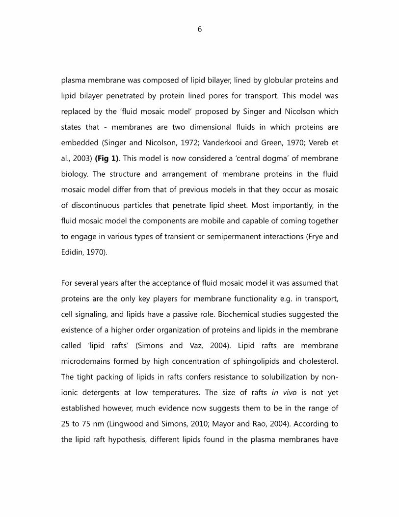
For several years after the acceptance of fluid mosaic model it was assumed that
proteins are the only key players for membrane functionality e.g. in transport,
cell signaling, and lipids have a passive role. Biochemical studies suggested the
existence of a higher order organization of proteins and lipids in the membrane
called „lipid rafts‟ (Simons and Vaz, 2004). Lipid rafts are membrane
microdomains formed by high concentration of sphingolipids and cholesterol.
The tight packing of lipids in rafts confers resistance to solubilization by non-
ionic detergents at low temperatures. The size of rafts in vivo is not yet
established however, much evidence now suggests them to be in the range of
25 to 75 nm (Lingwood and Simons, 2010; Mayor and Rao, 2004). According to
the lipid raft hypothesis, different lipids found in the plasma membranes have

7
different propensity to associate with each other due to differing biophysical
characteristics of the lipid. This causes phase separation which results in the
formation of distinct and stable domains by membranes. The combinatorial
association of different types of sphingolipids with cholesterol further accounts
for raft diversity (Simons and Gerl, 2010) (Fig 2). In recent years two types of
lipid rafts have been proposed: planar lipid rafts and non-planar lipid rafts (Allen
et al., 2007). Both types are enriched in sphingolipids and cholesterol. Planar
rafts are continuous with the plasma membrane, do not show invaginations and
often contain flotilin proteins. Non-planar rafts are made by caveolin proteins,
the so called „caveolae‟ (Allen et al., 2007). Both types of rafts are proposed to
cause localized cell signaling by recruiting specific proteins necessary for signal
transduction (Wachtler and Balasubramanian, 2006). They also provide cells with
a mechanism for sorting in the exocytic pathway (Lingwood and Simons, 2010).
4.1.2.Differences between yeast and higher eukaryotes plasma
membrane organization
The yeast plasma membrane appears simpler in composition compared to
mammalian cells. The inner leaflet of S cerevisiae is enriched in PE, PI, PS. In
contrast to higher eukaryotes, in which cholesterol is the most abundant sterol,
the yeast plasma membrane contains mainly ergosterol and minor amounts of
zymosterol. A striking feature of the yeast plasma membrane is the anomalously
slow mobility of lipids and proteins therein. This slow mobility does not appear
to depend on the cell wall or actin cytoskeleton as the mobility is only mildly

8
Fig 2.Raft based heterogeneity in cell membranes. (a) Nanoscale assemblies of lipids and
proteins can be modulated by actin structures. (b) Signaling and trafficking events initiate
formation of raft platforms. (c) Micrometre sized rafts are observed in model systems like giant
unilamellar vesicles and giant plasma membrane vesicles. (Simons and Gerl, 2010)

9
affected in spheroplasts (Valdez-Taubas and Pelham, 2003; van der Rest et al.,
1995; van Meer, 2002).
The organization of proteins and lipids on the yeast plasma membrane shows
many similarities with mammalian cells but also possesses some unique
features. Early studies from Kai Simon‟s lab showed that the yeast plasma
membrane contains lipid rafts. In yeast, lipid rafts are composed of
sphingolipids, ergosterol, inositolphosphoceramide and its mannosylated
derivatives. Lipid rafts in yeasts were identified on the basis of insolubility in 1%
Triton X-100 at 4 0C and thus called detergent resistant membranes (DRM)
(Schuck et al., 2003). This assay, though reproducible, suffers from possibilities
of artifacts. One limitation of this method is that partial insolubility need not
arise from differences in detergent sensitivity of coexisting domains, but could
also instead arise due to differences in the detergent sensitivity of the two
leaflets. Further, it has been recently observed that Triton X-100 promotes the
formation of ordered domains in model bilayers, so the presence of detergents
may induce segregation of components of the membrane bilayer. Current
advances in microscopy have allowed the direct visualization of these lipid rafts
by specific marker proteins. Three domains are now well recognized on the
yeast plasma membrane. They are named after marker proteins that
predominantly localize there (Fig 3).
The first identified microdomain in yeast was found to contain the highly
expressed yeast transporter – Pma1. Pma1 was found in the DRM fraction.

10
.
Fig 3. Yeast plasma membrane shows at least three kinds of mutually exclusive domains. MCP
(occupied by Pma1) shows network like organization, MCT (occupied by TORC2) shows punctate
organization and MCC (occupied by Can1) shows punctate organization (modified from
(Berchtold and Walther, 2009; Malinska et al., 2003)).
Microscopy studies showed that Pma1 forms network like pattern in the plasma
membrane and thus was called „membrane compartment occupied by Pma1‟ or
MCP. MCP domain covers a large area of the plasma membrane. Pma1 in this
domain freely moves within the plane of the membrane (Grossmann et al.,
2007; Malinska et al., 2003).
The second most studied domain is called MCC domain after the arginine
transporter Can1 that localizes there. Can1 in the MCC forms large patches of
roughly 300 nm diameter that appear as elongated membrane furrows by
freeze-fracture electron microscopy (Stradalova et al., 2009). It also behaves like
typical raft proteins as it found to be present in the DRM. The definition of raft
has been much discussed in many reviews and they are much smaller than 300
nm so it is suggested that Can1 fluorescence in the MCC region is due to raft
clustering. Consistent with the raft hypothesis, a sterol labeling dye filipin has
been observed to colocalize with Can1 foci (Grossmann et al., 2007; Wachtler et

11
al., 2003). MCC is also very stable and shows extremely low mobility if any. This
is observed in FRAP experiments where the bleached region does not recover
over the course of many minutes (Malinska et al., 2004). The organization of this
domain is independent of actin cytoskeleton as observed after treating cells
with Latrunculin A (Malinska et al., 2004). Recently it has been observed that
MCC domains correspond to furrow like invaginations. These invaginations are
elongated and do not colocalize with actin patches. Furthermore, membrane
potential changes affect the organization of MCC domain (Malinska et al., 2003).
Finally, the last well characterized domain is formed by the TORC2 complex -
MCT domain. MCT consists of small, mobile foci distinct from MCC. All TORC2
components localize as oligomeric foci to the plasma membrane, with two to six
copies of the subunits of the complex. In contrast to the MCP and MCC
domains, MCT components are highly dynamic. They show short range lateral
movement on the plasma membrane. The other events that contribute to the
dynamic movement of TORC2 are: appearance, disappearance, fusion and
splitting of foci. Interestingly, TORC2 foci at the plasma membrane do not
colocalize with actin patches and actin is not required for its motility (Berchtold
and Walther, 2009).
4.2.Endocytosis
Endocytosis is a universal process occurring in all cell types from bacteria to
mammalian cells. It can be defined as: „A complex process that controls the

12
Fig 4. Canonical endocytic pathway in yeast. (a) The four protein modules that are involved in
endocytic internalization in yeast are shown: coat complex (green), WASP-myosin complex
(yellow), actin network (red) and amphiphysin complex (blue). Components of these different
protein modules are assembled and disassembled dynamically. (b) The temporal localization of
the constituent proteins for each module in yeast. (c) The approximate temporal localization of
proteins during endocytic internalization in mammalian cells. Dashed lines indicate ambiguity in
the time frame of the protein dynamics. Endocytic protein modules have not been defined for
mammalian cells (Kaksonen et al., 2005).

13
composition of the plasma membrane, nutrient uptake and regulation of cell
signaling‟ (Ayscough, 2005). The word complex in this definition says that it is
not a single step process but requires multiple steps for it to be successful. For
any endocytic process to occur at least three steps are required. First, the site of
endocytosis is chosen. In eukaryotes, this is facilitated by proteins of the clathrin
family, FCHO domain containing proteins, receptor clustering and other
mechanisms. Clathrin and clathrin adaptors are central to the formation of
clathrin coated pits (CCP). Then there has to be a tubular invagination at the site
where endocytosis is initiated. This step in many organisms is mediated by
clathrin and amphiphysins. Finally, there scission occurs. In most organisms this
step is mediated by dynamin proteins. Yeast serves as a great model system to
study these processes because many of the endocytic effectors are conserved
between yeast and higher eukaryotes (Engqvist-Goldstein and Drubin, 2003)..
The most studied pathway is the actin dependent pathway. Recent studies have
shown that this route can be resolved into four modules on the basis of their
timing of arrival at the site of endocytosis – coat module, actin module, WASP
module and amphiphysin module (Fig 4).
4.2.1.Actin patch dependent endocytosis in yeast
The first step in the actin dependent pathway is mediated by proteins of the
coat module. Coat module proteins initiate cargo recruitment and invagination.
These processes are mediated independently of actin but their disassembly
requires actin polymerization. Assisting in this process is a multimeric protein
called clathrin, which consists of three heavy chains and three light chains

14
noncovalently bound to form a symmetric complex called a triskelion. The exact
role of clathrin in yeast is still under debate due to lack of in vivo evidence for
the formation of clathrin coated pits and clathrin coated vesicles in yeast.
Besides, it appears that CHC1 is the only gene in yeast encoding a clathrin
heavy chain. chc1 mutants are viable but grow worse than wildtype cells.
Actin polymerization is important as it is needed for membrane internalization.
The proteins of the second module initiate this important step of actin
polymerization. This module is composed of WASP homolog Las17, Myo5 and
Bbc1. The motor activity of myosin and the assembling actin drive the
invagination inward. It remains immobile at the plasma membrane at the site
where actin is being polymerized and disassembles after the coat movement
when actin is depolymerized. Myo5 mutants are defective in fluid phase
endocytosis and exhibit an increased number of invaginations on the
membrane. Bbc1 and Sla1 regulate coat-complex internalization at endocytic
sites, possibly by regulating actin nucleation via Las17 or Myo3/5 (Kaksonen et
al., 2005).
Proteins of the third module, Rvs161 and Rvs167, are then recruited which
contribute to the release of the forming vesicle. These two proteins belong to
the amphiphysin family (Lombardi and Riezman, 2001). Amphiphysins are
composed of BAR (Bin-Amphiphysin-Rvs) - domains. BAR domains sense
membrane curvature and assist in membrane tabulation (Habermann, 2004).
Both Rvs161 and Rvs167 have highly conserved BAR domain but only Rvs167

15
has the other conserved SH3 domain. SH3 domain binds proline rich regions
and is involved in cell signaling. They have a mobile fraction in the cytosol and
also form transient foci on the plasma membrane. These foci also colocalize
with actin patches (Youn et al., 2010). Rvs- proteins in vivo form homodimers or
heterodimers. They both play a role in endocytosis but Rvs167, because it has
an SH3 domain, also functions in cell division, which is mechanistically not very
clear (Youn et al., 2010). These proteins are suggested to be required for the
scission of the internalizing vesicles. Mutants of RVS- show retraction of the coat
complex after the initial internalization movement (Jonsdottir and Li, 2004). Even
though the amphiphysins are important for endocytosis, many studies in recent
years have found that its defects can be overcome by mutating the genes
involved in sphingolipid metabolism (Morgan et al., 2009). Germann et al
performed a suppressor screen using Rvs161 as bait and revealed proteins
involved in sphingolipid-dependent suppressor pathway (Germann et al., 2005).
They found mutants of SUR1, SUR2, SUR3 and SUR4 genes having the ability to
suppress the endocytic defects in RVS-mutants. All these could rescue defects in
the single mutants and also the double mutants. Sur1 is a catalytic subunit of
MIPC synthase. It forms a complex with Csg2 and functions in the spingolipid
biosynthesis. Sur2 is a sphinganine C4 – hydroxalase, catalyses the conversion of
sphinganine to phytosphinganine in the spingolipid biosynthesis. Sur4 is an
elongase in the sphingolipid biosynthesis, synthesizes very long chain 20-26-
carbon fatty acids from C18-CoA. Sur3 is physically unmapped. Another protein
of SUR (suppressor of RVS) family – Sur7, has been found to suppress the
defects in the amphiphysin mutants upon overexpression (Germann et al., 2005;

16
Lombardi and Riezman, 2001; McCourt et al., 2009; Morgan et al., 2009).
Mechanism of action of many of these proteins is not clear and also which step
of endocytosis do they suppress.
Amphiphysins only bend the membrane for invagination but next step of
scission is required to pinch off vesicle for intracellular trafficking. Proteins in the
fourth module, actin module, are suggested to perform this task. Actin assembly
forces may contribute in this process as evidenced by the presence of dense
actin filaments called „actin patches‟ around early vesicles. Actin patch
movement provides the force needed to direct the invaginating plasma
membrane into the cell against turgor pressure (Aghamohammadzadeh and
Ayscough, 2009) . The first observation of actin patches dates back to 1984
observation by Kilmartin et al (Drubin et al., 1988; Kilmartin and Adams, 1984).
They used antibodies to check for actin localization during cell cycle. Later
studies used fluorescent reporters to detect actin patches. Yeast cells show three
types of actin structures: the actin ring during cytokinesis, actin cables and actin
patches. The actomyosin ring in yeast starts early during cell cycle in G1 phase;
its contraction is coordinated temporally and spatially with septum formation;
surprisingly, the cells without actomyosin structure can divide, albeit less
efficiently. Actin cables are polarized structures in dividing cells oriented towards
mother-bud axis. These cables act as tracks on which the vesicle moves towards
the bud. Actin patches are 0.1 to 0.2 m in diameter and localize to the cell
cortex. In the non-dividing cells they are distributed everywhere in the cell, while
in the dividing cells oriented towards the mother bud axis. Each cell has about

17
10 to 100 patches. Actin patches are dynamic and localize with endocytic cargo.
The patches show a lifetime of approximately 15 seconds. At the molecular level
these actin patches are made of branched actin filaments. Actin patch formation
and movement appears to require actin polymerization nucleated by Arp2/3.
Ultrastructure studies have shown that they patches colocalize with membrane
invaginations. Genetic studies have shown that mutants of actin patch
components (e.g. act1-1, myo3∆myo5∆, sla1∆) have defects in cell wall assembly
and they grow larger than WT cells (Drubin et al., 1988; Smith et al., 2001).
4.2.2.Eisosomes organize yeast plasma membrane and are static sites
of endocytosis
Eisosomes underlie the MCC compartment on the plasma membrane. They are
large immobile protein complexes present on yeast plasma membrane. Each cell
has about 25 to 45 eisosomes. The core proteins that compose eisosomes are –
Pil1 and Lsp1. Pil1 and Lsp1 are 72% similar and the homologs are conserved
across fungal kingdom. Each eisosome foci comprises of 2000 – 5000 proteins
of each type. Eisosome proteins Pil1 and Lsp1 specifically interact with each
other in a 1:1 ratio (Fig 5). PIL1 deletion mutant shows a striking phenotype – a
complete loss of the membrane organization. This has been observed in thin
section electron micrographs and also by colocalization of a plasma membrane
marker dye with a fluorescently tagged protein. PIL1 mutants show the so-called
'eisosome remnants'. These remnants vary from cell to cell but are 1 to 5 per

18
Fig 5. Eisosomes organize plasma membrane. (a) Mid section of a cell is shown to represent the
colocalization of eisosomes proteins Pil1 and Lsp1. (b) Mid sections of cells expressing Lsp1-GFP
in WT, pil1 are shown. (c) Mid sections of cells expressing Sur7-GFP in WT, pil1, lsp1,
pil1lsp1 are shown (modified from (Walther et al., 2006)).
Fig 6. Eisosomes are sites of endocytosis. Mid sections of cells expressing Pil1 (green) is shown
and early intermediates of FM4-64 (red). All FM4-64 intermediates colocalize with eisosomes
(modified from (Walther et al., 2006).

19
cell. However, how and why only Pil1 causes this misorganization is still unclear,
as Pil1 and Lsp1 show strong similarity but only one gives this phenotype
(Walther et al., 2006).
Apart from eisosome's membrane organizing property, they also colocalize with
FM4-64 early intermediates at the plasma membrane (Fig 6). This colocalization
suggests their role in endocytosis, as FM4-64 is an endocytic tracer which is
known to traffic from plasma membrane to endosomal pathway to vacuoles.
FM4-64 serves a good example of lipid cargo. The colocalization of eisosomes
with Hxt2 intermediates of internalization suggest that also protein cargo is
internalized at eisosomes. Mammalian cells show a wide variety of mechanisms
of endocytosis and it is likely that yeast also has another pathway which is
independent of actin patches. This is supported by: (a) observation of zero
colocalization of eisosomes and actin patch marker Abp1, (b) The plasma
membrane FM4-64 intermediates localization is unaffected in cells treated with
LatA. The presence of two pathways could either allow spatial regulation, cargo
selective or temporal regulation in endocytosis. Intriguingly, these intermediates
of FM4-64 form only a few eisosomes – approximately about 4 in 40 eisosomes.
Experiments with Ste3-HA internalization have shown that eisosome mutants
have defects in endocytosis, so it conceivable that a pathway of endocytosis
originates at eisosomes.

20
4.3.Intracellular trafficking from plasma membrane to vacuoles
4.3.1.Sorting from the plasma membrane to vacuole
During intracellular trafficking the internalized cargo along with its coat is first
trafficked to early endosomes. At this step, the cargo can be either destined to
vacuoles or recycled to the plasma membrane. An important post translational
modification occurs in many cargoes early during internalization –
ubiquitinylation. It is the addition of 76 amino acid polypeptide, ubiquitin, to the
epsilon amino group of lysine residues in the target proteins. Polyubiquitin chain
serves as signal triggering endocytosis to early endosomes (Dupre et al., 2004;
Lin et al., 2008). Following their internalization, all endocytic ubiquitinylated
yeast plasma membrane cargoes are transported through several endocytic
intermediates known as post Golgi endosomes or late endosomes. There they
meet membrane proteins from Golgi apparatus where cargoes are sorted into
distinct domains depending on whether it is for recycling or degradation. Cargo
destined for vacuoles is sorted into vesicles which are budded into the interior
of the late endosomes resulting in the formation of multivesicular bodies (MVB)
(Katzmann et al., 2002). After fusion of MVBs with the vacuole, the vesicles are
released into the lumen of the vacuole and proteins associated with them are
degraded by vacuolar hydrolases (Fig 7).
4.3.2.AAA-ATPases for disassembly of complexes
The sorting of the MVB is mediated by a set of proteins called class E vps
(vacuolar protein sorting). The mutants of the members of this class accumulate

21
Fig 7. The schematic shows the intracellular trafficking in a typical cell. It shows cargo trafficking
through early, late and recycling endosomes (Orlando and Guo, 2009).
endosomal membranes and exhibit defects in the formation of MVB vesicles
(Hurley and Emr, 2006). Among class E vps proteins, yeast Vps4 has been shown
to play an essential role in the morphological and functional organization of the
endocytic system and is required for efficient transport from early to late
endosomes. Vps4 is a member of AAA-ATPase family and ATP binding regulates
the association of Vps4 with endosomal compartment (Wendland et al., 1998)
(Fig 8).
AAA–ATPases are universally present in all organisms from bacteria to
metazoans. Just like other P type ATPases, the characteristic features of AAA-
ATPases are:

22
Fig 8. AAA-ATPases in yeast. (a) Organization of domains in AAA-ATPase proteins. (b) Hexameric
oligomer of AAA-ATPase NSF. (c) Localization of yeast AAA-ATPase proteins (Hanson and
Whiteheart, 2005; White and Lauring, 2007).
Walker A, Walker B, sensor 1, sensor 2 (Frickey and Lupas, 2004) (Hanson and
Whiteheart, 2005). The Walker A motif is involved in the phosphate binding of
ATP, walker B motif is involved in the metal binding and ATP catalysis, sensor 1 is
involved in detection of nucleotide binding and ATP hydrolysis. Members of
AAA family assemble into ring-like oligomers that carry out non-covalent
conformational modifications of stable proteins and protein-protein complexes.
The chaperone like activity of AAA-ATPases promotes assembly and disassembly
of target protein complexes. This function is primarily achieved by application of
mechanical force on the host protein. Some proteins have only a single AAA
domain and others such as dyenin have more than one domain per protein. The
oligomers of the AAA proteins can be either hexamers or heptamers. Yeast has
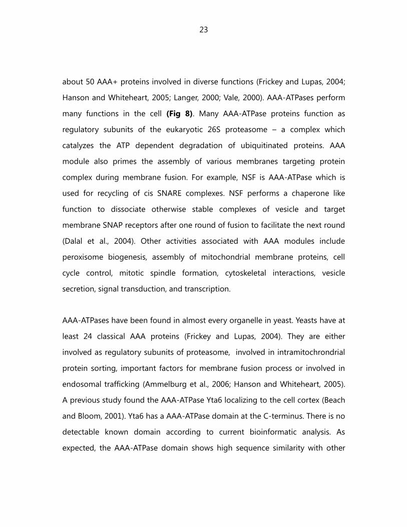
23
about 50 AAA+ proteins involved in diverse functions (Frickey and Lupas, 2004;
Hanson and Whiteheart, 2005; Langer, 2000; Vale, 2000). AAA-ATPases perform
many functions in the cell (Fig 8). Many AAA-ATPase proteins function as
regulatory subunits of the eukaryotic 26S proteasome – a complex which
catalyzes the ATP dependent degradation of ubiquitinated proteins. AAA
module also primes the assembly of various membranes targeting protein
complex during membrane fusion. For example, NSF is AAA-ATPase which is
used for recycling of cis SNARE complexes. NSF performs a chaperone like
function to dissociate otherwise stable complexes of vesicle and target
membrane SNAP receptors after one round of fusion to facilitate the next round
(Dalal et al., 2004). Other activities associated with AAA modules include
peroxisome biogenesis, assembly of mitochondrial membrane proteins, cell
cycle control, mitotic spindle formation, cytoskeletal interactions, vesicle
secretion, signal transduction, and transcription.
AAA-ATPases have been found in almost every organelle in yeast. Yeasts have at
least 24 classical AAA proteins (Frickey and Lupas, 2004). They are either
involved as regulatory subunits of proteasome, involved in intramitochrondrial
protein sorting, important factors for membrane fusion process or involved in
endosomal trafficking (Ammelburg et al., 2006; Hanson and Whiteheart, 2005).
A previous study found the AAA-ATPase Yta6 localizing to the cell cortex (Beach
and Bloom, 2001). Yta6 has a AAA-ATPase domain at the C-terminus. There is no
detectable known domain according to current bioinformatic analysis. As
expected, the AAA-ATPase domain shows high sequence similarity with other
24
AAA-ATPase domains. Yta6 shows maximum similarity to Spastin and Katanin
proteins, which in higher eukaryotes are involved in microtubule severing
(Connell et al., 2009; Hartman et al., 1998). At present the function of Yta6
remains completely uncharacterized.

25
4.4. Aims
Previous work showed that early intermediates of FM4-64 localize to the plasma
membrane independently of actin patches, but are dependent on eisosomes. In
the wildtype cells eisosomes were observed to colocalize with FM4-64
intermediates and the intermediates of Hxt2. This suggested the possibility of
alternative pathway of endocytosis independent of actin patches.
The goals of this work were to understand the mechanisms of endocytosis at
eisosomes. We took a candidate approach by studying two proteins – Yta6 and
Emp70. To understand the role of Yta6 in the biology of eisosomes we decided
to carry out biochemical experiments to know its interacting partners. We
carried out microscopy by confocal and TIR-FM to accurately determine the
localization of Yta6 in relation to eisosomes. Finally, to know its involvement in
endocytosis, we performed many types of endocytosis assays. To functionally
annotate Emp70, we performed live cell imaging of Emp70 with eisosomes to
obtain quantitative values for its localization with eisosomes. Emp70 was earlier
predicted to be a endosomal protein (Schimmoller et al., 1998). To this end, we
carried out microscopy and biochemical assays to determine if it is required for
endocytosis, recycling or for maintenance of intracellular compartments.
26
5.Materials and methods
5.1.Computational and data analyses
NCBI / Pubmed and Google / GoogleScholar were used for most literature
search. DNA/protein sequences of interest were extracted from SGD database.
In case of alignment or making phylogenetic trees or other sequence
manipulation, following softwares were used: NCBI BLAST, Uniprot, ExPaSy,
SMART, NCBI ORF finder, Geneious, APE.
Data obtained were either created in Microsoft office Excel or GraphPad Prism.
Using their internal options graphs, trendlines or plots were drawn. When
standard deviation was mentioned, it was manually incorporated. Histograms
were all drawn using the „Data analysis‟ Add-ins of Microsoft office Excel. All
tests of significance were performed in GraphPad Prism using its automated
method.
5.2.Microbiological and genetic techniques
5.2.1.Bacteria techniques
Chemically competent cells for transformation were prepared according to
standard protocols. Briefly, a single colony was grown in 200 mL LB media for 2-
3 hrs with vigorous shaking (37 0C, 370 rpm). This was in triplicate, and done till
OD reached 0.6. The cultures were then chilled on ice for 30 mins. Cells
suspension was then applied into two 500 ml sterile centrifuge tubes and
centrifuged at 3000 rpm for 10 min at 4 0C. Pelleted cells were washed with the
original volume of ice-cold ddH2O, and spin at 3000 rpm for 10 min at 4 0C.
27
Washed cells were resuspended with 200 ml (about 1/3 of original volume) of
sterile, ice-cold 0.1 M CaCl2 by gentle pipetting. They were left on ice for 1-2hrs
before spinning down at 3000 rpm for 10 min at 4 0C. Cells were then
resuspended in 6 ml of sterile, ice-cold 0.1 M CaCl2 15% glycerol (1/100 of the
original volume). 10-50 l of cells was aliquoted by dispensing into sterile
microcentrifuge tubes (the tubes should be placed on dry ice previously in order
to freeze cell immediately). Finally, aliquots were frozen at -70 ºC until use.
For molecular biology cloning related work the XL1Blue or DH5alpha E coli
strains were used. pTOPO, pET, yeast shuttle vectors were transformed and
standard protocols were followed. For protein expression related work BL21
strain of E coli was used. For transformation, following procedure was followed:
an aliquot of competent cells was put on ice and mixed with 1 g of DNA
solution followed by incubation for 15 mins there, a heat shock was given for 90
sec and vial was put back on ice for another 30 sec, then 300 l of LB media was
added and vial was put on 37 0C incubator. Finally, after 30 mins cells were
plated.
5.2.2.Yeast techniques
5.2.2.1.Gene tagging and deletion
Gene tagging with fluorescent reporter proteins or non-fluorescent tags was
performed by transformation PCR – product (Janke et al., 2004; Longtine et al.,
1998). This method was also used to create yeast strains with gene deletions. It
relies on homologous recombination of the transformed PCR product with the

28
regions in the yeast genome. The positive cells were identified by microscopy,
western blotting or colony PCR.
5.2.2.2.Yeast transformation
A LioAc based method was used for all yeast transformation methods. First a
culture of 5 ml was started from a single colony on plate for overnight. Early
morning another 50 ml culture was started using the overnight culture from 0.2
OD. After the culture reached 0.7 to 0.8 OD, it was pelleted. The pellet was
mixed with 10 l of PCR product or 1-2 g of plasmid DNA and 100 l of 0.2 M
LioAc (Sigma), followed by addition of 400 l of PEG 4000 (Serva). This mix was
incubated at room temperature before addition of 20 l of DMSO and then heat
shock at 42 0C for 20 mins. After heat shock cells were washed with YPD and
incubated at 30 0C for more than 4 hrs, before plating on plates with
appropriate resistance or auxotrophy.
5.2.2.3.Yeast growth assays
Two types of growth assays were used to check for growth defects in yeast
strains. In the growth assay on plates, serially diluted cells were plated starting
from 0.2 OD cells. The plates contained appropriate chemical related to the
experiment and were incubated at 30 0C or as per the experiment requirements.
In the growth assay in culture, an automated machine connected to software for
growth measurement was used. The software allowed to export to a .csv file
which was used to show growth curves using Microsoft Excel.

29
5.2.2.4.Yeast mating and sporulation
To mate a- and - types of yeast, they were first individually grown to mid log
phase. Small amount of culture of each type was then pelleted, mixed in 20l
and incubated on plate till any zygotes were visible when observed in
microscope. These zygotes were manually separated from each other on the
dissection microscope, and incubated at 30 0C for three days to grow. Upon
growth a small amount was put for overnight culture in YPD media. The culture
was then pelleted and put in rich sporulation media, incubated at 30 0C for few
days. Upon visible sporulation the spores were dissected using the dissection
microscope. For tetrad dissection following protocol was followed: centrifuge
100 l of sporulation culture, resuspend in 20 l of zymolase buffer with 2 l of
zymolyase, incubate at RT for 15 mins, add 100 l zymolase buffer to stop the
reaction, perform dissection at the microscope and let spores grow at 30 0C. The
mating type was tested using the mating type PCR which a- and - type gave
different band sizes upon PCR amplication. Amplification was performed using
following primer: F (AGTCACATCAATCGTTTATGG), R – a specific
(GCACGGAATATGGGACTACTTCG), R – - specific (ACTCCACTTCAAGTAA-
GAGTTTG). A band of 544 bp is expected from Mat a- type strain while from Mat
- 404 bp band is expected.
5.2.2.5.Yeast genomic DNA isolation
To isolate genomic DNA from yeast 5.0 OD cells were pelleted, then
resuspended in 200 l of breaking buffer (2% Triton X-100, 1% SDS, 100 mM
NaCl, 10 mM Tris-Cl pH 7.5, 1 mM EDTA). Small amount of zirconium beads were
30
added and cells were lysed by vortex for 8 mins. 200 l of Phenol-chloroform
(1:1) was added and mixed. This was spin down for 10 mins at RT and upper
layer was transferred to another eppendorf. 10 l of sodium acetate was then
added, followed by equal amount of isopropanol. This was put on rotator for 10
mins for precipitates to form, followed by spin down. The genomic DNA
containing pellet was then dissolved in water and stored.
5.2.2.6.Yeast lysate preparation
For general purpose yeast lysate preparation following method was used. 5.0
OD cells were pelleted. To denature proteins and cell wall 8 M urea was added.
A small amount of zirconium beads were added to this and they were vortexed
for 8 mins. This was followed by addition of 20 l of 20% SDS and 20% 5X
Lamelli buffer. The mix was then heated at 65 0C for 5 mins before loading 20 l
in the PAGE.
5.2.2.6.Yeast DNA preparation for colony PCR
1 ml of yeast culture was pelleted and 40 l water was added. A small amount of
zirconium beads were added before vortex for 8 mins. 1 l of that mix was then
used for PCR reaction as template. PCR reaction of 25 l containing – water (16
l), 10X Buffer (2.5 l), 25 mM MgCl2 (3 l), F primer (1.5l of 10 pM), R primer
(1.5l of 10 pM), DNA template (1 l) and Taq polymerase (1 l), was performed.

31
5.3. Microscopy
5.3.1.Imaging
For fluorescence microscopy, yeast cells were grown to an OD600nm = 0.6 in YPD
media at 30 0C and observed in SC media unless and otherwise stated. Cells
were mounted onto coverslips previously coated with concanavalin-A and
directly imaged with an ANDOR / TiLL iMIC CSU22 spinning disk confocal
microscope, 100X 1.4 NA oil immersion objective. For quantification of the
relative numbers of Yta6 molecules making foci, the individual images of Yta6-
GFP, Cse4-GFP, and Spc105-GFP were collected with same settings.
For FRAP experiments, the FRAP module of the Andor software was used. ROI of
interest were selected. After initializing the time lapse imaging, the selected ROI
were bleached with high power laser. Using ImageJ data was imported to
Microsoft excel and manually the curves were created.
5.3.2. Image processing
We collected 16-bit images using Andor Image iQ 1.9 in the linear range of the
camera. For presentation, images were filtered with a smoothening filter
averaging two pixels, converted to 8-bit images and cropped using ImageJ
(http://rsb.info.nih.gov/ij/) or a Guassian filter plugin with .75 pixels was used.
For quantitation of colocalization, we collected stacks and extracted four-
dimensional images for individual cells. Turboreg plugin of ImageJ was used to
correct for drift of cell in making movie of colocalization of Hxt3-RFPmars and

32
Lsp1-RFPmars. Quantification was always performed on original images. Finally,
images were put together using Adobe Illustrator.
5.4. Assays
5.4.1.FM4-64 uptake assay
Cells exponentially growing at an OD600 = 0.7 (1 ml) were harvested,
resuspended in 50 μl of medium and chilled on ice for 5 min. FM4-64 was
added to a final concentration of 10 μM and incubated for another 10 min. Cells
were washed with ice-cold medium, resuspended and incubated for different
time points, after which cells were killed by 10 mM NaN3 and 10 mM NaF and
immediately analyzed by microscopy.
5.4.2.Actin staining by phalloidin
Phalloidin staining was performed to stain actin structures in yeast. Following
procedure was followed: Grow an overnight culture, next morning inoculate
again starting at 0.2 OD till mid log phase, spin down 200 l of cells and fix with
3.6% formaldehyde for 30 mins at ice cold temperature, spin down at 1200 rpm
to wash out the formaldehyde, resuspend in PBS with 5 M phalloidin, incubate
for 30 mins at ice cold temperature, wash out phalloidin and perform
microscopy.
5.4.3.CPY secretion assay
The CPY secretion colony blot assay was performed as described using anti-CPY
antibodies (Invitrogen-A6428) (Mullins and Bonifacino, 2001).
33
5.4.4.Can1 degradation assay
For Can1 localization and western blot analysis cells were grown in log phase in
SC-Arginine medium containing 1.5% raffinose and 0.5% glucose. To induce
Yta6 overexpression, cells were then shifted to galactose containing medium for
4 hrs followed by addition of 150 M CHX. The addition of CHX was taken as 0
time point followed by observation or preparation of cells for western blotting
as described previously (Frohlich et al., 2009). Equal amounts were loaded on
PAGE.
5.4.5.Hxt3 degradation assay
Cells expressing Hxt3-GFP in wildtype or in mutants were grown in 2% glucose
containing YP media. To induce degradation, the cells were transferred to media
containing 2% galactose. Equivalent of cells were harvested at mentioned and
prepared for either microscopy or western blotting.
5.5.Molecular biology techniques
5.5.1.PCR
Polymerase chain reaction (PCR) was used to amplify fragments of DNA. PCR
was performed for either gene cloning, gene tagging / deletion using PCR
method, making mutants or colony PCR from yeast. For a 50 l reaction
following was used: water (32 l), 10X Buffer (5 l), 25 mM MgSO4 (6 l), 10 mM
dNTP (3 l), 10 pM forward primer (3 l), 10 pM reverse primer (3 l), DNA
polymerase (1 l), Template (1 l). Except for colony PCR and gene tagging /

34
deletion, where Taq polymerase was used, Phusion polymerase was used
(Finnzymes).
5.5.2.Restriction digestion and ligation
For cloning purposes DNA was digested according to standard procedures.
Appropriate restriction enzymes (obtained from NEB or fermentas) were used to
specifically digest DNA and create sticky / blunt ends suitable for ligation. After
over 2 hr digestion, the mix was run on 1% TAE agarose gel. Using methods and
protocols from Qiagen, DNA was extracted and ligation was performed. T4 DNA
ligase from Fermentas was used in all cases for ligation performed overnight at
16 0C.
5.5.3.DNA sequencing
All sequencing was performed at the Core Facility (Max Planck Institute of
Biochemistry) using an ABI 3730 sequencing machine. A volume of 7 l
containing – 100 ng DNA, 1 l of 10 pM primer and water was provided to the
facility. Facility performed the sequencing reaction with the DYEnamic ET
terminator cycle sequencing kit (Amersham-Pharmacia) according to
manufacturer‟s instructions.
5.6.Biochemical techniques
5.6.1.Western blotting
For all western blotting procedures a 10% PAGE was used unless otherwise
stated. The gel was first incubated in transfer buffer containing methanol. Dry
blotting or wet transfer was used to transfer proteins to the nitrocellulose

35
membrane. The membrane was then blocked with 5% milk, incubated with
primary antibody according to the company suggestions, washed with TBST
with tween, incubated with HRP conjugated secondary antibody for 1 hr and
washed last time with TBST with tween. The washed blot was incubated with a
solution to cause chemiluminisence which was imaged in the Fiji image reader
setup.
List of antibodies:
Primary antibody Company
Anti-HA SantaCruz biotech
Anti-Myc SantaCruz biotech
Anti-GFP Molecular probes
Anti-GFP SantaCruz biotech
Anti-CPY Invitrogen
Secondary antibody Company
Anti-mouse SantaCruz biotech
Anti-rabbit SantaCruz biotech
5.6.2.Mass spectometry
Protein extracts from 70 ODs of 'light' and 'heavy' labeled cells were obtained as
described (Walther et al., 2007). For immunopurification, equivalent amounts of
proteins were incubated with anti-GFP AB-conjugated magnetic nanobeads
(Miltenyi Biotech) for 5 mins at 4 0C and loaded on μMacs columns (Miltenyi
Biotech) in a magnetic μMacs Separator (Miltenyi Biotech), washed three times
with 1 ml of lysis buffer with 1% (v/v) Triton-X100, three times with 1 ml of lysis
buffer without Triton X-100 and eluted by TEV cleavage. Eluates were mixed,
diluted 5x in 8 M urea, reduced for 20 min at room temperature (22 0C) in 1 mM
DTT and then alkylated for 30 min by 5.5 mM iodoacetamide in the dark. Then,
the eluates were digested, desalted and concentrated as described. Peptides
were separated on-line using an Easy nLC system (Proxeon Biosystems, Odense,

36
Denmark). Samples (5 μl) were loaded as described. Peptides were eluted with a
segmented gradient of 2–60% solvent B for 102 mins with a constant flow of
250 nl min−1. The HPLC system was coupled to an LTQ-Orbitrap Velos mass
spectrometer (Thermo Fisher Scientific) via a nanoscale LC interface (Proxeon
Biosystems). The spray voltage was 2.2 kV, and the temperature of the heated
capillary was 180 0C. Survey full scan spectra (m/z = 300–1600) were acquired in
positive ion mode with a resolution of 60,000 at m/z = 400 after accumulation
of 1,000,000 ions. Up to ten most-intense ions were sequenced by collision-
induced dissociation in the LTQ. Precursor ion charge-state screening was
enabled, and all unassigned charge states as well as singly charged peptides
were rejected. The dynamic exclusion list was restricted to a maximum of 500
entries with a maximum retention period of 90 secs and a relative mass window
of 10 p.p.m. Orbitrap measurements were performed enabling the lock mass
option for survey scans to improve mass accuracy. Data were acquired using the
Xcalibur software (version 2.1.0, Thermo Fisher Scientific) and MaxQuant, version
1.0.1. The data was searched against the yeast database concatenated with
reversed copies of all sequences and supplemented with frequent contaminants
using Mascot (version 2.2.0, Matrix Science). Carbamidomethylated cysteines
were set as fixed, whereas oxidation of methionine and N-terminal acetylation
were set as variable modifications. Maximum allowed mass deviation for MS/MS
peaks and missed cleavages were 0.5 and 3 Da, respectively. Maximum false-
discovery rates (FDR) were 0.01 both on peptide and protein levels. Minimum
required peptide length was six residues. Proteins with at least two peptides
were considered identified (Walther et al., 2007).

37
5.6.3.Lipid isolation
Two methods of lipid isolation were used. For the cold lipid extraction method
(non-radioactive), 25 ml cultures of the appropriate strains were incubated with
shaking at 30 0C to OD 0.5. To serve as controls for the success of the method,
Myriocin / Aureobasidin was added independently to wildtype strains for 1 hr.
Cells were harvested and washed. They were lysed in 1 ml lysis buffer and
transferred to glass vials. A small amount of zirconium beads was added and
tubes were vortex for 2 mins, followed by incubation at 60 0C for 20 mins. The
extracts were then spinned down at 3000 rpm for 10 mins and dried under
nitrogen gas, max 40 0C. Dried pellet was resuspended in 500 l water -
saturated butanol and 250 l of waster was added. This mix was vortexed and
then spinned down at 4000 rpm for 5 mins. The upper organic phase was then
transferred to a new glass vial and let dry under nitrogen gas. During the drying
time, the silica plates (Whatman) were activated. Silica plates were heated for 10
mins in microwave and then let cool. On the silica plates, the 100 l lipids mixed
in CHCL3/MetOH/water was dropwise loaded. To run the plate, the TLC plate was
dipped in 3 cm TLC solution in a closed glass chamber. The TLC was performed
for 2 hrs. To develop, the TLC plate was dipped in cerium molybdate stain and
heated at 200 0C.
For the radioactive method following procedure was performed. Cells were
grown in synthetic media containing either glucose (SC-Glu) or galactose (SC-
Gal) for 6 h to OD~0.7, washed twice and resuspended in SC-Ser and SC-Gal-
Ser. Aureobasidin A treatment was started 20 min prior to labelling. Cells were

38
labeled with [3H] serine for 90 mins and normalized by OD. Lipid extraction, mild
alkaline hydrolysis and desalting were performed as described in (Guan and
Wenk, 2006). The dried lipid extracts were reconstituted in
chloroform:methanol:water (16:16:5, v/v/v), applied to a silica LK6D thin-layer
chromatography (TLC) plate (Whatman) and resolved in
chloroform:methanol:4.2 N ammonium hydroxide (9:7:2, v/v/v). Radioactive
bands were visualized by Kodak Biomax MR film (Sigma-Aldrich) after
EN3HANCE (PerkinElmer) treatment.
5.7. Preparation of yeast ultrathin sections and electron
microscopy
Cells were grown in yeast peptone dextrose medium to 0.7 - 0.8 OD. 10 OD of
cells were harvested by centrifugation. Cells were fixed by a solution containing
1% Glutaraldehyde, 0.2% Paraformaldehyde in PBS for 50 mins, followed by
washing twice with 0.9% NaCl. Pellet was then resuspended in 2% KMnO4 and
incubated at room temperature for 45 mins, followed by dehydration through
graded alcohols – 50%, 70%, 80%, 90%, 95%, 100% (10 – 15 mins each). This
was followed by incubation in propylene oxide for 15 mins twice, then
embedding media / propylene oxide (1:1), Embedding media / propylene oxide
(1:1), embedding media / propylene oxide (2:1) and finally with embedding
medium overnight. Embedding media (R1165) was obtained from Agar Scientific
Ltd, England. Sectioning was done with a Reichert Ultracut (Leica/ Reichert,
Vienna, Austria). A poststaining step was performed using 1% Uranyl acetate
(prepared in filtered water) for 30 mins at room temperature. Electron

39
microscopy images were taken with a Philips CM 120 (FEI / Philips, Eindhoven,
Netherlands). Adjustments of image size, brightness and contrast were
performed on GIMP and ImageJ.
5.8. Composition of buffers and media used
Laemmli sample buffer:
• 2% (w/v) SDS
• 20% (v/v) glycerol
• 100 mM Tris base
• 60 mM EDTA
• 0.1% (w/v) bromophenol blue
DNA loading buffer
• 40% sucrose
• 1 mg Bromophenol blue
50X TAE
• 2 M Tris
• 50 mM EDTA
10X SDS PAGE Running buffer
• 25 mM Tris, pH 8.3
• 192 mM Glycine
• 0.1%SDS
50X Stock Solution of TAE Buffer:
• 57.1 ml glacial acetic acid
• 242 g Tris base
• 100 ml of 0.5M EDTA
Add water to 1L and adjust pH to 8.5
with KOH
Coomassie brilliant blue solution:
• 20% (v/v) methanol
• 10% (v/v) acetic acid
• 0.1% (w/v) Coomassie Brilliant Blue
R-250
Destaining solution:
• 20% (v/v) methanol
• 10% (v/v) acetic acid
Transfer buffer:
• 250 mM Tris base
• 1.92 M glycine
• 0.1% (w/v) SDS
• 20% (v/v) methanol
TBST:
• 25 mM Tris-HCl, pH 7.5
• 137 mM NaCl
• 2.6 mM KCl
Stripping buffer:
• 4% (w/v) SDS
• 100 mM beta-mercaptoethanol
• 62.5 mM Tris/HCl, pH 6.8
40
• 0.1% (v/v) Tween 20
LB-medium (and plates)
• 1% (w/v) tryptone (Difco)
• 0.5% (w/v) yeast extract (Difco)
• 1% (w/v) NaCl
• 1.5% (w/v) agar (plates)
• sterilized by autoclaving
YPD:
• 1% (10 g/l) yeast extract (Difco)
• 2% (20 g/l) bacto-peptone (Difco)
• 2% (20 g/l) D-(+)-glucose
• 2% (20 g/l) agar (for plates)
• sterilized by autoclaving
SC-media/plates:
• 0.67% (6.7 g/l) yeast nitrogen base
(Difco)
• 0.2% (2 g/l) drop out amino acid mix
• 2% (20 g/l) glucose, raffinose, or
galactose
• 2% (20 g/l) agar (for plates)
• sterilized by autoclaving
Sporulation media:
• 1% glucose
• 7.3 mM KH2PO4
Lipid lysis buffer
• 95% ethanol/water/diethylether-
/pyridine/NH4Cl (15:15:5:1:0.018)
TLC running buffer solution
• 180 ml CHCL3/MetOH/4.2N NH4OH
(9:7:2)
Cerium molybdate stain (Hanessian‟s stain)
• 5 g CeSO4
• 25 g (NH4) Mo7O24 4xH2O
• 50 ml sulfuric acid
• 450 ml water
5.9. Yeast strain list
TWY138 Mata ura3 trp1 leu2 his3 ade2 can1-100 Walther et al 2006
TWY250 Mat ura3 trp1 leu2 his3 ade2 can1-100 rvs161::KAN Walther,TC
TWY252 Mat ura3 trp1 leu2 his3 ade2 can1-100 rvs167::KAN Walther, TC
TWY425 Mat ura3 trp1 leu2 his3 ade2 can1-100 yta6::KAN Walther,TC

41
TWY471 Mat his3 leu2 lys2 ura3 Yta6-TAP::KAN
Ghaemmaghami, S
et al 2003
TWY821 Mata ura3 trp1 leu2 his3 ade2 can1-100 Yta6-GFP::HIS This study
TWY834 Mat ura3 trp1 leu2 his3 ade2 can1-100 Sla1-RFPMars::NAT This study
TWY965 Mat ura3 trp1 leu2 his3 ade2 can1-100 Yta6-TAP::KAN This study
TWY971
Mata ura3 trp1 leu2 his3 ade2 can1-100 GAL inducible N-term GFP Yta6
(gigYta6) This study
TWY980 Mat ura3 trp1 leu2 his3 ade2 can1-100 gigYta6::HIS pil1::NAT This study
TWY1011 Mat ura3 trp1 leu2 his3 ade2 can1-100 Sla1-RFPMars::NAT gigYta6::HIS This study
TWY1158 Mata his3 leu2 lys2 ura3 Can1-GFP::HIS Huh et al, 2003
TWY1205
Mata ura3 trp1 leu2 his3 ade2 can1-100 GAL inducible N-term HA tagged
Yta6(giHAYta6) This study
TWY1234 Mat ura3 trp1 leu2 his3 ade2 can1-100 Yta6-TAP::KAN, Pil1-Myc::HIS This study
TWY1235 Mat ura3 trp1 leu2 his3 ade2 can1-100 Yta6-TAP::KAN Lsp1-Myc::HIS This study
TWY1238
Mat ura3 trp1 leu2 his3 ade2 can1-100 Yta6-TAP::KAN, Pil1-Myc::HIS
lsp1::NAT This study
TWY1239
Mat ura3 trp1 leu2 his3 ade2 can1-100 Yta6-TAP::KAN Lsp1-Myc::HIS
pil1::NAT This study
TWY1321 Mat ura3 trp1 leu2 his3 ade2 can1-100 Yta6-GFP::HIS pil1::NAT This study
TWY1533 Mat his3 leu2 lys2 ura3 Can1-GFP::HIS giHA-Yta6::KAN Huh et al, 2003
TWY1742 Mat ura3 trp1 leu2 his3 ade2 can1-100 Yta6-GFP::HIS lsp1::HPH This study
TWY1743 Mat ura3 trp1 leu2 his3 ade2 can1-100 gigYta6::HIS lsp1::NAT This study
TWY1757 Mat ura3 trp1 leu2 his3 ade2 can1-100 gigYta6::HIS pil1::NAT lsp1::HPH This study
TWY1900 Mat ura3 trp1 leu2 his3 ade2 can1-100 Hxt2-Mars::NAT Yta6-GFP::KAN This study
TWY2042 Mata ura3 trp1 leu2 his3 ade2 can1-100 gigYta6::HIS, pRS306 Pil1-GFP::URA This study
TWY2076 Mat his3 leu2 lys2 ura3 Can1-GFP::HIS, rvs161::NAT This study
TWY2078 Mat his3 leu2 lys2 ura3 Can1-GFP::HIS, rvs161::NAT giHAYta6::KAN This study

42
TWY2072 Mata his3 leu2 lys2 ura3 Hxt3-GFP::HIS Huh et al, 2003
TWY2207 Mata his3 leu2 lys2 ura3 Hxt3-GFP::HIS, lsp1::NAT This study
TWY2209 Mata his3 leu2 lys2 ura3 Hxt3-GFP::HIS, pil1::HpH This study
TWY2211 Mata his3 leu2 lys2 ura3 Hxt3-GFP::HIS, lsp1::NAT, pil1::HpH This study
TWY2213 Mata his3 leu2 lys2 ura3 Hxt3-GFP::HIS, yta6::NAT This study
TWY2215 Mata his3 leu2 lys2 ura3 Hxt3-GFP::HIS, giHAYta6::KAN This study
TWY2216 Mata his3 leu2 lys2 ura3 Hxt3-GFP::HIS, giHAYta6::Kan, rvs161::NAT This study
TWY2217 Mat his3 leu2 lys2 ura3 vrp1::KAN Winzeler et al., 1999
TWY2218 Mat his3 leu2 lys2 ura3 vrp1::KAN, giHAYta6::HIS This study
TWY2224 Mata his3 leu2 lys2 ura3 Hxt3-GFP::HIS, rvs161::KAN This study
TWY2279 Mat ura3 trp1 leu2 his3 ade2 can1-100 lsp1::NAT, pil1::HPH, Yta6-GFP This study
TWY2274
Mat ura3 trp1 leu2 his3 ade2 can1-100 gigYta6, rvs161::KAN, sla1-
RFPmars::NAT This study
TWY2272 Mat ura3 trp1 leu2 his3 ade2 can1-100 rvs161::KAN, sla1RFPmars::NAT This study
TWY2229 Mata his3 leu2 lys2 ura3 Hxt3-GFP::HIS, Lsp1-RFPmars::NAT This study
TWY1194
Mat a Kex2-RFP-Mars::NATR, EMP70-GFP::KAN
R ura3 trp1 leu2 his3
ade2 can1-100 This study
TWY1044 Mat a EMP70-GFP::KANR SNF7-RFP-Mars::NAT
R ura3 trp1 leu2 his3
ade2 can1-100 This study
TWY1063 Mat a EMP70-GFP::KANR ura3 trp1 leu2 his3 ade2 can1-100 This study
TWY871 Mat EMP70-GFP::KANR, LSP1-RFP-Mars::NAT
R ura3 trp1 leu2 his3
ade2 can1-100 This study
TWY1319
Mat a YLR413w-RFP-Mars::NATR, EMP70-gfp::KAN
R ura3 trp1 leu2
his3 ade2 can1-100 This study
TWY1320
Mat a YLR413w-RFP-Mars:.NATR, EMP70-gfp::KAN
R, del_pil1::KANR
ura3 trp1 leu2 his3 ade2 can1-100 This study
TWY1195
Mat a KEX2-GFP::KANR, emp70::NAT
R ura3 trp1 leu2 his3 ade2
can1-100 This study

43
TWY138 Mat a ura3 trp1 leu2 his3 ade2 can1-100 This study
TWY1360 Mat a emp70::NATR ura3 trp1 leu2 his3 ade2 can1-100 This study
TWY1223 Mat a vps1::HPHR ura3 trp1 leu2 his3 ade2 can1-100 This study
TWY1230
Mat a tmn1::NATR tmn2::HPH
R tmn3::HPH
R ura3 trp1 leu2 his3
ade2 can1-100 This study
TWY1183
Mat a VPS5-RFP-Mars::NATR, EMP70-GFP::KAN
R ura3 trp1 leu2 his3
ade2 can1-100 This study
TWY450 Mat pil1 ::kan ura3 trp1 leu2 his3 ade2 can1-100 This study
TWY1184 Mat a tmn2 ::hph ura3 trp1 leu2 his3 ade2 can1-100 This study
TWY1182 Mat tmn3 ::hph ura3 trp1 leu2 his3 ade2 can1-100 This study
TWY1207 Mat emp70 :: nat, tmn3 ::hph This study
TWY1206 Mat a emp70::nat, tmn2 ::hph ura3 trp1 leu2 his3 ade2 can1-100 This study
TWY1814 Mat a tmn2 ::hph, tmn3 ura3 trp1 leu2 his3 ade2 can1-100
TWY1064
Mat a Emp70-GFP ::kan, Lsp1-Cherry::HIS ura3 trp1 leu2 his3 ade2
can1-100
TWY1369
Mat a/
ura3 trp1 leu2 his3 ade2 can1-100
cdc28::natNT2 This study
TWY1370
Mat a/
ura3 trp1 leu2 his3 ade2 can1-100
cdc28::natNT2, [pRS316-CDC28] This study
TWY1371
Mat
ura3 trp1 leu2 his3 ade2 can1-100
cdc28::natNT2, [pRS316-CDC28] This study
TWY1372
Mat
ura3 trp1 leu2 his3 ade2 can1-100
cdc28::natNT2, [pRS316-CDC28; pRS314] This study
TWY1373
Mat
ura3 trp1 leu2 his3 ade2 can1-100
cdc28::natNT2, [pRS316-CDC28; pRS314-CDC28] This study
TWY1374
Mat
ura3 trp1 leu2 his3 ade2 can1-100
cdc28::natNT2, [pRS316-CDC28;pRS314-cdc28K40R] This study
TWY1375
Mat
ura3 trp1 leu2 his3 ade2 can1-100
cdc28::natNT2, [pRS316-CDC28; pRS314-cdc28K40Q] This study

44
TWY1376
Mat a
S288c
Cdc28-GFP::HISMX6
Huh, Won-Ki et al,
Nature 2003
5.10.Yeast vectors
Accession no. Plasmid Tag Marker
P30299 pYM13 TAP kanMX4
P30300 pYM14 6HA kanMX4
P30301 pYM15 6HA HIS3MX6
P30302 pYM16 6HA hphNT1
P30303 pYM17 6HA natNT2
P30304 pYM18 9myc kanMX4
P30305 pYM19 9myc HIS3MX6
P30306 pYM20 9myc hphNT1
P30307 pYM21 9myc natNT2
P30308 pYM22 3HA klTRP1
P30235 pYM23 3myc klTRP1
P30236 pYM24 3HA hphNT1
P30239 pYM27 EGFP kanMX4
P30240 pYM28 EGFP HIS3MX6
P30279 pYM-
N25
GAL1 Promoter,
yeGFP
natNT2
pYM27.1 RFPmars natNT2
pYM27.2 RFPmars KanMX6
pFA6a GAL1 Promoter, GFP KanMX6
P30347 pFA6- No tag – deletion hphNT1
P30346 pFA6- No tag - deletion natNT2
pFA6a GAL1 Promoter, GFP HIS3MX6
(Janke et al., 2004; Longtine et al., 1998)

45
6.Results
6.1. Yta6 is involved in endocytosis
6.1.1. Yta6 dynamically localizes to the plasma membrane
The uncharacterized AAA-ATPase Yta6 was previously found to localize in a
peculiar pattern reminiscent of eisosome localization after over-expression
(Beach and Bloom, 2001). To test whether endogenous Yta6 localizes to the
plasma membrane we created Yta6 with C-terminal GFP fusion using
homologous recombination to introduce the corresponding sequence into the
genome. When we determined the localization of Yta6-GFP in respect to a
plasma membrane marker Hxt2-RFPmars, we found that Yta6 forms foci on the
plasma membrane (Fig 9a). We then performed time lapse imaging to
determine if Yta6 foci are static or dynamic. It showed that many Yta6 foci
remain at the plasma membrane for approximately two minutes before
disappearing (Fig 9b,c). To know whether Yta6 foci at the plasma membrane
exchange with cytosolic Yta6, we performed FRAP (fluorescence recovery after
photobleaching) experiments. Three foci were imaged every two seconds. This
revealed that Yta6 foci recover in approximately two minutes (Fig 9d,e).
6.1.2. Yta6 forms oligomeric foci on the plasma membrane
Yta6 belongs to the AAA-ATPase family of proteins. These proteins have a highly
similar AAA-ATPase domain but unique domains for localization to respective

46
Fig 9. Yta6 dynamically localizes to the plasma membrane. (a) A representative image of
endogenous Yta6 tagged with GFP and Hxt2 tagged with RFPmars is shown. A merged image is
shown for colocalization. (b) Sum of images of 70 time frames containing Yta6 foci along with
kymograph is shown to represent their dynamics. (c) A plot of Yta6 foci disappearance vs time is
made to show the residence time of Yta6 foci. (d) Representative images of FRAP of a foci of
Yta6 as marked in blue. (e) Quantification of FRAP from three foci (Bar = 2.5 m).
Fig 10. Yta6 forms approximately seven foci per cell. (a) Comparison of the cytoplasmic Yta6
intensity and Yta6 foci intensity is shown which was used to specify Yta6 foci. (b) Quantification
of average Yta6 foci per cell. (c) A plot of the average Yta6 foci intensity (n=50) vs average foci
intensity of Cse4 (32 molecules; n=20), Spc105 (80 molecules; n=20), and LacI (128 molecules;
n=18) is shown. Error bars in all cases indicates standard deviation.

47
cellular compartments. Most of the AAA-ATPases form oligomers. To know if
Yta6 forms oligomers, we used a microscopy based method. First, we
determined the brightness of Yta6 foci vs cytosolic signal (Fig 10a). Using this
quantification we then determined the average number of Yta6 foci per cell (Fig
10b). We then compared the brightness of Yta6 foci with foci made by three
different proteins: Cse4 (32 molecule foci), Spc105 (80 molecule foci) and LacI
repressor (128 molecule foci). Using this method we found that Yta6 forming
foci on average contain six of its molecules (Fig 10c).
6.1.3. Regions of Yta6 required for localization to the plasma
membrane
AAA-ATPases usually have one or more AAA-ATPase domain and another
domain which provides specificity for localization. Yta6 has one AAA-ATPase
domain and there is another potential uncharacterized domain at the N-
terminus for its localization. To determine if its N-terminus is required for
localization to the plasma membrane, we created a mutant without the AAA-
ATPase domain. We found that its GAL-inducible overexpressed N-terminus (1-
456) is sufficient for localization and foci formation at the plasma membrane
(Fig 11). To test if AAA-ATPase domain is involved in localization we created a
Walker A mutant of GAL-inducible overexpressed Yta6 (K517A). This showed
that AAA-ATPase of Yta6 affects its localization (Fig 12).

48
Fig 11. N-terminus of Yta6 is sufficient for its plasma membrane localization. (a) Representative
image of WT cell expressing GFP-Yta6 from gal-inducible promoter is shown. (b) Representative
image of N term mutant of GFP Yta6 (1-456) from gal-inducible promoter is shown (Bar=2.5
m).
Fig 12. Walker A mutant of Yta6 is unable to normally localize to the plasma membrane. Left
panel shows a representative WT cell with GFP-Yta6 from gal-inducible promoter (oeYta6). Right
panel shows representative picture of a cell expressing mutant Yta6 K517A (Bar = 2.5 m).
6.1.4. Yta6 forms ‘punctae’ on the plasma membrane
Confocal microscopy showed dim but specific membrane fluorescence signal in
Yta6 GFP expressing cells. To investigate this population at a higher time
resolution we used total internal reflection fluorescence microscopy (TIR-FM). It
revealed many, transient Yta6 spots that were highly mobile but less bright (Fig
13). Quantification showed that this population of Yta6 localizes to the plasma
membrane with a residence time of 60 ms.

49
Fig 13. Yta6 shows fast moving punctae. Representative TIR-FM brightfield image and Yta6-GFP
along with kymograph for 5 min is shown. The pink arrow is to indicate static foci and blue
arrows indicate punctae. Far right panel shows quantification of lifetime of punctae. Error bar
indicates 25th
percentile, median and 75th
percentile (Bar = 2.5 m).
6.1.5. Yta6 deletion mutant shows canavanine resistance
To determine if Yta6 mutants have any defects, we tested its growth by plating
serial dilutions at 37 0C and 24 0C. We did not observe any changes in growth
under both conditions compared to WT. To test if yta6∆ is sensitive to
canavanine (3 g/ml) we compared growth of wildtype BY4741, yta6 in BY4741
and W303. BY4741 has endogenous Can1 expression but W303 is mutated for
Can1, thus served as a control. We found that yta6 strains are resistant to
canavanine (Fig 14).
Fig 14. yta6 shows resistance to canavanine. Serially diluted cells of wt, yta6 and W303 were
plated on plates with or without 3 g/ml canavanine to test for its sensitivity.

50
6.1.6. Yta6 colocalizes with eisosomes
Because the Yta6-GFP signal previously reported was reminiscent of the
eisosome pattern, we determined the endogenous localization of Yta6-GFP in
respect to eisosomes, marked by Pil1-RFPmars and found it colocalized at a
subset of eisosomes in optical top and midsections (88.6 ± 4.2% (SD) Yta6-GFP
foci completely overlap with eisosome signal) (Fig 15). The high overlap of Yta6
foci with eisosomes suggests that Yta6 specifically localizes to the eisosomes.
Fig 15. Yta6 colocalizes with eisosomes. (a) Representative image of GFP tagged endogenous
Yta6 and RFPmars tagged endogenous Pil1 is shown. Merged image is shown for colocalization.
(b) Line scan over the midsection of the mother cell in (a). (c) Quantification of colocalization is
shown. Error bar indicates standard deviation. (Bar = 2.5 m).
6.1.7. Yta6 physically interacts with eisosome core components
To determine whether co-localization of Yta6 and eisosome components is due
to a physical interaction between the proteins, we purified Yta6 fused to a
tandem affinity purification (TAP) tag from yeast cells by affinity
chromatography. We used stable isotope labeling with amino acids in cell
culture (SILAC) for the Yta6-TAP tagged strain to discriminate specific interactors
from background binders present in eluates from control purifications (Walther
et al., 2007).

51
Fig 16. Eisosome components copurify in Yta6 co-immunoprecipitation experiments. (a) Affinity
purification of Yta6-TAP expressing cells labeled with Lys8 and untagged control cells labeled
with Lys0 were analyzed by quantitative LC-MS/MS. Averaged ratios of heavy to light intensities
for proteins are plotted against the intensity of signal from the heavy labeled sample. Significant
outliers (P> 0.0000001) for the ratio are shown in blue (b) Yta6 interacts with Pil1 independent
of Lsp1. Pil1-Myc and Lsp1-Myc were immunoprecipitated and probed for the presence of Yta6-
TAP in WT cells (left panels) or pil1 or lsp1 (right panels).
Fig 16 shows that Yta6 had an abundance ratio of roughly 50, indicating that it
mostly was purified from labeled cells. We also detected the major eisosome
core components Pil1 and Lsp1, as well as the known eisosome binding protein
Mrp8 (Wang et al., 2009) with significantly higher ratios than background,
indicating that they purify specifically from the strain expressing Yta6-TAP. To
independently confirm the interaction between Yta6 and eisosomes, we
performed a reverse experiment and immuno-purified Myc-tagged eisosome
core components. We specifically found Yta6-TAP in purifications from these
cells, but not in the ones performed in parallel from untagged controls. To
further test whether Yta6 requires a particular core component for its interaction
with eisosomes, we immuno-isolated Pil1-Myc or Lsp1-Myc from cells with a
deletion of the other major eisosome component, LSP1 or PIL1, respectively.
Comparison of the eluates from each purifications shows that Yta6 requires Pil1,
but not Lsp1, for its interaction with eisosomes. From these data, we conclude

52
that Yta6 directly or indirectly interacts physically with eisosome components
and that this is dependent on the presence of Pil1.
6.1.8. Yta6 requires Pil1 for localization to the plasma membrane
To test whether the interaction of Yta6 with eisosomes is required for normal
localization of Yta6 at the cell cortex, we investigated endogenously tagged
Yta6-GFP in lsp1∆ or pil1∆ cells by fluorescence microscopy. Consistent with our
biochemical data, deletion of PIL1, but not LSP1, resulted in the loss of foci from
the plasma membrane (Fig 17a). However, some residual Yta6-GFP fluorescence
remained at the plasma membrane in pil1∆ cells and lsp1∆pil1∆ cells that was
visible only by TIR-FM microscopy (Fig 17b). This most likely indicates that Yta6
is targeted to the cell cortex and then independently interacts with eisosomes
there. To validate this hypothesis, we over-expressed GFP-tagged Yta6 and
found that it forms foci in wildtype (WT), as well as lsp1∆, pil1∆ and pil1∆ lsp1∆
cells. We also found that Yta6 requires actin for localization to the plasma
membrane (Fig 17c).
6.1.9. YTA6 genetically interacts with PIL1
To determine if Yta6 shows genetic interaction with Pil1 we used a condition
where Pil1 shows phenotype. It has been reported previously that pil1∆ is
resistant to 5 M Myriocin (Walther et al., 2007) . Myriocin is an inhibitor of
serine palmitoyl transferase, thus it inhibits sphingolipid synthesis. We used this
phenotype and made a double mutant of PIL1 and YTA6. We compared the

53
Fig 17. Yta6 has many mechanisms for localizing to the plasma membrane. (a) Endogenous Yta6
requires eisosomes to form foci at the plasma membrane. Representative confocal mid sections
of WT, lsp1, pil1 and lsp1pil1 cells expressing endogenous Yta6 (upper panel) or oeYta6
(overexpressed Yta6- gal inducible) (lower panel) are shown. (b) Yta6 punctae localize
independently of eisosome components. (c) Yta6 requires actin for localization to the plasma
membrane. Representative confocal mid sections of untreated or cells treated with LatA for 30
mins are shown (Bar = 2.5 m).
growth of cells in culture of cells with or without Myriocin. We found that Yta6
suppressed pil1 resistance of growth in Myriocin (Fig 18).
6.1.10. Yta6 colocalizes with endocytic sites marked by FM4-64
Yta6 localizes to a subset of eisosomes at any given time. Similarly, FM4-64, a
styryl dye widely used as an endocytic tracer forms punctate foci that

54
Fig 18. Yta6 is partially required for the suppression of the Myriocin growth defect by Pil1.
Growth curves are shown of cells of WT, yta6, pil1, yta6pil1, WT + Myr, yta6 + Myr, pil1
+ Myr, yta6pil1 + Myr. 1 M Myriocin was used. Error bars indicate standard deviation from
three experiments in all cases.
Fig 19. Yta6 colocalizes with endocytic sites marked by FM4-64 foci. (a) Representative confocal
mid and top sections of Yta6-GFP localization (green) in respect to plasma membrane FM4-64
intermediates (red) is shown. (b) Yta6-GFP is not internalized with FM4-64. Images of the
colocalized signal of Yta6-GFP (green) and FM4-64 (red) during a chase for 60 mins are shown.
Bar = 2.5 m.
colocalize with some but not all eisosomes (Walther et al., 2006). We therefore
tested whether Yta6 colocalizing with eisosomes also have FM4-64 foci. We

55
found that more than 60% of Yta6-GFP foci colocalized with FM4-64
intermediates at the plasma membrane. FM4-64 is primarily used as an
endocytic tracer in many cell types. In yeast it is often used to study trafficking
from plasma membrane to endosomes to vacuole. Vacuole is the final
destination of this dye. To observe if Yta6 is internalized as a cargo we observed
its colocalization with the dye at later time points. We found that at later time
points during FM4-64 uptake, more and more of the dye was located in the cell
interior, where it did not overlap with the Yta6-GFP signal (Fig 19).
6.1.11. Yta6 overexpression induces increased FM4-64 foci formation
and endocytosis
The spatial correlation of Yta6 punctae and FM4-64 foci suggests a functional
relationship between the two. To test the hypothesis that Yta6 is required for
FM4-64 focus formation, we performed FM4-64 uptake pulse-chase
experiments in yta6∆ cells. At the initial time points of the experiments, we
measured a modest but significant and reproducible reduction in the number of
FM4-64 foci at the plasma membrane in yta6∆ compared to WT control cells
(Fig 20). Presumably, this leads to the mild reduction of FM4-64 uptake
observed after 60 min of chase. To test whether Yta6 induces the formation of
FM4-64 foci, we over-expressed Yta6 from the GAL-promoter and counted FM-
64 foci at the plasma membrane, as well as uptake of the dye. Fig 20 shows the
dramatic increase in the number of early FM4-64 foci in cells overexpressing
Yta6 compared to controls, leading to increased FM4-64 signal at the vacuolar
membrane after 60 min chase.

56
Fig 20. Yta6 overexpression induces increased FM4-64 foci formation and endocytosis. (a) Yta6
deletion mutant shows reduced plasma membrane FM4-64 intermediates. Representative
confocal mid sections of the early time point of FM4-64 labeled yta6 cells are shown. Left
panels show representative images, right panel shows quantification of number of FM4-64 foci
per cell. (b) Yta6 deletion mutant shows mild reduction in endocytosis. Representative pictures
of FM4-64 after chase of 60 mins in WT and yta6 are shown. (c) Formation of FM4-64 focal
intermediates seen early during its uptake is strongly increased in cells overexpressing Yta6
(oeYta6). Left panels show representative images, right panels the number of FM4-64 foci per
cell. (d) Representative pictures of FM4-64 after chase of 60 mins in WT and oeYta6 are shown.
(e) Protein cargo Can1 accumulates faster in the vacuoles after a chase for 3 hrs with CHX (Bar =
2.5 m).
6.1.12. Yta6 is required for membrane swirl formation
The induction of FM4-64 foci formation by Yta6 overexpression provides an
opportunity to characterize their structure in more detail. To this end, we
analyzed thin sections by transmission electron microscopy, which revealed

57
many flask-shaped membrane swirls next to the plasma membrane in cells
overexpressing Yta6 (Fig 21). These membrane swirls were also present in some
sections of WT cells, albeit at a much lower frequency. Due to their localization
at the plasma membrane and induction by Yta6 overexpression, we conjecture
that these membrane swirls correspond to the FM4-64 foci. Apart from
membrane swirls we observed finger-like-projections in the EM pictures. To
compare their dimensions we quantified and compared both the swirls and
finger-like projections. We also performed the converse experiment where we
deleted YTA6 and observed membrane swirls. In this case we found that their
numbers were reduced compared to WT (Fig 22).
6.1.13. Pil1 is required for the increased FM4-64 intermediate
formation in overexpression of Yta6
Colocalization of Yta6 and FM4-64 at eisosomes suggests that the induction of
foci might be dependent on eisosomes. We tested this model by overexpressing
Yta6 in cells where Pil1 is missing and found that this leads to strong reduction
of the number of FM4-64 intermediates compared to controls, indicating that
eisosomes are required for their Yta6-induced formation. The remaining FM4-64
signal was mostly found in one large cluster that was also labeled by Lsp1-GFP
and thus likely represents eisosomes remnants (Fig 23).

58
Fig 21. Overexpressed Yta6 shows increased membrane swirls (a) Representative electron
micrographs of either WT cells in galactose or oeYta6 in galactose are shown. Yellow squares
show membrane swirls in zoomed view. Right panel shows one zoomed in membrane swirl.
(b) A representative WT cell is shown with zoomed finger like projections and membrane
swirls are shown on the right panel. (c) Quantification and comparison of membrane swirls
with finger-like-projections. (d) oeYta6 affects the distribution of eisosomes. Left panel
shows a WT cell with Pil1-RFPmars. Right panel shows a representative image of a cell
overexpressing Yta6 with Pil1-RFPmars.
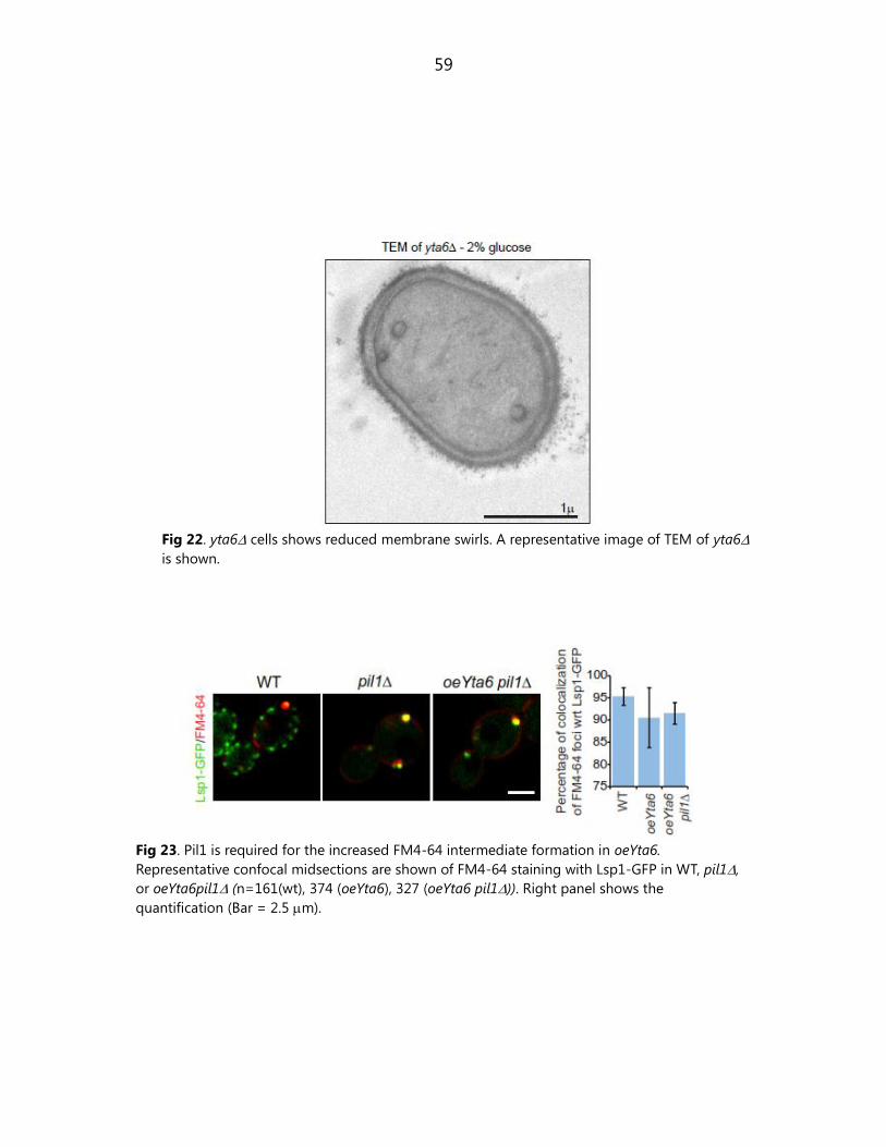
59
Fig 22. yta6 cells shows reduced membrane swirls. A representative image of TEM of yta6
is shown.
Fig 23. Pil1 is required for the increased FM4-64 intermediate formation in oeYta6.
Representative confocal midsections are shown of FM4-64 staining with Lsp1-GFP in WT, pil1,
or oeYta6pil1 (n=161(wt), 374 (oeYta6), 327 (oeYta6 pil1)). Right panel shows the
quantification (Bar = 2.5 m).

60
6.1.14. Yta6 overexpression rescues endocytic defects in yeast
amphiphysin mutants
Yta6 overexpression induces FM4-64 foci formation and uptake, but its deletion
only mildly affects FM4-64 internalization. In contrast, mutations of many genes
encoding actin-patch proteins, which participate in the most studied pathway of
endocytosis in yeast, impair FM4-64 uptake (Munn et al., 1995; Young et al.,
2002). A possible explanation might be that Yta6 functions in a pathway parallel
to actin patches that plays a modest role in FM4-64 endocytosis under normal
conditions. To test this hypothesis, we assayed FM4-64 uptake in yeast
amphiphysin mutants rvs161∆ and rvs167∆ cells. Consistent with previous
results from other groups, each of these conditions led to a strong reduction of
FM4-64 uptake (Douglas et al., 2009; Heese-Peck et al., 2002; Wendland et al.,
1996) (Fig 24). Yta6 overexpression reproducibly rescues FM4-64 uptake in
rvs161∆ and rvs167∆ mutants. Absence of the corresponding proteins also led
to a reduction of FM4-64 foci. This could be mediated by due to some effect on
eisosomes or by Yta6. To test if eisosomes are affected in yeast amphiphysin
mutants we observed qualitatively and quantitatively the eisosomes numbers
and fluorescence. We did not observe any change (Fig 25a,b). We found that
Yta6 localization is affected in the amphiphysin mutants suggesting that
reduction of foci may be mediated due to reduced Yta6 foci formation (Fig
25c). Finally, to ensure that the 4 0C incubation used for pulse labeling in these
experiments has no effect on the results, we performed the FM4-64 assay
without incubation on ice and found consistent rescue of the inhibition of FM4-
64 endocytosis in rvs161∆ cells by Yta6 overexpression (Fig 26).

61
Fig 24. Yta6 overexpression rescues endocytic defects in yeast amphiphysin mutants. (a) Yta6
overexpression rescues FM4-64 uptake defects in rvs161 cells. Representative confocal
midsections of FM4-64 uptake at different time points of a 60 min chase are shown for WT
(upper row), rvs161 (second row from top) cells and cells overexpression Yta6 (oeYta6, third
row from the top) or overexpressing Yta6 in rvs161 cells (bottom row) are shown. (b) Yta6
overexpression rescues FM4-64 uptake defects in rvs167 cells. Representative confocal
midsections of FM4-64 uptake at different time points of a 60 min chase are shown for WT,
rvs167, and oeYta6rvs167 (Bar = 2.5 m).

62
Fig 25. Reduced FM4-64 foci in oeYta6rvs161 and oeYta6rvs167 are not dependent on
eisosomes. (a) Eisosomes localization is not affected in RVS mutants. (b) Quantification of
number of eisosomes, eisosomes fluorescence intensity and cytosolic fluorescence in WT,
rvs161 and rvs167. (c) oeYta6 localization is affected in rvs161 and rvs167 (Bar = 2.5 m).

63
Fig 26. Endocytic rescue mediated by oeYta6 occurs efficiently in room temperature in cells
pulsed with FM4-64. Representative confocal midsections of a 30 min FM4-64 chase experiment
is shown in WT, rvs161, oeYta6, oeYta6rvs161 (Bar = 2.5 m).

64
6.1.15. Eisosomes are not required for rescue of endocytosis
mediated by Yta6 overexpression
To test if eisosomes are required for rescue of endocytosis mediated by Yta6
overexpression in amphiphysin mutants, we observed FM4-64 uptake in
oeYta6rvs161pil1. We compared the FM4-64 uptake in WT, rvs161,
oeYta6rvs161 and oeYta6rvs161pil1. Consistent with previous results we
found that oeYta6 rescued FM4-64 uptake defects in rvs161 but combination
Fig 27. Endocytic rescue mediated by oeYta6 is independent of eisosomes. Representative
confocal midsections of FM4-64 chase in WT, rvs161, oeYta6rvs161 and oeYta6rvs161pil1
are shown for a 60 min chase experiment (Bar = 2.5 m).

65
pil1 did not reduce the delivery of FM4-64 uptake. We thus conclude that PIL1
is not required for rescue mediated by Yta6.
6.1.16. Yta6 overexpression endocytic defects of protein cargoes in
amphiphysin mutants
To test whether the effect of Yta6-overexpression is specific just for the FM4-64
dye or may be relevant also for endogenous protein cargoes, we investigated
the uptake the plasma membrane arginine transporter Can1, which is
internalized by endocytosis. To this end, we blocked Can1 synthesis and delivery
to the plasma membrane, by addition of the translation blocking drug
cycloheximide (CHX), which induces its endocytosis. Time course analysis of
Can1-GFP internalization after CHX treatment by microscopy showed its uptake
and delivery to the vacuole. As expected, Can1 endocytosis was efficiently
delayed and the protein stabilized at the plasma membrane in rvs161∆ cells.
Analogous to experiments using FM4-64, over-expression of Yta6 in otherwise
WT cells had only a mild effect, if any, on Can1-GFP uptake and transport to the
vacuole. However, over-expression of Yta6 in rvs161∆ cells rescued the defects
in Can1-GFP uptake.
To confirm these results, we probed for Can1-GFP degradation after endocytosis
by Western blot. At the beginning of the CHX chase, WT, rvs161∆, Yta6
overexpressing and Yta6 overexpressing rvs161∆ cells all expressed similar levels
of Can1-GFP. After 4 h CHX chase, Can1-GFP was almost completely degraded in

66
Fig 28. oeYta6 can rescue endocytosis of protein cargoes in yeast amphiphysin mutants. (a)
Representative images of midsections of cells expressing Can1-GFP are shown in the time
course experiments in WT and mutants. (b) Representative images of midsections of cells
expressing Hxt3-GFP are shown in the time course experiments in WT and mutants (Bar = 2.5
m).
wild type cells, consistent with our observations by confocal microscopy. Can1-
degradation was almost completely blocked by rvs161∆ and this could be
reversed by Yta6 over-expression, similar to our observation for FM4-64 uptake.
To test whether suppression of the endocytic defects of rvs-mutants by elevated
levels of Yta6 is specific for Can1, or maybe is more general, we also
investigated other cargoes, such as the sugar transporter Hxt3-GFP. This protein
mostly localizes at the plasma membrane in high glucose containing medium,

67
but is internalized after shift to galactose containing medium. Hxt3-GFP uptake
after such a shift in conditions was strongly reduced in rvs161∆ cells, but
rescued by over-expression of Yta6 (Fig 28).
6.1.17. Deletion of Yta6 does not affect rate of endocytosis of Can1
and Ste3
Our data on the rescue of Hxt3 and Can1 endocytic defects in rvs-mutants
indicate that both proteins can access an uptake pathway activated by increased
Yta6 levels. We next determined the contribution of this pathway,

68
Fig 29. Yta6 deletion does not affect the endocytosis of Can1 and Ste3. (a) Representative
confocal midsections of Can1-GFP are shown in WT and yta6 cells. Can1-GFP levels on the
plasma membrane are shown before (upper panels) and after (lower panels) 4 h chase. (b)
Western blot analysis of GalP::Ste3-HA shut down (shift to 2% glucose containing medium) in
WT and yta6. Quantification of the Ste3-HA degradation as shown in the lower panel (Bar = 2.5
m).
and specifically Yta6, to the endocytosis of these transporters. When we
analyzed uptake of Can1 in yta6∆ cells in the presence of CHX, we found no
significant reduction. Similarly, constitutive uptake of the a-factor pheromone
receptor Ste3 was not impaired in yta6∆ cells (Fig 29).
6.1.18.Yta6 mediated rescue does not affect early markers of clathrin
dependent endocytosis
Two interpretations might explain the suppression of the endocytic defect due
to RVS161 deletion by Yta6 over-expression: i) increased Yta6 levels could lead
to re-activation of actin patches defective in endocytosis due to absence of
Rvs161 or ii) Yta6 could activate an actin patch-independent pathway operating
in parallel. If mutant actin patches are re-activated by Yta6, we expect that their
distribution, lifetimes or dynamics, which are compromised in the rvs161∆,
rvs167∆ and vrp1∆ mutants, are rescued by increased Yta6 levels. We first tested
the effect of Yta6 overexpression on actin patch distribution in WT or rvs161∆
cells. Compared to WT cells, rvs161∆ mutants have depolarized actin patches,
but this was not affected by the overexpression of Yta6. We next investigated
the lifetime of actin patches marked with Sla1, which is an early arriving
component of actin patches and indication of endocytic efficiency. Consistent

69
with previous reports, we observed an increased average residence time of Sla1-
RFPmars foci at the plasma membrane of rvs161∆ cells, but increasing Yta6
levels had no influence on this phenotype of RVS161 deletion (Fig 30).
Fig 30. Overexpression of Yta6 has no effect on stalled Sla1-RFPmars patches after LatA
treatment. (a) WT cells or cells overexpressing Yta6 (oeYta6) harboring Sla1-RFPmars were
imaged by confocal time lapse microscopy before or after LatA treatment. Representative
confocal midsections (top row) or kymographs (lower panels) are shown. (b) Quantification of
Sla1-RFPmars life time in WT, rvs161, and rvs161oeYta6 is shown (n=31 (wt), 31 (rvs161), 31
(oeYta6 rvs161) (Bar = 2.5 m).
6.1.19. Yta6 mediated rescue does not affect late markers of clathrin
dependent endocytosis
To also confirm our findings for a component of actin patches that arrives later
during the endocytic reaction, we also determined the lifetime of Abp1-GFP
(Kaksonen et al., 2005). Consistent with prior reports, we observed increased
lifetime of Abp1-GFP in rvs161∆ cells compared to WT, but overexpression of
Yta6 had no effect on this defect. Another prediction from the hypothesis that
overexpression reactivates actin patches in the investigated mutants is that

70
Fig 31. Late marker Abp1 of canonical endocytic pathway in yeast is unaffected by oeYta6. (a)
Actin patch distribution is not altered in cells overexpressing Yta6 compared to rvs161 cells.
Actin patches labeled by Abp1-GFP were followed for 10 seconds and maximum projections of
such a movie are shown. (b) Actin patch life time is not altered by overexpressing Yta6.
Representative actin patches are shown over time in WT, rvs161 cells overexpressing Yta6
(oeYta6) cells as indicated. Right panel shows the kymograph of time series as shown in left
panel. (c) Quantification of Abp1 life times as shown in (c) (n=31 (wt), 31 (rvs161), 31 (oeYta6
rvs161). (d) Yta6 does not colocalize with Abp1. A representative picture of a cell expressing
endogenous Yta6-GFP and Abp1-RFPmars is shown. (e) Representative kymographs are shown
of the cells in (d) (f) Right panel shows a representative picture of a cell overexpressing GFP Yta6
and Abp1-RFPmars. (g) Representative kymographs are shown (Bar = 2.5 m).

71
Fig 32. Hxt3 endocytic intermediates colocalize with eisosomes. (a) Live imaging of single cell
expressing Lsp1-RFPmars and Hxt3-GFP is shown in mid sections to show colocalization of
uptake of Hxt3 at eisosomes. Zoomed panel (bottom) shows the cargo aggregation at the
eisosomes at 0 sec, invagination at 2 sec, scission at 4 sec, internalization of cargo containing
vesicle at 6 sec. Scale bars = 2.5 m. A kymograph of the marked region is shown below. (b)
Hxt3 accumulates at eisosomes upon chase in LatA (Bar = 2.5 m).

72
Fig 33. Yta6 is not involved in the pathway of rescue of RVS mutants which is mediated by SUR4.
(a) Plating of serial dilutions WT or combinations of yta6, sur4, rvs161 is shown for the study
of any crosstalk with Sur4 mediated rescue of RVS mutants. (b) Yta6 mutants do not affect
sphingolipid levels. The bands were assigned after comparison with previously published work
(Tabuchi et al., 2006).

73
these proteins colocalize. However, our analysis of the localization of Yta6-GFP
expressed at either endogenous or strongly increased levels in respect to Abp1
showed no overlap in single images or time series. These data together show
that there is no effect of increased Yta6 levels on dynamics or localization of
actin patches under conditions where uptake of FM4-64 is rescued and that Yta6
is not a component of actin patches. We therefore rule out that the suppression
of rvs161∆ cells endocytic defects by increased Yta6 is due to re-activation of
actin patches, but instead is most likely due to a bypass of actin patches by
activating an alternative endocytic pathway (Fig 31).
6.1.20. Yta6 colocalizes with endocytic intermediates of protein cargo
Hxt3
To determine the localization of Hxt3-GFP during internalization from the
plasma membrane, we followed it by fluorescence microscopy. Before the chase
period, Hxt3-GFP is localized uniformly in the plasma membrane. During the
chase period, when its uptake is induced, however, Hxt3-GFP formed clusters
that localized at eisosomes marked with fluorescent Lsp1-RFPmars. When we
followed the fate of these foci by fluorescence time-lapse microscopy, we found
that many of these clusters connected with internal signal and apparently were
internalized within few seconds, whereas other persisted for a longer and
variable time. Fig 32 shows that the internalization of Hxt3-GFP from the one
such cluster at the plasma membrane at eisosomes into the cell interior during a
90 sec time course. Importantly, the Hxt3-GFP signal inside the cells is does not

74
reflect an internal pool of the protein that is recruited to the plasma membrane
because in kymographs we see clusters appear in the plasma membrane, but
never localizing from a previously existing cell-internal pool. After their
appearance, Hxt3 clusters are also seen to migrate into the cell interior,
apparently reflecting their internalization. To test whether Hxt3 uptake still
requires actin, we performed Hxt3 endocytosis experiments in the absence of
polymerized actin due to the presence of Latrunculin A (LatA). Under these
conditions, plasma membrane clusters still formed, often with brighter signal,
but their internalization was blocked. In addition, Yta6 was not localized to the
plasma membrane any longer. This shows that the Yta6 and actin patches
intersect and suggests that endocytosis of cargo mediated by Yta6 might also
require the actin cytoskeleton after initial cluster formation. The formation of
Hxt3-GFP clusters at eisosomes during its uptake together with the requirement
for eisosome proteins and Yta6 for this reaction strongly suggests that Hxt3-GFP
is endocytosed by a pathway originating there.
6.1.21. Yta6 mediated rescue does not require SUR genes
One potential mechanism of the rescue of endocytosis by Yta6 could occur
similar to that mediated by SUR genes. We tested this by employing mutants of
Sur4. First we observed if Yta6 is involved in the mechanism of rescue of Sur4 of
the rvs161∆. To this end we compared the growth of WT, sur4∆, rvs161∆,
sur4∆rvs161∆, rvs161∆ yta6∆, sur4∆yta6∆ and sur4∆ rvs161∆ yta6∆. As
expected we observed the rescue of growth of sur4∆rvs161∆ but this was not
affected in combination with yta6∆ (Fig 33a). Independently we performed

75
Fig 34. Yta6 overexpression shows reduced local and total amounts of Sur7. (a) Representative
mid sections of cells expressing Sur7-GFP is shown in WT, yta6, WT (galactose grown) and
oeYta6 (galactose grown). (b) Lysates of cells expressing Sur7 –GFP in WT, yta6∆, WT (galactose
grown), oeYta6 (galactose grown) were prepared and subjected to immunoblotting and probing
with anti-GFP and anti-Pgk1 (Bar = 2.5 m).
semiquantitative lipid analysis by thin layer chromatography (TLC) and found
that Yta6 deletion or overexpression mutants do not show any changes in the
lipid levels (Fig 33b). Next we tested if Yta6 has any crosstalk with Sur7 as
overexpression of Sur7 is known to suppress RVS defects. We performed
western blot to quantify the total amount of Sur7 in oeYta6 and found that its
total levels are reduced suggesting that Sur7 may be another cargo that is
endocytosed at eisosomes. Microscopy of Sur7 foci further revealed its lower
amounts per foci in comparison to WT (Fig 34). It is thus likely that endocytic
rescue effect mediated by Yta6 overexpression is independent of Sur7 and it is
possibly a cargo which requires Yta6 for endocytosis.

76
6.2. Emp70 is required for organization of intracellular
compartments in trafficking
6.2.1. Emp70 dynamically localizes to eisosomes
To address functional relationships between plasma membrane processes,
genetic interactions among a set of 374 genes involved in plasma membrane
biology were selected (Aguilar et al., 2010). In the PIL1 genetic interaction
network of the plasma membrane E-MAP, we found that EMP70 is the strongest
candidate for a functional relationship with PIL1 since: i) the two genes have a
high genetic profile correlation (Correlation = 0.366; highest score for PIL1; ii)
the two genes are part of a triangle of positive interactions, with all partners
showing positive interactions with each other iii) the Emp70 homologue Tmn2
was found in a systematic study to be required for normal Pil1-GFP localization
(Frohlich, Moreira et al. 2009). In addition, our unbiased statistical analysis
identified EMP70 and PIL1 as part of the same module. These genetic links
prompted us to investigate the EMP70 in more detail. To this end, we
fluorescently tagged Emp70 with GFP and analyzed it‟s localization by confocal
microscopy. We performed quantification of colocalization of Emp70 and
eisosome component Lsp1 and found that approximately half of Emp70 foci
colocalize with Lsp1 (Fig 35).

77
6.2.2. Emp70 is an endosomal protein
Emp70-GFP localizes in a complex pattern consisting of central ring reminiscent
of vacuoles and several bright foci in the cytoplasm that often seem connected
Fig 35. Emp70 foci localize to the cell periphery. Emp70-GFP (green) was expressed in cells
harboring the fluorescent eisosomes marker Lsp1-RFPmars. Representatives mid- (left) and top
sections (right) are shown. Boxes highlight selected areas of colocalization (Bar=2.5 m).
to the vacuole. Emp70 was previously found in a membrane fraction enriched
for endosomal membranes (Singer-Krueger, JBC 1993). We therefore tested
whether the Emp70 foci in the cytoplasm are endosomal compartments. To this
end we used a number of endosomal markers and found that Emp70-GFP foci

78
localize with Kex2 marking the TGN/early endosome, but not for example the
late endosomal/ pre-vacuolar marker Vps5 (Fig 36, Fig 38a).
Fig 36. Emp70 colocalizes with Kex2. Emp70-GFP and Kex2-RFPmars were coexpressed and
imaged. Representative confocal midsections are shown (Bar=2.5 m).
Fig 37. Emp70 localizes to FM4-64 marked endocytic compartment. (a) Cells expressing Emp70-
GFP were pulse labeled with FM4-64 (red) and imaged for 1 hr. Images of midsections of cells at
selected time are shown as indicated. (b) Quantification of the organelle distribution of Emp70.
Emp70-GFP was imaged in live cells and analyzed for colocalization with Kex2-RFPmars (n=100),
vacuolar FM4-64 (n=91), Snf7-RFPmars (n=93), diploid strain expressing one tagged Snf7 allele)
and Lsp1-Cherry (n=107). The relative area of overlap between signals was quantified as a
percentage of total area occupied by Emp70 signal. Box plots representing maxima, 75th
percentile, media, 25th
percentile and minima are shown for the colocalization with each marker.
(Bar = 2.5 m.)

79
Fig 38. Emp70 localization in respect to endosomes. (a) Strains expressing Emp70-GFP and
Vps5-RFPmars from the endogenous locus were imaged by confocal microscopy. Representative
images are shown. (b) FM4-64 uptake experiment was performed at 16 0C and representative
confocal midsections are shown. (Bar = 2.5 m.)
Fig 39. Emp70 is required for normal endosome function. (a) Emp70 is required for normal
localization of Kex2-GFP. Kex2-GFP was expressed in either WT or emp70 cells, and
representative confocal midsections are shown. (b) Emp70 family members are required for late
endosomal protein retrieval. Mutants of EMP70, TMN2 or TMN3 were tested alone or in
combination for CPY secretion. A representative colony blot is show. (Bar = 2.5 m.)
80
6.2.3. Emp70 colocalizes with FM4-64 intermediates at the plasma
membrane and intracellularly
To independently test whether the labeled compartments are part of the
endocytic route, we used the endocytosis tracer FM4-64 in pulse-chase
experiments. FM4-64 uptake experiments in cells expressing GFP-labeled
Emp70, revealed co-localization of Emp70 foci with the earliest endocytic
intermediates that we could observe (Fig 35). As the dye migrated through the
endocytic system, it colocalized with a subset of Emp70 positive foci towards the
end of the reaction, but markedly less at intermediate time points. At the final
time point, FM4-64 clearly labeled the vacuole delimiting membrane where it
colocalized with the Emp70-GFP ring staining. Together this indicates that
Emp70 localizes to early endosomes and the vacuolar membrane. Trafficking
from the early endosome can be blocked by incubation of cells at 16 0C, which
leads to an accumulation of FM4-64 in this compartment. To confirm that
Emp70 indeed localizes with early endosomes, we localized it in cells where
FM4-64 was blocked in the 16 0C compartment and found near perfect overlap
between the GFP and the FM4-64 signal, further arguing that Emp70 localizes to
an early endosome (Fig 37, Fig 38b).
6.2.4. Emp70 is required for normal endosome functions
To test whether Emp70 is important for the function of early endosomes, we
analyzed the localization of Kex2-GFP in emp70 strain. This deletion resulted
in a dramatic re-localization of Kex2 from the steady state early endosome

81
localization to the vacuole (Fig 36). Under normal conditions, Kex2 sorting
depends on signals that send it to the early endosome, which subsequently
matures into a late endosome, from where it is actively retrieved. It is therefore
possible that the defect in Emp70 results either from a defect in retrieval of the
protein from the late endosome, or a complex trafficking defect that results in a
defect of early endosomes (Fig 39a). If retrograde trafficking is affected this
implies that the functionality of the receptor, Vps10, is impaired leading to a
secretion of the normally vacuolar sorted CPY. To test this possibility, we
assayed CPY secretion and found that EMP70 deletion alone did not result in
miss-sorting and secretion of CPY. This was in clear contrast to the effect of
vps1∆, a mutation in the dynamin-like GTPase known to affect endosomal
recycling (Fig 39b).
6.2.5. Emp70 deletion mutant does not affect endocytosis
To test the role of Emp70 in endocytosis we tested its deletion mutant for
endocytic defect. We tested the endocytosis of styryl dye FM4-64 and protein
cargo Ste3-HA. We did not observe any significant delay of Ste3-HA in emp70
compared to WT (Fig 40).
Fig 40. emp70 does not affect the rate of
Ste3-HA degradation. Graph shows the
quantification from three independent
experiments of the Ste3-HA chase in WT and
emp70 cells.

82
Fig 41. PIL1 is required for normal Emp70 localization to the cell periphery. Emp70-GFP was
expressed in cells expressing the plasma membrane marker Ylr413w-RFPmars, and foci
overlaying this marker were counted in more than 100 WT and pil1 cells. Results are shown as
a histogram of number of spots opposed to the plasma membrane in each cell.
6.2.6. Pil1 is required for normal Emp70 localization to the cell
periphery
We also detected Emp70 foci in the cellular periphery that did not localize to
eisosomes. To test whether this association has a functional relevance, we
investigated the localization of Emp70 in pil1∆ cells. In these cells we observed
a marked reduction of foci close to the plasma membrane. To quantify this
effect, we recorded confocal mid-sections, and had several observers count
the number of peripheral foci in each cell. The resulting histogram confirmed
the visual impression that Emp70 foci in the cell periphery are greatly reduced in
pil1∆ cells (Fig 41).

83
6.2.7. Yta6 partially colocalizes with Emp70
Finally to test if Emp70 is involved in Yta6 mediated pathway, we tested its
colocalization with Emp70. To this end we performed time lapse imaging and
observed their colocalization. We found a transient colocalization of Emp70 and
Yta6 (Fig 42).
Fig 42. Yta6 partially colocalizes with Emp70. Cells coexpressing Yta6-GFP and Emp70-RFPmars
were followed in time lapse imaging. Such a cell showing their colocalization is shown (t = sec;
Bar = 2.5 m).

84
7. Discussion
In this study we performed detailed analysis of localization and function of AAA-
ATPase Yta6 that was earlier shown to localize to the cell cortex (Beach and
Bloom, 2001). Previously discovered AAA-ATPase proteins in yeast have been
found in almost all cell organelles but only one recently discovered AAA-ATPase
AipA was found to localize to cell cortex (Higuchi et al., 2011). AipA was found
to colocalize with Abp1 which is a bonafide marker of canonical endocytosis
mediated by actin patches. It has been suggested to be a negative regulator of
endocytosis as its overexpression displays impaired growth. Sequence
homology with other yeast AAA-ATPase proteins revealed its similarity to Yta6
and Sap1. Our studies revealed that Yta6 localizes to the plasma membrane but
in contrast to AipA which colocalizes with members of the canonical pathway of
endocytosis, Yta6 colocalizes with eisosomes. Due to unique localization of most
AAA-ATPase proteins it is likely that they have a targeting sequence and in case
of Yta6 – plasma membrane targeting. We found for Yta6 that its N-terminal
domain without the AAA-ATPase domain is sufficient for its localization to the
plasma membrane. Due to low sequence similarity it is unclear which other
AAA-ATPase proteins would likely localize to the plasma membrane and thus
better bioinformatic analysis is required to recognize domain if any in its N-
terminus. The closest homolog of Yta6 is Sap1 in yeast and that does not
localize to the plasma membrane (Noguchi and Noguchi, 2007). Spastin and
katanin are its other homologs found in higher eukaryotes. Katanin has been
primarily suggested to function in microtubule severing consistent with its

85
localization. Spastin function is currently unclear. A recent study has implicated
its role in endocytosis and intracellular trafficking (Connell et al., 2009). Further
studies are required to understand if Spastin does in higher eukaryotes what
Yta6 performs in yeasts.
We showed by microscopy and immunoprecipitation experiments that Yta6
specifically localizes and interacts with eisosomes, respectively. Consistent with
our immunoprecipitation experiments we observed that endogenous Yta6
localization depends on eisosomes. However, GAL promoter driven
overexpressed Yta6 did not show mislocalization in pil1∆ strain. In light of our
data from confocal and TIR-FM imaging, it is likely that punctae of Yta6 first
localize to the plasma membrane and then oligomerize to form foci at
eisosomes. Interestingly, localization of Yta6 also depends on actin. This could
be either by interaction with actin patches or actin cables. We did not observe
any colocalization of Yta6 with Abp1 which marks the actin patches, thus it
maybe mediated by actin cables.
Previously, we have linked eisosomes to endocytosis primarily based on the
observation that intermediates which form at the plasma membrane early
during FM4-64 uptake colocalize with eisosomes and such eisosomes are called
„active eisosomes‟. This work showed that Yta6 maybe functions in activating
eisosomes and likely functions in an alternative endocytic pathway originating at
eisosomes. However, at any given time point, only a fraction of eisosomes carry
such an FM4-64 intermediate. This raises the question of what distinguishes
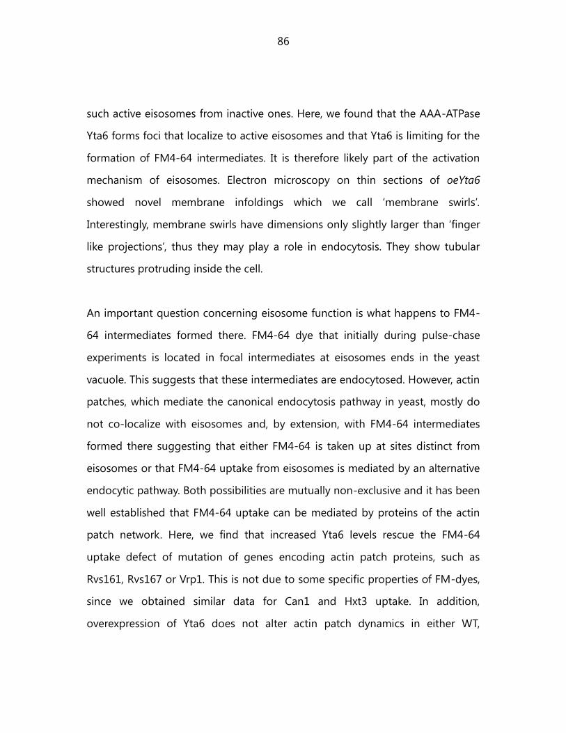
86
such active eisosomes from inactive ones. Here, we found that the AAA-ATPase
Yta6 forms foci that localize to active eisosomes and that Yta6 is limiting for the
formation of FM4-64 intermediates. It is therefore likely part of the activation
mechanism of eisosomes. Electron microscopy on thin sections of oeYta6
showed novel membrane infoldings which we call „membrane swirls‟.
Interestingly, membrane swirls have dimensions only slightly larger than „finger
like projections‟, thus they may play a role in endocytosis. They show tubular
structures protruding inside the cell.
An important question concerning eisosome function is what happens to FM4-
64 intermediates formed there. FM4-64 dye that initially during pulse-chase
experiments is located in focal intermediates at eisosomes ends in the yeast
vacuole. This suggests that these intermediates are endocytosed. However, actin
patches, which mediate the canonical endocytosis pathway in yeast, mostly do
not co-localize with eisosomes and, by extension, with FM4-64 intermediates
formed there suggesting that either FM4-64 is taken up at sites distinct from
eisosomes or that FM4-64 uptake from eisosomes is mediated by an alternative
endocytic pathway. Both possibilities are mutually non-exclusive and it has been
well established that FM4-64 uptake can be mediated by proteins of the actin
patch network. Here, we find that increased Yta6 levels rescue the FM4-64
uptake defect of mutation of genes encoding actin patch proteins, such as
Rvs161, Rvs167 or Vrp1. This is not due to some specific properties of FM-dyes,
since we obtained similar data for Can1 and Hxt3 uptake. In addition,
overexpression of Yta6 does not alter actin patch dynamics in either WT,

87
rvs161∆ or of Sla1 dynamics in latrunculin A treated cells, arguing that the
suppression is due to a bypass of the actin patch requirement. Together, these
data strongly suggest that there is an alternative endocytic pathway, which
starts at eisosomes to which Yta6 localizes. Intriguingly, only some protein
cargoes - such as Hxt3 - require this pathway for efficient uptake, whereas
others - such as Can1 or Ste3 - may use it and can access it when Yta6 is over-
expressed, but are not dependent on it under the conditions tested.
The suppression of the rvs161∆ endocytic phenotype by Yta6 overexpression is
reminiscent of its suppression by deletions of genes encoding enzymes of the
sphingolipid synthesis pathway, collectively named SUR (Suppressors of RVS-
deletions) (Germann et al., 2005). Loss of any of these genes‟ function results in
rescue of viability of rvs∆-cells upon starvation. In addition, such deletions also
suppress the endocytic defect of rvs161∆ or rvs167∆ cells. Suppression of RVS-
mutations in these cases could be due to Yta6 activation. This could be
mediated for example by sphingolipid signaling, since bioactive synthesis
intermediates of these lipids accumulate in sur mutants. In addition, it was
previously demonstrated that sphingoid base synthesis is required for the
internalization step of endocytosis and that this requirement reflects a signaling
function of sphingolipids (Friant et al., 2001; Zanolari et al., 2000). How these
findings relate to actin patch-dependent versus Yta6-dependent endocytosis is
yet unclear. Intriguingly, phosphorylation of eisosome components Pil1 and
Lsp1 by Pkh-kinases is controlled by alterations in sphingolipid metabolism,
particularly evident after a block of complex sphingolipid synthesis.
Overexpression of Pkh-kinases also rescues the sphingoid base synthesis

88
requirements for endocytosis (Friant et al., 2001). It is therefore possible that this
sphingolipid signaling network is used to trigger plasma membrane endocytosis
after specific stimuli at eisosomes and that at least part of this activation is
mediated by Yta6. Consistent with this notion, we found Yta6 phosphorylation
strongly decreased after blocking sphingolipid synthesis, maybe reflecting post-
translational regulation of the protein‟s activity (observed by Florian Fröhlich).
Activation of the Yta6-pathway by signals to take-up specific proteins would
explain why YTA6 mutation has only a modest effect on endocytosis of some
tested cargoes. Alternatively, another protein could share a redundant function
with Yta6, therefore masking its phenotype on the uptake of some proteins. A
candidate for such a protein is the highly abundant AAA-ATPase Cdc48, since it
and its adaptors Shp1 and Ufd1 are required for suppression of the rvs161∆
endocytic defects on some transporters by sphingolipid synthesis mutants
(Morgan et al., 2009). Based on known functions of AAA-ATPase domains, both
Yta6 and Cdc48 could disassemble oligomeric complexes and therefore perform
a mechanical function of disassembling complexes during endocytosis (Sauer et
al., 2004). Alternatively and not mutually exclusively, overexpression or
inactivation of Yta6 (and maybe Cdc48) may participate in signaling that in turn
activates endocytosis. Despite the evidence that endocytic defects of rvs-
mutants are suppressed by alterations in sphingolipid metabolism, it has not
been tested whether this rescue is due to a bypass or re-activation of the Rvs-
dependent, actin patch-mediated pathway. Our data now suggests that at least
Yta6-mediated suppression of endocytic defects by-passes the normal actin-

89
dependent uptake machinery because (i) Yta6-mediated uptake functions in the
absence of actin polymerization and (ii) Yta6 overexpression does not rescue the
Sla1 dynamics after actin depolymerization and (iii) Yta6 overexpression does
not restore the dynamics of Abp1-GFP labeled actin-patches in rvs161∆ cells.
What could be the physiological function of the Yta6-mediated endocytic
pathway? As YTA6 mutants do not strongly affect endocytosis under normal
conditions, it is thus likely that this pathway is activated only in certain
conditions. For example, heat shock generates a transient increase of
sphingolipid base levels that signals to trigger uptake the uracil permease Fur4
(Chung et al., 2000). Many transporters in yeast, including Fur4, localize to the
MCC. If domain organization is disrupted, turnover of some transporters is
increased. Domain organization could therefore perform two related functions:
A first function could be to sequester and protect proteins from endocytic
uptake by the constitutive actin patch machinery under normal conditions. A
second function would be to rapidly internalize transporters by the Yta6-
mediated pathway after a change in conditions, for example during stress
situations.
In yeast, only the canonical endocytic pathway mediated by clathrin and actin
patches has been described. Our data now suggest that at least part of this
pathway can be by-passed. How does the alternative pathway relate to
endocytic mechanisms in other systems? Mammalian cells have at least ten
distinguishable pathways. The closest homologues of Yta6 in mammalian cells

90
are spastin and related AAA-ATPases. In contrast to these proteins though, Yta6
does not have a motif to interact with microtubules and likely does not have
microtubule severing activity. Intriguingly though, Spastin is localized to
endosomes in mammalian cells, is required for efficient degradation of the EGF
receptor and was suggested to couple membrane trafficking to microtubule
remodeling (Bakowska et al., 2007; Connell et al., 2009). In addition, depletion of
Spastin was found to inhibit BMP-signaling and this is linked to endocytosis of
the BMP-receptor II, mediated by NIPA1. Both NIPA1 and Spastin are encoded
by genes mutated in hereditary spastic paraplegia and their trafficking functions
are likely important for the development of the disease (Fink, 2006). Yta6 might
thus share an ancient function in trafficking with these orthologous AAA-
ATPases. A better understanding of its molecular function, downstream targets
and the pathway that it activates may therefore contribute to our understanding
of the cell biological foundations of the development of these diseases.
All cargo after internalization from plasma membrane needs further trafficking
from the endosomal pathway to their final destination for either recycling or
degradation. In yeast the degradative organelle is the vacuole which performs
similar functions as lysosomes in mammalian cells. We found from E-MAP
(epistatic mini array profile) analysis of PIL1 with other genes that a protein
Emp70 has strong negative genetic interaction with eisosome component Pil1.
Emp70 is a transmembrane protein; specifically it has nine transmembrane
regions. Emp70 homologs are present in mammals and show functions related
to endocytosis. It has two more homologs in yeast - Tmn2 and Tmn3. Tmn2 has
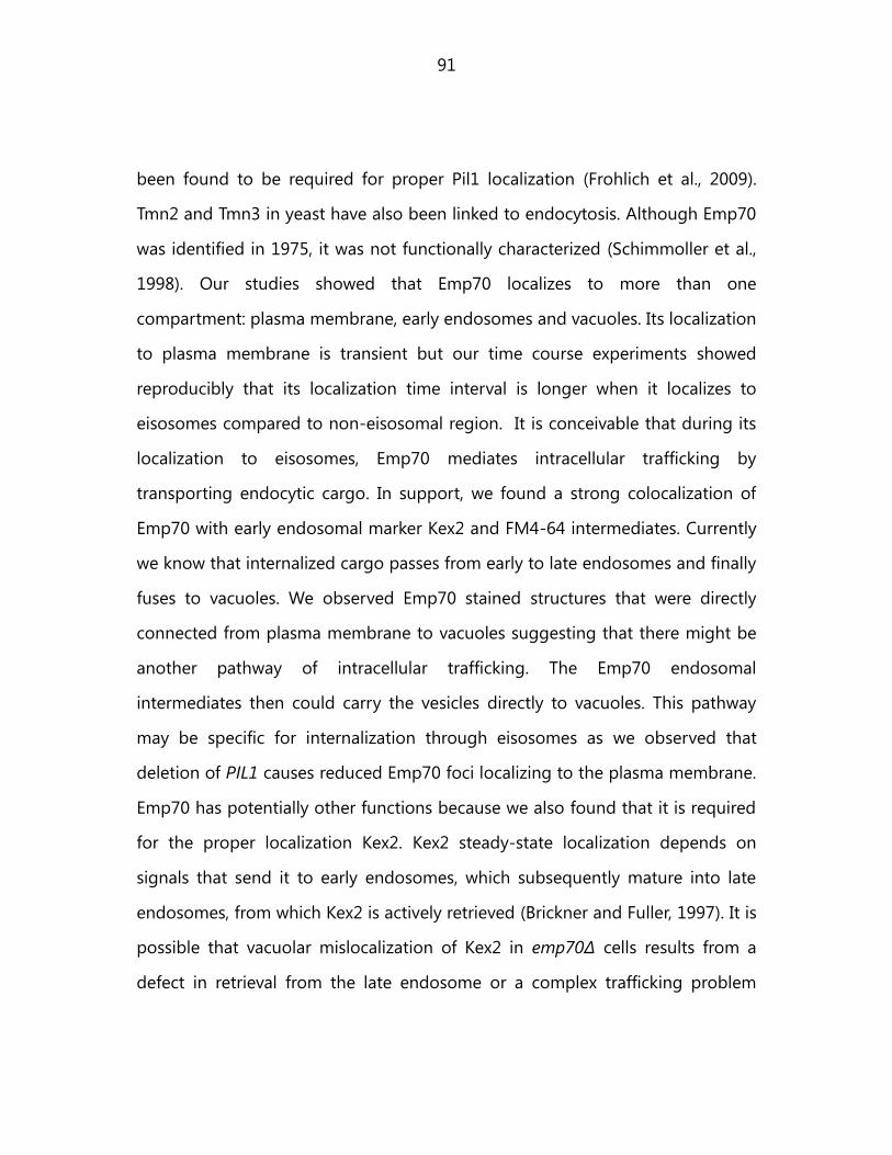
91
been found to be required for proper Pil1 localization (Frohlich et al., 2009).
Tmn2 and Tmn3 in yeast have also been linked to endocytosis. Although Emp70
was identified in 1975, it was not functionally characterized (Schimmoller et al.,
1998). Our studies showed that Emp70 localizes to more than one
compartment: plasma membrane, early endosomes and vacuoles. Its localization
to plasma membrane is transient but our time course experiments showed
reproducibly that its localization time interval is longer when it localizes to
eisosomes compared to non-eisosomal region. It is conceivable that during its
localization to eisosomes, Emp70 mediates intracellular trafficking by
transporting endocytic cargo. In support, we found a strong colocalization of
Emp70 with early endosomal marker Kex2 and FM4-64 intermediates. Currently
we know that internalized cargo passes from early to late endosomes and finally
fuses to vacuoles. We observed Emp70 stained structures that were directly
connected from plasma membrane to vacuoles suggesting that there might be
another pathway of intracellular trafficking. The Emp70 endosomal
intermediates then could carry the vesicles directly to vacuoles. This pathway
may be specific for internalization through eisosomes as we observed that
deletion of PIL1 causes reduced Emp70 foci localizing to the plasma membrane.
Emp70 has potentially other functions because we also found that it is required
for the proper localization Kex2. Kex2 steady-state localization depends on
signals that send it to early endosomes, which subsequently mature into late
endosomes, from which Kex2 is actively retrieved (Brickner and Fuller, 1997). It is
possible that vacuolar mislocalization of Kex2 in emp70Δ cells results from a
defect in retrieval from the late endosome or a complex trafficking problem

92
affecting early endosome function. Normally, if retrieval is compromised,
vacuolar sorted carboxypeptidase Y (CPY) is secreted. We tested this and found
that, in contrast to the control vps1Δ, emp70Δ alone does not lead to CPY
secretion (Robinson et al., 1988; Rothman et al., 1989). EMP70 has two
homologs in the genome, TMN2 and TMN3. To address whether they could
compensate for Emp70 function in its absence, we tested CPY secretion in
strains with different combinations of the family members deleted. TMN2
deletion alone had no effect, and TMN3 deletion alone only a weak effect, on
CPY sorting. In contrast, combining emp70Δ with either tmn2Δ or tmn2Δ tmn3Δ
resulted in CPY secretion, showing that Emp70 is functionally redundant with
Tmn2 in vacuolar protein sorting and that the Emp70 protein family is required
for normal endosomal function.

93
8. Curriculum Vitae
Personal Data
Name: Michael Rehman
Date and Place of Birth: 28.09.1981, India
Marital status: Married
Education and research experience:
2002 Bachelor in Biosciences
2004 Masters in Biotechnology
2004-2006 Junior Research Fellow, National Centre for
Biological Sciences, India
Nov 2006- Feb 2007 Research experience, Emil Fischer Zentrum,
Erlangen
Mar 2007 – present Doctorate candidate at the Max Planck
Institute of Biochemistry in the group of
Prof. Tobias C Walther.
Title of thesis: Analysis of endocytosis at
eisosomes
Publications
1. N E Ziolkowska, L Karotki, M Rehman, J T Huiskonen, T C Walther. “ Eisosome-driven
plasma membrane organization is mediated by BAR domains”. (Nature Structural and
Molecular Biology accepted)
2. M Rehman, P S Aguilar, F Frohlich, M Shales, I Ulitsky, A Olivera-Couto, H Braberg, R
Shamir, P Walter, M Mann, C S Ejsing, N J Krogan, T C Walther. “A Plasma Membrane E-MAP
Reveals Links Between the Eisosome, Sphingolipid Metabolism and Endosomal Trafficking”,
Nature Structural and Molecular Biology 17(7): 901-908 (first author – equal contribution)
3. S Srivastava, B Ramdass, S Nagarajan, M Rehman, G Mukherjee, S Krishna. "Notch1
regulates the functional contribution of RhoC to cervical carcinoma progression." Br J Cancer
102(1): 196-205. 2010
4. C Choudhary, C Kumar, F Gnad, M L Nielsen, M Rehman, T C Walther, J V Olsen, M
Mann. "Lysine acetylation targets protein complexes and co-regulates major cellular
functions." Science 325(5942): 834-840. 2009
5. B Ramdass, TT Maliekal, S Lakshmi, M Rehman, P Rema, P Nair, G Mukherjee, BK
Reddy, S Krishna, M R Pillai "Coexpression of Notch1 and NF-kappaB signaling pathway
components in human cervical cancer progression." Gynecol Oncol 104(2): 352-361.2007

94
9. Declaration of individual contributions in publications
Rehman, M., Aguilar, P. S., Frohlich, F., Shales, M., Ulitsky, I., Olivera-Couto, A.,
Braberg, H., Shamir, R., Walter, P., Mann, M., Ejsing, C. S., Krogan, N. J.,Walther, T.
C. (2010). "A plasma-membrane E-MAP reveals links of the eisosome with
sphingolipid metabolism and endosomal trafficking." Nat Struct Mol Biol 17(7):
901-908. (MR, PSA, FF, MS – equally contributed)
Michael Rehman was responsible for performing all experiments related
to Emp70 and Pil1 genetic interaction (Fig 5, Fig 6).
Choudhary, C., Kumar, C., Gnad, F., Nielsen, M. L., Rehman, M., Walther, T. C.,
Olsen, J. V., Mann, M. (2009). "Lysine acetylation targets protein complexes and
co-regulates major cellular functions." Science 325(5942): 834-840.
Michael Rehman was responsible for performing experiments of
functional importance of Cdc28 K40 acetylation (Fig 4a). He was partially
involved in the experiments related to Fig S7, where Cdc28-GFP pulldown
was required for mass spectrometry analysis of its acetylation.
N E Ziolkowska, L Karotki, M Rehman, J T Huiskonen, T C Walther. “Eisosome-
driven plasma membrane organization is mediated by BAR domains”. Nat Struct
Mol Biol
Michael Rehman was responsible for bioinformatics analysis figures: Fig
2b, supplementary figure 3. He was also involved in all the yeast work with
NE Ziolkowska.

95
10. Acknowledgements
This thesis arose out of research that was done in the Walther laboratory from
2007 to 2011. It gives me a great pleasure to acknowledge all the people who
helped me during this period. In the first place I would like to thank Dr Tobias C
Walter for his wonderful supervision. Before joining Walther laboratory I had
gained research experience in India for two years but it not teach me how to
design a good experiment. Tobi taught me what types of questions are more
important to ask, what are good hypotheses, the importance of making models
in science and last but not the least the importance of presenting the work to
the outside world. I have already started to apply many of his teachings and
they will be remembered for many years from now.
Walther lab also provided a very scientific and friendly atmosphere during those
years. I would like to thanks Ulli and Peter for their excellent technical help. Ulli
almost got me started into yeast gene tagging by helping me to obtain the
endogenous Yta6 GFP tagged strain. I am grateful to Doris who helped me with
many microscopy related issues. Her enthusiasm and care of the microscopy
setup enabled us to have more data and fewer frustrations. Florian Frohlich‟s
cheerfulness and helpful nature enabled me to get through with SILAC based
proteomics experiment. I am also thankful to Pablo Aguilar, Lena, Pablo
Mardones, Natalie, Dilip, Jerry, Alvaro, Tina, Anoop, Giesla, Nikola, Tina, Shravan,
Christian, Georgios, Romain and Flo W for many discussions. I want to specially
thank Felix for many discussions and kicker games mixed with further
discussions which made Giesla confused if we were playing or brain-storming. I
96
gratefully acknowledge Dr Matthias Mann and Dr Chunaram Choudhary for the
collaboration work and support.
As is true for any endeavor, my research period also had its tough times. I want
to take this opportunity to thank my parents, Elena, relatives and Roshan for
their love, care, support and being there at all times.

97
11. References
Aghamohammadzadeh, S., and K.R. Ayscough. 2009. Differential requirements
for actin during yeast and mammalian endocytosis. Nat Cell Biol. 11:1039-1042.
Aguilar, P.S., F. Frohlich, M. Rehman, M. Shales, I. Ulitsky, A. Olivera-Couto, H.
Braberg, R. Shamir, P. Walter, M. Mann, C.S. Ejsing, N.J. Krogan, and T.C. Walther.
2010. A plasma-membrane E-MAP reveals links of the eisosome with
sphingolipid metabolism and endosomal trafficking. Nat Struct Mol Biol. 17:901-
908.
Alberts, B., Johnson, A., Lewis, J, Raff, M., Roberts, K., and Walter, Peter. 2002.
Molecular biology of the cell. . Garland science. 4th edition.
Allen, J.A., R.A. Halverson-Tamboli, and M.M. Rasenick. 2007. Lipid raft
microdomains and neurotransmitter signalling. Nat Rev Neurosci. 8:128-140.
Ammelburg, M., T. Frickey, and A.N. Lupas. 2006. Classification of AAA+ proteins.
J Struct Biol. 156:2-11.
Ayscough, K.R. 2005. Defining protein modules for endocytosis. Cell. 123:188-
190.
Bakowska, J.C., H. Jupille, P. Fatheddin, R. Puertollano, and C. Blackstone. 2007.
Troyer syndrome protein spartin is mono-ubiquitinated and functions in EGF
receptor trafficking. Mol Biol Cell. 18:1683-1692.
Beach, D.L., and K. Bloom. 2001. ASH1 mRNA localization in three acts. Mol Biol
Cell. 12:2567-2577.
Berchtold, D., and T.C. Walther. 2009. TORC2 plasma membrane localization is
essential for cell viability and restricted to a distinct domain. Mol Biol Cell.
20:1565-1575.

98
Brickner, J.H., and R.S. Fuller. 1997. SOI1 encodes a novel, conserved protein that
promotes TGN-endosomal cycling of Kex2p and other membrane proteins by
modulating the function of two TGN localization signals. J Cell Biol. 139:23-36.
Chung, N., G. Jenkins, Y.A. Hannun, J. Heitman, and L.M. Obeid. 2000.
Sphingolipids signal heat stress-induced ubiquitin-dependent proteolysis. J Biol
Chem. 275:17229-17232.
Connell, J.W., C. Lindon, J.P. Luzio, and E. Reid. 2009. Spastin couples microtubule
severing to membrane traffic in completion of cytokinesis and secretion. Traffic.
10:42-56.
Dalal, S., M.F. Rosser, D.M. Cyr, and P.I. Hanson. 2004. Distinct roles for the AAA
ATPases NSF and p97 in the secretory pathway. Mol Biol Cell. 15:637-648.
Douglas, L.M., S.W. Martin, and J.B. Konopka. 2009. BAR domain proteins Rvs161
and Rvs167 contribute to Candida albicans endocytosis, morphogenesis, and
virulence. Infect Immun. 77:4150-4160.
Drubin, D.G., K.G. Miller, and D. Botstein. 1988. Yeast actin-binding proteins:
evidence for a role in morphogenesis. J Cell Biol. 107:2551-2561.
Dupre, S., D. Urban-Grimal, and R. Haguenauer-Tsapis. 2004. Ubiquitin and
endocytic internalization in yeast and animal cells. Biochim Biophys Acta.
1695:89-111.
Engqvist-Goldstein, A.E., and D.G. Drubin. 2003. Actin assembly and endocytosis:
from yeast to mammals. Annu Rev Cell Dev Biol. 19:287-332.
Fink, J.K. 2006. Hereditary spastic paraplegia. Curr Neurol Neurosci Rep. 6:65-76.
Friant, S., R. Lombardi, T. Schmelzle, M.N. Hall, and H. Riezman. 2001. Sphingoid
base signaling via Pkh kinases is required for endocytosis in yeast. EMBO J.
20:6783-6792.

99
Frickey, T., and A.N. Lupas. 2004. Phylogenetic analysis of AAA proteins. J Struct
Biol. 146:2-10.
Frohlich, F., K. Moreira, P.S. Aguilar, N.C. Hubner, M. Mann, P. Walter, and T.C.
Walther. 2009. A genome-wide screen for genes affecting eisosomes reveals
Nce102 function in sphingolipid signaling. J Cell Biol. 185:1227-1242.
Frye, L.D., and M. Edidin. 1970. The rapid intermixing of cell surface antigens
after formation of mouse-human heterokaryons. J Cell Sci. 7:319-335.
Germann, M., E. Swain, L. Bergman, and J.T. Nickels, Jr. 2005. Characterizing the
sphingolipid signaling pathway that remediates defects associated with loss of
the yeast amphiphysin-like orthologs, Rvs161p and Rvs167p. J Biol Chem.
280:4270-4278.
Grossmann, G., M. Opekarova, J. Malinsky, I. Weig-Meckl, and W. Tanner. 2007.
Membrane potential governs lateral segregation of plasma membrane proteins
and lipids in yeast. EMBO J. 26:1-8.
Habermann, B. 2004. The BAR-domain family of proteins: a case of bending and
binding? EMBO Rep. 5:250-255.
Hanson, P.I., and S.W. Whiteheart. 2005. AAA+ proteins: have engine, will work.
Nat Rev Mol Cell Biol. 6:519-529.
Hartman, J.J., J. Mahr, K. McNally, K. Okawa, A. Iwamatsu, S. Thomas, S.
Cheesman, J. Heuser, R.D. Vale, and F.J. McNally. 1998. Katanin, a microtubule-
severing protein, is a novel AAA ATPase that targets to the centrosome using a
WD40-containing subunit. Cell. 93:277-287.
Heese-Peck, A., H. Pichler, B. Zanolari, R. Watanabe, G. Daum, and H. Riezman.
2002. Multiple functions of sterols in yeast endocytosis. Mol Biol Cell. 13:2664-
2680.

100
Higuchi, Y., M. Arioka, and K. Kitamoto. 2011. Functional analysis of the putative
AAA ATPase AipA localizing at the endocytic sites in the filamentous fungus
Aspergillus oryzae. FEMS Microbiol Lett.
Hurley, J.H., and S.D. Emr. 2006. The ESCRT complexes: structure and mechanism
of a membrane-trafficking network. Annu Rev Biophys Biomol Struct. 35:277-298.
Janke, C., M.M. Magiera, N. Rathfelder, C. Taxis, S. Reber, H. Maekawa, A.
Moreno-Borchart, G. Doenges, E. Schwob, E. Schiebel, and M. Knop. 2004. A
versatile toolbox for PCR-based tagging of yeast genes: new fluorescent
proteins, more markers and promoter substitution cassettes. Yeast. 21:947-962.
Jonsdottir, G.A., and R. Li. 2004. Dynamics of yeast Myosin I: evidence for a
possible role in scission of endocytic vesicles. Curr Biol. 14:1604-1609.
Kaksonen, M., C.P. Toret, and D.G. Drubin. 2005. A modular design for the
clathrin- and actin-mediated endocytosis machinery. Cell. 123:305-320.
Karp, G. 2004. Cell and Molecular biology: Concepts and experiments.
Katzmann, D.J., G. Odorizzi, and S.D. Emr. 2002. Receptor downregulation and
multivesicular-body sorting. Nat Rev Mol Cell Biol. 3:893-905.
Kilmartin, J.V., and A.E. Adams. 1984. Structural rearrangements of tubulin and
actin during the cell cycle of the yeast Saccharomyces. J Cell Biol. 98:922-933.
Langer, T. 2000. AAA proteases: cellular machines for degrading membrane
proteins. Trends Biochem Sci. 25:247-251.
Lin, C.H., J.A. MacGurn, T. Chu, C.J. Stefan, and S.D. Emr. 2008. Arrestin-related
ubiquitin-ligase adaptors regulate endocytosis and protein turnover at the cell
surface. Cell. 135:714-725.
Lingwood, D., and K. Simons. 2010. Lipid rafts as a membrane-organizing
principle. Science. 327:46-50.
101
Lombardi, R., and H. Riezman. 2001. Rvs161p and Rvs167p, the two yeast
amphiphysin homologs, function together in vivo. J Biol Chem. 276:6016-6022.
Longtine, M.S., A. McKenzie, 3rd, D.J. Demarini, N.G. Shah, A. Wach, A. Brachat, P.
Philippsen, and J.R. Pringle. 1998. Additional modules for versatile and
economical PCR-based gene deletion and modification in Saccharomyces
cerevisiae. Yeast. 14:953-961.
Malinska, K., J. Malinsky, M. Opekarova, and W. Tanner. 2003. Visualization of
protein compartmentation within the plasma membrane of living yeast cells.
Mol Biol Cell. 14:4427-4436.
Malinska, K., J. Malinsky, M. Opekarova, and W. Tanner. 2004. Distribution of
Can1p into stable domains reflects lateral protein segregation within the plasma
membrane of living S. cerevisiae cells. J Cell Sci. 117:6031-6041.
Mayor, S., and M. Rao. 2004. Rafts: scale-dependent, active lipid organization at
the cell surface. Traffic. 5:231-240.
McCourt, P.C., J.M. Morgan, and J.T. Nickels, Jr. 2009. Stress-induced ceramide-
activated protein phosphatase can compensate for loss of amphiphysin-like
activity in Saccharomyces cerevisiae and functions to reinitiate endocytosis. J
Biol Chem. 284:11930-11941.
Morgan, J., P. McCourt, L. Rankin, E. Swain, L.M. Rice, and J.T. Nickels, Jr. 2009.
Altering sphingolipid metabolism in Saccharomyces cerevisiae cells lacking the
amphiphysin ortholog Rvs161 reinitiates sugar transporter endocytosis.
Eukaryot Cell. 8:779-789.
Mullins, C., and J.S. Bonifacino. 2001. Structural requirements for function of
yeast GGAs in vacuolar protein sorting, alpha-factor maturation, and
interactions with clathrin. Mol Cell Biol. 21:7981-7994.
102
Noguchi, C., and E. Noguchi. 2007. Sap1 promotes the association of the
replication fork protection complex with chromatin and is involved in the
replication checkpoint in Schizosaccharomyces pombe. Genetics. 175:553-566.
Orlando, K., and W. Guo. 2009. Membrane organization and dynamics in cell
polarity. Cold Spring Harb Perspect Biol. 1:a001321.
Patton, J.L., and R.L. Lester. 1991. The phosphoinositol sphingolipids of
Saccharomyces cerevisiae are highly localized in the plasma membrane. J
Bacteriol. 173:3101-3108.
Robinson, J.S., D.J. Klionsky, L.M. Banta, and S.D. Emr. 1988. Protein sorting in
Saccharomyces cerevisiae: isolation of mutants defective in the delivery and
processing of multiple vacuolar hydrolases. Mol Cell Biol. 8:4936-4948.
Rothman, J.H., I. Howald, and T.H. Stevens. 1989. Characterization of genes
required for protein sorting and vacuolar function in the yeast Saccharomyces
cerevisiae. EMBO J. 8:2057-2065.
Sauer, R.T., D.N. Bolon, B.M. Burton, R.E. Burton, J.M. Flynn, R.A. Grant, G.L.
Hersch, S.A. Joshi, J.A. Kenniston, I. Levchenko, S.B. Neher, E.S. Oakes, S.M.
Siddiqui, D.A. Wah, and T.A. Baker. 2004. Sculpting the proteome with AAA(+)
proteases and disassembly machines. Cell. 119:9-18.
Schimmoller, F., E. Diaz, B. Muhlbauer, and S.R. Pfeffer. 1998. Characterization of
a 76 kDa endosomal, multispanning membrane protein that is highly conserved
throughout evolution. Gene. 216:311-318.
Schuck, S., M. Honsho, K. Ekroos, A. Shevchenko, and K. Simons. 2003.
Resistance of cell membranes to different detergents. Proc Natl Acad Sci U S A.
100:5795-5800.
Simons, K., and M.J. Gerl. 2010. Revitalizing membrane rafts: new tools and
insights. Nat Rev Mol Cell Biol. 11:688-699.

103
Simons, K., and W.L. Vaz. 2004. Model systems, lipid rafts, and cell membranes.
Annu Rev Biophys Biomol Struct. 33:269-295.
Singer, S.J., and G.L. Nicolson. 1972. The fluid mosaic model of the structure of
cell membranes. Science. 175:720-731.
Smith, M.G., S.R. Swamy, and L.A. Pon. 2001. The life cycle of actin patches in
mating yeast. J Cell Sci. 114:1505-1513.
Stradalova, V., W. Stahlschmidt, G. Grossmann, M. Blazikova, R. Rachel, W. Tanner,
and J. Malinsky. 2009. Furrow-like invaginations of the yeast plasma membrane
correspond to membrane compartment of Can1. J Cell Sci. 122:2887-2894.
Tabuchi, M., A. Audhya, A.B. Parsons, C. Boone, and S.D. Emr. 2006. The
phosphatidylinositol 4,5-biphosphate and TORC2 binding proteins Slm1 and
Slm2 function in sphingolipid regulation. Mol Cell Biol. 26:5861-5875.
Valdez-Taubas, J., and H.R. Pelham. 2003. Slow diffusion of proteins in the yeast
plasma membrane allows polarity to be maintained by endocytic cycling. Curr
Biol. 13:1636-1640.
Vale, R.D. 2000. AAA proteins. Lords of the ring. J Cell Biol. 150:F13-19.
van der Rest, M.E., A.H. Kamminga, A. Nakano, Y. Anraku, B. Poolman, and W.N.
Konings. 1995. The plasma membrane of Saccharomyces cerevisiae: structure,
function, and biogenesis. Microbiol Rev. 59:304-322.
van Meer, G. 2002. Cell biology. The different hues of lipid rafts. Science.
296:855-857.
Vanderkooi, G., and D.E. Green. 1970. Biological membrane structure, I. The
protein crystal model for membranes. Proc Natl Acad Sci U S A. 66:615-621.
Vereb, G., J. Szollosi, J. Matko, P. Nagy, T. Farkas, L. Vigh, L. Matyus, T.A.
Waldmann, and S. Damjanovich. 2003. Dynamic, yet structured: The cell

104
membrane three decades after the Singer-Nicolson model. Proc Natl Acad Sci U
S A. 100:8053-8058.
Wachtler, V., and M.K. Balasubramanian. 2006. Yeast lipid rafts?--an emerging
view. Trends Cell Biol. 16:1-4.
Wachtler, V., S. Rajagopalan, and M.K. Balasubramanian. 2003. Sterol-rich plasma
membrane domains in the fission yeast Schizosaccharomyces pombe. J Cell Sci.
116:867-874.
Walther, T.C., P.S. Aguilar, F. Frohlich, F. Chu, K. Moreira, A.L. Burlingame, and P.
Walter. 2007. Pkh-kinases control eisosome assembly and organization. EMBO J.
26:4946-4955.
Walther, T.C., J.H. Brickner, P.S. Aguilar, S. Bernales, C. Pantoja, and P. Walter. 2006.
Eisosomes mark static sites of endocytosis. Nature. 439:998-1003.
Wang, H., B. Kakaradov, S.R. Collins, L. Karotki, D. Fiedler, M. Shales, K.M. Shokat,
T.C. Walther, N.J. Krogan, and D. Koller. 2009. A complex-based reconstruction of
the Saccharomyces cerevisiae interactome. Mol Cell Proteomics. 8:1361-1381.
Wendland, B., S.D. Emr, and H. Riezman. 1998. Protein traffic in the yeast
endocytic and vacuolar protein sorting pathways. Curr Opin Cell Biol. 10:513-
522.
Wendland, B., J.M. McCaffery, Q. Xiao, and S.D. Emr. 1996. A novel fluorescence-
activated cell sorter-based screen for yeast endocytosis mutants identifies a
yeast homologue of mammalian eps15. J Cell Biol. 135:1485-1500.
White, S.R., and B. Lauring. 2007. AAA+ ATPases: achieving diversity of function
with conserved machinery. Traffic. 8:1657-1667.
Youn, J.Y., H. Friesen, T. Kishimoto, W.M. Henne, C.F. Kurat, W. Ye, D.F. Ceccarelli, F.
Sicheri, S.D. Kohlwein, H.T. McMahon, and B.J. Andrews. 2010. Dissecting BAR

105
domain function in the yeast Amphiphysins Rvs161 and Rvs167 during
endocytosis. Mol Biol Cell. 21:3054-3069.
Zanolari, B., S. Friant, K. Funato, C. Sutterlin, B.J. Stevenson, and H. Riezman.
2000. Sphingoid base synthesis requirement for endocytosis in Saccharomyces
cerevisiae. EMBO J. 19:2824-2833.