Analog Model for Gaze-Evoked Nystagmus - OMLABomlab.org/Personnel/lfd/Jrnl_Arts/025_Analog Model for...
5
IEEE TRANSACTIONS ON BIO MEDICAL ENGINEER ING, VOL. BME-25, NO.1, JANUARY 1978 71 Analog Model for Gaze-Evoked Nystagmus L A. ABEL, MEMBER, IEEE, L F_ DELL'OSSO, SENIOR MEMBER , IEEE, AND K R DAROFF Abstract-A relatively simple model of the fast eye movement (sac- cadic) system has been developed and simulated on an analog computer. e model is capable of producing normal saccadic eye movements and has several provisions which permit the simulaon of clinical sis of ge-evoked nystaus. According to the model, gaze-evoked nystag- mus may result from various abnoalities in the neuron pool respon- sible for inteation of the pulse of high frequency firing which initiates each saccade. Simulation of these inferred neuronal deficits produced subtle differences in saccadic behavior as well as gaze-evoked nystaus. These differences occurred both in the nystaus-free range of gaze angles and after the nystagmus appeared. The subtlety of the deficits and the sparsity of studies of gaze-evoked nystaus using modem oculographic recording techniques, probably explains why such saccadic behavior has not been previously noted. If one, or more than one, of these simulated mechanisms is, in fact, an accurate model of the actual physiolocal deficit, future studies of such paents should reveal the corresponding abnormal saccadic behavior. L INTRODUCTION I N recent years bioengineers have studied eye movements with considerable interest. The ability to precisely control the visual input signal and the feasibility of quantitative mea- surement of the eye movement output structures makes it an ideal motor system for analysis_ As the physiological mecha- nisms of ocular motor control unravel, model construction with components corresponding to the actual biological system become more feasible_ Manipulation of various parameters of the model can then be utilized to provide valuable insights into the behavior of the actual ocular motOl: system in diseased states. Zee and associates utilized computer simulation to explain the possible mechanisms of both down-beating nystag- mus [1] and slow saccades [2] _ A similar application led Ochs et a1. [3] to postulate a change in saccadic gain as the under- lying defect of certain ocular oscillations consequent to cerebellar disease. The ability to simulate known ocular disturbances by model manipulation affords unique potential for model validation_ A model could hardly be tenable if alterations did not result in naturally occurring abnormalities_ Whereas the aforementioned models were directed at unusual, albeit important, ocular motor defects, we will report on our study of gaze-evoked nystaus, the most common ocular motor abnormality encountered in clinical practice_ A brief nosological discussion is provided for clarification. Nystagmus is an ocular oscillation divided into pendular and jerk types depending upon waveform characteristics [4] _ The "jerk" refers to the faster saccadic phase which brings the eyes back to the intended spatial position after the deviation created by the slow phase_ As distinct from "fixation" nystag- mus, which is present in primary (straight-ahead) position and is often pendular, gaze-evoked nystagmus occurs when the eyes are maintained in an eccentric (gaze) position, is invariably of the jerk type, and usually persists as long as the deviated position is intended_ Patients who suffer a transient paralysis of gaze in a given direction recover in a characteristic fashion, as noted by clinical observation [5] _ The eyes seem unable to maintain gaze in the direction of the previous paralysis, drift slowly toward primary position, and a corrective saccade repositions the eyes eccentrically_ Repetition of this pattern results in a nystagmus, aptly designated "gaze-paretic." Gaze-evoked nystagmus is not always "paretic," prompting Jung and Kornhuber [6], utilizing oculographic studies, to define gaze-paretic nystag- mus as that type of gaze nystagmus with a low (1-2 Hz) frequency. Determination that a pulse increase in neuronal firing fre- quency was responsible for the initiation of saccades and that its integral, the step, was the firing pattern responsible for maintaining ocular deviation [7], [8], inspired the obvious conclusion that an inadequate step must account for the slow phase of gaze-paretic nystagmus. Furthermore, the orbital plant dynamics [9] required the slow phase to be a decreasing velocity exponential [10] as distinct from the linear slow phase of vestibular nystagmus. The two extant criteria (frequency less than 2 Hz and exponential slow phase), however, are not invariably associated. Many high frequency nystagmus patterns have the paretic-type slow phases and 1-2 Hz nystagmus may have linear slow phases, obscuring the specificity of "gaze-paretic" nystaus. Indeed, Kornhuber [11] and Dichgans and lung [12] have refrained from defining precisely the "paretic" variety of gaze-evoked nystagmus; the latter designating "large amplitude" as the distinguishing factor. It was upon this background of imprecise clinical conceptual- ization, as well as the evolution of eye movement modeling from Young and Stark [13] to Robinson [14], that we began to consider the problem of modeling saccadic generation, eye position maintenance, and gaze-evoked nystagmus. We under- took the construction of an analog computer model that could simulate normal eye movements and produce the nystagmus in response to alterations corresponding to pathological conditions. II. MODEL The model herein described is a simplified representation of
Transcript of Analog Model for Gaze-Evoked Nystagmus - OMLABomlab.org/Personnel/lfd/Jrnl_Arts/025_Analog Model for...
IEEE TRANSACTIONS ON BIO MEDICAL ENGINEER ING, VOL. BME-25, NO.1, JANUARY 1978 71
Analog Model for Gaze-Evoked Nystagmus
L A. ABEL, MEMBER, IEEE, L F_ DELL'OSSO, SENIOR MEMBER , IEEE, AND K R DAROFF
Abstract-A relatively simple model of the fast eye movement (sac
cadic) system has been developed and simulated on an analog computer. The model is capable of producing normal saccadic eye movements and has several provisions which permit the simulation of clinical signs of gaze-evoked nystagmus. According to the model, gaze-evoked nystagmus may result from various abnormalities in the neuronal pool responsible for integration of the pulse of high frequency firing which initiates each saccade. Simulation of these inferred neuronal deficits produced subtle differences in saccadic behavior as well as gaze-evoked nystagmus. These differences occurred both in the nystagmus-free range of gaze angles and after the nystagmus appeared. The subtlety of the deficits and the sparsity of studies of gaze-evoked nystagmus using modem oculographic recording techniques, probably explains why such saccadic
behavior has not been previously noted. If one, or more than one, of
these simulated mechanisms is, in fact, an accurate model of the actual physiological deficit, future studies of such patients should reveal the corresponding abnormal saccadic behavior.
L INTRODUCTION
IN recent years bioengineers have studied eye movements with considerable interest. The ability to precisely control
the visual input signal and the feasibility of quantitative measurement of the eye movement output structures makes it an ideal motor system for analysis_ As the physiological mechanisms of ocular motor control unravel, model construction with components corresponding to the actual biological system become more feasible_ Manipulation of various parameters of the model can then be utilized to provide valuable insights into the behavior of the actual ocular motOl: system in diseased states. Zee and associates utilized computer simulation to explain the possible mechanisms of both down-beating nystagmus [1] and slow saccades [2] _ A similar application led Ochs et a1. [3] to postulate a change in saccadic gain as the underlying defect of certain ocular oscillations consequent to cerebellar disease. The ability to simulate known ocular disturbances by model manipulation affords unique potential for model validation_ A model could hardly be tenable if alterations did not result in naturally occurring abnormalities_
Whereas the aforementioned models were directed at unusual, albeit important, ocular motor defects, we will report on our study of gaze-evoked nystagmus, the most common ocular motor abnormality encountered in clinical practice_ A brief nosological discussion is provided for clarification.
Nystagmus is an ocular oscillation divided into pendular and jerk types depending upon waveform characteristics [4] _ The "jerk" refers to the faster saccadic phase which brings the eyes back to the intended spatial position after the deviation
created by the slow phase_ As distinct from "fixation" nystagmus, which is present in primary (straight-ahead) position and is often pendular, gaze-evoked nystagmus occurs when the eyes are maintained in an eccentric (gaze) position, is invariably of the jerk type, and usually persists as long as the deviated position is intended_
Patients who suffer a transient paralysis of gaze in a given direction recover in a characteristic fashion, as noted by clinical observation [5] _ The eyes seem unable to maintain gaze in the direction of the previous paralysis, drift slowly toward primary position, and a corrective saccade repositions the eyes eccentrically_ Repetition of this pattern results in a nystagmus, aptly designated "gaze-paretic." Gaze-evoked nystagmus is not always "paretic," prompting Jung and Kornhuber [6], utilizing oculographic studies, to define gaze-paretic nystagmus as that type of gaze nystagmus with a low (1-2 Hz) frequency.
Determination that a pulse increase in neuronal firing frequency was responsible for the initiation of saccades and that its integral, the step, was the firing pattern responsible for maintaining ocular deviation [7], [8], inspired the obvious conclusion that an inadequate step must account for the slow phase of gaze-paretic nystagmus. Furthermore, the orbital plant dynamics [9] required the slow phase to be a decreasing velocity exponential [10] as distinct from the linear slow phase of vestibular nystagmus. The two extant criteria (frequency less than 2 Hz and exponential slow phase), however, are not invariably associated. Many high frequency nystagmus patterns have the paretic-type slow phases and 1-2 Hz nystagmus may have linear slow phases, obscuring the specificity of "gaze-paretic" nystagmus. Indeed, Kornhuber [11] and Dichgans and lung [12] have refrained from defining precisely the "paretic" variety of gaze-evoked nystagmus; the latter designating "large amplitude" as the distinguishing factor.
It was upon this background of imprecise clinical conceptualization, as well as the evolution of eye movement modeling from Young and Stark [13] to Robinson [14], that we began to consider the problem of modeling saccadic generation, eye position maintenance, and gaze-evoked nystagmus. We undertook the construction of an analog computer model that could simulate normal eye movements and produce the nystagmus in response to alterations corresponding to pathological conditions.
II. MODEL
The model herein described is a simplified representation of
72 IEEE TRANSACTIONS ON BIOMEDICAL ENGINEERING, VOL. BME-25, NO.1, JANUARY 1978
PULSE GENERATOR
threshold INTERNAL
8T ___ -� MONITOR
�I+
'""'>-t--.� N E U R A L
+ INTEGRATOR
LVVW:::=J leak
inhibition
eaClual
PLANT
S
edestled SATURAT ION
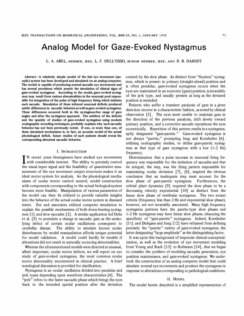
Fig. 1. Block diagram of the model used to simulate gaze-evoked nystagmus. The input target (IJT) is compared by the internal monitor to the eye position command (IJCMD) and the necessary saccade initiated in the pulse generator. The monitor contains an error threshold below which no correction is called for. It also is prevented by a refractory circuit from calling for a correction during a sllc�ll�e. Ti!e pulse generator c6iltiU!ls nonllnearities that reproduce the physiological variations in pulse height and duration. The output of the pulse generator is integrated in the neural integrator. The pulse and step are summed, with the resulting innervation used to drive the plant to produce the desired eye position (IJ E). Both a leaky integrator and saturating integrator (SAT) are indicated as ways to produce gaze-evoked nystagmus. The inset shows typical saturation characteristics used (soft, S; intermediate, J; hard, H).
Fig. 2. Analog computer simulation· of gaze-evoked nystagmus. The pulse generator is simulated by Al-A3, 11, 12, Cl, C3, NLl, NL2 and Kl, the neural integrator pool by A4 -A6, the summing junction by A 7 and the plant by AS and A9. The internal monitor consists of AID, C2, C5 and associated circuitry. Sl applies a target input (IJT) and S2 initiates a saccade (IJ E). The frequency of gaze-evoked nystagmus is set by T.
increase in the firing rate of neurons in the pontine paramedian reticular formation (PPRF) [16]. The pulse of increased firing rate is then integrated, possibly also in the PPRF, producing the step increase. The pulse and step are then summed at an unknown pre-motoneuronal site and activate the agonist muscles to initiate and complete the saccade. The neural activity is measured by an "internal monitor" [17], which can institute a correction if the command is insufficient to generate the desired eye movement.
Our model is capable of unidirectional, conjugate versional eye movement only, a limitation imposed solely to conserve computer modules. Given additional computer capacity, we could readily make the model capable of bidirectional movements. The results herein presented can be appropriately generalized. We will present the actual implementation of the different elements of the block diagram, beginning with the pulse generator (Fig. 2).
The pulse generator (Al-A3, 11, 12, Cl, C3, NLl, NL2 and
ABEL et al.: GAZE·EVOKED NYSTAGMUS
Kl) is the most complex component of the model. It is a
bootstrap circuit, sampling the error at the output of the internal monitor, operating until this error i� reduced to zero, and then turning itself off until the next saccade is required. The voltage levels correspond to neural firing rates in the pulse generator, neural integrator (A4, AS) and summing stages (A6, A 7). A partial exception is the nonlinearity which determines pulse height, NL2; this includes both the increased firing rate present in each motoneuron and the recruitment of additional neurons as saccadic size increases. Another nonlinearity, NLl, controls the pulse duration as a function of saccadic size. Both height and duration can be independently adjusted to produce physiological saccades.
The output of the pulse generator serves as the input to the neural integrator which, when functioning normally, produces a step output whose height is proportional to the desired gaze angles. This is modeled by two integrators with individually adjustable outputs summed together. We divided the integrator neuronal pool into two parts to permit simulation of pathological changes affecting only a portion of the neurons. Each integrator has associated control elements monitoring its output and placing it in the hold state as long as the proper level is maintained.
The output of the neural integrator stage is summed with the pulse and the resulting pulse-step of innervation drives the extraocular muscles. The eye "plant" is represented as a linear second order lag system [14] with one fast and one slow time constant, the characteristics of which are reflected in the exponential slow phase of the nystagmus produced by the model.
Although there is compelling evidence for the existence of an internal monitor [17], [18] the intricacies of its function can only be surmised. The monitor could provide a mechanism for the faster initiation of corrective eye movements than possible with visual feedback alone. In the model, the monitor compares the output of the neural integrator with the desired eye position, calling for a corrective saccade if the error exceeds its threshold level. Also included are the comparators responsible for determining whether the elements of the neural integrator are at their proper output and, if so, maintaining that level. Small errors may be ignored by the monitor and left uncorrected. Clinically the visual loop would initiate the corrective saccade when the error reached a critical size.
III. PATHOLOGY
Various defects in the functioning of the neural integrator may cause gaze-evoked nystagmus. One such abnormality would be the inability of a partial population of the constituent neurons to maintain the firing rate reached after integration of the pulse; that is, their output frequency would decay back toward zero. We simulated this in the model by placing a resistor in parallel with the integrator capacitor, producing a "leaky integrator " [19]. A saccade made by a system so affected will initially reach the proper gaze angle but drift back to a more medial position determined by the percent of non-leaky integrators. With persisting refixational effort, this error will be sensed by the internal monitor when it next samples the step level (the sum of the outputs of leaky, and non-leaky integrators) and a command to make a corrective saccade will be issued. In this manner, gaze-evoked nystagmus
73
will be produced, the amplitude of which will be a function of gaze angle. The greater the population of defective integrator neurons, the farther will the drift be back toward primary position, thus giving the appearance of what Dichgans and Jung [12] would probably call "gaze-paretic" nystagmus.
Another possible nystagmus-causing defect of the integrator would be the production of a less than normal step size in response to a pulse input. This might result either from an inability of the integrator neurons to achieve the proper firing rate or from a failure in recruitment reflected by fewer active neurons at a given gaze angle than is required.
A third possible defect (suggested by David Robinson in a personal communication) would be an integrator neuronal pool functioning normally for &.
az� angles below a certain value, but producing inadequate firing frequencies for larger deviations. These saturating integrators would lead to a model with abnormal behavior only at gaze angles where the integrator response was inadequate. The point at which saturation begins would determine the nystagmus-free gaze zone.
Although the conditions described above are all capable of producing gaze-evoked nystagmus, each predicts different behavior both in the nystagmus field and the region of gaze without nystagmus.
1. When a small percentage of the integrator neuronal pool is leaky, a nystagmus would result where the slow phases of eye movement would decay to a more central steady state gaze angle determined by the percentage of normal remaining integrator neurons. Furthermore, with a small proportion of leaky neurons, there would be an appreciable region of gaze without nystagmus, since the error might be less than the dead zone of the internal monitor. Such a small leak would lead to a mismatch between the pulse and step in the nystagmusfree area, resulting in apparent slight overshooting of saccades within this range. The overshoot is only apparent, for the eyes initially reach the target and then drift back to a more central position. The size of the nystagmus-free field of gaze would be a function of gaze angle, the percentage of leaky integrator neurons, and the size of the internal monitor dead zone.
2. When all the integrator neurons have large leaks, there would be no step component of innervation to the extraocular muscles. The eyes would drift back toward primary position even after very small amplitude gaze deviations. Nystagmus would ensue when the dead zone of the internal monitor was exceeded and corrective saccade initiated. If the time constant of the leak were shorter than that of the eye plant, the nystagmus waveform would be an exponential decay determined by the latter. Conversely, if the time constant of the leak was longer, the waveform would be a function of both the leak and the plant, and might appear nearly linear.
3. When the total neural integrator output is some percentage of the correct value, consequent to either a small portion of the neurons failing to function entirely or most of them firing at a fixed percentage of their required output, the expected clinical manifestations would be identical to those of #1 above.
4. When the integrator stage functions normally for gaze angles below a certain value but becomes progressively inadequate as larger saccades are attempted, the field of gaze
74 IEEE TRANSACTIONS ON BIOMEDICAL ENGINEERING, VOL. BME-25, NO.1, JANUARY 1978
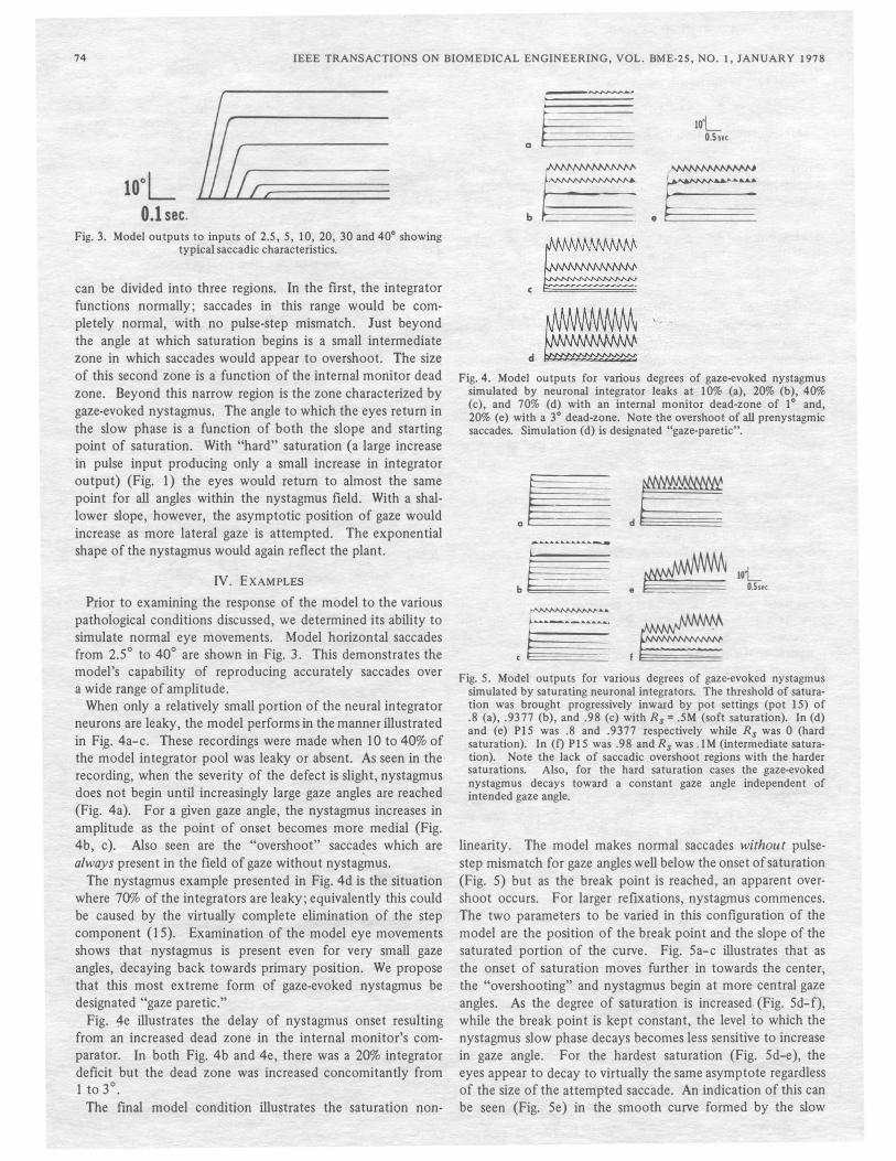
Fig. 3. Model outputs to inputs of 2.5, 5, 10, 20, 30 and 40° showing typical saccadic characteristics.
can be divided into three regions. In the first, the integrator functions normally; saccades in this range would be completely normal, with no pulse-step mismatch. Just beyond the angle at which saturation begins is a small intermediate zone in which saccades would appear to overshoot. The size of this second zone is a function of the internal monitor dead zone. Beyond this narrow region is the zone characterized by gaze-evoked nystagmus. The angle to which the eyes return in the slow phase is a function of both the slope and starting point of saturation. With "hard" saturation (a large increase in pulse input producing only a small increase in integrator output) (Fig. 1) the eyes would return to almost the same point for all angles within the nystagmus field. With a shallower slope, however, the asymptotic position of gaze would increase as more lateral gaze is attempted. The exponential shape of the nystagmus would again reflect the plant.
IV_ EXAMPLES
Prior to examining the response of the model to the various pathological conditions discussed, we determined its ability to simulate normal eye movements. Model horizontal saccades from 2.5° to 40° are shown in Fig. 3. This demonstrates the model's capability of reproducing accurately saccades over a wide range of amplitude.
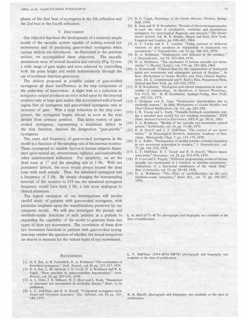
When only a relatively small portion of the neural integrator neurons are leaky, the model performs in the manner illustrated in FiE. 4a-c. These recordings were made when 10 to 40% of the model integrator pool was leaky or absent. As seen in the recording, when the severity of the defect is slight, nystagmus does not begin until increasingly large gaze angles are reached (Fig. 4a). For a given gaze angle, the nystagmus increases in amplitude as the point of onset becomes more medial (Fig. 4b, c). Also seen are the "overshoot" sacca4es which are always present in the field of gaze without nystagmus.
The nystagmus example presented in Fig. 4d is the situation where 70% of the integrators are leaky; equivalently this could be caused by the virtually complete elimination of the step component (15). Examination of the model eye movements shows that nystagmus is present even for very small gaze angles, decaying back towards primary position. We propose that this most extreme form of gaze-evoked nystagmus be designated "gaze paretic."
Fig. 4e illustrates the delay of nystagmus onset resulting from an increased dead zone in the internal monitor's comparator. In both Fig. 4b and 4e, there was a 20% integrator deficit but the dead zone was increased concomitantly from 1 to 3°.
.
The final model condition illustrates the saturation non-
.�
b�.�
~
lo'L 0.5 set.
� �-
d � Fig. 4. Model outputs for various degrees of gaze-evoked nystagmus
simulated by neuronal integrator leaks at 10% (a), 20% (b), 40% (c), and 70% (d) with an internal monitor dead-zone of 1° and, 20% (e) with a 3° dead-zone. Note the overshoot of all prenystagmic saccades. Simulation (d) is designated "gaze-paretic".
.l!!! d�
b�.�
<�f�
wL 0.5set.
Fig. 5. Model outputs for various degrees of gaze-evoked nystagmus simulated by saturating neuronal integrators. The threshold of saturation was brought progressively inward by pot settings (pot 15) of .8 (a), .937 7 (b), and .98 (c) with Rs = .5M (soft saturation). In (d) and (e) PIS was .8 and .937 7 respectively while Rs was 0 (hard saturation). In (f) PIS was .98 and Rs was .IM (intermediate saturation). Note the lack of saccadic overshoot regions with the harder saturations. Also, for the hard saturation cases the gaze-evoked nystagmus decays toward a constant gaze angle independent of intended gaze angle.
linearity. The model makes normal saccades without pulsestep mismatch for gaze angles well below the onset of saturation (Fig. 5) but as the break point is reached, an apparent overshoot occurs. For larger reflXations, nystagmus commences. The two parameters to be varied in this configuration of the model are the position of the break point and the slope of the saturated portion of the curve. Fig. 5a-c illustrates that as the onset of saturation moves further in towards the center, the "overshooting" and nystagmus begin at more central gaze angles. As the degree of saturation is increased, (Fig. 5d-f), while the break point is kept constant, the level to which the nystagmus slow phase decays becomes less sensitive to increase in gaze angle. For the hardest saturation (Fig. 5d-e), the eyes appear to decay to virtually the same asymptote regardless of the size of the attempted saccade. An indication of this can be seen (Fig. 5e) in the smooth curve formed by the slow
ABEL et 01.: GAZE·EVOKED NYSTAGMUS
phases of the first beat of nystagmus in the 5th refixation and the 2nd beat in the fourth refixation.
V. DISCUSSION
Our objective has been the development of a relatively simple model of the saccadic system capable of making normal eye movements and of producing gaze-evoked nystagmus when various defects are introduced. As illustrated in the previous
section, we accomplished these objectives. The saccadic simulations were of normal duration and velocity (Fig. 3) over a wide range of gaze angles and were achieved by controlling both the pulse height and width independently through the
use of nonlinear function generators. The defects proposed as possible causes of gaze-evoked
nystagmus all share insufficiency in the step component of the pUlse-step of innervation. A slight leak or a reduction in integrator output produces an error acted upon by the internal monitor only at large gaze angles; this is correlated with a broad region free of nystagmus and gaze-evoked nystagmus only at extremes of gaze. With a large reduction in the step component, the nystagmus begins almost as soon as the eyes deviate from primary position. This latter variety of gazeevoked nystagmus, representing a maximum defect in the step function, deserves the designation "gaze-paretic" nystagmus.
The .onset and frequency of gaze-evoked nystagmus in the model is a function of the sampling rate of the internal monitor. These correspond to variable factors in human subjects dependent upon mental set, attentiveness, vigilance, and undoubtedly other undetermined influences. For simplicity, we set the dead zone at 10 and the sampling rate at 5 Hz. With our postulated defects, the error would always exceed the dead zone with each sample. Thus, the simulated nystagmus had a frequency of 5 Hz. By simply changing the intersampling intervals of the monitor to 333 ms, the simulated nystagmus frequency would have been 3 Hz, a rate more analogous to clinical situations.
The logical extension of our investigations will involve careful study of patients with gaze-evoked nystagmus, with particular emphasis upon the manifestations predicted by our computer model. We will also investigate the pursuit and vestibulo-ocular functions of such patients as a prelude to expanding the capability of the model to generate these two types of slow eye movements. The correlation of these slow eye movement functions in patients with gaze-evoked nystagmus may resolve the question of whether the neural integrators are shared or separate for the various types of eye movements.
REFERENCES
[ 1) D. S. Zee, A. R. Friendlich, D. A. Robinson "The mechanism of downbeat nystagmus," Arch. Neurol., vol. 30, pp. 227-237, 1974.
(2) D. S. Zee, L. M. Optican, J. D. Cook, D. A. Robinson and W. K. Engel, "Slow saccades in spinocerebellar degeneration," Arch. Neurol., voL 33, pp. 243-251, 1976.
(3) A. L. Ochs, J. B. Seihorst, W. F. Hoyt and L. Stark, "Simulation of abnormal eye movements in cerebellar disease," Brain, to be published.
(4) L. F. Dell'Osso and R. B. Daroff, "Congenital nystagmus waveforms and foveation strategies," Doc. Ophthal., vol. 39, pp. 155-182,1975.
7S
(5) D. G. Cogan, Neurology of the Ocular Muscles, Thomas, Springfield, 1956.
(6) R. Jung and H. H. Kornhuber, "Results of electronystagmography in man: the value of optokinetic, vestibular and spontaneous nystagmus for neurological diagnosis and research," The Oculomotor System, Ed. M. B. Bender, Harper and Row, New York, Evanston and London, pp. 428-48 1,1964.
(7) A. F. Fuchs and E. S. Luschei, "Firing patterns of abducens neurons of alert monkeys in relationship to horizontal eye movements," J. Neurophysiol., vol. 33, pp. 382-392, 1970.
(8) D. A. Robinson, "Oculomotor unit behavior in the monkey," J. Neurophysiol., vol. 33, 393-404, 1970.
(9) D. A. Robinson, "The mechanics of human saccadic eye movements," J. Physiol. (Lond.), vol. 174, pp. 245-264, 1964.
(10) G. Kommerell, "Clinical clues for the organization of horizontal quick eye movements and subsequent periods of fixation." In Basic Mechanisms of Ocular Motility and Their Clinical Implications, Ed. G. Lennerstrand and P. Bach-y-Rita, Pergammon Press, Oxford and New York, pp. 325-335, 1975.
[11) H. H. Kornhuber, "Nystagmus and related phenomena in man: an outline of otoneurology. In Handbook of Sensory Physiology, Vol Vl/2, Ed. H. H. Kornhuber, Springer-Verlag, New York, pp. 193-232, 1974.
(12) J. Dichgans and R. Jung, "Oculomotor abnormalities due to cerebellar lesions." In Basic Mechanisms of Ocular Motility and Their Clinical Implications, Op. Cit., pp. 281-298.
(13) L. R. Young and L. Stark, "Variable feedback experiments testing a sampled data model for eye tracking movements," IEEE Trans. Human Factors in Electronics, HFE-4: pp. 38-51, 1963.
(14) D. A. Robinson, "Models of the saccadic eye movement control system," Kybernetik, vol. 14, pp. 7 1-84, 1973.
(15) R. B. Daroff and L. F. Dell'Osso, "The control of eye movements." In Neurological Reviews, American Academy of Neurology, Minneapolis, Chap. 7, pp. 143-170, 1976.
(16) E. L. Keller, "Participation of medial pontine reticular formation in eye movement generation in monkey," 1. Neurophysiol., vol. 37, pp. 316-332, 1974.
(17) L. F. Dell'Osso, B. T. Troost and R. B. Daroff, "Macro square wave jerks," Neurology, vol. 25, pp. 975-979,1975.
(18) D. Frost and E. Poppel, "Different programming modes of human saccadic eye movements as a function of stimulus eccentricity: Indications of a functional subdivision of the visual field," Bioi. Cybernetics, vol. 23, pp. 39-48, 1976.
(19) D. A. Robinson, "The effect of cerebellectomy on the cat's vestibulo-ocular integrator," Brain Res., vol. 7 1, pp. 195-207, 1974.
t. A. Abel (S'71-M'76) photograph and biography not available at the time of publication.
L. F. Dell'Osso (S'64-M'66-SM'76) photograph and biography not available at the time of publication.
R. B. Daroff, photograph and biography not available at the time of publication.