An Update on Pancreas Neoplasms - pathcme.com · 5/21/2018 7 Intraductal Papillary Mucinous...
33
5/21/2018 1 An Update on Pancreas Neoplasms Arief Suriawinata, M.D. Professor of Pathology and Laboratory Medicine Geisel School of Medicine at Dartmouth Department of Pathology and Laboratory Medicine Dartmouth-Hitchcock Medical Center Pancreas - normal Pancreas - tumors Lines of Differentiation in Pancreatic Neoplasms Differentiation Special Stain IHC EM Ductal Mucin stain Glycoprotein markers (CEA, B72.3, Ca19.9, CA125) MUC1, MUC3, MUC4, MUC5AC, CK7 60% loss of SMAD4 Mucigen granules Acinar PAS-D, butyrate esterase Enzyme markers (trypsin, chymotrypsin, lipase, others) Zymogen granules, irregular fibrillary granules Neuroendocrine Grimelius stain Neuroendocrine markers (chromogranin, synaptophysin, CD56) Peptide markers (insulin, glucagon, etc.) Dense core granules
Transcript of An Update on Pancreas Neoplasms - pathcme.com · 5/21/2018 7 Intraductal Papillary Mucinous...

5/21/2018
1
An Update on
Pancreas Neoplasms
Arief Suriawinata, M.D.
Professor of Pathology and Laboratory Medicine
Geisel School of Medicine at Dartmouth
Department of Pathology and Laboratory Medicine
Dartmouth-Hitchcock Medical Center
Pancreas -
normal
Pancreas -
tumors
Lines of Differentiation in Pancreatic Neoplasms
Differentiation Special Stain IHC EM
Ductal Mucin stain
Glycoprotein markers
(CEA, B72.3, Ca19.9, CA125)
MUC1, MUC3, MUC4, MUC5AC, CK7
60% loss of SMAD4
Mucigen
granules
AcinarPAS-D, butyrate
esterase
Enzyme markers
(trypsin, chymotrypsin, lipase, others)
Zymogen
granules,
irregular
fibrillary
granules
Neuroendocrine Grimelius stain
Neuroendocrine markers
(chromogranin, synaptophysin, CD56)
Peptide markers
(insulin, glucagon, etc.)
Dense core
granules

5/21/2018
2
Pancreas Ductal Adenocarcinoma (PDAC)
Synonyms Tubular adenocarcinoma, infiltrating duct carcinoma
Epidemiology 60-80 y.o.
4th leading cancer death in US (increasing to 2nd by 2030)
50% higher in men than women
5 year survival rate – 7%
Etiology Tobacco smoking (3X), chronic pancreatitis (10X), obesity, alcohol,
diabetes mellitus
Signs &
Symptoms
Jaundice, pruritus, back pain, weight loss
New onset diabetes mellitus in 70% of patients
Late symptoms: liver metastasis, duodenal invasion (gastric outlet
obstruction), peritoneal cavity involvement (ascites)
Tests Serum tumor markers (CA19-9, CEA)
CT (best method) – mass, “double duct sign”, vascular invasion
assessment
EUS – heterogenous mass, lymph node assessment, FNA biopsy
Localization Head of pancreas (60-70%), body (5-15%), tail (10-15%)
Pancreatic adenocarcinoma
5/21/2018
3
Osteoclastic Giant Cell Carcinoma
CD68 Keratin
Recent Developments on PDAC
• EUS – FNA has become an established method in the
initial diagnosis of pancreatic tumors
• Cytopathology evaluation
• Tissue procurement
• 2 tier grading system on precursor lesions
• PanIN, IPMN, MCN
• AJCC Cancer Staging Manual, 8th edition
• Neoadjuvant chemoradiation has been widely
administered
• Resectable and nonresectable PDAC
• Complete surgical resection remains the only potentially
curative option
• Median overall survival of 28 months after resection and
adjuvant chemotherapy
• Borderline resectable or locally advanced disease may
have margin negative resection after neoadjuvant
chemotherapy
• Similar overall survival with resectable disease
• Neoadjuvant chemoradiation
• Reduces micrometastases
• Increases likelihood of complete resection and
survival
PDAC Treatment

5/21/2018
4
Treatment and Median Survival
Gillen et al. PLoS 2010
• Patients with BR/LA-PDAC who had a pCR after neoadjuvant
chemoradiation had a significantly prolonged survival compared
with those who had nCR or a limited response
• Pancreatic intraepithelial neoplasia (PanIN)
• Intraductal papillary mucinous neoplasm (IPMN)
• Mucinous cystic neoplasm (MCN)
PDAC Precursors

5/21/2018
5
• 2 tier system for all precursor lesions – low grade & high grade
• PanIN
• IPMN
• MCN
• PanIN of any grade at margin in pancreas with invasive
carcinoma does not have prognostic implications
• Intraductal lesions 0.5 to 1 cm can be either large PanINs or
small IPMNs
• "Intraductal spread of invasive carcinoma" (aka, "colonization")
is invasive carcinoma invading back into and extending along
the ductal system, may morphologically mimic high-grade
PanIN
Molecular Alterations in Pancreatic Ductal Lesions
K-ras 35% 45% 65% 85% 90%
p53 0% 0% <5% 20% 70%
HER-2/neu 82% 86% 92% 100% 69%
p16 24% 19% 55% 71% 95%
SMAD4/DPC4 0% 0% 0% 31% 55%
Mucinous Papillary Atypical CIS Invasive
Metaplasia Hyperplasia Hyperplasia Carcinoma
Wilentz et al., Cancer Res 60: 2002
Basturk et al., Am J Surg Pathol 2015
PanIN 1A PanIN 1B PanIN 2 PanIN 3
Low grade PanINHigh grade
PanIN
Hereditary Pancreatic Carcinoma
Heritable factors involved in 10%
Familial pancreatic carcinoma
At least two first degree relatives with PDAC and not associated with
other known hereditary syndromes
PDAC-associated hereditary syndromes:
Peutz-Jeghers syndrome (STK11/LKB)
Hereditary pancreatitis [PRSS1 (cationic trypsinogen), SPINK1]
Familial atypical multiple mole melanoma syndrome [CDKN2A (p16-
Leiden deletion)]
Lynch syndrome (hMSH2; hMLH1)
Familial adenomatous polyposis
Hereditary breast-ovarian cancer syndrome (BRCA2)
Ataxia telangiectasia
5/21/2018
6
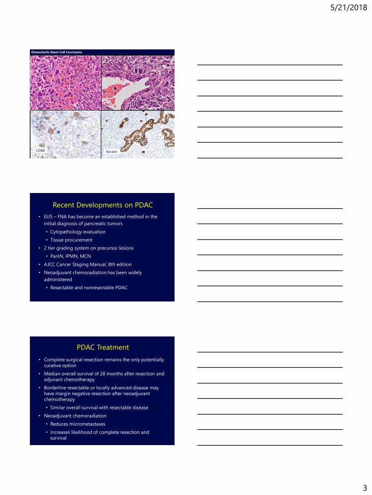
Pancreatic Carcinoma Screening
• High risk individuals
• Familial pancreatic carcinoma
• Hereditary pancreatitis
• Peutz Jeghers syndrome
• Chronic pancreatitis
• Identification of precursor lesions and
early resectable PDAC
• Cancer of the Pancreas Screening-5
CAPS5)Study (CAPS5)
https://clinicaltrials.gov/ct2/show/study/NCT0
2000089
• Pancreatic fluid mutations & circulating
pancreatic epithelial cells
• Alternative pathway – KRAS mutation
• Metaplasia – dysplasia – cancer sequence
• Seen in individuals with familial pancreatic cancer
Acinar Ductal Metaplasia & Atypical Flat Lesions
Basturk, et al. Am J Surg Pathol 2015
ADM
AFL

5/21/2018
7
Intraductal Papillary Mucinous Neoplasms
▪ Definition:
“Grossly and radiographically visible (>1cm) epithelial tumor arising
from the main pancreatic duct or duct branches, causing dilatation and
mucin production”
▪ Often lack invasive carcinoma
▪ 30% of surgically resected IPMNs harbor invasive carcinoma
▪ 5 year survival is better than PDAC
▪ IPMN alone 30-50%
▪ IPMN + invasive ca = 70-90%
Genetic Features K-ras p53 DPC4 p16STK11/L
KB11GNAS RNF43
Ductal adenocarcinoma >95% 50-70% 40-60% 95% 5% 5% 5%
IPMN 13-100% 0-50% 5% 0-20% 32% 50-79% 23-36%
Intraductal Papillary Mucinous Neoplasm
Synonyms Mucinous duct ectasia, duct ectatic mucinous cystadenoma/carcinoma,
mucin producing tumor, villous adenoma, papillary adenoma/carcinoma
Epidemiology 3% of pancreatic exocrine and 20% of cystic neoplasms
50-90 y.o., 8.7% in individuals >80 y.o, incidence is increasing (incidental,
asymptomatic), male = female
Etiology Cigarette smoking, Peutz-Jeghers syndrome, FAP, family history of
pancreatic carcinoma, McCune-Albright syndrome
Signs and
symptoms
Nonspecific abdominal pain, chronic pancreatitis, weight loss, new onset
diabetes mellitus, jaundice
Clinical findings CA19-9 and CEA elevated in cases with invasive carcinoma
Endoscopy – mucin extrusion from ampulla of Vater
Radiology – ectasia and cystic dilatation of pancreatic duct
Localization Predominantly in head of pancreas; localized, multicentric or diffusely
involving the entire pancreas ductal system, extending to ampulla
or common bile duct
IPMN Classification
Old, WHO 2000, AFIP Fascicle 4th
series termsWHO 2010 terms
New 2-tier system
Baltimore Consensus 2015
Intraductal papillary mucinous
adenoma, IPMN with low
grade dysplasia
IPMN with low-grade
dysplasia
IPMN low grade
IPMN, borderline; IPMN with
moderate dysplasia
IPMN with intermediate-
grade dysplasia
Intraductal papillary mucinous
carcinoma (in situ); intraductal
papillary mucinous carcinoma,
noninvasive
IPMN with high-grade
dysplasiaIPMN high grade
Intraductal papillary mucinous
carcinoma, invasive
IPMN with an associated
invasive carcinoma
IPMN, ….grade, with an
associated invasive
carcinoma
Invasive carcinoma with an
associated IPMN

5/21/2018
8
5/21/2018
9
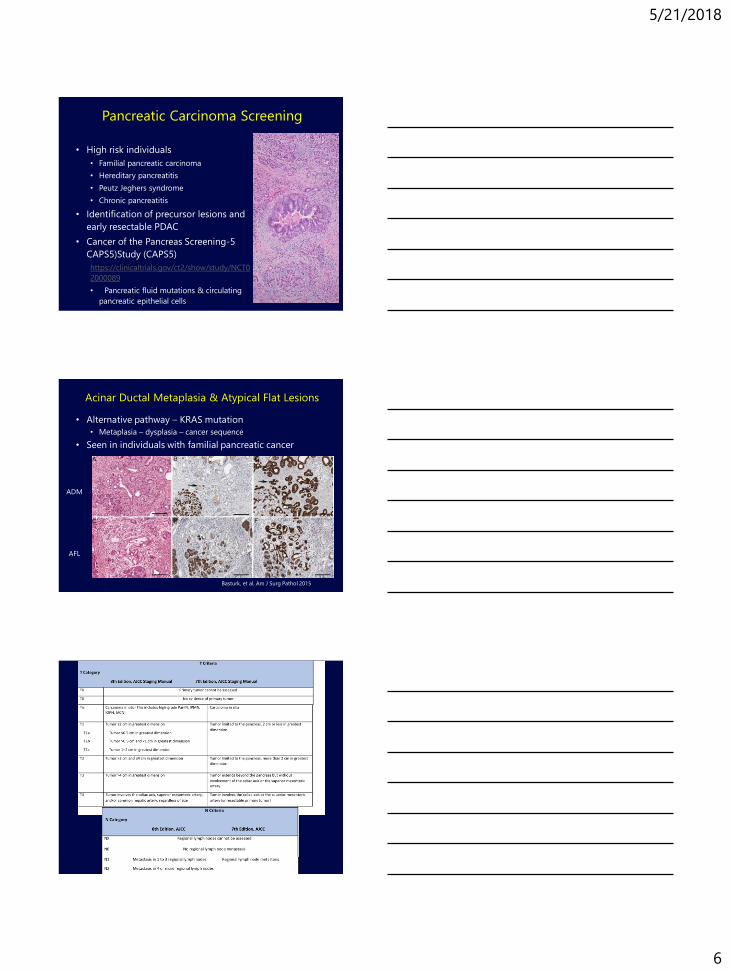
Gastric foveolar
Intestinal type
Pancreatobiliary
Oncocytic
MUC1 MUC2 MUC5AC
- -
-
-
+
++
+
+
+
focal+ focal+
H&E MUC6
Tubulopapillary
CDX2
+
+
+
+
+
- -
- -
-
-- -

5/21/2018
10

5/21/2018
11
IPMN – Carcinogenesis Pathway
Source: Katabi & Klimstra 2008
Tan, Basturk, Klimstra, 2015
GNAS
KRAS
IPMN - Main vs. Branch Ducts
• 70% involve main duct, 30% confined to branch ducts
• Branch duct type (Terris, et al)
• Younger patients & low risk of progression
• Main duct type: 20% CIS, 37% invasive carcinoma
• Branch duct type: 15% CIS, 0% invasive carcinoma
• 5 year risk of progression(Levy, et al)
• MD 63% vs BD 15%

5/21/2018
12
Prognosis & Treatment
• Presence and size of invasive component
- Mural nodules
- Solid masses
- Large tumor size
- Dilated main pancreatic duct
- Cyst wall thickening
- Increase serum CA19-9
• Complete surgical resection is curative for IPMN
• Partial pancreatectomy
- Extent
- Status of margin – frozen section difficulties
- Risk of local recurrence – skip nature
- Surveillance
• 2 tier system for all precursor lesions
• PanIN
• IPMN
• MCN
• Clinical significance of dysplasia at resection margin of IPMN lacking
invasive carcinoma remains to be determined
• Increased risk of recurrence after prolonged follow up
• Further resection recommended on high grade dysplasia at margin
• “Incipient IPMN” are lesions 0.5-1.0cm with intestinal or oncocytic
papillae or GNAS mutations (intestinal or gastric type)
• “Simple mucinous cyst” are cysts > 1 cm with gastric-type flat
mucinous lining at most minimal atypia without ovarian-type stroma
• Synonym: IPMN, oncocytic type
• Form large cystic lesion with friable papillary growths in large
pancreatic ducts
• Distinctive feature:
- oncocytic cytoplasm (mitochondria)
- eccentric nucleoli
- intraepithelial lumina, often containing mucin, producing
cribriform architecture
• Difference from conventional IPMNs:
MUC6 +, HepPar1 +, lack of KRAS mutation
Intraductal Oncocytic Papillary Neoplasm

5/21/2018
13

5/21/2018
14
• Synonym: Intraductal tubular neoplasm
• Definition: Grossly visible intraductal lesion composed
of tubule-forming epithelial neoplasm with high grade
dysplasia and ductal differentiation without overt
production of mucin
• 3% of IPMN’s
• Solid, nodular masses in dilated pancreatic ducts, no
mucin
• 40% ITPN’s harbor invasive carcinoma, usually localized
Intraductal Tubulopapillary Neoplasm

5/21/2018
15

5/21/2018
16
• Definition:
Cyst forming epithelial neoplasm that commonly does
not communicate with the pancreatic ductal system,
lined by mucin-producing epithelium and with
associated ovarian-type subepithelial stroma
• Almost exclusively in women, 40-50 y.o.
• Patients with MCNs with associated invasive carcinoma are 5-10 years
older
• >95% in body and tail of pancreas
• Incidental finding, less likely to present with pancreatitis,
jaundice or new onset diabetes mellitus
Mucinous Cystic Neoplasm (MCN)
• Noninvasive tumor: CK 7,8,18, 19, CA19-9, EMA,
CEA, MUC5AC, MUC2 (goblet cells), KRAS
mutation
• Invasive tumor: MUC1, loss of SMAD4,
alterations of p16 and p53
• Similar molecular alterations to PanIN
Mucinous Cystic Neoplasm

5/21/2018
17
Old terms WHO 2010New 2-tier system
Baltimore Consensus 2015
Mucinous cystadenomaMCN with low-grade
dysplasiaMCN low grade
Mucinous cystic tumor,
borderline
MCN with intermediate-
grade dysplasia
Mucinous
cystadenocarcinoma
MCN with high-grade
dysplasiaMCN high grade
Mucinous
cystadenocarcinoma,
invasive
MCN with an associated
invasive carcinoma
MCN, …grade, with an
associated invasive carcinoma
Invasive carcinoma associated
with MCN
Mucinous Cystic Neoplasm Grading

5/21/2018
18
5/21/2018
19
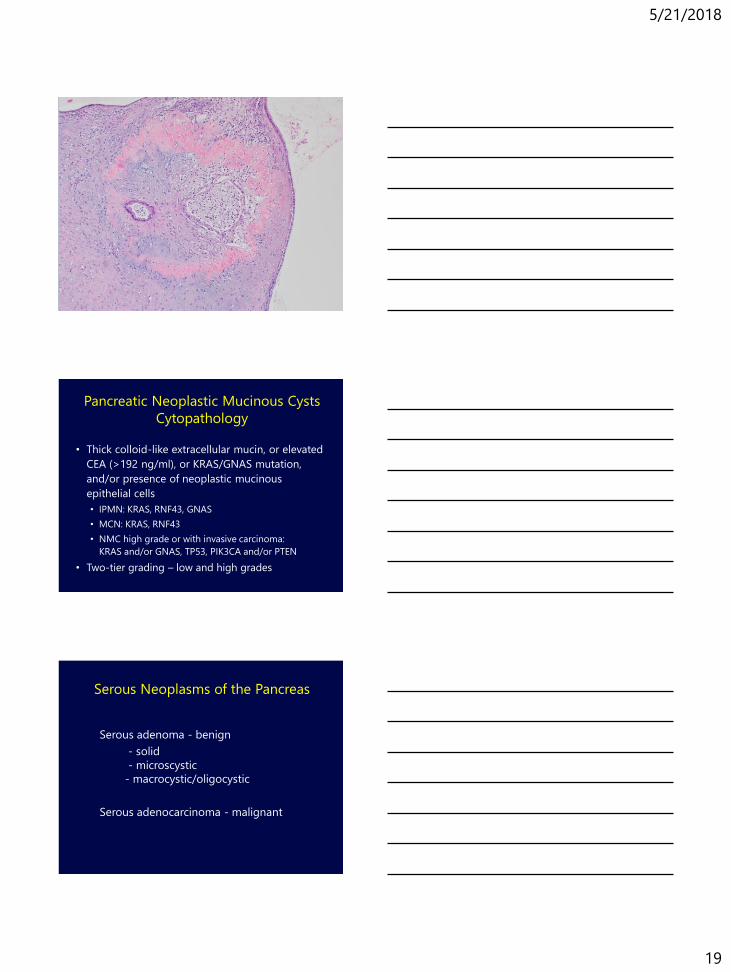
Pancreatic Neoplastic Mucinous Cysts
Cytopathology
• Thick colloid-like extracellular mucin, or elevated
CEA (>192 ng/ml), or KRAS/GNAS mutation,
and/or presence of neoplastic mucinous
epithelial cells
• IPMN: KRAS, RNF43, GNAS
• MCN: KRAS, RNF43
• NMC high grade or with invasive carcinoma:
KRAS and/or GNAS, TP53, PIK3CA and/or PTEN
• Two-tier grading – low and high grades
Serous Neoplasms of the Pancreas
Serous adenoma - benign
- solid
- microscystic
- macrocystic/oligocystic
Serous adenocarcinoma - malignant

5/21/2018
20
Serous Neoplasms of the Pancreas
Serous adenoma
- 1-2% pancreatic neoplasms
- mean age 60 y.o., slight female predominance
- 50-75% in body or tail
- associated with Von Hippel Lindau syndrome
- CK 7,8,18 and 19; EMA, inhibin, MUC6 and NSE +
- no KRAS or P53 mutation
Serous adenocarcinoma
- exceedingly rare
- direct invasion into adjacent organ or
metastasize

5/21/2018
21

5/21/2018
22
Pancreatic Neurondocrine Neoplasms
• Non-syndromic (non-functioning), but IHC +
- alpha-cell/glucagon producing NET
- beta-cell/insulin producing NET
- G-cell/gastrin-producing NET
• Syndromic (functioning) – “..oma”
- Insulinoma
- Glucagonoma
- Somatostatinoma
- Gastrinoma
- VIPoma
Pancreatic Endocrine Neoplasms
Associated Conditions
• Multiple Endocrine Neoplasia I
• von Hippel-Lindau Syndrome
• Tuberous Sclerosis
• Pheochromocytoma
• Cushing’s Syndrome

5/21/2018
23
Pancreatic Neuroendocrine Neoplasms
Criteria for Malignancy
• Traditional Criteria
• Metastases
• Gross invasion
• Vascular invasion
• Correlation with clinical syndromes
• Predictors of aggressive behavior
• Mitotic rate
• Ki-67 index
• Invasion
• Necrosis
• Poorly Differentiated Neuroendocrine Carcinoma
Pancreatic Neuroendocrine Neoplasm Grading
WHO 2010 WHO 2017
Well differentiated neuroendocrine tumorWell differentiated neuroendocrine tumor
(PanNET)
Neuroendocrine tumor (PanNET) G1
(<2 mitoses/10hpf and/or =<2% Ki67 index)
Neuroendocrine tumor (PanNET) G1
(<2 mitoses/10hpf and/or <3% Ki67 index)
Neuroendocrine tumor (PanNET) G2
(2-20 mitoses/10hpf and/or 3-20% Ki67 index)
Neuroendocrine tumor (PanNET) G2
(2-20 mitoses/10hpf and/or 3-20% Ki67 index)
Neuroendocrine tumor (PanNET) G3
(>20 mitoses/10hpf or >20% Ki67 index)
Poorly differentiated neuroendocrine carcinoma
(PanNEC)
Poorly differentiated neuroendocrine carcinoma
(PanNEC)
Neuroendocrine carcinoma (PanNEC) G3
(>20 mitoses/10hpf or >20% Ki67 index)
Neuroendocrine carcinoma (PanNEC) G3
(>20 mitoses/10hpf or >20% Ki67 index)Small cell type
Large cell type
Mixed adeno-neuroendocrine carcinoma (MANEC)Mixed neuroendocrine-non-neuroendocrine
neoplasm (MINEN)
• Well differentiated NEN - low, intermediate or
high grade
• Minimal to moderate atypia
• Typical organoid patterns, lacking necrosis
• General neuroendocrine markers +
• Associated with hormonal syndrome/functioning
tumor
Pancreatic Neuroendocrine Tumor
(PanNET)

5/21/2018
24

5/21/2018
25

5/21/2018
26
Pancreatic Neuroendocrine Carcinoma
(PanNEC)
• Poorly differentiated high grade NEN
• Highly atypical small cells or intermediate to
large cells
• General neuroendocrine markers +
• Exocrine markers -
• Rarely associated with hormonal syndromes
• TNM classification follows PDAC

5/21/2018
27
WD G3 PanNETs PD G3 PanNECs
MEN1, DAX, ATRX mutations
IHC loss of DAXX or ATRX
P53, RB1, KRAS mutations
IHC loss of RB
Recognizable as NETs Small cell or large cell type
Often evolve from a recognizable
lower grade component (1 or 2)
No lower grade component
No upper limit given, but usually
ki67<40 to 55%, mitotic count
<20/10hpf
Must have ki67 index >20%, no lower
limit given but usually >55%
Plasmacytoid morphology, smooth
nuclear contour, round nuclei
Pleomorphism, nuclear molding,
necrosis
WD G3 PanNETs vs PD G3 PanNECs
• Plasmacytoid morphology
• Smooth nuclear contour
• Abundant cytoplasm
• Apoptosis
• Nuclear tangles

5/21/2018
28
Mixed Neuroendocrine-Non-Neuroendocrine Neoplasm
(MINEN)
• Mixed neoplasm with components of a nonendocrine
carcinoma (mostly ductal adenocarcinoma or acinar cell
carcinoma) combined with a neuroendocrine neoplasm
• Usually both components are high grade malignant
carcinomas (G3), but ocasionally one of the two or both
components may belong to the G1/G2 category
• Each component comprises >30% of the tumor
Solid-Pseudopapillary Tumor
2-3% of pancreatic tumors
Tumor of young females
F:M = 9.5:1; Mean age = 30.3 yrs
Symptoms usually related to presence of mass
Detected during pregnancy, after trauma,
incidentally
Low grade malignant neoplasm
Metastases in 15%
Long survival after metastases
Uncertain histogenesis
Monomorphic epithelial cells with solid and
pseudopapillary structures
Solid-Pseudopapillary Tumor:
Molecular Features
APC / b-catenin pathway upregulation (95%)
b-catenin mutations
Overexpression of cyclin D1
Upregulation of genes required in Notch, Hedgehog, and
androgen receptor signaling pathways
E-cadherin expression changes from a membranous to
intracytoplasmic localization
No abnormalities in “ductal carcinoma genes”
K-ras
p53
DPC4

5/21/2018
29
Solid Pseudopapillary Tumor:
Staining Profile
Stain % Positive
Trypsin 0
Chymotrypsin 0
Lipase 0
Chromogranin 0
Synaptophysin 30
Neuron Specific Enolase 80
CD56 95
Mucicarmine 0
CEA 5
Keratin 30
Vimentin 100
α-1-antitrypsin 84
CD10 100
Beta catenin 97
Progesterone receptor 90

5/21/2018
30

5/21/2018
31
Acinar Cell Carcinoma
Clinical Features
1-2% of pancreatic tumors
Male predominance, mean age = 61
Non-specific presenting symptoms
Jaundice rare
Lipase hypersecretion syndrome

5/21/2018
32
Trypsin
Acinar Cell Carcinoma
Genetic Features
Al-Hader A et al, World J Gastroenterol 2017

5/21/2018
33
• BRAF fusion in 24% acinar
tumors, including pure and mixed
types
• BRAF partners:
• SND1 (50%)
• HERPUD1 (18%)
• Potential for targeted therapy to
inhibit MAPK pathway activity
Treatment
• Aggressive surgical resection
• 5-year survival on resected patients 72%
• Resection for metastases
• No defined treatment guidelines for cure
• May be chemoresponsive to agents that have
activity against pancreatic adenocarcinomas and
colorectal carcinomas
• Targeted therapy
• MMR deficient – PD-1 receptor blocker
• SND1 -BRAF fusion – MEK inhibitor
• JAK-1 mutation – JAK-1 & 2 inhibitor