An unusual case of pheochromocytoma and unruptured...
6
Vlaams Diergeneeskundig Tijdschrift, 2017, 86 Case report 99 INTRODUCTION Ante-mortem diagnosis of abdominal aortic aneu- rysms (AAAs) in dogs has been rarely reported in the literature. Currently, little is known about the events leading to the development of AAAs in the canine spe- cies. The term “aneurysm” is derived from the Greek ανευρυσµα (aneurism), meaning “widening”, and is defined as the permanent and irreversible localized dilation of a vessel (Sakalihasan et al., 2005). Large- An unusual case of pheochromocytoma and unruptured abdominal aortic aneurism in a male Yorkshire terrier Ongebruikelijk voorkomen van een feochromocytoom en niet-geruptureerd aneurysma van de abdominale aorta bij een mannelijke yorkshireterriër 1* B.Á. Rodrigues, 2 Q.G. Grangeiro, 2 C. Scaranto, 3 G. Konradt, 3 M.V. Bianchi, 4 D. Driemeier, 5 J.L.R. Rodrigues 1* Av. Cristo rei 134; 93020-350 São Leopoldo, RS Brazil 2 Medvet Veterinary Clinic, Av. Boqueirão 1004, Canoas, RS-Brazil 3 Department of Pathology, Faculty of Veterinary Medicine- Federal University of Rio Grande do Sul (UFRGS). Av. Bento Gonçalves, 9090 Prédio 42505 – Agronomia – Porto Alegre, RS- Brazil 4 Department of Pathology, Faculty of Veterinary Medicine- Federal University of Rio Grande do Sul (UFRGS). Av. Bento Gonçalves, 9090 Prédio 42505 – Agronomia – Porto Alegre, RS- Brazil 5 Laboratory of Embryology and Biotechniques of Reproduction, Faculty of Veterinary Medicine- Federal University of Rio Grande do Sul (UFRGS). Av. Bento Gonçalves, 9090-Agronomia- Porto Alegre, RS- Brazil [email protected]; [email protected] vessel aneurysm is a disorder of the tunica media of the arterial wall, characterized by destruction of the extracellular matrix components associated with re- modelling of the vessel wall in an aneurysmal fashion (Michel, 1998). The usual pathophysiology of an aor- tic aneurysm involves disruption of the aortic intima and media, along with the formation of a hematoma (Waldrop et al., 2003). Hemodynamic stress facilitates the atheromatous process and impairs the mechanical properties of the abdominal aorta (Michel, 1998). BSTRACT A six-year-old, male Yorkshire terrier was presented with acute vomiting, anorexia, depres- sion, watery diarrhea and sudden blindness. On the basis of a transabdominal ultrasonographic examination, the presence of a prominent aortic aneurysm was established. The aneurysm of the aorta was confirmed at post-mortem examination. Unexpectedly, a pheochromocytoma of the left adrenal gland was found to be involved with the aneurysm. In this case report, the unusual occurrence of a large, unruptured abdominal aortic aneurism (AAA) concurrent with a pheo- chromocytoma in a male Yorkshire terrier dog is discussed. SAMENVATTING Een zes jaar oude, mannelijke yorkshireterriër werd aangeboden met acuut braken, anorexia, de- pressie, waterige diarree en plotse blindheid. Op basis van een transabdominaal uitgevoerd echogra- fisch onderzoek werd een prominent aneurysma van de abdominale aorta vastgesteld. Bij post- mortemonderzoek werden de aanwezigheid en de locatie van het aneurysma bevestigd. Bovendien werd een feochromocytoom van de linkerbijnier vastgesteld dat betrokken was bij het aneurysma. In deze casuïstiek wordt het uitzonderlijk voorkomen van een groot, niet-geruptureerd aneurysma van de abdominale aorta (AAA) samen met een feochromocytoom van de bijnier bij een mannelijke york- shireterriër beschreven. A
Transcript of An unusual case of pheochromocytoma and unruptured...

Vlaams Diergeneeskundig Tijdschrift, 2017, 86 99Vlaams Diergeneeskundig Tijdschrift, 2017, 86 Case report 99
INTRODUCTION
Ante-mortem diagnosis of abdominal aortic aneu-rysms (AAAs) in dogs has been rarely reported in the literature. Currently, little is known about the events leading to the development of AAAs in the canine spe-cies. The term “aneurysm” is derived from the Greek ανευρυσµα (aneurism), meaning “widening”, and is defined as the permanent and irreversible localized dilation of a vessel (Sakalihasan et al., 2005). Large-
An unusual case of pheochromocytoma and unruptured abdominal aorticaneurism in a male Yorkshire terrier
Ongebruikelijk voorkomen van een feochromocytoom en niet-geruptureerdaneurysma van de abdominale aorta bij een mannelijke yorkshireterriër
1*B.Á. Rodrigues, 2Q.G. Grangeiro, 2C. Scaranto, 3G. Konradt, 3M.V. Bianchi, 4D. Driemeier,5J.L.R. Rodrigues
1*Av. Cristo rei 134; 93020-350 São Leopoldo, RS Brazil2Medvet Veterinary Clinic, Av. Boqueirão 1004, Canoas, RS-Brazil
3Department of Pathology, Faculty of Veterinary Medicine- Federal University of Rio Grandedo Sul (UFRGS). Av. Bento Gonçalves, 9090 Prédio 42505 – Agronomia – Porto Alegre,
RS- Brazil4Department of Pathology, Faculty of Veterinary Medicine- Federal University of Rio Grande
do Sul (UFRGS). Av. Bento Gonçalves, 9090 Prédio 42505 – Agronomia – Porto Alegre,RS- Brazil
5Laboratory of Embryology and Biotechniques of Reproduction, Faculty of Veterinary Medicine-Federal University of Rio Grande do Sul (UFRGS). Av. Bento Gonçalves, 9090-Agronomia-
Porto Alegre, RS- Brazil
[email protected]; [email protected]
vessel aneurysm is a disorder of the tunica media of the arterial wall, characterized by destruction of the extracellular matrix components associated with re-modelling of the vessel wall in an aneurysmal fashion (Michel, 1998). The usual pathophysiology of an aor-tic aneurysm involves disruption of the aortic intima and media, along with the formation of a hematoma (Waldrop et al., 2003). Hemodynamic stress facilitates the atheromatous process and impairs the mechanical properties of the abdominal aorta (Michel, 1998).
BSTRACT
A six-year-old, male Yorkshire terrier was presented with acute vomiting, anorexia, depres-sion, watery diarrhea and sudden blindness. On the basis of a transabdominal ultrasonographic examination, the presence of a prominent aortic aneurysm was established. The aneurysm of the aorta was confirmed at post-mortem examination. Unexpectedly, a pheochromocytoma of the left adrenal gland was found to be involved with the aneurysm. In this case report, the unusual occurrence of a large, unruptured abdominal aortic aneurism (AAA) concurrent with a pheo-chromocytoma in a male Yorkshire terrier dog is discussed.
SAMENVATTING
Een zes jaar oude, mannelijke yorkshireterriër werd aangeboden met acuut braken, anorexia, de-pressie, waterige diarree en plotse blindheid. Op basis van een transabdominaal uitgevoerd echogra-fisch onderzoek werd een prominent aneurysma van de abdominale aorta vastgesteld. Bij post- mortemonderzoek werden de aanwezigheid en de locatie van het aneurysma bevestigd. Bovendien werd een feochromocytoom van de linkerbijnier vastgesteld dat betrokken was bij het aneurysma. In deze casuïstiek wordt het uitzonderlijk voorkomen van een groot, niet-geruptureerd aneurysma van de abdominale aorta (AAA) samen met een feochromocytoom van de bijnier bij een mannelijke york-shireterriër beschreven.
A

100 Vlaams Diergeneeskundig Tijdschrift, 2017, 86
Many factors contribute to the pathogenesis of AAAs. Local inflammation, apoptosis and oxidative stress, which activate proteases and degrade the aor-tic wall, play a role (Rizas et al., 2009). This struc-tural instability leads to weakening of the aortic wall and to aortic aneurysm. Additionally, hypertension has consistently been shown to be a major risk factor for AAAs in humans (Vardulaki et al., 2000) and in mice (Kanematsu et al., 2010). Pheochromocytomas can cause episodic or continuous systemic hyperten-sion in 43% of dogs (Barthez et al., 1997; Loste et al., 2013). These tumors are uncommon in dogs (preva-lence of 0.17-0.76%) (Myers, 1997).
Canine cases of AAAs have been associated with systemic fungal disease (Gershenson et al., 2011; Mu-rata et al., 2015), spirocercosis (van der Merwe et al., 2008; Kirberger et al., 2013), and familial inheritance in Leonberger dogs (Chetboul et al., 2003). The co-existence of an AAA with a pheochromocytoma has been previously reported as uncommon in humans (Kota et al., 2013). The aim of this case report is to discuss the unusual occurrence of a concurrent large unruptured abdominal aortic aneurism (AAA) and a pheochromocytoma in a male Yorkshire terrier dog.
CASE REPORT
A six-year-old, male Yorkshire terrier presented with acute vomiting, anorexia, depression, watery diarrhea and sudden blindness. Physical examination revealed that the dog was very dehydrated and had pale mucous membranes. The rectal temperature was within normal limits, and the body condition score was 2/5. The skin of the ventral abdomen showed petechiae and multifocal ecchymosis. The hind limbs had mild subcutaneous edema. Routine (red and white) blood cell count was within normal limits for the species. Increased alkaline phosphatase (ALP) ac-tivity (583 IU/L; reference interval 20-156 IU/L) and increased urea (73.5 mmol/L; reference interval 10.7-
28.6 mmol/L) were observed on the serum chemistry panel. Additionally, thrombocytopenia (0 /μL; refer-ence interval 200.000- 500.000/µL) and platelet ag-gregation (+++) were identified in the blood sample.
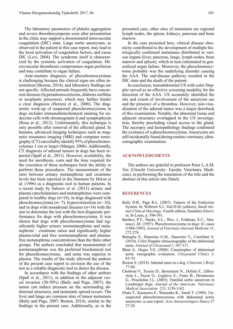
An abdominal ultrasonographic (US) examination was performed using a multiple frequency (5-8 MHz) convex transducer. Imaging results revealed moderate peritoneal effusion, bilaterally enlarged kidneys (di-mensions of the left and right kidney were 5.24 cm and 5.21 cm, respectively; the normal reference in-terval for dogs weighing 0-4 kg is 3.2-3.3 cm) (Finn- Bodner, 1995) with mild left and right pyelectasis and urolithiasis in the bladder and the prostatic urethra. A hypoechoic, irregular nodular lesion (3.08 cm x 3.11 cm) was present in the pancreas. The pancreatic le-sion was confirmed as a metastasis with necrosis at histopathology. Multiple enlarged lymph nodes with dimensions varying between 1.13 cm and 2.18 cm were visualized. The ultrasonographic features of the lymph nodes were that they were round in shape, hy-poechoic relative to the surrounding fat and showed a homogeneous echotexture. Additionally, US exami-nation revealed a pulsatile anechoic structure, which reflected a focal dilation of the abdominal aorta (4.39 cm x 3.94 cm) at the level of the branching of the left celiac artery (Figure 1A). Within the dilation, the blood flow was turbulent, with an appearance partial-ly resembling a yin-yang sign; this pattern is observed on color Doppler as blood within one side of the an-eurysm travelling towards the probe (colored red) and blood on the other side travelling away from the probe (colored blue) (Figure 1B). Additionally, remarkable invasion of the aorta by a hypoechoic thrombus was visualized (Figure 1A). Standard transverse and lon-gitudinal views of the aneurysm components by US followed previous descriptions in the literature (Bhatt and Dogra, 2008). On the basis of the US findings, the diagnosis of an aortic aneurysm was established.
The patient’s initial treatment was supportive and consisted of isotonic crystalloids (lactated Ringer’s solution; sodium chloride 0.9% 40 mL/kg IV per day)
Figure 1B. Color Doppler scan of the unruptured AAA (partial yin-yang pattern) (white arrow) (B.A. Rodri-gues).
Figure 1A. B-mode scan of AAA (4.39 cm x 3.94 cm) in a male Yorkshire terrier. A large thrombus (T) surround-ing the central lumen lines the aneurysmal wall (B.A. Rodrigues).
Vlaams Diergeneeskundig Tijdschrift, 2017, 86 101
administered in an alternately basis, empirical anti-biotic treatment (enrofloxacin 2.5% and amoxicillin, 5 mg/kg sid SC and 10 mg/kg bid SC, respectively), fructose with vitamin B complex (one ampule IV per day), ondansetron (0.1 mg/kg bid IV) and predniso-lone (1 mg/kg IM per day). After two days without signs of clinical improvement, the dog died. A full necropsy was then authorized and performed.
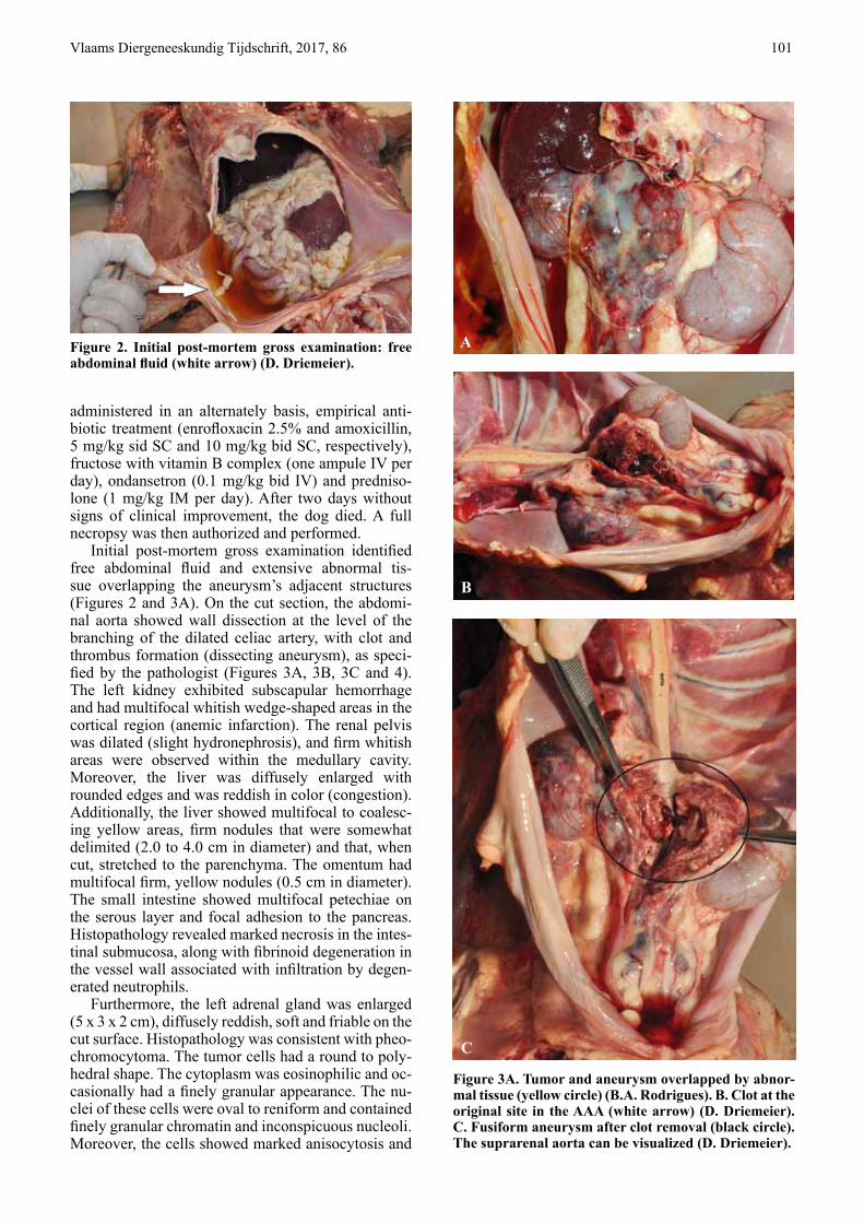
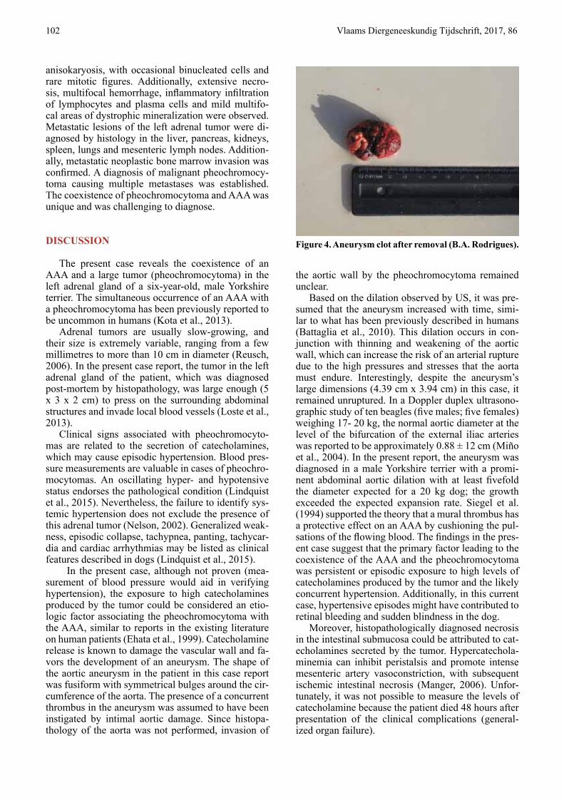
Initial post-mortem gross examination identified free abdominal fluid and extensive abnormal tis-sue overlapping the aneurysm’s adjacent structures (Figures 2 and 3A). On the cut section, the abdomi-nal aorta showed wall dissection at the level of the branching of the dilated celiac artery, with clot and thrombus formation (dissecting aneurysm), as speci-fied by the pathologist (Figures 3A, 3B, 3C and 4). The left kidney exhibited subscapular hemorrhage and had multifocal whitish wedge-shaped areas in the cortical region (anemic infarction). The renal pelvis was dilated (slight hydronephrosis), and firm whitish areas were observed within the medullary cavity. Moreover, the liver was diffusely enlarged with rounded edges and was reddish in color (congestion). Additionally, the liver showed multifocal to coalesc-ing yellow areas, firm nodules that were somewhat delimited (2.0 to 4.0 cm in diameter) and that, when cut, stretched to the parenchyma. The omentum had multifocal firm, yellow nodules (0.5 cm in diameter). The small intestine showed multifocal petechiae on the serous layer and focal adhesion to the pancreas. Histopathology revealed marked necrosis in the intes-tinal submucosa, along with fibrinoid degeneration in the vessel wall associated with infiltration by degen-erated neutrophils.
Furthermore, the left adrenal gland was enlarged (5 x 3 x 2 cm), diffusely reddish, soft and friable on the cut surface. Histopathology was consistent with pheo-chromocytoma. The tumor cells had a round to poly-hedral shape. The cytoplasm was eosinophilic and oc-casionally had a finely granular appearance. The nu-clei of these cells were oval to reniform and contained finely granular chromatin and inconspicuous nucleoli. Moreover, the cells showed marked anisocytosis and
Figure 2. Initial post-mortem gross examination: free abdominal fluid (white arrow) (D. Driemeier).
Figure 3A. Tumor and aneurysm overlapped by abnor-mal tissue (yellow circle) (B.A. Rodrigues). B. Clot at the original site in the AAA (white arrow) (D. Driemeier). C. Fusiform aneurysm after clot removal (black circle). The suprarenal aorta can be visualized (D. Driemeier).
A
B
C
102 Vlaams Diergeneeskundig Tijdschrift, 2017, 86
anisokaryosis, with occasional binucleated cells and rare mitotic figures. Additionally, extensive necro-sis, multifocal hemorrhage, inflammatory infiltration of lymphocytes and plasma cells and mild multifo-cal areas of dystrophic mineralization were observed. Metastatic lesions of the left adrenal tumor were di-agnosed by histology in the liver, pancreas, kidneys, spleen, lungs and mesenteric lymph nodes. Addition-ally, metastatic neoplastic bone marrow invasion was confirmed. A diagnosis of malignant pheochromocy-toma causing multiple metastases was established. The coexistence of pheochromocytoma and AAA was unique and was challenging to diagnose.
DISCUSSION
The present case reveals the coexistence of an AAA and a large tumor (pheochromocytoma) in the left adrenal gland of a six-year-old, male Yorkshire terrier. The simultaneous occurrence of an AAA with a pheochromocytoma has been previously reported to be uncommon in humans (Kota et al., 2013).
Adrenal tumors are usually slow-growing, and their size is extremely variable, ranging from a few millimetres to more than 10 cm in diameter (Reusch, 2006). In the present case report, the tumor in the left adrenal gland of the patient, which was diagnosed post-mortem by histopathology, was large enough (5 x 3 x 2 cm) to press on the surrounding abdominal structures and invade local blood vessels (Loste et al., 2013).
Clinical signs associated with pheochromocyto-mas are related to the secretion of catecholamines, which may cause episodic hypertension. Blood pres-sure measurements are valuable in cases of pheochro-mocytomas. An oscillating hyper- and hypotensive status endorses the pathological condition (Lindquist et al., 2015). Nevertheless, the failure to identify sys-temic hypertension does not exclude the presence of this adrenal tumor (Nelson, 2002). Generalized weak-ness, episodic collapse, tachypnea, panting, tachycar-dia and cardiac arrhythmias may be listed as clinical features described in dogs (Lindquist et al., 2015).
In the present case, although not proven (mea-surement of blood pressure would aid in verifying hypertension), the exposure to high catecholamines produced by the tumor could be considered an etio-logic factor associating the pheochromocytoma with the AAA, similar to reports in the existing literature on human patients (Ehata et al., 1999). Catecholamine release is known to damage the vascular wall and fa-vors the development of an aneurysm. The shape of the aortic aneurysm in the patient in this case report was fusiform with symmetrical bulges around the cir-cumference of the aorta. The presence of a concurrent thrombus in the aneurysm was assumed to have been instigated by intimal aortic damage. Since histopa-thology of the aorta was not performed, invasion of
the aortic wall by the pheochromocytoma remained unclear.
Based on the dilation observed by US, it was pre-sumed that the aneurysm increased with time, simi-lar to what has been previously described in humans (Battaglia et al., 2010). This dilation occurs in con-junction with thinning and weakening of the aortic wall, which can increase the risk of an arterial rupture due to the high pressures and stresses that the aorta must endure. Interestingly, despite the aneurysm’s large dimensions (4.39 cm x 3.94 cm) in this case, it remained unruptured. In a Doppler duplex ultrasono-graphic study of ten beagles (five males; five females) weighing 17- 20 kg, the normal aortic diameter at the level of the bifurcation of the external iliac arteries was reported to be approximately 0.88 ± 12 cm (Miño et al., 2004). In the present report, the aneurysm was diagnosed in a male Yorkshire terrier with a promi-nent abdominal aortic dilation with at least fivefold the diameter expected for a 20 kg dog; the growth exceeded the expected expansion rate. Siegel et al. (1994) supported the theory that a mural thrombus has a protective effect on an AAA by cushioning the pul-sations of the flowing blood. The findings in the pres-ent case suggest that the primary factor leading to the coexistence of the AAA and the pheochromocytoma was persistent or episodic exposure to high levels of catecholamines produced by the tumor and the likely concurrent hypertension. Additionally, in this current case, hypertensive episodes might have contributed to retinal bleeding and sudden blindness in the dog.
Moreover, histopathologically diagnosed necrosis in the intestinal submucosa could be attributed to cat-echolamines secreted by the tumor. Hypercatechola-minemia can inhibit peristalsis and promote intense mesenteric artery vasoconstriction, with subsequent ischemic intestinal necrosis (Manger, 2006). Unfor-tunately, it was not possible to measure the levels of catecholamine because the patient died 48 hours after presentation of the clinical complications (general-ized organ failure).
Figure 4. Aneurysm clot after removal (B.A. Rodrigues).
Vlaams Diergeneeskundig Tijdschrift, 2017, 86 103
The laboratory parameters of platelet aggregation and severe thrombocytopenia soon after presentation in the clinic may support a disseminated intravascular coagulation (DIC) state. Large aortic aneurysms, as observed in the patient in this case report, may lead to the local activation of coagulation factors, and cause DIC (Levi, 2004). The syndrome itself is character-ized by the systemic activation of coagulation. Mi-crovascular thrombosis compromises organ perfusion and may contribute to organ failure.
Ante-mortem diagnosis of pheochromocytomas is challenging because the clinical signs are often in-termittent (Boston, 2014), and laboratory findings are not specific. Affected animals frequently have concur-rent diseases (hyperadrenocorticism, diabetes mellitus or neoplastic processes), which may further hinder a clear diagnosis (Herrera et al., 2008). The diag- nostic work-up of suspected pheochromocytoma in dogs includes immunohistochemical staining for en-docrine cells with chromogranin A and synaptophysin (Rosa et al., 2012). Unfortunately, this technique is only possible after removal of the affected gland. In humans, advanced imaging techniques such as mag-netic resonance imaging (MRI) and computer tomo- graphy (CT) can reliably identify 95% of pheochromo- cytomas 1 cm or larger (Manger, 2006). Additionally, CT diagnosis of adrenal tumors in dogs has been re-ported (Spall et al., 2011). However, availability, the need for anesthesia, costs and the time required for the execution of these techniques limit the ability to perform these procedures. The measurement of the ratio between urinary metanephrine and creatinine levels has been reported in the literature by Héron et al. (1996) as a diagnostic tool in human patients. In a recent study by Salesov et al. (2015) urinary and plasma catecholamines and metanephrines were com-pared in healthy dogs (n=10), in dogs diagnosed with pheochromocytoma (n= 7), hypercortisolism (n= 10), and in dogs with nonadrenal diseases (n=14) with the aim to determine the test with the best diagnostic per-formance for dogs with pheochromocytoma. It was shown that dogs with pheochromocytoma had sig-nificantly higher urinary normetanephrine and meta-nephrine : creatinine ratios and significantly higher plasma-total and free normetanephrine and plasma-free metanephrine concentrations than the three other groups. The authors concluded that measurement of normetanephrine was the preferred biochemical test for pheochromocytoma, and urine was superior to plasma. The results of the study allowed the authors of the present case report to envision the use of the test as a reliable diagnostic tool to detect the disease.
In accordance with the findings of other authors (Spall et al., 2011), in addition to the adjacent ves-sel invasion (30-50%) (Baily and Page, 2007), the tumor can induce pressure on the surrounding ab-dominal structures, and metastatic spread occurs. The liver and lungs are common sites of tumor metastasis (Baily and Page, 2007; Boston, 2014), similar to the findings in the present case. Additionally, as in the
presented case, other sites of metastasis are regional lymph nodes, the spleen, kidneys, pancreas and bone marrow.
In the case presented here, clinical disease chro-nicity contributed to the development of multiple his-tologically confirmed metastases distributed in vari-ous organs (liver, pancreas, lungs, lymph nodes, bone marrow and spleen), which in turn culminated in gen-eralized organ failure. Moreover, the pheochromocy-toma probably was the underlying disorder causing the AAA. The end-disease pathway resulted in the DIC state and the death of the patient.
In conclusion, transabdominal US with color Dop-pler served as an effective screening modality for the detection of the AAA. US accurately identified the site and extent of involvement of the aneurysm sac and the presence of a thrombus. However, non-visu-alization of the adrenal tumor was a major drawback of this examination. Notably, the abnormal tissue and adjacent structures overlapped in the US investiga-tion, thereby precluding visual access to the gland. The necropsy and histopathology findings confirmed the existence of a pheochromocytoma. Aneurysms are still incidentally found during routine veterinary, ultra- sonographic examination.
ACKNOWLEDGMENTS
The authors are grateful to professor Peter L.A.M. Vos (Utrecht University- Faculty Veterinary Medi-cine), in performing the translation of the title and the abstract of this article into Dutch.
REFERENCES
Baily D.B., Page R.L. (2007). Tumors of the Endocrine System. In: Withrow S.J., Vail D.M. (editors). Small Ani-mal Clinical Oncology. Fourth edition, Saunders-Elsevi-er, St Louis, p. 590-591.
Barthez, P.Y., Marks, S.L., Woo, J., Feldman, E.C., Mat-teucci ,M. (1997). Pheochromocytoma in dogs: 61 cases (1984-1995). Journal of Veterinary Internal Medicine 11, 272-278.
Battaglia S., Danesino G.M., Danesino V., Castellani S. (2010). Color Doppler ultrasonography of the abdominal aorta. Journal of Ultrasound 1, 107-117.
Bhatt S., Dogra V.S. (2008). Catastrophes of abdominal aorta: sonographic evaluation. Ultrasound Clinics 3, 83–91.
Boston S. (2014). Adrenal mass in a dog. Clinician´s Brief, 77-80.
Chetboul V., Tessier D., Borenstein N., Delisle F., Zilber-stein L., Payen G., Leglaive E., Franc B., Derumeaux G., Pouchelon J.L. (2003). Familial aortic aneurysm in Leonberger dogs. Journal of the American Veterinary Medical Association 223, 1159-1162.
Ehata T., Karasawa F., Watanabe K., Satoh T. (1999). Un-suspected pheochromocytoma with abdominal aortic aneurysm--a case report. Acta Anestesiologica Sinica 37 27-28.

104 Vlaams Diergeneeskundig Tijdschrift, 2017, 86
Finn-Bodner S.T. (1995). The kidneys. In: Cann C.C. (edi-tor). Practical Veterinary Ultrasound. First edition. Wil-liams & Wilkins Philadelphia, p.166.
Gershenson R.T., Melidone R., Sutherland-Smith J., Rog-ers C.L. (2011). Abdominal aortic aneurysm associated with systemic fungal infection in a dog. Journal of the American Animal Hospital Association 47, 45-49.
Héron, E., Chatellier, G., Billaud, E,, Foos, E., Plouin, P.F. (1996). The Urinary Metanephrine-to-Creatinine Ratio for the Diagnosis of Pheochromocytoma. Annals of In-ternal Medicine 125, 300-303.
Herrera, M.A., Mehl, M.L., Kass, P.H., Pascoe, P.J., Feld-man, E.C., Nelson, R.W. (2008). Predictive factors and the effect of phenoxybenzamine on outcome in dogs un-dergoing adrenalectomy for pheochromocytoma. Jour-nal of Veterinary Internal Medicine 22, 1333-1339.
Kanematsu Y., Kanematsu M., Kurihara C., Tsou T.L., Nuki Y., Liang E., Makino H., Hashimoto T. (2010). Pharma-cologically Induced Thoracic and Abdominal Aortic An-eurysms in Mice. Hypertension 55, 1267-1274.
Kirberger R.M., Stander N., Cassel N., Pazzi P., Mukorera V., Christie J., Carstens A., Dvir E. (2013). Computed tomographic and radiographic characteristics of aortic le-sion in 42 dogs with spirocercosis. Veterinary Radiology & Ultrasound 54, 212-222.
Kota S. K., Kota S. K., Meher L. K., Jammula S., Mohapa-tra S., Modi K. D. (2013). Coexistence of pheochromo-cytoma with abdominal aortic aneurysm: An untold asso-ciation. Annals of Medical and Health Science Research 3, 258-261.
Levi, M. Current understanding of disseminated intravas-cular coagulation (2004). British Journal of Haematol-ogy 124, 567–576.
Lindquist E., Frank J., Modler P., Lobetti R. (2015). Adre-nal tumors. In: The Curbside Guide-Diagnosis & Treat-ment of Common Sonographically Detected Disease: Canine & Feline. First edition. Sonopath, New Jersey, p. 121-128.
Loste A., Borobia M., Borobia M., Lacasta D., Carbonell M., Basurco A., Marca M.C. (2013). Adrenal gland tu-mours. Different clinical presentations in three dogs: a case report. Veterinarni Medicina 58, 377-384.
Manger, W.M. (2006). Diagnosis and management of pheo-chromocytoma – recent advances and current concepts. Kidney International 70, 30–35.
Michel J.-B. (1998). Acquired abdominal aortic aneurysm. Nephrology Dialysis Transplantation 13, 20-24.
Miño N., Espino L., Suárez M., Santamarina G., Barreiro A. (2004). Estudio de la aorta abdominal mediante dop-pler espectral pulsado en perros. Archivos de Medicina Veterinária 36, 87-92.
Murata Y., Chambers J.K., Uchida K., Nakashima K., Hanafusa Y., Ikezawa M., Sugita T., Nakayama H. (2015). Mycotic aneurysm caused by Graphium species
in a dog. The Journal of Veterinary Medical Science 77, 1285–1288.
Myers N.C. (1997). Adrenal incidentalomas. Veterinary Clinics of North America: Small Animal Practice 27, 381–399.
Nelson R.W. Diagnostic Approach to the Incidental Ad-renal Mass. In WSAVA 2002 Vin. Available from http://www.vin.com/apputil/content/defaultadv1.aspx?pId=11147&catId=29491&id=3846171&print=1 Acessed 12 Oct.2016.
Reusch, CE. Adrenal tumors in dogs. In WSAVA/FE-CAV/CSAVA 2006. Available from http://www.ivis.org/proceedings/wsava/2006/lecture9/Reusch2.pdf?LA=1 Acessed 05 Oct. 2015.
Rizas K.D., Ippagunta N., Tilson M.D. III. (2009). Immune cells and molecular mediators in the pathogenesis of the abdominal aortic aneurysm. Cardiology in Review 17, 201-210.
Rosa, C., Schoeman, J.P., Dvir, E. (2012). Budd-Chiari-like syndrome associated with a pheochromocytoma invad-ing the right atrium in a dog. Israel Journal of Veterinary Medicine 67, 180-185.
Sakalihasan N., Limet R., Defawe O.D. (2005). Abdominal aortic aneurysm. Lancet 365, 1577-1589.
Salesov, E., Boretti, F.S., Sieber-Ruckstuhl, N.S., Rentsch, K.M., Riond, B., Hofmann-Lehmann, R.,. Kircher, P., Grouzmann, E. Reusch, C.E. (2015). Urinary and plasma catecholamines and metanephrines in dogs with pheo-chromocytoma, hypercortisolism, nonadrenal disease and in healthy dogs. Journal of Veterinary Internal Medi-cine 29, 597-602.
Siegel C.L., Cohan R.H., Korobkin M., Alpern M.B., Cour-neya D.L., Leder R.A. (1994). Abdominal aortic aneu-rysm morphology: CT features in patients with ruptured and nonruptured aneurysms. American Journal of Roent-genology 163, 1123–1129.
Spall B., Chen A.V., Tucker R.L., Lahmers K.K., Righter D.J., Hayles J. (2011). Imaging diagnosis-metastatic ad-renal pheochromocytoma in a dog. Veterinary Radiology & Ultrasound 52, 534-537.
Van der Merwe L.L., Kirberger R.M., Clift S., Williams M., Keller N., Naidoo V. (2008). Spirocerca lupi infection in the dog: a review. The Veterinary Journal 176, 294-309.
Vardulaki K.A., Walker N.M., Day N.E., Duffy S.W., Ash-ton H.A., Scott R.A.P. (2000). Quantifying the risks of hypertension, age, sex and smoking in patients with ab-dominal aortic aneurysm. British Journal of Surgery 87, 195-200.
Waldrop J.E., Stoneham A.E., Tidwell A.S., Jakowski R.M., Rozanski E.A., Rush J.E. (2003). Aortic dissec-tion associated with aortic aneurysms and posterior pa-resis in a dog. Journal of Veterinary Internal Medicine 17, 223–229.