An investigation of an infection with a protozoan parasite...
92
An investigation of an infection with a protozoan parasite causing mortalities in Little Penguins (Eudyptula minor) on Penguin Island, Western Australia Kamahl Campbell, BA BSc BVMS (Hons) This thesis is presented for the degree of Master of Science of Murdoch University, 2015.
Transcript of An investigation of an infection with a protozoan parasite...
An investigation of an infection with a protozoan parasite causing mortalities in
Little Penguins (Eudyptula minor) on Penguin Island, Western Australia
Kamahl Campbell, BA BSc BVMS (Hons)
This thesis is presented for the degree of Master of Science of Murdoch University, 2015.


I declare that this thesis is my own account of my research and contains as its main content work
that has not previously been submitted for a degree at any tertiary education institution.
....................................
Kamahl Campbell
1
Abstract
Since at least 2001, routine post mortems of deceased penguins from Penguin Island, Western
Australia, have been conducted at Murdoch University Veterinary Hospital (MUVH). In late 2011 and
early 2012, a cluster of 12 cases presented with similar and characteristic gross and microscopic
changes, namely birds in good body condition with hepatomegaly and splenomegaly, multifocal
hepatic and splenic necrosis and numerous, small, 1-2µm diameter protozoan parasites within the
necrotic foci. A review of earlier reports in the MUVH archive identified isolated similar cases from
2006 and 2008, which had been provisionally diagnosed as Avian Malaria, that is, Plasmodium spp.
infection. An investigation was established in order to: a) definitively identify the parasite causing
the mortalities, with the additional aims of: b) evaluating the live population of Penguin Island Little
Penguins for the presence of parasitaemia, and c) investigating the possibility that another bird
species present on Penguin Island might represent a reservoir of infection.
Ninety-four blood smears were made from 79 individual Little Penguins collected from winter to
summer of 2012 and in the early spring of 2013. One smear identified intraerythrocytic organisms
consistent with the blood stages of an apicomplexan parasite, for example, merozoites or early
gametocytes of Haemoproteus or Plasmodium, or merozoites or sporozoites of Babesia. Fifty-one
blood smears were made from 51 Bridled Terns (Sterna anaethetus) captured during two visits to
the island in November 2012 and March 2013, with no parasites detected in these smears.
Electron microscopy of the protozoan parasite identified it as belonging to the phylum Apicomplexa.
Further identification to the level of genus was not possible.
Molecular identification of the parasite using Polymerase Chain Reaction (PCR) methodology gave
inconsistent results. PCR performed by an independent laboratory identified a novel Haemoproteus
spp. organism in 4 of 10 cases from this group; however, these results could not be replicated in our
laboratory. Additional PCR using a variety of primers aimed at detecting members of the
Apicomplexa identified a parasite from the family Sarcocystidae, which was subsequently identified
as Toxoplasma. Immunohistochemistry of formalin fixed tissues also identified Toxoplasma in the
hepatic and splenic lesions.

2
The distinctive mortalities which were observed in this group of penguins, and which have occurred
sporadically since, appear to be attributable to a fulminant toxoplasmosis, with or without a
concurrent haemoproteosis in some cases. The significance of the apparent polyparasitism in some
of the birds is unknown, as the relative contribution of concurrent Haemoproteus infection to the
lesion aetiopathogenesis cannot be quantified at this time. Though the clinical signs of infection are
unknown, the gross and microscopic appearance at post mortem is sufficiently characteristic to
allow a diagnosis to be made on these features. Definitive confirmation of infection may be made by
immunohistochemistry or PCR.

3
Table of Contents
An investigation of an infection with a protozoan parasite causing mortalities in Little
Penguins (Eudyptula minor) on Penguin Island, Western Australia .......................................... 0
Abstract ......................................................................................................................................................... 1
Acknowledgments ...................................................................................................................................... 5
1. Literature review ................................................................................................................................... 7
Preamble .................................................................................................................................................................................7
Little Penguins ......................................................................................................................................................................7
Penguin Island and threats to the population ........................................................................................................8
Haemoparasitic diseases of birds .............................................................................................................................. 13
Haemoparasitic diseases of penguins...................................................................................................................... 15
Other protozoan parasites infecting penguins .................................................................................................... 17
Summary .............................................................................................................................................................................. 17
2. Post mortem examinations ............................................................................................................... 19
Introduction ....................................................................................................................................................................... 19
Materials and methods .................................................................................................................................................. 20
Routine penguin necropsy procedure ............................................................................................................. 20
Results ................................................................................................................................................................................... 21
Case1: 06/1172 ........................................................................................................................................................... 22
Case 2: 08/1075 .......................................................................................................................................................... 22
Case 3: 11/484 ............................................................................................................................................................ 23
Case 4: 11/624 ............................................................................................................................................................ 24
Case 5: 11/626 ............................................................................................................................................................ 25
Cases 6 and 7: 11/627 and 11/628 ................................................................................................................... 26
Case 8:12/020 ............................................................................................................................................................. 26
Case 9: 12/021 ............................................................................................................................................................ 27
Case 10: 12/338 .......................................................................................................................................................... 28
Case 11: 12/362 .......................................................................................................................................................... 29
Case 12: 12/375 .......................................................................................................................................................... 29
Case 13: 12/385 .......................................................................................................................................................... 32
Case 14: 12/473 .......................................................................................................................................................... 32
Case 15: 12/517 .......................................................................................................................................................... 33
Case 16: 12/524 .......................................................................................................................................................... 34
Case 17: 13/081 .......................................................................................................................................................... 35

4
Case 18: 13/670 .......................................................................................................................................................... 36
Other ................................................................................................................................................................................. 36
Summary .............................................................................................................................................................................. 39
3. Fieldwork ............................................................................................................................................... 42
Introduction ....................................................................................................................................................................... 42
Materials and methods .................................................................................................................................................. 43
Blood collection and blood smear preparation ........................................................................................... 43
Results ................................................................................................................................................................................... 48
Penguins ......................................................................................................................................................................... 48
Bridled Terns ............................................................................................................................................................... 49
Summary .............................................................................................................................................................................. 49
4. Electron microscopy............................................................................................................................ 51
Introduction ....................................................................................................................................................................... 51
Materials and Methods .................................................................................................................................................. 51
Results ................................................................................................................................................................................... 52
Discussion ............................................................................................................................................................................ 58
5. Molecular Identification of the Parasite ....................................................................................... 61
Introduction and Aims ................................................................................................................................................... 61
Materials and Methods .................................................................................................................................................. 62
DNA extraction ............................................................................................................................................................ 62
Polymerase Chain Reaction ................................................................................................................................... 65
DNA sequencing .......................................................................................................................................................... 68
Results ................................................................................................................................................................................... 69
Immunohistochemistry .................................................................................................................................................. 72
Results ............................................................................................................................................................................. 73
Discussion ............................................................................................................................................................................ 75
6. Discussion .............................................................................................................................................. 78
References: ................................................................................................................................................. 84

5
Acknowledgments
Thanks are due to:
My two excellent supervisors, Nahiid Stephens and Belinda Cannell, for your enthusiasm, direction,
wisdom, advice, readiness to discuss the new developments and follow where they apparently were
leading, for good company and for the occasional gentle prod when it was required. This project
took more of a winding (perhaps even anfractuous) path than anticipated, but it was never a chore,
mostly thanks to your efforts and support in keeping the workload manageable.
Nic Dunlop and Sandy McNeill, for your indispensable help with collecting samples from the Bridled
Terns, and, though it didn’t quite work out as planned, from the Caspian Terns.
Peter Fallon for demystifying electron microscopy, and Louise Pallant for demystifying the electron
micrographs.
Andrea Paparini, for all of your guidance and practical help making the PCR stage of the investigation
so much better than it might have been and no worse than it had to be; Adriana Botero-Gomez for
cheerfully performing the Toxoplasma PCR; Tim Hyndman for advice, encouragement and lab space;
and Peter Irwin for chipping in with vital contributions when needed most.
Michael Slaven and Gerard Spoelstra for your virtuosity in the art and science of histochemistry, and
for taking an interest in the penguins.
Mandy O’Hara for your practical advice and your straight talking at all times.
The several West Australian government departments (CALM, DEC and DPaW) that submitted and
paid for many of the penguin post mortems in the first place.
All in the pathology department who haven’t already been mentioned (Jo Moore, Cheryl Moller,
Celia Smuts, Zi Lim) for helping to make sure that none of this work was undertaken in a
hypoglycaemic or an insufficiently caffeinated state.
And above all to Rachel, Elliot and Alex for giving me a reason.

6

7
1. Literature review
Preamble
The subject of and material for this thesis arose from observations made during routine post
mortem examinations of Little Penguins (Eudyptula minor) conducted at Murdoch University
Veterinary Hospital. The penguins, most of which were found deceased on Penguin Island as well as
along the coast in the Perth and Rockingham areas, were, for the most part, identifiable as
inhabitants of Penguin Island either by wingband or microchip, or by virtue of where they were
found. In those birds that lacked definitive identifiers, a small number may have originated from
nearby Garden Island instead.
Little Penguins
Little Penguins are the smallest species of penguin, and the only penguin native to Australia. Their
distribution takes in the southern coastline of mainland Australia, the coastlines of Tasmania and the
Bass Strait islands, most of New Zealand, and a number of its associated islands, including Stewart
and Chatham Islands. The total population worldwide is estimated to be 500-600,000 individuals,
with the largest populations on the coasts of Victoria, the Bass Strait islands and Tasmania34,79.
According to the International Union for Conservation of Nature, the current conservation status of
this species is designated Least Concern1. However, many individual colonies of these birds are
threatened, particularly mainland colonies, and their overall numbers are considered to be in
decline2,8,34,44,79,88.
As true seabirds, Little Penguins spend extensive periods of time at sea, coming to land principally
for the purposes of breeding and their annual moult. Locally, the breeding cycle sees courtship,
laying and chick rearing occurring from mid-autumn to early summer, with eggs being laid from as
early as April to as late as December51. Egg incubation, and the subsequent guarding of the chicks
after hatching, requires an equal contribution from both parents. Little Penguins typically lay eggs in
clutches of 2, with a 2-7 day interval between the first and the second egg, though occasionally only
a single egg is produced48,88. Successful hatching of the eggs and rearing to fledglings depends on
the capacity of a pair to provide continuous protection for the approximately 35 day incubation
period10,48, and the post-hatching guard stage, which lasts from 8 to 38 days, during which the chicks
are not left alone10,36. The parents take turns foraging for food and attending the nest; when food is
scarce, the foraging parent may be unable to gather adequate food near enough to the nest that it is
able to return in time to relieve the nesting parent, and in this case hunger eventually forces the

8
nesting parent to abandon the eggs or chicks. The diet of the Little Penguin may include many small
species of fish, squid, prawns and other small crustaceans7,65,86, but among the local birds the bulk of
their diet is made up of the sandy sprat (Hyperlophus vittatus), southern sea garfish (Hyporhamphus
melanochir), blue sprat (Spratelloides robustus) and pilchard (Sardinops neopilchardus, Sardinops
sagax)50,68. Thus, the breeding success of the penguins is closely linked to the abundance of these
fish species in their local foraging grounds. And obviously, breeding success is pivotal to the ability
of a colony to maintain its numbers or to replenish numbers after a population decline.
Populations of Little Penguins have been reported to be in decline from a number of locations
around Australia2,8,88. In many cases, the causes for the population declines have been attributed to
human activities, such as habitat alteration, reduction in fish stocks, pollution or entanglement in
fishing paraphernalia56,79. In one study of 213 Little Penguins from the Otago region of New
Zealand’s South Island, the most commonly identified causes of death were starvation (16%); road,
rail or unspecified trauma (23%); and predation by dog or ferret (23%)38. Since they are vulnerable
to predation from introduced species, Little Penguins have abandoned or been forced from most
onshore nesting sites and now nest mainly on offshore islands.
Penguin Island and threats to the population
The largest colony of Little Penguins in Western Australia breeds on Penguin Island, located roughly
40km south of the city of Perth (see Fig 2)56. Besides this large colony, other smaller colonies in the
local area are found on Garden Island and Carnac Island a few kilometres to the north from Penguin
Island. Other Western Australian colonies are found on islands near Augusta, Albany and in the
Recherche Archipelago near Esperance (see Fig 1).

9
Figure 1 – Location of substantial colonies of Little Penguins in Western Australia, Penguin Island in
red, other colonies in yellow
Penguin Island itself is a limestone island, one of the largest of a chain remaining after submersion of
a dune ridge system now lying offshore from the City of Rockingham62. Many of the islands in this
chain and the surrounding sea are encompassed within the Shoalwater Islands Marine Park, a
conservation park, gazetted in 1990, which extends from Point Peron in the north to Warnbro Sound
in the south3. Penguin Island is the largest of the islands within the Marine Park. The management
of the marine park currently falls to the Western Australian government’s Department of Parks and
Wildlife, formerly the Department of Environment and Conservation, formerly the Department of
Conservation and Land Management – for simplicity, these several incarnations of the responsible
government agency will be referred to as ‘the Department’. In the Department’s current
management plan, Penguin Island is designated as lying within a Special Purpose Zone (Wildlife
Conservation)3. Though it is uninhabited, and in fact only ever had one long-term resident (Seaforth
McKenzie, who lived on the island from about 1914 to 1926), Penguin Island has a long history of use
as a recreation and holiday destination. Recreational use continues to this day, except during the
peak breeding season of the Little Penguins, and access to the island for the community is identified
as an important component of the Department’s management plan. Under the terms of the plan,

10
the status of the colony of Little Penguins is one of several Key Performance Indicators used to gauge
the efficacy of the management of the Marine Park. Specifically, the breeding success and
abundance of the birds, as well as the number of entanglements of penguins in fishing equipment
per year, are the stated measures by which the colony’s health is quantified3.
Figure 2 – Location of Penguin Island (identified by arrows) in relation to Perth and to the city of
Rockingham, Western Australia
The geology of Penguin Island is not well-suited to typical Little Penguin nesting habits, consisting as
it does of low shrubs growing on a sandy substrate. Here, the penguins tend not to make burrows as
they do in most other colonies, since the sand is too unstable for this purpose; instead, the majority
of birds nest under the low, scrubby Tetragonia or Rhagodia bushes which cover the island, with a
few making nest sites from other vegetation, relatively infrequent natural structures such as
limestone caves, or the remnants of human building materials and structures49. Since 1986, artificial
nest-boxes made of plywood have been placed at various sites around the island with recorded
occupancy rates ranging from 49% to 94%49. The good occupancy rates for the nest boxes suggest
that they are at least adequate for the penguins’ requirements, though there is evidence that
redesign of the boxes to prevent high ambient temperatures, with the potential to induce
hyperthermia, may improve their utility75.

11
Naturally enough, the Little Penguins are recognised to play a key ecological role in the marine park.
They are, however, notable for other reasons as well. Not only does this colony lie at the western,
and near the northern, limit of the species’ range56, but the birds are also significantly larger than
those from other populations87, and this colony also has a longer breeding season than many others
do. By these and other criteria, this colony is given the highest conservation status out of 256 Little
Penguin colonies identified Australia-wide18. Given the importance of this colony, and its fortuitous
location near Western Australia’s largest population centre and academic institutes, there has been
extensive academic and, more recently, government research and monitoring activity conducted
over a relatively extended period of time11-13,16,22,49-51,59,75,87.
In the last few decades, several population estimates have been made for different locations on
Penguin Island, as well as for the whole island, using methodologies of different degrees of
accuracy87 12. Conveniently, the penguins display a great deal of site fidelity in their choice of landing
sites, reliably departing from and arriving at the same beach for the duration of the breeding
season12,51, which allows the island, small though it is, to be evaluated as a group of population
zones. Estimates using a mark-recapture method that recorded birds returning to the island at five
landing sites gave partial population estimates of approximately 700 in 1987, 425 in 1988, 620 in
1989, 450 in 1990 and 525 in 199187. More recently, the most accurate island-wide population
estimates have been made using a mark-recapture methodology which involved capturing penguins
at four landing sites, repeating the capture process three times (2007) or four times (2008) at two to
three week intervals, and evaluating the data in combination with a head-count of penguins arriving
at a total of 14 sites12. By this method, the total population of Penguin Island was estimated to be
2369 individuals in 2007. The following year, the total population was estimated at 2069 birds. In
2011, another population estimate was obtained using a slightly modified methodology. Due to
some violations in the assumptions of the models used with the 2011 data, a Robust Design model
was used to compare abundance between years. With these adjustments, the comparable
population estimates over a shorter time frame were 1695, 1413 and 964 penguins in 2007, 2008
and 2011 respectively (Cannell, unpubl. data). While these numbers are higher than those obtained
from the years 1987 to 1991, the earlier estimates are for a part of the island only, and are therefore
presumed to underestimate the total population; also, direct comparisons with the 1987 to 1991
estimates cannot be made, as the estimates were made using substantially different methodologies
(see12). Overall, the data suggest that the population is decreasing substantially, and to make
matters worse, indicators of breeding success (the number of chicks produced per pair, the

12
proportion of eggs that hatched, the proportion of eggs that led to fledglings, and the average mass
of chicks at the time of fledging) have also declined in recent years in association with elevated sea
surface temperatures, suggesting that further population decline can be expected11.
The decline in population is attributable to a range of pressures that are reducing both the expected
life span of individual birds and the breeding success of those that remain. In recent years, poorer
breeding in this colony has been associated with higher sea surface temperatures and an increased
Leeuwin current11. The Leeuwin current is a nutrient-poor seasonal current which carries water
south from the tropical north, reducing the capacity of the water to support sea flora and fauna
alike11. This effect, combined with reduced fish stocks due to commercial and recreational fishing,
the disturbance of fish nurseries by water craft and their launch sites, and reduced water quality
have been identified as some of the most important anthropogenic threats to the penguins’
wellbeing3. As noted above, when local fish stocks are inadequate, penguins must forage further
afield which keeps them away from their nests for longer periods and reduces overall breeding
success (Cannell, unpubl. data). Other significant threats to individual penguins include trauma from
watercraft strikes, entanglement in plastic detritus, and land habitat disturbances, whether
intentional or inadvertent, from human visitors to Penguin Island57.
One component of the Department’s monitoring strategy is to request post mortem examinations to
determine the cause of death for Little Penguins found dead in Western Australia, with a particular
focus on those birds found between Perth and Dunsborough. The post mortems have been carried
out by veterinary pathologists at Murdoch University Veterinary Hospital (MUVH), and, over the past
ten years, the most common causes of death in this population have been found to be starvation,
traumatic injuries (frequently attributed to watercraft strike) and a range of infectious conditions
(Cannell et al, in prep). In 2011/2012, however, 12 penguins were found to have evidence of a
protozoan parasite infection, principally affecting the liver and spleen, which caused significant
necrosis of these tissues and which was deemed to have caused the deaths of the birds.
Morphologically, the parasites in the tissues were identifiable as apicomplexans, a group of protozoa
with a slightly elongate form and an eccentric nucleus. On the basis of the parasite morphology and
the pattern of infection predominantly affecting the reticuloendothelial system, the infections were
provisionally diagnosed as Avian malaria. Avian malaria, a haemoparasitic disease of birds caused by
Plasmodium species parasites, has a worldwide distribution6 and is common in captive penguin
populations (see discussion below), but by virtue of presenting as an outbreak associated with an
apparent spike in penguin mortality, it appeared to represent a new threat to this population. One-

13
off cases of suspected Avian malaria in Little Penguins had previously been diagnosed at MUVH in
2006 and 2008, though each diagnosis was made on histologic morphology alone, and neither these
nor the later cases had been confirmed to be Plasmodium infections. A complete discussion of the
cases forms the basis of Chapter 2 of this thesis.
Haemoparasitic diseases of birds
Haemoparasites are a closely related group of common vector-borne protozoan parasites infecting
birds, reptiles and mammals, with the common feature that at least one stage in their lifecycles
occurs in the host’s erythrocytes. Avian haemoparasites include species from the families
Haemospororida (e.g., Plasmodium, Haemoproteus and Leucocytozoon) and Aconoidasida (e.g.,
Babesia). Infection of free-living birds with parasites from the Haemospororida has been reported
worldwide and such infections appear to be common by most reports42,43,72,80,83. Note that the term
“Avian malaria” is used to refer specifically to infections with Plasmodium spp. parasites, though,
occasionally, infections with Haemoproteus spp. are also included under this general heading (for
example, 73,74). However, given that there are substantial differences in pathogenicity, vectors and
host specificity, the restricted definition has been argued to be both more appropriate and more
useful83. In this document the term will be applied to infections with Plasmodium spp. only;
haemoproteosis and leucocytozoonosis will be used to indicate infections with Haemoproteus and
Leucocytozoon respectively. Similarly, babesiosis indicates infection with Babesia spp.
Figure 3 - Generalised life cycle of haemosporidian parasites

14
The haemoparasite life cycle typically involves a vertebrate and an invertebrate host. The parasites
are invariably transmitted by an arthropod vector, which injects protozoal sporozoites into the
vertebrate host during feeding. The sporozoites migrate through tissues and in the blood to infect
macrophages and endothelial cells in various tissues (typically including liver, spleen, muscle and
lung) where they reproduce as merozoites. New merozoites emerge from ruptured host cells, infect
erythrocytes and develop as gametocytes, which are then ingested by the arthropod vector during a
blood meal. Note that a characteristic point of distinction between Haemoproteus and Plasmodium
is that Plasmodium parasites have additional asexual reproduction stages, which take place in
erythrocytes resulting in the further production of merozoites. Sexual reproduction with the
development of ookinetes and oocysts takes place in the arthropod’s intestinal tract, ultimately
producing schizonts which migrate to the arthropod’s salivary glands ready for transmission to a new
vertebrate host6,83. Plasmodium parasites are transmitted by mosquitoes, Haemoproteus are
transmitted by ceratopogonid and hippoboscid biting flies, and Leucocytozoon are transmitted by
Simuliid biting flies6. Babesia spp. are distinct in that they are pure red cell parasites, that is, they
have no tissue phase, and they are transmitted by ticks.
The clinical signs and post mortem findings arising from Plasmodium, Haemoproteus and
Leucocytozoon infections are relatable to the life cycle of the parasite in the vertebrate host.
Asexual reproduction of merozoites in macrophages of the liver and spleen lead to necrosis and
enlargement of these tissues with an associated inflammatory response and systemic illness5. The
erythrocyte stages induce the removal of infected erythrocytes from the circulation and haemolysis
in the liver and spleen, resulting in anaemia. In some cases, post mortem examination also reveals
intracoelomic haemorrhage from rupture of organs, often the liver31. Microscopically, the parasites
are detected as small bodies encysted in the cytoplasm of macrophages or free in necrotic tissues.
Parasites may also be seen in the cytoplasm of erythrocytes in blood smears or occasionally in
cytological preparations (impression smears or fine needle aspirate biopsies). Speciation of the
parasites may be done on erythrocyte stage (i.e., gametocyte) morphology83 or by molecular
techniques including Polymerase Chain Reaction, with or without sequencing,85 or other specific
tests such as serology. As a red cell parasite, the clinical signs of babesiosis are principally due to the
destruction or removal of erythrocytes from circulation and the generation of inflammatory
mediators. Typical clinical manifestations include anaemia, fever, jaundice, malaise, lethargy and
anorexia77.

15
Many Plasmodium species parasites have a low host specificity, with the result that cross-infection
between avian species occurs quite commonly (though there is substantial variation in specificity
between Plasmodium species)6,83. Among the most important in terms of avian disease worldwide
are Pl. relictum, which has been reported to infect at least 419 species from 70 avian families, and Pl.
elongatum, with a relatively restricted host range of 67 avian species6. Haemoproteus and
Leucocytozoon parasites, by contrast, tend to be highly host specific6,83. For the most part, this
means that the avian hosts are relatively well adapted to these parasites and infections are usually
subclinical, though not necessarily without health consequences6,70. Aberrant infections, however,
do occur, and disease and deaths due to haemoproteosis and leucocytozoonosis have been reported
in captive birds, especially in mixed aviaries and zoos where birds may be exposed to haemoparasite
species which they do not normally encounter31 20,61,63.
The prevalence of haemoparasite infections in seabirds is somewhat lower than that in other bird
populations, which has been attributed to factors directly and indirectly related to reduced exposure
to the invertebrate vectors, and to natural immunity19,71,72. Even so, penguins are among the
seabirds which have been found often to have haemoparasite infections, with a mean prevalence in
one review of 14.4%72.
Haemoparasitic diseases of penguins
Avian malaria is an important disease in captive penguins. Several species of Plasmodium have been
implicated, though the most common are Pl. relictum, Pl. elongatum or Pl. cathemerium6,45. For
example, Pl. relictum was found to infect Magellanic Penguins (Spheniscus magellanicus) in the Sao
Paulo zoo in Brazil9 in an outbreak characterised by acute onset of clinical signs and a high mortality
rate. Infection of a Magellanic penguin in a South Korean theme park collection has also been
reported as due to a Plasmodium spp. infection, though the parasite was not identified to the
species level52. Pl. elongatum and Pl. relictum were reported to cause both clinical and subclinical
infections in African Black-footed Penguins kept (Spheniscus demersus) in the Baltimore Zoo in the
USA17, and Pl. relictum was associated with high mortality in Magellanic Penguins in a zoo in Des
Moines, USA32.
There have also been a number of reports of infections with haemosporidian parasites in free-living
penguins, though their association with disease is less clear than it is in captive penguins, which are
inevitably under closer scrutiny. Deaths attributed to Pl. juxtanucleare have been reported in five
African Black-footed penguins that had been admitted to a rehabilitation facility in South Africa35.

16
Studies reporting the seroprevalence of plasmodium infections have been published in Jackass
Penguins (aka African Black-footed Penguins, Spheniscus demersus) from South Africa33, and in
Galapagos Penguins (Spheniscus mendiculus) from the Galapagos Islands64. The average
seroprevalence in the Jackass Penguins was 39%, with a range in several studies of samples taken
from wild and rescued birds of 20% to 62%. In the Galapagos Penguins, 97.2% (176/181) of the
penguins tested positively using an ELISA assay to detect plasmodium antibodies. PCR screening of
blood samples from the same population, however, returned only 9.4% (17/181) positive results. In
another study of wild Galapagos penguins, Plasmodium parasites were detected by PCR of blood
samples, reporting a prevalence of 5% (19/362)54; in this survey, the DNA from one of the penguins
which returned a positive PCR was sequenced to identify a Haemoproteus infection rather than
Plasmodium. In 1999, Jones and Shellam published a survey of haemoparasites in wild living
penguins from locations in Antarctica, South America and Australia, including the Little Penguins of
Penguin Island46. They examined blood smears from 194 penguins of 4 species (Emperor Penguin
(Aptenodytes forsteri), Little Penguin, Humboldt Penguin (Spheniscus humboldti) and Adelie Penguin
(Pygoscelis adeliae)) and did not detect haemoparasites in any of the smears. Evidence as to the
presence of Plasmodium infection in Yellow-Eyed Penguins (Megadyptes antipodes) in New Zealand
identifies seropositivity for Plasmodium antibodies in some studies, and negative PCR findings in
others80. Overall, these results suggest that, in many penguin populations, exposure to Plasmodium
is in fact high, but that parasitaemia, and perhaps also clinical disease, is far less common.
Leucocytozoon has been reported in Yellow-Eyed Penguins from New Zealand, with the prevalence
of infection, as measured by PCR, varying by location from 74% to 11%4,37. In these birds, the
infection is considered to be of generally low pathogenicity; occasional individual deaths are
attributed to leucocytozoonosis, as are some mass mortality events, though the causal connection
linking the parasites to the deaths is far from certain. Leucocytozoon has also been reported in wild
Fiordland Crested Penguins (Eudyptes pachyrhynchus) in New Zealand29. In captive penguins,
infection with Leucocytozoon has been reported only in 3 Macaroni penguins (Eudpytes
chrysolophus) from an English zoo, and then the infection was not associated with disease, but was
detected during an investigation into the death of one other bird from this collection due to a
Plasmodium infection69. Haemoproteus infection has not been reported in penguins with two
exceptions: the single Galapagos penguin noted above, and recently in four Little Penguins from
Penguin Island14, of which there will be more discussion in chapter five of this thesis.

17
Other protozoan parasites infecting penguins
The haemosporidian parasites discussed to this point belong to the phylum Apicomplexa, a taxon in
which the members have in common an apical complex consisting of rhoptries, micronemes, a
conoid and a polar ring associated with microtubules15. Among apicomplexan parasites other than
the Haemospororida (Plasmodium, Haemoproteus and Leucocytozoon) few have been reported to
cause infections in penguins. Probably the most notable is a piroplasmid infection in African Black-
Footed Penguins, in which Babesia peircei is considered to be an enzootic infection27. There is also a
single report of Little Penguins in Australia infected with a parasite which was morphologically
identified as consistent with Babesia (cited in Duignan, 200125), though further details are lacking.
Infections with Theileria have not been reported in penguins.
One further apicomplexan parasite which has been reported to cause disease in a Little Penguin is
Toxoplasma gondii56; notably, the infection in this case resulted in marked hepatitis and splenitis,
and was acutely fatal. While it is not a haemoparasite as such, this protozoan can infect the
nucleated erythrocytes of avian species78, a feature which does not occur in mammalian species.
While reports of toxoplasmosis in penguins are rare, it has been reported to cause fatal infections in
juvenile Black Footed penguins71. The exposure of seabirds to Toxoplasma would be expected to be
low, as the definitive host of this parasite is the cat. Curiously, however, a survey of Galapagos
Penguins found that penguins living on an island inhabited by cats (Isabela Island) had a lower
seroprevalence for Toxoplasma than those living on an adjacent, cat-free island (Fernandina
Island)19.
Summary
The evidence from the literature suggests that haemoparasite infections in wild penguins are not
rare, but that the pathogenicity of the infections is probably low in adapted populations. Exposure
of naïve populations to new infections, however, as has been observed in many captive penguin
populations, may result in overt disease and deaths. With regard to the Little Penguins of Penguin
Island, the temporal cluster of fatal protozoan infections in this wild population is suggestive of the
introduction of the infection to non-adapted hosts. The true incidence of disease in the population
is unknown, as the sample of birds found and presented for post mortem in a timely manner is likely
to represent a very small proportion of overall deaths in this group. In general terms, a new
infection might be introduced to a population in several ways. These include: by the introduction of
a new host carrying its own burden of infectious organisms that can be spread to other species; by a

18
migratory host already present in that environment which has itself become exposed to a new
infection in another location from which it migrates; or by altered climatic conditions that permit the
extension of a new vector into the environment28.
The aims of this project are three-fold:
1. to document the post mortem findings that are associated with this protozoan infection
2. to identify the parasite at least to the level of genus, and
3. to commence collection of data intended to assess the prevalence of the disease in the live
penguin population and in potential avian hosts other than the penguins.

19
2. Post mortem examinations
Introduction
On behalf of WA government bodies and academic researchers, routine post mortem examinations
of Little Penguins have been conducted by Murdoch University Veterinary Hospital’s (MUVH)
pathology department since at least 2001. In many instances, the birds have been found dead on or
around Penguin Island, though they are also found on Perth beaches and in other locations as far
away as Busselton, approximately 190km to the south. Sometimes, penguins have been found
unwell or moribund, and have been taken to private veterinary clinics for assessment followed by
treatment or euthanasia according to the attending veterinarian’s judgement. For the most part,
however, the time period elapsed between the bird’s death and its discovery and submission for
post mortem is unknown, and consequently many of the birds are severely autolysed on
presentation, greatly reducing the ability to diagnose a cause of death or to reliably detect gross and
histologic lesions. In addition, many penguins had been stored frozen prior to presentation for post
mortem, in which case artefacts associated with freezing and thawing also distort the tissues.
Despite these limitations, the examination often permits useful conclusions to be drawn, or at least
rules in or out common causes of death such as starvation and major traumatic injury.
During the latter months of 2011 (November and December), four penguins were found within the
space of 2 months to have died with similar, distinctive lesions. In each case, the birds were
diagnosed to have died from, or at least to have tissue changes consistent with, Avian malaria
(though it was noted in each case that this diagnosis was provisional rather than confirmed). A
review of penguin post mortems since 2001 revealed that a similar infection had been presumptively
diagnosed in a single case in 2006, and another in 2008. Subsequently a further 8 cases were
diagnosed, out of a total of 40 penguins submitted, in 2012, and 2 more cases were diagnosed from
23 post mortems in 2013. An abridged version of each post mortem report from these cases is
presented here to encompass the relevant information, and the major findings are summarised in
Table 1.

20
Materials and methods
Routine penguin necropsy procedure
From 2011 onwards, post mortems were conducted in accordance with a standardised protocol
devised jointly between Dr Belinda Cannell and the veterinary pathologists at MUVH (see appendix
1). Prior to 2011 (that is, in the 2 cases from 2006 and 2008) the post mortem was conducted
according to the experience and judgment of the pathologist on duty at the time of submission.
Under the standardised protocol, each penguin was weighed and examined externally for evidence
of injuries and ectoparasites. A body condition score was estimated based on the bird’s bodyweight,
degree of pectoral musculature and the presence of subcutaneous and abdominal fat stores, with a
score of 1 indicating emaciation and a score of 5 indicating obesity. In most cases, swabs were made
of the oropharynx and cloaca, placed in viral transport medium; these were submitted to the
Department of Agriculture and Fisheries Animal Health Laboratory in Kensington, Western Australia
for PCR assay for Avian Influenza virus and Newcastle Disease virus antigen. Bacteriology was not
included in the protocol as the great majority of submissions were found well after the time of death
and post mortem bacterial growth would have made the results virtually uninterpretable. Penguins
were placed in dorsal recumbency and a midline incision was made from the beak to the cloaca,
reflecting the skin to the lateral midline. The coelomic cavity was incised and the gross position of
the internal organs was assessed. Where present, the abdominal fat pad surrounding the cloaca in
the coelomic cavity was dissected free and weighed, then wrapped in aluminium foil and stored at -
20°C. The sternum was removed by cutting through the ribs dorsally and through the clavicles and
humeri bilaterally. The heart was removed and assessed grossly by transecting the great vessels
near their insertion. One half of the heart was fixed in 10% formalin, and the other half was
wrapped in aluminium foil and stored at -20°C. The liver was removed and weighed, then thinly
sliced to identify possible lesions in the parenchyma. At least one section of liver was fixed in
formalin, and a second sample was stored in aluminium foil at -20°C. The tongue was externalised
ventrally by incising through its attachments on the medial aspects of the mandibles, then the
tongue, oesophagus and trachea together were retracted after cutting through the hyoid apparatus.
The oesophagus was dissected free from the trachea, and the entire gastrointestinal tract was
removed. The oesophagus and stomach were opened and inspected to assess for the presence of
gastric parasites (in particular the gastric nematode Contracaecum). The spleen was inspected,
weighed and one half was fixed in formalin while the other half was stored at -20°C. The kidneys
were removed and inspected, one entire kidney fixed in formalin, the other stored at -20°C. The

21
gonads were inspected. The brain was removed and half was fixed in formal, the other half stored at
-20°C.
In two cases (11/627 and 11/628), samples of liver and spleen were placed in Optimal Cutting
Temperature compound, snap frozen in liquid nitrogen, and then stored at -80°C. For three cases
(12/362, 12/375, 12/385), samples of liver and spleen were fixed in glutaraldehyde and stored at 4°C
in preparation for electron microscopy.
Unless the tissues were judged to be too autolysed, routine histology (4µm tissue sections stained
with haematoxylin and eosin) was performed on liver, spleen, heart, lung, kidney and brain.
Histology of other tissues, including skeletal muscle, skin and proventriculus was included if gross
lesions had been detected.
The weights of the liver, spleen and abdominal fat pad were interpreted with reference to a
published survey of E. minor organ weights compiled from birds submitted for post mortem from
the Otago coast in New Zealand’s South Island39. In this paper, the weights of these organs, along
with heart, kidney and genitalia, are tabulated as normal organ weights for adult and juvenile Little
Penguins. The reported weights for spleens in the adult penguins range from 0.23 to 5.94g, a 25.8-
fold range, which the author justifiably declares to be biologically uninterpretable. In all probability,
the larger spleens were not normal at all. Organs from animals which have died naturally cannot be
assumed to be normal, and splenomegaly is associated with many disease processes. With this
ambiguity in mind, some spleens in the cases compiled below were not identified as being of
abnormal size because they fell within this reported range, while others of similar size were reported
as being enlarged, essentially as a subjective judgement made by the pathologist, particularly if
other gross pathological changes were noted. For this reason, ‘splenomegaly’ is a consistent finding
inconsistently reported in these reports. To a lesser degree, but for comparable reasons,
hepatomegaly is an inconsistent finding inconsistently reported.
Results
All penguin post mortems conducted at Murdoch University Veterinary Hospital from 2001 to 2013,
the period for which a searchable database exists, were reviewed, a total of 168 cases. The
following are those which were diagnosed with protozoal infections of spleen and/or liver. In the
second case, 08/1075, the pathologist’s comment included an observation that the protozoa are
most likely Plasmodium, the causative agents of Avian malaria, and in subsequent cases this
attribution was usually followed. The cases are presented in order of increasing pathology accession
number, that is, the order in which they were accepted at MUVH. Many of the birds had been
22
frozen for some time prior to examination, so the sequence does not necessarily indicate the order
of deaths.
Case1: 06/1172
Gross findings:
The bird was in good body condition and there were mild autolytic changes. There was mild
splenomegaly.
Histopathology:
Spleen: there is moderate multifocal necrosis of the splenic parenchyma and moderate multifocal
to coalescing histiocytosis. Within the foci of necrosis are aggregates of necrotic cells with
pyknotic or karyorrhectic nucleus, shrunken and fragmented densely eosinophilic cytoplasm,
eosinophilic proteinaceous material and abundant oval to round protozoal organisms, 1-2µm across
with an apical densely basophilic nucleus. The protozoa are also seen within the cytoplasm of
macrophages.
Liver: there is mild multifocal random necrosis of hepatocytes surrounded by small aggregates of
Kupffer cells and rare heterophils. The necrotic hepatocytes have pyknotic or karyorrhectic
nucleus and shrunken densely eosinophilic cytoplasm. There are moderate numbers of protozoa
within the necrotic foci. They are also seen within the cytoplasm of normal hepatocytes and
Kupffer cells, particularly around periportal areas.
Protozoa were also observed surrounding a pericardial artery, in the pulmonary interstitium and in
the adipose tissue surrounding the carotid artery in the thoracic inlet.
Final comment:
The pathologist’s final comment indicated that protozoan infections of Little Penguins had not
previously been reported in Australia, and that infection with Plasmodium juxtanucleare had been
reported in Spheniscus demersus (the African Black Footed Penguin) (J Zoo Wildl Med. 2003
Sep;34(3):250-5). The possibility of molecular assays was raised, but there is no indication that this
was followed through. The term ‘Avian malaria’ was not used.
Case 2: 08/1075
Gross findings:
External examination: Blood was seen on the surface of the feathers around the mouth and on the
chest. The eyes were absent. Body weight was 1.3 kg. The bird was well feathered and the skin
was very thick with a layer of subcutaneous fat. There were no penetrating wounds into the skin.
Alimentary system: Overall the liver felt soft and was fragile, weight was 69.42g. The right liver
lobe had focal metallic yellow speckling in a linear arrangement extending from the lobe edge to
just over half of the lobe length (urate precipitate on the capsular surface). The underside to the

23
same liver lobe was diffusely discoloured greenish black colour (post-mortem pseudomelanosis).
All other lobes were also discoloured but only along the edges. The bile duct was elongated (8.5
cm) and distended with bile.
Histopathology:
Spleen: There are multifocal aggregates of macrophages with one to four intracytoplasmic bodies
resembling apicomplexan parasites that are approximately 2µm diameter with a dense eccentrically
located central body.
Heart: There are a few foci where cardiac myocytes are disrupted by aggregates of
intracytoplasmic apicomplexans.
Liver: There is multifocal degeneration and necrosis of hepatocytes with intracytoplasmic
apicomplexan parasites, occasionally arranged in a ring form. There is moderate diffuse
extramedullary haematopoiesis.
Lung: There are diffusely increased numbers of macrophages in the interstitium, some with
intracytoplasmic apicomplexans.
Final Comment:
The intracytoplasmic bodies resembling apicomplexan parasites are most likely the tissue phase of
Plasmodium sp. (the causative agents of avian malaria). However, [it is surprising] to find disease
due to Plasmodium species, because a penguin that is a member of a resident population would not
normally develop severe clinical disease and succumb to the parasite species present in its usual
range. Clinical avian malaria is more usually encountered in zoological collections where penguins
are kept in a foreign environment. Alternative aetiologic agents include other apicomplexan
parasites such as Toxoplasma or Sarcocystis sp. In addition the appearance of the intra-cytoplasmic
bodies in semi-putrefied tissue sections is indistinguishable from amastigotes of Leishmania or
Trypanosoma sp. It will be interesting to monitor the population for any clinical disease or deaths
with similar findings.
Case 3: 11/484
Gross findings:
The bird weighed 1350g and there was a large amount of subcutaneous adipose tissue present. The
alimentary system contained no ingesta or digesta.
Histopathology:
The kidney and liver sections had moderate to marked autolytic changes. Many hepatocytes
contained 1-2 medium, occasionally large, clear vacuoles in the cytoplasm (vesicular change,
hepatic lipidosis).
Final comment:
24
The absence of ingesta/digesta in the gastrointestinal tract and hepatic lipidosis are supportive of
acute starvation as the cause of death. No further evidence of underlying disease was found.
Note: during a later review of post mortem findings conducted by Dr Belinda Cannell, it was
considered that the high bodyweight and abundant adipose tissue in this bird made the diagnosis of
death due to starvation unlikely, the more so given that Little Penguins regularly endure periods of
inanition during breeding and moulting without apparent adverse physiological consequences other
than weight loss. On reviewing the slides, it was observed that there were multifocal histiocytic
infiltrates in the liver, and that in these areas there were numerous 1-2µm ovoid organisms, free
and within macrophages. There was no spleen sample to evaluate, nor was there any remark made
on the presence of splenomegaly or measurement made of the weight of the spleen. Though the
evidence is incomplete, the death of this penguin is now presumed to have been due to the
protozoal hepatitis, rather than starvation.
Case 4: 11/624
Gross findings:
The penguin was in reasonable to good body condition with subjectively adequate to good muscle
bulk and a convex pectoral profile; it weighed 1370g. The spleen was markedly enlarged (spleen
weight = 9.61g; congestion) and had a granular appearance to the cut surface, with multitudinous
pinpoint off-white foci scattered throughout the parenchyma (lymphoid hyperplasia e.g. secondary
to chronic antigenic stimulation versus neoplasia, necrosis or inflammatory cellular infiltrate).
Histopathology:
Lung (left and right): The pulmonary vasculature is diffusely hyperaemic. Autolysis and freezing has
somewhat disrupted the appearance of the parenchyma; however it appears that the pulmonary
interstitium is diffusely hypercellular owing to the presence of increased numbers of inflammatory
cells, which appear to be a mixture of predominantly macrophages with lesser numbers of
granulocytes (heterophils). Occasionally the cytoplasm of macrophages contains several
intracytoplasmic bodies resembling apicomplexan parasites (notably the tissue phase of
Plasmodium spp.).
Spleen: Autolysis and freezing has somewhat disrupted the appearance of the parenchyma. There is
widespread, multifocal to coalescing splenic parenchymal necrosis with an associated marked
multifocal to coalescing increase in heterophils and macrophages. Numerous macrophages and
many heterophils contain numerous intracytoplasmic bodies resembling apicomplexan parasites
(notably the tissue phase of Plasmodium spp.; presumably having phagocytosed merozoites that
have been released from schizonts).
Liver: There is diffuse hyperaemia. Autolysis and freezing has somewhat disrupted the appearance
of the parenchyma. Numerous individual macrophages (Kupffer cell hypertrophy) are scattered
throughout the sinusoids and parenchyma and they frequently contain intracytoplasmic bodies

25
resembling apicomplexan parasites (notably the tissue phase of Plasmodium spp.). Frequently the
hepatocytes exhibit mild microvesicular vacuolar hepatopathy and both they and Kupffer cells
contain a small amount of golden-brown pigment (haemozoin/malarial pigment, haemosiderin).
Final Comment:
Histopathology of the liver, lungs and spleen in this case revealed the presence of macrophages
containing intracytoplasmic bodies resembling apicomplexan parasites; they resemble the tissue
form of Plasmodium spp. These protozoal organisms are associated in this case with severe splenic
necrosis and inflammation, and the findings are consistent with disseminated Avian Malaria. This
case bears close resemblance to pathology no. 08/1075. It would be interesting to know if there has
been a recent sudden increase in mortality, particularly in birds in relatively good body condition
that could be attributable to a possible outbreak of Plasmodium spp.
Case 5: 11/626
Gross findings:
Bodyweight 1105.3g. The spleen was subjectively moderately enlarged (7.53g) and was friable and
bloody. The liver appeared mildly enlarged and weighed 61.19g; there were multifocal pinpoint
white spots on all lobar capsular surfaces (necrosis, fibrosis, foci of cellular infiltrate i.e.
hepatitis).
Histopathology:
Liver: There are numerous multifocal, randomly placed foci of hepatocellular necrosis. Degenerate
leukocytes within these areas as well as the hepatocytes themselves appear to contain
intracytoplasmic bodies resembling apicomplexan parasites. There is a multifocal to coalescing,
mild to moderate, mixed periportal inflammatory infiltrate. Some of the leukocytes appear to
contain intracytoplasmic bodies resembling apicomplexan parasites.
Spleen: There is widespread, multifocal to coalescing splenic parenchymal necrosis with an
associated marked multifocal to coalescing increase in degenerate leukocytes (probably heterophils
and macrophages). These degenerate leukocytes contain numerous intracytoplasmic bodies
resembling apicomplexan parasites.
Final Comment:
Histopathology of the liver and spleen in this case revealed the presence of degenerate leukocytes
(likely macrophages) containing intracytoplasmic bodies resembling apicomplexan parasites; they
resemble the tissue form of Plasmodium spp. Numerous hepatocytes also contain the same
structures. These protozoal organisms are associated in this case with hepatic and splenic necrosis
and inflammation, and the findings are consistent with disseminated Avian Malaria. This case bears
close resemblance to pathology no. 08/1075 and 11/624 (the latter penguin submitted and post-
mortemed in this batch of penguins).

26
Cases 6 and 7: 11/627 and 11/628
Gross findings:
Body weights = 1160g (11/627) and 1225g (11/628). The liver in the case of both penguins
appeared mildly enlarged and weighed 79.73g (11/627) and 63.73g (11/628) respectively; there
were a few pinpoint white spots on each lobar capsular surface in the case of both livers (not
evident within the parenchyma) (necrosis, fibrosis, foci of cellular infiltrate i.e. hepatitis). The
spleens from both penguins appeared normal in size (5.03g, 11/627 and 4.21g, 11/628) and were
friable (PM change, necrosis).
Histopathology:
Liver: The livers from both birds exhibit numerous small multifocal randomly scattered foci of
hepatocellular necrosis with an associated mild, multifocal to coalescing patchy mixed degenerate
leukocyte infiltration. Scattered aggregates of hepatocytes as well as associated degenerate
leukocytes contain intracytoplasmic bodies resembling apicomplexan parasites.
Spleen: The spleens from both birds exhibit widespread, multifocal to coalescing splenic
parenchymal necrosis with an associated marked multifocal to coalescing increase in degenerate
leukocytes (probably heterophils and macrophages). These degenerate leukocytes contain
numerous intracytoplasmic bodies resembling apicomplexan parasites.
Heart: 11/627 was unremarkable. 11/628 exhibited a focally extensive area of acute myocardial
necrosis with an accompanying mild mixed inflammatory infiltrate. Numerous cardiac myocytes in
this area as well as the associated leukocytes contain intracytoplasmic bodies resembling
apicomplexan parasites.
Final comment:
Histopathology of the liver and spleen in both of these cases (and, additionally, the heart in
11/628) revealed the presence of degenerate leukocytes (likely macrophages) containing
intracytoplasmic bodies resembling apicomplexan parasites; they resemble the tissue form of
Plasmodium spp. Numerous hepatocytes in both cases, as well as cardiac myocytes in the case of
11/628 also contain identical organisms. These protozoal organisms are associated in these 2 cases
with hepatic and splenic necrosis and inflammation (and, in the case of 11/628, a focus of cardiac
muscle necrosis), and the findings are consistent with disseminated Avian Malaria. This case bears
close resemblance to pathology no. 08/1075, 11/624 and 11/626 (the latter 2 penguins submitted
and post-mortemed in this batch of penguins).
Case 8:12/020
Gross findings:
27
Bodyweight 1230g. The pectoral muscles formed a convex surface and all bony prominences were
adequately covered by soft tissues and muscle, giving the bird a body condition score of 3/5.
Spleen - the spleen measured 3.1 x 2 x 1.3cm and weighed 3.8grams.
Histopathology:
Liver – within the parenchyma there are multifocal, small to moderate (up to 1mm diameter)
approximately circular areas in which hepatocellular detail is lost, leaving mostly amorphous
eosinophilic debris and pyknotic and karyorrhectic nuclei (necrosis). Within the necrotic areas
there are many small extracellular organisms resembling apicomplexan parasites. The organisms
measure approximately 1-2µm in diameter with a dense central nucleus surrounded by a round,
clear capsule, and they are often present in small clusters or chains (presumptive Plasmodium
spp.). Surrounding the smaller necrotic foci, but mostly absent from the larger ones, are
infiltrations of lymphocytes, plasma cells and macrophages. In some areas, organisms can be seen
in macrophages. Throughout the section and mostly within hepatocytes or sinusoids, there is a
moderate amount of slightly refractile, yellow-brown pigment.
Spleen – throughout the section there is a diffuse loss of all red pulp architecture, with
lymphocytes, plasma cells and macrophages infiltrating in greater or lesser numbers against a
background of amorphous eosinophilic debris and pyknotic and karyorrhectic nuclei (necrosis). The
section contains a multitude of extracellular organisms as described above. The white pulp
structure is lost.
Final Comment:
The severe tissue necrosis seen in the spleen and liver are presumed to have resulted in a state of
acute shock, causing the death of this otherwise apparently healthy bird. A presumptive diagnosis
of Avian Malaria is made based on the morphological characteristics of the observed organisms in
the liver and spleen, though molecular tests are required for definitive diagnosis. This case appears
similar to that of a previous case, pathology number 11/624 reported on 16/11/2011, in which it
was noted that Avian malaria (P. relictum) is a common cause of captive penguin mortality in the
Northern hemisphere and in African Black-Footed Penguins; clinical disease due to Plasmodium spp.
has been reported in free-living Blue (aka Little) Penguins previously25. Also, Avian Malaria has been
reported to cause mortalities with a rapid clinical course in Magellanic Penguins in captivity9.
Case 9: 12/021
Gross findings:
Bodyweight 1300g, body condition score of 3/5. The liver weighed 58.6g and the spleen weighed
3.3g.
Histopathology:

28
Liver – the architecture and cytological detail of the liver are diffusely lost (autolysis). Scattered
throughout the parenchyma, there are occasional small extracellular organisms resembling
apicomplexan parasites. The organisms measure approximately 1-2µm in diameter with a dense
central nucleus surrounded by a round, clear capsule, and they are at times present in small
clusters or chains (presumptive Plasmodium spp.).
Spleen – the architecture and cytological detail of the spleen are diffusely lost (autolysis). There
are occasional small clusters of organisms as described above.
Final Comment:
Marked autolytic changes prevent any reliable histological assessment of the tissues examined. The
significance of the splenic and liver parasites is difficult to judge. Recent similar cases have seen
such parasites, likely the agents of Avian Malaria, in areas of splenic and hepatic necrosis.
However, in this case distinguishing possible pre mortem necrosis from post mortem autolysis is
impossible. In addition, the numbers of parasites observed in this case are relatively low. The
infection may have been subclinical, or it may have been a contributing factor in the bird’s death.
On the available evidence, the cause of death cannot be ascertained, though starvation, trauma
and/or predation can be ruled out.
Case 10: 12/338
Gross findings:
With a bodyweight of 1.19kg and good soft tissue coverage of the keel bone, a body condition score
of 3/5 was given. The liver weighed 62.4g. The spleen was uniformly enlarged, weighing 6.9g
Ancillary Tests: Newcastle disease virus antigen suspect positive.
Histopathology:
Liver and spleen: while both samples are moderately to severely autolysed, areas of loss of all
cellular detail can be identified amongst the autolysed tissue (antemortem necrosis), these
accompanied by many small (1-2μm), round bodies consisting of a densely basophilic centre
surrounded by a non-staining capsule (apicomplexan parasites).
Final Comment:
The protozoal organisms observed in the liver and spleen are similar to those reported for case
12/020 among others. The organisms are considered to be most consistent with Plasmodium
species, though this diagnosis is yet to be confirmed. In any case, acute necrotising splenitis and
hepatitis with probable systemic consequences is considered to be responsible for the bird’s death.

29
Case 11: 12/362
Gross findings:
Bodyweight 1240g. Good soft tissue coverage of bony prominences, convex pectoral muscles, giving
a body condition score of 3.5/5. At 59g, the liver comprised 4.8% of bodyweight, putting it at the
upper limit of the reported range for adult males (range 2.8 to 4.8%39). On the capsular surface
and the cut surfaces throughout the parenchyma, there were multiple, randomly distributed, 0.5 to
1mm, tan to white, moderately well-delineated flat discolourations (necrosis). At 5.9g, the spleen
was at the upper end of the reported range for spleen weights in the little penguin. Measuring 33 x
24 x 12mm, it appeared uniformly enlarged. On the capsular surface there were similar lesions to
those seen in the liver.
Histopathology:
Liver – throughout the parenchyma, there are frequent foci, occasionally extensive, in which the
hepatic architecture has been lost and replaced by amorphous eosinophilic debris containing
pyknotic and karyorrhectic nuclei (necrosis). Within these foci there are abundant small (2-4μm),
round to ovoid organisms, each with an eccentric nucleus (protozoal parasites). Primarily around
blood vessels, but also scattered throughout the parenchyma, there are moderate to marked
infiltrates of lymphocytes and macrophages with lesser numbers of heterophils. Away from the
necrotic and inflammatory foci, the hepatocytes are moderately swollen with poorly defined
cytoplasmic margins and compression of sinusoids.
Spleen – the splenic parenchyma is a virtually uninterrupted network of small to large necrotic foci
containing myriad protozoal parasites as described above. The remainder of the tissue consists of
lymphocytes, macrophages, erythrocytes and islands of intact splenic cords.
Heart – there are sporadic small foci in which the cardiac myocytes are hypereosinophilic with
pyknotic nuclei, and which are accompanied by small clusters of protozoal parasites as described
above.
Final Comment:
The severe necrotising splenitis and hepatitis with a superabundance of protozoal parasites will
undoubtedly have resulted in a systemic inflammatory response leading to a state of shock and
ultimately to the death of the penguin. Morphologically, the parasites are consistent with
Plasmodium spp. as has been previously reported, though this diagnosis remains to be confirmed.
Case 12: 12/375
Gross findings:
The penguin was in reasonable to good body condition with subjectively adequate to good muscle
bulk and a convex pectoral profile; it weighed 1300g. The liver was markedly diffusely enlarged
(liver weight = 73g) with diffusely rounded edges. Disseminated over the capsular and cut surfaces

30
were innumerable pinpoint to 3mm cream to white flat spots (necrosis, inflammation)(Fig 1.) The
entire liver was extremely friable, rupturing easily in the process of gentle removal. The spleen was
moderately enlarged, extremely friable (spleen weight = 6.62g) and had a granular appearance to
the cut surface, with myriad pinpoint to 3mm cream to white foci scattered throughout the
parenchyma and over the capsular surface (necrosis, inflammation). On the heart, there were
multifocal (~6-7) pinpoint to 2mm pale pink to cream flat discolourations over the surface of both
ventricles, as well as a focally extensive area of coalescing 3mm long linear streaks; these did not
grossly appear to penetrate into the myocardium for any appreciable distance (necrosis,
inflammation). The parietal pericardium is diffusely slightly opaque (inflammation). The air sacs
appear diffusely opaque white and slightly thickened (inflammation).
Figure 1 – case 12, enlarged liver with multiple, pinpoint cream to white spots
Histopathology:
Spleen: There is widespread, multifocal to coalescing, severe splenic parenchymal necrosis (~40% of
the parenchyma is necrotic) with an associated marked multifocal to coalescing increase in
heterophils and macrophages (which appear degenerate in the worst areas) as well as multifocal to
coalescing haemorrhage. Numerous macrophages and many heterophils contain numerous
intracytoplasmic bodies resembling apicomplexan parasites (notably the tissue phase of
Plasmodium spp.; presumably having phagocytosed merozoites that have been released from
schizonts); myriads of the same organisms are also free in the tissue, often at the periphery of the
necrotic areas and appear well-preserved.
Liver: The tissue appears well-preserved. There are numerous multifocal, randomly placed foci of
hepatocellular necrosis (occupying ~20% of the parenchyma). Degenerate leukocytes within these

31
areas as well as the hepatocytes themselves appear to contain intracytoplasmic bodies resembling
apicomplexan parasites; the parasites are often free within and on the periphery of the necrotic
tissue also. There is a multifocal to coalescing, moderate, mixed periportal inflammatory infiltrate
as well as random intrasinusoidal extending to intraparenchymal mixed infiltrate; the cell
population appears largely composed of heterophils and macrophages. Some of the leukocytes
appear to contain intracytoplasmic bodies resembling apicomplexan parasites.
Heart: There are numerous multifocal small foci of heterophilic and histiocytic inflammation
separating individual/clusters of cardiac myocytes; many of the macrophages contain apicomplexan
parasites as seen in the liver/spleen and some of these appear free as well. Occasional
individual/small aggregates of cardiac myocytes are necrotic, fragmented and are in the process of
being phagocytosed by macrophages; heterophils are also present, and again, apicomplexan
parasites are seen within the cytoplasm of macrophages in these areas.
Air sacs and parietal pericardium: Within the fibrous connective basement tissue of the air sac
there is a small focal area of histiocytic and heterophilic inflammatory infiltrate. A couple of
histiocytes appear to contain a few intracytoplasmic objects resembling the apicomplexan parasites
seen in the liver, spleen and myocardium; however there is no notable necrosis as seen elsewhere.
Lungs: There is diffuse hyperaemia and diffuse increased cellularity due to an inflammatory
infiltrate which is predominantly heterophilic and histiocytic. Rare individual macrophages
scattered amongst the parenchyma contain 1-2 apicomplexan organisms. There are occasional small
areas of coagulative necrosis focussed on the interstitium; and in these areas and/or at their
periphery, numerous free as well as intracytoplasmic apicomplexans can be seen.
Pectoral skeletal muscle (D): There are rare individual degenerate myofibres accompanied by a
mild histiocytic infiltrate localised solely to these areas; apicomplexans are not seen associated
with these areas.
Final comment:
Histopathology of the liver, spleen, heart, lungs and possibly also air sac (organism numbers were
low and the lesion was small so difficult to examine adequately) in this case revealed the presence
of intracytoplasmic bodies (particularly within histiocytes but also seen in hepatocytes; not,
however, in erythrocytes) resembling apicomplexan parasites; they resemble the tissue form of
Plasmodium spp. These protozoal organisms are associated in this case with hepatic, splenic,
cardiac and pulmonary necrosis and inflammation, and the findings are consistent with
disseminated Avian Malaria. This case bears close resemblance to several other cases seen through
our diagnostic service from this penguin colony recently. Thankfully the tissues from this case were
well-preserved and the carcase had not been frozen; various samples for future investigative
techniques are available.
Avian malaria (P. relictum) is a common cause of captive penguin mortality in the Northern
hemisphere and African Black-Footed Penguins; clinical disease due to Plasmodium spp. has been
reported in free-living Blue (aka Little) Penguins previously; although typically, clinical avian
malaria is most often seen in captivity. As the amastigotes of Leishmania and Trypanosoma spp can

32
appear morphologically very similar to Plasmodium spp, molecular testing is required for definitive
speciation of the parasite.
Case 13: 12/385
Gross findings:
With a body weight of 1175g, convex pectoral musculature and good soft tissue coverage of all bony
prominences, the bird had a body condition score of 3/5. The liver was moderately large (60.73g)
and on the capsular surface there were multiple 0.5–1mm flat, cream lesions (necrosis). The spleen
was moderately to markedly enlarged (3.78g) with a mottled red to dark red capsular surface. On
the epicardial surface of the heart, there were several, linear, 5-10mm pale red discolourations
(necrosis, epicarditis).
Histopathology:
Liver – scattered randomly throughout the parenchyma, there are areas in which the hepatocytes
are swollen and eosinophilic with condensed and fragmented nuclei (necrosis). Associated with the
regions of necrosis are small numbers and occasional aggregates of small (2-4μm), round to ovoid
organisms, each with an eccentric nucleus (protozoal parasites).
Spleen – the splenic parenchyma comprises large numbers of histiocytic cells. In many small
patches, affecting approximately 30-40% of the tissue, the cells are swollen and eosinophilic with
condensed and fragmented nuclei (necrosis). Protozoal organisms as described above are present
in small to moderate numbers throughout the tissue.
Heart – in occasional small patches, cardiac myocytes are swollen and eosinophilic with fragmented
nuclei; these patches invariably contain small numbers of protozoal parasites as described.
Final comment:
The penguin’s death is attributable to a state of shock arising from the extensive splenic and
hepatic necrosis associated with an acute protozoal infection. Morphologically, the parasites are
consistent with Plasmodium sp. as has been previously reported, though this diagnosis remains to
be confirmed.
Case 14: 12/473
Gross findings:
With a body weight of 1030g, convex pectoral musculature and good soft tissue coverage of all bony
prominences, the bird had a body condition score of 3/5. The liver was moderately enlarged
(56.4g), and randomly scattered over the capsular surface and extending into the parenchyma
there were multiple 0.5–2mm flat, cream areas of discolouration (necrosis). The spleen was
moderately to markedly enlarged (4.6g) and was dark red with speckled tan areas of discolouration
on the capsular surface (necrosis) (Fig 2.)
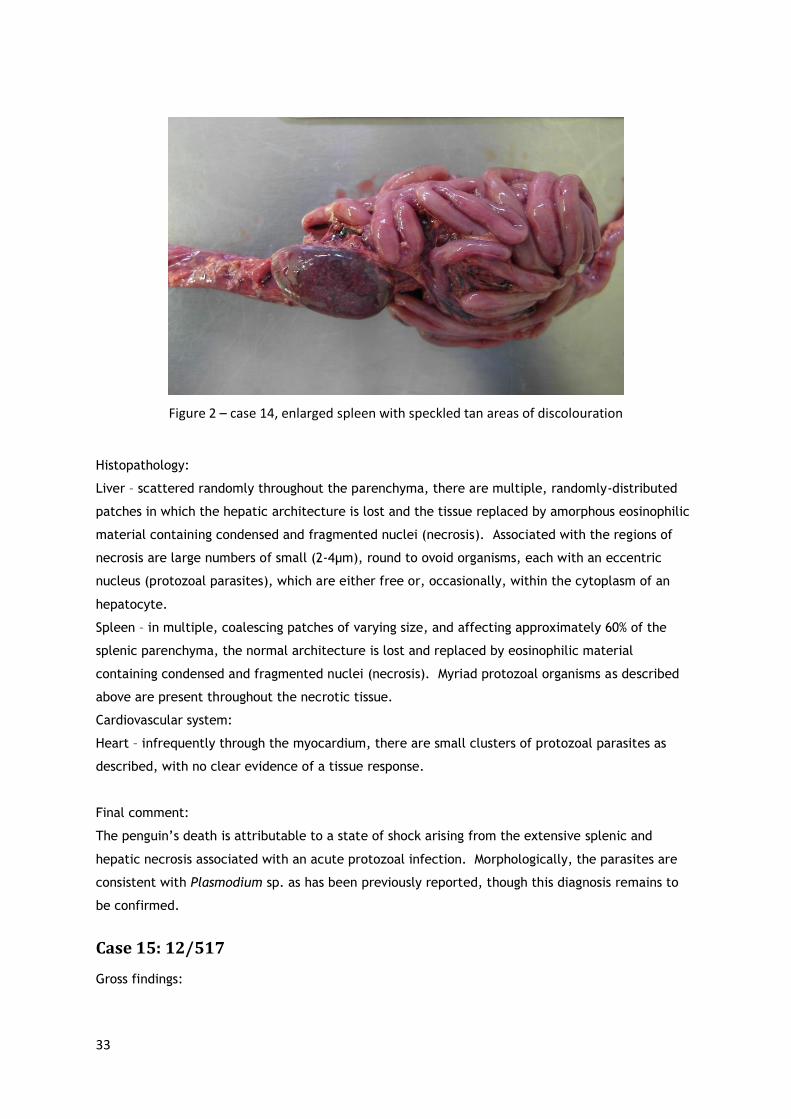
33
Figure 2 – case 14, enlarged spleen with speckled tan areas of discolouration
Histopathology:
Liver – scattered randomly throughout the parenchyma, there are multiple, randomly-distributed
patches in which the hepatic architecture is lost and the tissue replaced by amorphous eosinophilic
material containing condensed and fragmented nuclei (necrosis). Associated with the regions of
necrosis are large numbers of small (2-4μm), round to ovoid organisms, each with an eccentric
nucleus (protozoal parasites), which are either free or, occasionally, within the cytoplasm of an
hepatocyte.
Spleen – in multiple, coalescing patches of varying size, and affecting approximately 60% of the
splenic parenchyma, the normal architecture is lost and replaced by eosinophilic material
containing condensed and fragmented nuclei (necrosis). Myriad protozoal organisms as described
above are present throughout the necrotic tissue.
Cardiovascular system:
Heart – infrequently through the myocardium, there are small clusters of protozoal parasites as
described, with no clear evidence of a tissue response.
Final comment:
The penguin’s death is attributable to a state of shock arising from the extensive splenic and
hepatic necrosis associated with an acute protozoal infection. Morphologically, the parasites are
consistent with Plasmodium sp. as has been previously reported, though this diagnosis remains to
be confirmed.
Case 15: 12/517
Gross findings:

34
With a body weight of 1200g, convex pectoral musculature and good soft tissue coverage of all bony
prominences, the bird had a body condition score of 3/5. At 5.62% of bodyweight (reference range
2.30 – 5.34), the liver was moderately enlarged; randomly scattered over the capsular surface and
extending into the parenchyma there were multiple 0.5–2mm flat, cream areas of discolouration
(necrosis). The spleen was markedly enlarged (6.18g) and was dark red with speckled tan areas of
discolouration on the capsular surface (necrosis). There was a moderate amount of viscous clear
fluid containing yellow, flocculent material in a branching linear pattern on the parietal surface of
the lungs bilaterally (mucus, inflammatory exudate)
Histopathology:
Liver – scattered throughout the parenchyma, there are multiple, randomly-distributed patches in
which the hepatic architecture is lost and the tissue replaced by amorphous eosinophilic material
containing condensed and fragmented nuclei (necrosis). Associated with the regions of necrosis are
large numbers of small (2-4μm), round to ovoid organisms, each with an eccentric nucleus
(protozoal parasites), which are either free or, occasionally, within the cytoplasm of an
hepatocyte. Besides the necrotic areas, there are also multiple, randomly-distributed mild to
moderate infiltrates of primarily macrophages, with fewer lymphocytes.
Spleen – in multiple, coalescing patches of varying size, and affecting approximately 40% of the
splenic parenchyma, the normal architecture is lost and replaced by eosinophilic material
containing condensed and fragmented nuclei (necrosis). Myriad protozoal organisms as described
above are present throughout the necrotic tissue.
Heart – occasionally through the myocardium, there are small to moderate clusters of protozoal
parasites as described, with no clear evidence of a tissue response.
Lungs – scattered throughout the parenchyma, there are infrequent small clusters of protozoal
parasites as described, with a moderate number of air capillary stromal cells having pyknotic nuclei
(apoptosis, necrosis).
Final comment:
The penguin’s death is attributable to a systemic inflammatory response, likely leading to a state of
shock, arising from the extensive splenic and hepatic necrosis associated with an acute protozoal
infection. Morphologically, the parasites are consistent with Plasmodium spp. as has been
previously reported, though this diagnosis remains to be confirmed. No histological evidence of
pleuritis was found.
Case 16: 12/524
Gross findings:
With a body weight of 1240g, flattened pectoral musculature and with prominent keel and femurs,
the bird had a body condition score of 2.5/5. At 7.6% of bodyweight (reference range 2.30 – 5.34),
and extending 20mm beyond the xiphoid process, the liver was moderately enlarged. Randomly
scattered over the capsular surface and throughout the parenchyma there were multiple 1–2mm

35
flat, off-white areas of discolouration (necrosis). Weighing 6.12g and, across the short axis,
spanning 3.5 times the diameter of adjacent small intestine, the spleen was markedly enlarged.
Also, there were speckled 0.5-3mm areas of tan discolouration on the capsular surface (necrosis).
Cytology:
Impression smears of liver and spleen were stained with Wright’s-Giemsa stain. The smears are
highly cellular and contain, respectively, many erythrocytes and splenic and hepatic parenchymal
cells against a dense, proteinaceous background. At the edges of the hepatic smear, numerous 3-
5μm, oval bodies with a thin outer membrane, palely basophilic cytoplasm and a sharply
demarcated, irregular, brightly eosinophilic nucleus are seen (apicomplexan parasites).
Histopathology:
By request of the submitter, histological examination of the moderately decomposed tissues was
not conducted.
Final comment:
In view of the moderately advanced decomposition of the penguin, an abbreviated post mortem
examination was requested and performed. However, given the gross similarities of this case with
the numerous penguins diagnosed in the past 10 months with Avian Malaria, and given the
protozoan parasites seen on cytological examination of the liver impression smear, it is reasonable
to conclude that this penguin’s death was attributable to a systemic inflammatory response, likely
leading to a state of shock, arising from splenic and hepatic necrosis associated with an acute
protozoal infection. Morphologically, the parasites are consistent with Plasmodium spp. as has
been previously reported, though this diagnosis remains to be confirmed.
Case 17: 13/081
Gross findings:
With a bodyweight of 1320g, good soft tissue coverage of bony prominences and slightly convex
pectoral muscles, a body condition score of 3/5 was given. Scattered over the capsular surface and
throughout the parenchyma on the cut surface of the liver, there were multiple, circular, yellow-
white spots of up to 2mm diameter (necrosis). At 5.2% of bodyweight, the liver is at the upper
limit of normal size. At 5.68g, the spleen was markedly enlarged and scattered across its capsular
surface were multiple circular yellow-white spots of up to 1mm diameter, similar to those seen in
the liver (necrosis).
Histopathology:
Liver – the tissue is markedly autolysed, with virtually no cellular detail discernible. At scattered
points throughout the tissue, there are clusters of intact small (1-2µm) ovoid nucleated organisms
consistent with apicomplexan protozoal parasites within small foci of hepatocellular necrosis.

36
Spleen – though all cellular elements exhibit signs of degeneration (cytoplasmic swelling, condensed
or distorted nuclei), there are multiple foci scattered randomly throughout the parenchyma in
which all cellular detail is lost (necrosis). In many of the necrotic foci, there are clusters of ovoid
organisms as seen in the liver, both free within the necrotic debris and in the cytoplasm of
macrophages.
Final comment:
Marked autolysis complicates a thorough histological evaluation of this bird’s tissues, though the
gross findings and the presence of apicomplexan parasites in the necrotic centres in the spleen and
in scattered locations in the liver effectively confirm a diagnosis of haemoproteosis, an infection
which has been documented frequently in birds from this population in the last couple of years.
Case 18: 13/670
Gross findings:
With a bodyweight of 1090g and flat pectoral muscles, a body condition score of 3/5 was given.
Liver – there were occasional, poorly-defined 1-2mm diameter roughly circular patches of pale
brown discolouration on the capsular surface (necrosis) and at 59.8g the liver was moderately
enlarged. At 4.7g, the spleen was markedly and uniformly enlarged, with multiple, pale-red, 0.5-
1mm diameter spots on the capsular surface (splenomegaly and necrosis).
Histopathology:
Liver – affecting the entirety of the liver, the cells are swollen and eosinophilic with a loss of all
nuclear detail, though the general architectural arrangement of the tissue is retained (post-mortem
autolysis). Randomly distributed throughout the tissue, there are patches in which all cellular
detail is lost (antemortem necrosis). Associated with these patches are numerous clusters of intact
small (1-2µm) ovoid nucleated organisms consistent with apicomplexan protozoal parasites
(presumed Haemoproteus sp.)
Spleen – there are multiple foci scattered randomly throughout the parenchyma in which all
cellular detail is lost (necrosis). In these necrotic foci, there are clusters of ovoid organisms as
seen in the liver, both free within the necrotic debris and in the cytoplasm of macrophages.
Final Comment:
The gross and histological findings effectively confirm the diagnosis of haemoproteosis, an infection
that has been seen intermittently in this population since late 2011.
Other
In one additional case, 13/083, there was histological evidence of mononuclear cell hepatitis with
rare ovoid bodies consistent with protozoal organisms and a mildly enlarged spleen. In this case the
bird had become trapped in wire mesh and had been found dead. Gross and histopathological

37
examination could not ascertain the cause of death, though haemoproteosis was considered
unlikely.

38
Parasites detected
Case # Hepatomegaly Splenomegaly Liver Spleen Heart Lung Other
1 - 06/1172 Parasites in adipose tissue, pericardial artery
2 - 08/1075 Bile duct distension
3 - 11/484 No spleen sample or weight recorded
4 - 11/624
5 - 11/626
6 - 11/627
7 - 11/628
8 - 12/020
9 - 12/021
10 - 12/338 Newcastle disease virus, suspect +ve
11 - 12/362
12 - 12/375 Parasites in air sacs, pericardium. Histiocytic myositis (no parasites in the muscle lesions)
13 - 12/385
14 - 12/473
15 - 12/517
16 - 12/524 No histology, parasites detected on cytology
17 - 13/081
18 - 13/670

39
Summary
The common features in these cases are birds in good body condition with livers tending to be large,
moderately to markedly enlarged spleens, multiple, randomly-distributed areas of necrosis in the
liver and spleen with small protozoal organisms in the necrotic lesions in these two tissues. Other
tissues in which the organisms are reasonably often detected include the heart (7 of 18) and lungs (4
of 18) and much less commonly adipose tissue (1 of 18) and pericardium (2 of 18). Figures 3 and 4
below show typical histopathological findings, and figure 5 shows typical cytological findings from
several cases with the protozoan organisms clearly visible in figures 2 and 3.
There are few associated comorbidities. One penguin (case 10 - 12/338) was suspect positive for
Newcastle disease virus by PCR to detect antigen. One penguin (case 12 - 12/375) had a histiocytic
myositis, though without evidence of parasites in this tissue. In one case (case 16 – 12/524), the
diagnosis was made without histology, but on the gross changes and cytological evidence. Note that
in cases 17 and 18, the pathologist’s final comment indicated a diagnosis of haemoproteosis; these
cases were reported after early Polymerase Chain Reaction results had identified infection with a
novel Haemoproteus spp. parasite in some penguins from Penguin Island; this will be discussed more
fully in chapter 5.
Protozoan parasites are readily-detectable by light microscopy if a directed effort is made to find
them, though they could be overlooked if a protozoal infection is not suspected, or if cellular debris
in necrotic tissues makes them difficult to identify. Note, though, that the resolution of external
morphology and internal features at the scale of these organisms means that identification beyond
the level of phylum (i.e., Apicomplexa) requires additional techniques such as Polymerase Chain
Reaction (PCR), In-situ Hybridisation (ISH) or electron microscopy. While the majority of the reports
compiled here suggest that the infection is likely to be due to Plasmodium spp. parasites, this
suggestion was made on the basis that captive penguins have often been reported to be susceptible
to this infection with similar post mortem findings. Clear evidence of erythrocyte infection is lacking
in all of these cases. On morphological evidence other Haemospororida such as Haemoproteus and
Leucocytozoon, and other Apicomplexa such as Toxoplasma and Sarcocystis must also be considered
as potential agents of disease.

40
Figure 3 – case 14, liver, necrotic focus (the area of relative pallor, within which numerous organisms
were identifiable)(5μm section, Haematoxylin and Eosin stain, 200x)
Figure 4 – case 14, liver, intact and necrotic hepatocytes and numerous protozoa (arrows), free and
within cysts

41
(2μm section, Martius Scarlet Blue stain, 1000x)
Figure 5 – case 11, splenic impression smear, erythrocytes (E), splenic stromal cells (S) and numerous
protozoa (arrows)(Wright’s Giemsa stain, 1000x, case 11 – 12/362)

42
3. Fieldwork
Introduction
From the post mortem examinations, it had been established that a disease existed in this
population of Little Penguins in which the key post mortem findings of mild to moderate
hepatomegaly, marked splenomegaly and multifocal hepatic and splenic necrosis were associated
with the presence of intralesional protozoal parasites in those tissues, and occasionally in
erythrocytes. As previously discussed, these observations were suggestive of an infection with a
haemoparasite in which the tissue phase resulted in such extensive and acute tissue necrosis as to
induce a state of shock.
Similar gross and microscopic lesions had not been observed in other penguins presented for post
mortem. It appeared that, at least in the sample population we had, exposure to this infection was,
or at least could be, rapidly fatal. Working hypotheses were developed which stated that:
1. The penguins were immunologically naïve to the infection, and thus were aberrant hosts
in the life cycle of this parasite
2. Another bird species present on Penguin Island, whether permanently or intermittently,
was the definitive host for this parasite
3. The penguins had recently become exposed to this new infection either because there
was a greater prevalence of parasitaemia in the definitive host, or because there was a
new competent invertebrate vector by which the parasite could be transmitted to other
hosts
We wished to investigate whether exposure to the parasite was invariably fatal for the penguins, or
whether some that had been infected had survived to become parasitaemic. Also, we wished to
investigate which other bird species that used Penguin Island might be carriers of the disease.
Besides the Little Penguin, bird species that breed on Penguin Island include Bridled Terns (Sterna
anaethetus), Caspian Terns (Hydroprogne caspia), Silvereyes (Zosterops lateralis), Singing
Honeyeaters (Lichenostomus virescens), Silver Gulls (Chroicocephalus novaehollandiae), and Buff-
banded Rails (Gallirallus philippensis)67. Other birds that make visits to the island include Welcome
Swallows (Hirundo neoxena), Willy Wagtails (Rhipidura leucophrys), Ravens (Corvus coronoides),
Australian Kestrels (Falco cenchroides), Little Eagles (Hieraaetus morphnoides), Swamp Harriers
(Circus approximans), Barn Owls (Tyto alba), Boobook Owls (Ninox novaeseelandiae) and Pelicans

43
(Pelecanus conspicillatus)67. Conceivably, any one of these species may represent the host of the
parasite that came to be infectious to the penguins.
In order to achieve the above stated aims, we decided to collect peripheral blood samples from the
penguins and selected other species and to make blood smears which could be assessed for the
postulated erythrocytic stages of the parasite. Examination of blood smears is a simple and easily-
performed technique which is routinely used to diagnose Plasmodium infections (malaria) in
humans.
Two avian species which breed on Penguin Island were prioritised as candidates for blood sampling,
the Bridled Tern and the Caspian Tern. The Bridled Tern is a seabird which breeds on Penguin Island
in the period from September to May. During the winters, the terns migrate to the north-west
Sulawesi Sea between the Indonesian and Philippine archipelagos26. In this period, the birds are
likely to be exposed to vector-borne infectious organisms which they may then carry back to Penguin
Island for the next breeding season.
The Caspian Tern is another migratory seabird which breeds in the Perth metropolitan area from July
to November57. Local breeding and roosting sites include Rottnest, Carnac, Bird, Gull and Seal
islands, as well as Penguin Island, and their nest sites vary from year to year57. Outside of the
breeding season, they migrate to estuarine habitats, which presumably exposes them to a variety of
invertebrate vectors.
Materials and methods
Blood collection and blood smear preparation
Penguins
An online sample size calculator (http://sampsize.sourceforge.net/iface/index.html#prev) was used
to determine the appropriate sample size for a disease prevalence survey, using the following
parameters:
1. Precision - 10%
2. Prevalence estimate - 17% (based on the 9 of 54 penguins presenting for post mortem
which had had evidence of protozoan infection)

44
3. Population size - 950 (current Penguin Island population estimate)
4. Confidence interval - 95%
The recommended sample size derived from the calculation was 52. Based on this calculation, it was
proposed to collect samples from 52 penguins in 2012 and to repeat the collection in 2013, making a
total of 104 samples across the two years.
Prior to the commencement of fieldwork, the proposed capture and blood collection protocols were
approved by the Murdoch University Animal Ethics Committee (Permit No. RW2510/12). All bird
handling and blood collection procedures were performed by one of the named operators on the
Animal Ethics Permit or, on one occasion, by a veterinarian and Animal Welfare Officer from the
Animal Ethics Committee (Dr Cree Monaghan) who was in attendance to monitor the procedures
and offer guidance. Seven separate visits were made to Penguin Island (32° 18' S, 115° 41' E) to
collect blood from penguins; four of these visits were made during the day on 26/07/12 (12
samples), 21/08/12 (14 samples), 14/09/12 (17 samples) and 15/10/12 (11 samples), and three were
overnight visits on 24/11/12 (7 samples), 06/12/12 (15 samples) and 29/09/13 (17 samples).
A total of 94 penguin blood smears were made and examined. Of these, several were repeat blood
smears from previously sampled animals. Penguin #208116 was sampled on two occasions
(21/08/12 and 29/09/13), as were penguin #208357 (24/11/12 and 29/09/13), penguin #211387
(21/08/12 and 14/09/12), penguin #215672 (14/09/12 and 15/10/12), penguin #264455 (26/07/12
and 21/08/12), penguin #458030 (14/09/12 and 06/12/12), penguin #460705 (21/08/12 and
15/10/12), and penguin #461519 (06/12/12 and 29/09/13). Penguin #213543 was sampled on three
occasions (26/07/12, 21/08/12 and 14/09/12), as was penguin #399204 (21/08/12, 14/09/12 and
15/10/12). One penguin, #252549, was sampled on four occasions (26/07/12, 21/08/12, 15/10/12
and 24/11/12). Thus, the 94 smears represent samples from 79 individual penguins, and they were
collected from the austral winter to summer of 2012 and in the early austral spring of 2013.
Sampling during these periods was intended to detect infections in different seasons, in the event
that there was a seasonal component to exposure, for example, increased vector numbers during
warmer weather. Also, the majority of cases seen at post mortem had been presented in late
austral spring and summer, so it was considered important to include these seasons in the collection
periods.
During the daytime collections, penguins were captured during checks of the nestboxes which are
scattered around Penguin Island. Any penguin present in a nestbox was gently removed (providing it

45
did not have very young chicks, in which case it was left otherwise undisturbed). Each penguin was
weighed and scanned for a microchip if no flipper band was present. Blood collection was performed
immediately, and as near to the nest box as possible, then the penguin was either returned to the
nest box straight away, or placed in a small enclosure (specifically, a plastic cat carrier) for no longer
than 15 minutes for the purpose of collecting a faecal sample (for a separate study concurrently
being conducted by Dr Belinda Cannell). While in the cat carrier, the penguins were attended at all
times until they were returned to their nestboxes.
For night collections, the penguins were captured at or shortly after dusk when they returned to the
island from foraging at sea. By habit, the penguins make landfall at specific beaches that are closest
to their nests. To corral the birds, the methodology described in Cannell et al (2011) was followed.
Briefly, low fences constructed from rolls of shadecloth and plastic lattice were erected at various
landing sites at two of the major arrival beaches for the penguins - the southern and eastern
beaches. The fences were oriented so as to channel any penguins coming ashore at those sites into
small corrals which could then be closed. The penguins were then removed to holding pens
consisting of large, open-bottomed cardboard boxes placed out of sight of the corral so as not to
deter other penguins arriving. The birds were collected over a period of 30-60 minutes. At the end
of this time, they were removed one at a time for weighing, scanning for microchips (or recording of
flipper band ID) and blood collection; any birds without a microchip or flipper band were
microchipped at this time. After sampling, the birds were released close to the beach at which they
had arrived.
Peripheral blood was collected from the metatarsal vein on the dorsal aspect of one or the other
foot. During the first four collection trips, the birds were restrained by hand and were partially
wrapped in a calico bag to cover their heads and upper bodies. After gaining approval from the
Research Ethics and Integrity office, the penguins were subsequently restrained using an equine
knee/skid boot which consists of a sturdy yet soft, approximately cylindrical support, open at each
end and along one side where it can be secured by three Velcro straps. The penguins were placed
lengthwise in this device with their backs uppermost, and the straps were then secured to form an
open-ended tube with the penguin safely encased within (Fig 1). From this position, a foot could be
grasped, externalised and restrained. After swabbing with methylated spirit, a 23 or 25 gauge
butterfly needle was inserted into the metatarsal vein, and up to 1mL of blood was collected into a 1
or 2mL syringe with slight negative pressure. After sampling, a fresh dry swab was applied to the
venipuncture site with gentle pressure until there was no evident bleeding. No animals were injured

46
during capture or sampling, and any signs of mild distress (e.g., vocalisation) ceased on the birds’
release.
Figure 1 – equine knee/skid boot used for penguin restraint (1:1 scale model penguin in situ)
In each case, a standard thin blood smear was made at the time of blood collection and the slide was
air dried and stored for later staining; any remaining blood was expelled into a labelled 2mL plastic
blood tube containing potassium EDTA, and this tube was retained until the prepared smears had
been stained in case additional smears were required. In many cases, only enough blood to make a
single blood smear was obtained.
Within five days of smear preparation, the slides were stained with Wright’s Giemsa stain on an
automatic stainer, then manually cover-slipped.
To assess each slide for the presence of parasites, a minimum of 20 oil immersion fields were
examined. Red cells in the monolayer were observed for the presence of intracytoplasmic
organisms.

47
Bridled Terns
Blood collection and its protocols were approved by the Murdoch University Animal Ethics
Committee (Permit No. RW2510/12). Bridled Terns (Sterna anaethetus) were captured at night
during two visits to the island on 24/11/12 (21 samples) and 02/03/13 (31 samples, no repeats). To
capture a tern, one of two registered bird banders (Dr Nic Dunlop or Ms Sandy McNeill) would shine
the light from a headlamp towards the bird, and then place a soft net over the bird while it sat
immobile. The terns were restrained by holding their wings against their bodies, which is easily
accomplished with a single hand in this small species. Birds were checked for leg bands, the number
on the bands was identified, and those without a leg band were then banded.
Blood was collected from the either the leg vein (where visible) as it ran over the lateral tarsus, or,
more frequently, from the wing vein. The birds were held in dorsal recumbency with a wing, usually
the right, held in extension, and the wing vein was identified as it ran over the medial aspect of the
humerus just proximal to the elbow. After swabbing with methylated spirit, a 25 gauge needle
attached to a 1mL syringe was inserted into the vein and up to 0.1mL of blood was collected into the
syringe. A blood smear was made at the time of blood collection and air dried for later staining.
After wing venipuncture, a small (typically 5 x 5mm) square of Kaltostat dressing (ConvaTec Inc.,
Skillman, NJ, USA) was applied to the puncture site and held in place for approximately 30 seconds.
Kaltostat is a calcium alginate dressing which assists in the formation of a haemostatic plug at
bleeding wound sites. All birds were able to fly without difficulty on release.
Within five days of smear preparation, the slides were stained with Wright’s Giemsa stain on an
automatic stainer, then manually cover-slipped.
To assess each slide for the presence of parasites, a minimum of 20 oil immersion fields were
examined. Red cells in the monolayer were observed for the presence of intracytoplasmic
organisms.
Caspian Terns
Two visits were made to Penguin Island with the intention of collecting blood samples from Caspian
Terns (Hydroprogne caspia). On both occasions, inclement weather reduced the time available for
attempting to capture the birds. Additionally, these attempts were made relatively late in the
Caspian Tern’s breeding season, and the birds had become wary of the traps used to capture them

48
(in many instances they had previously been trapped for banding). Ultimately, no birds were
trapped on either occasion, and no samples could be collected from this species.
Results
Penguins
Intraerythrocytic parasites were detected in one blood smear. The smear was collected from
penguin #215057, which was captured on the island’s south beach on 06/12/12 (i.e., during the
austral summer). This penguin was one of a group captured and held for approximately 30 minutes
before sampling. It was in excellent body condition, with a body mass of 1850g. This is well above
the average mass of males on Penguin Island, which is 1348 ± 200g (n=118) (Cannell, unpublished
data), and it was most likely that this bird was amassing fat in preparation for its annual moult
(Cannell pers. comm.) No bird in this group showed signs of being systemically unwell (e.g., lethargy,
ataxia, respiratory distress) at the time of capture, blood collection or release.
The blood smear for this penguin showed evidence of a regenerative erythrocyte response, with
abundant immature erythrocytes identifiable by their morphology (large, round nuclei, relatively
round cells with basophilic cytoplasm). In the cytoplasm of a small percentage of cells there were
one or more 1-3µm long ovoid bodies with clear cytoplasm and an apical nucleus (see Fig 2).
These bodies are consistent with the blood stages of apicomplexan parasites, for example,
merozoites or early gametocytes of Haemoproteus or Plasmodium, or merozoites or sporozoites of
Babesia. The evidence of erythrocyte regeneration suggests a resolving anaemia, presumably due to
haemolysis in response to the erythrocytic parasites.
a b

49
Figure 2, a to d: protozoan parasites in the cytoplasm of erythrocytes (penguin #215057, collected
06/12/2012) (Wright’s Giemsa stain, 1000x)
Bridled Terns
No parasites were detected in any blood smears from Bridled Terns.
Summary
A confident estimate of the prevalence of haemoparasite infection in Penguin Island Little Penguins
cannot be made on the evidence of a single positive bird in 94 samples from 79 individuals.
However, it appears that the overall prevalence of parasitaemia in both Little Penguins and Bridled
Terns is low.
The morphology of the parasites seen in penguin #215057 does not allow identification to the level
of genus. While they are somewhat consistent with the gametocytes of Haemoproteus parasites,
there are reasons to suspect that is not what they are. Haemoproteus gametocytes develop within
7-10 days to occupy most of the cytoplasm of the erythrocyte6, and all of the parasites seen in this
bird are of approximately the same size. Conceivably, a mass release of merozoites could lead to
erythrocyte infection by a single, very early stage of parasites, though this must be considered
improbable.
An alternative explanation is that the parasites in this smear are a haemoparasite such as the
piroplasms Babesia or Theileria, which would be expected to show relatively little morphological
variation throughout this life stage40. In support of this hypothesis is the evidence of red cell
c d

50
regeneration on this smear; this may indicate recovery from a haemolytic anaemia, a typical
consequence of piroplasm infection. Also, while it is not a haemoparasite (in that it does not
ordinarily have a blood phase) Toxoplasma is another apicomplexan parasite with a comparable
morphology; Toxoplasma does not infect the anucleate erythrocytes of mammals, though it can
infect erythrocytes in avian hosts22.
No haemoparasites were detected in the Bridled Tern smears. This may indicate an absence of
haemoparasite infection, which is not unexpected in seabirds, or it may be due to seasonal variation
in infection status, with infections occurring at other times of the year. Alternatively, it may be that
there is a latent haemoparasite infection, and that parasitaemia is immunologically suppressed. This
final possibility could be investigated with serology or with PCR to detect parasite antigen or
antibodies. In any case, the hypothesis that Bridled Terns might be implicated as the source of the
penguin infection can neither be confirmed nor refuted on this evidence.

51
4. Electron microscopy
Introduction
The very small size of the parasite infecting the penguins limits the extent of the morphological
assessment that can be made from light microscopy. To improve the resolution and to allow
evaluation of subcellular structures, representative samples were prepared for transmission electron
microscopy. For this purpose, samples from 3 penguins were fixed in 5% glutaraldehyde at the time
of post mortem. The samples were: liver and spleen from case 12/362; heart, spleen and liver from
12/375; spleen and liver from 12/385.
Materials and Methods
Samples measuring approximately 1 x 1 x 3mm were collected from fresh carcasses (i.e., not frozen)
at the time of post mortem examination and fixed in glutaraldehyde at 4°C until required
(approximately 4 months).
Processing for transmission electron microscopy was performed with the spleen and liver samples
from case 12/362 only. This case was selected as it had a very high concentration of parasites visible
by light microscopy, so it was considered that it offered the greatest probability of detecting the
parasites in the very small tissue sections electron microscopy requires. The sample was processed
in accordance with the standard protocols for the transmission electron microscope at Murdoch
University.
The glutaraldehyde fixative was removed from the sample tube and replaced with Dalton’s chrome
osmic acid, then the sample was covered and allowed to fix for approximately 90 minutes. The
sample was washed four times in 70% alcohol, followed by a 5 minute wash in 90% alcohol, then a 5
minute wash in 95% alcohol and then three washes in absolute ethanol for 3.5 minutes each. The
sample was then washed in two changes of propylene oxide over 10 minutes before being left in a
60:40 solution of propylene oxide and epoxy resin for 1 hour. Finally, the sample was left on a
rotator overnight.
The following day, the sample was placed in a vacuum chamber, at a negative pressure of
approximately 30 inches of mercury, before it was embedded in fresh epoxy resin and baked at 60°C
for 24 hours.

52
After baking, the resin blocks were trimmed to the margins of the sample, and then placed in a
Reichert Om U3 ultramicrotome. A fresh glass knife, made on an LKB Bromma 7800 Knifemaker, was
used for each block to cut thick sections at 1µm thickness. Sections were collected on a glass slide
and stained with toluidine blue for initial light microscopic assessment to assess whether adequate
numbers of host cells and parasites were present so as to be suitable to make thin sections. Thin
sections were then made at 90nm thickness on a Reichert Ultracut E ultramicrotome, also using a
freshly made knife. These sections were collected onto 200 mesh copper grids and stained with lead
citrate and uranyl acetate.
Lead citrate stain was prepared by placing approximately 0.3g of lead citrate into a vial. The lead
citrate was dissolved by adding 10mL of distilled water to the vial, followed by 0.1mL of 10N sodium
hydroxide. The mixture was shaken, transferred to a centrifuge tube and centrifuged at 2,700rpm
for 10 minutes. 10mL of stock saturated uranyl acetate solution was placed in a second centrifuge
tube and centrifuged at the same time as the lead citrate solution.
To stain the sections, single drops of each solution were placed on a wax coated petri dish. The grids
were first introduced to the base of the uranyl acetate drop, with the grid facing up, and allowed to
stand for 7 minutes. They were then washed by dipping repeatedly in distilled water, and allowed to
dry on filter paper. Once dry, the grids were placed on the top of the lead citrate drop, with the grid
facing down, and allowed to float there for 4 minutes. The grids were washed again and allowed to
dry on filter paper. Once stained, the grids were stored at room temperature until required.
Grids were examined using a Philips CM100 BioTwin Transmission Electron Microscope (Eindhoven,
The Netherlands). Images were captured on photographic film and scanned to create digital copies.
Results
The following images were photographed from the electron microscopy sessions.

53
Figure 1 (and detail) – four parasites in a cyst within the cytoplasm of a host cell, spleen, x3810, bar =
2μm
Image left: E - erythrocyte, Ph - phagocyte, P - protozoa; image right (detail): N - nucleus
In Figure 1 there is a host erythrocyte (E) adjacent to a large, possibly phagocytic, cell (Ph). In the
cytoplasm of the large cell, abutting its nucleus, there is a cluster of four protozoal organisms (one
marked with a P), each approximately 1.5 to 2μm across. Though a membrane cannot be seen, the
clear space surrounding the parasites is distinctly different from the large cell’s cytoplasm,
suggesting that the parasites are isolated in a vacuolar space. The left-most organism, which is
enlarged in the detail image, is oriented to show the typical, slightly elongated apicomplexan
morphology with a rounded base, a basal nucleus (N) and a cluster of organelles congregating at the
tapered apex (arrow).

54
Figure 2 - Single extracellular parasite, spleen, x15000, bar = 1μm
Figure 2 demonstrates the pellicle, the outer wall of the organism, which consists of an outer and a
continuous inner membrane15. Three acidocalcisomes and a crescent of rough endoplasmic
reticulum are also present15,58.
Figure 3 – apical complex, spleen, x15000, bar = 1μm

55
In Figure 3, several of the components of the apical complex are visible at the lower left hand side of
the organism. The numerous short dark rods are micronemes14. Adjacent to these are several dense
granules, and there is a larger elongate body next to one of the dense granules consistent with a
rhoptry15.
Figure 4 – two protozoa within a host cell, spleen, x6500, bar = 2μm.
P - parasites, N - nucleus (host cell)
In Figure 4 there are two distinct parasites (P) contained within the boundaries of another cell, which
appears to be a host cell with a large, electron dense nucleus (N).

56
Figure 5 – a cluster of extracellular protozoa, spleen, x4800, bar = 2μm
Arrow - dividing protozoan
In Figure 5, numerous protozoa, one of which is dividing (indicated by arrow) are clustered in a
background of amorphous material, presumed to be cellular debris from necrotic host tissues.
Figure 6 – early division, spleen, x15000, bar = 1μm

57
Figure 7 – early division, spleen, x20000, bar = 500nm
Figure 8 – late division, spleen, x11000, bar = 2μm
Arrow – membrane dissecting between dividing parasite cells

58
In figures 6 to 8, two distinct cells can be seen enclosed within a single pellicle. In figures 6 and 7,
there are two nuclei enclosed in nascent pellicles with little cytoplasm and few organelles. The
majority of the cytoplasm belongs to the mother cell. In figure 8, there are 2 virtually complete cells
contained within a single pellicle, with a membrane dissecting between them (arrow).
Discussion
The identifiable ultrastructural features of this protozoan parasite include a pellicle, acidocalcisomes,
dense granules, micronemes, rhoptries, and reproduction by a process of binary fission.
A pellicle is the name given to the plasma membrane of a protozoan organism when it is
accompanied by an additional, continuous underlying membrane15. The pellicle is characteristic of
the motile stages of sporozoan and ciliate parasites15.
Acidocalcisomes are intracellular calcium stores that appear as round, clear spaces in the cytoplasm,
often, though not invariably, containing a smaller, concentric or eccentric electron dense body.
Acidocalcisomes are non-specific organelles which are highly conserved across a very broad range of
species from bacteria to humans15.
Dense granules are spherical, electron dense bodies that may occur anywhere in the cytoplasm of
apicomplexan parasites. Their function is incompletely characterised, but some identified proteins
have been associated with virulence40.
Micronemes are small, elongate, electron dense secretory organelles that are confined to the apical
segment of the parasite. These organelles are characteristic of apicomplexans and are involved in
parasite attachment to host cells40.
Rhoptries are another component of the apical complex. These secretory organelles are also
incompletely characterised, though there is evidence that they are involved in the penetration of
host cells and in the establishment of the parasitophorous vacuole in which parasites establish and
divide in the host cell40.
Considered together, the ultrastructural features of the protozoan parasite in the spleen of penguin
12/362 unambiguously identify it as an apicomplexan. However, these features are broadly
characteristic of the phylum, and further identification to the level of genus, or even family, is not
possible on this evidence. The most likely pathogenic apicomplexans to consider are the
59
haemosporidians (Plasmodium, Haemoproteus and Leucocytozoon), the piroplasmid Babesia and
various genera of coccidia including Toxoplasma, Eimeria, Isospora, Atoxoplasma and Sarcocystis6.
The morphologic features of the disease identified by post mortem (that is, random multifocal
necrosis in the liver and spleen) make Babesia infection relatively unlikely, given that Babesia is
strictly an erythrocyte parasite in the vertebrate host, and it does not replicate in fixed cells in
tissues40. With no evidence of involvement of the gastrointestinal tract, or of development to micro-
and macrogamete stages, the likelihood of Eimeria infection is greatly reduced6. Sarcocystis is rarely
pathogenic in birds, and acute pathological changes, when they do occur, tend to be most severe in
the lungs6. Of the remaining organisms, the gross and microscopic findings are non-specific and may
be compatible with an infection with any one of them.
The apicomplexans are a diverse and complex group, and their life cycles resist generalisation.
However, at a very simple level, these protozoa have both sexual and asexual stages of
reproduction. Sexual reproduction involves the fusion of macrogametes to microgametes by a
process of syngamy76, and by definition it occurs in the definitive host, though the macro- and
microgametes themselves may have formed in either the definitive host or an intermediate host.
Sexual reproduction results in the production of infective sporozoites, which, in the appropriate host
tissue, multiply by endopolygeny to produce one or more generations of merozoites. At some point,
merozoites divide to form macro- and microgametes for the next round of sexual reproduction. In
Toxoplasma asexual reproduction occurs by a process of endodyogeny, a special form of
endopolygeny in which only two daughter cells are produced both by incorporating some organelles
from the mother cell and by de novo synthesis of others35,60. Endodyogeny is not unique to
Toxoplasma, however, having been reported in the apicomplexan genera Cytoisospora21, Isospora55,
Sarcocystis66, Besnoitia24 and Neospora81. Thus, the several instances identified in these
photographs in which a pair of parasites is visible within a larger cell are suggestive of, but not
definitive for, Toxoplasma infection.
Many of the protozoa seen in these images are extracellular. The asexual stages of the
apicomplexan life cycle involve penetration of sporozoites or merozoites into host cells followed by
reproduction in the host cell cytoplasm or in a cyst. After reproduction, the parasites lyse the host
cell and emerge to the extracellular environment whereupon they may infect new cells. Thus, a
heavy parasite burden can cause significant host tissue necrosis, which, if widespread, or if multiple
tissues are involved, can induce a systemic inflammatory response. While the parasites in the
images above are often well-preserved, the background cells are usually degenerate, which is

60
consistent with the light microscopic findings and the hypothesis that the penguins ultimately
succumbed to a state of shock mediated by cytokine overproduction.
In conclusion, the infection in the penguins is confirmed to be due to an apicomplexan parasite.
Further identification on morphology alone is not expected to be definitive. Molecular identification
by techniques such as the Polymerase Chain Reaction (PCR) are required.

61
5. Molecular Identification of the Parasite
Introduction and Aims
From the light microscopy and electron microscopy investigations to this point, the morphological
characteristics of the parasite in the penguin tissues allow it to be identified as a protozoan parasite
in the phylum Apicomplexa. However, definitive identification at least to the level of genus is
necessary to allow an appropriate understanding of the disease. The most definitive means by
which this may be achieved is amplification and sequencing of the organism’s DNA using the
Polymerase Chain Reaction (PCR) technique. PCR is a process whereby oligonucleotide primers are
used to induce the targeted replication of segments of DNA in large numbers, permitting that DNA
then to be dyed and visualised as distinctive bands on an agarose gel. The amplified segments of
DNA can be collected, the nucleotide sequence can be determined, and analysis of that sequence
allows a comparison to be made with a database of known DNA sequences that are characteristic of
specific organisms.
With respect to the penguins, samples of multiple tissues, usually including liver, spleen, kidney,
heart, brain and adipose tissue, have for some years been routinely collected and stored at -20°C for
all penguin post mortems conducted at Murdoch University, primarily for the purposes of
toxicological assays. Thus, tissue samples were available from this archive allowing DNA extraction
for PCR. In principle, to identify the agent causing the deaths of these penguins, it should be
demonstrable that DNA from a single parasite is present in all of those birds diagnosed with
protozoal infections. Thus, the aims of PCR in this context were:
1. To prove that an apicomplexan parasite can be detected and identified in the affected
tissues
2. To identify the parasite to at least the level of genus
3. To validate an in-house PCR that can be carried out at Murdoch University on these and
future cases for definitive diagnosis
All case numbers referred to in this section are as presented in Table 1 in chapter 2. Prior to
undertaking PCR for this project, tissue samples from 10 cases (cases 1, 2, and 4 to 11 inclusive) had
been submitted to an external laboratory (Center for Evolutionary and Theoretical Immunology,
University of New Mexico, Albuquerque, NM, USA) for PCR assessment with the intention to
demonstrate Plasmodium and/or Haemoproteus cytochrome-b DNA. From these ten samples,
positive findings were obtained from four: cases 5, 8, 9 and 1014. The PCR products from each of

62
these positive cases were sequenced, and they were identified as representing a novel species of
Haemoproteus in the subgenus Parahaemoproteus. Samples from these cases were included in the
current PCR investigation described in this thesis, and the four positive samples were considered to
represent positive controls for Haemoproteus. The PCR protocol used in this project was based on
that published in Cannell et al, 201314. Consequently, a supplementary aim was derived from this
publication, namely:
4. To verify the results from the external laboratory that found Haemoproteus in the
penguin tissues
In addition to testing for Haemoproteus, the penguin DNA was also used in PCR assays with primers
designed to detect piroplasmids, apicomplexans generally, and Toxoplasma. The reasons for
running these assays are discussed more fully in chapter six of this thesis; in essence, however, they
were performed to broaden the investigation as a consequence of the consistently negative results
obtained from the Haemoproteus assays (described below).
Materials and Methods
DNA extraction
DNA was extracted from a frozen sample of liver when available, or from spleen when liver was not
kept. Samples were collected from 15 birds, all of them with microscopic evidence of protozoan
parasite infection. The 15 cases were: 1 to 10 and 12 to 16. Nine of these (1, 2 and 4 to 10) were
from the same cases that had been tested in New Mexico; one case (case 11) was excluded as no
archived frozen tissue could be found for this bird. Six additional samples were included as they had
been received for post mortem after the previous group of samples had been submitted to New
Mexico (cases 12 to 16), or, in one case, because the diagnosis of protozoan infection was not made
until a later review of cases (case 3).
DNA was extracted using a DNeasy Blood & Tissue Kit (Qiagen), following the manufacturer’s
instructions. Briefly, for each sample approximately 25mg of tissue was chopped finely with a fresh
scalpel blade and collected into a 1.5mL microcentrifuge tube. After the addition of 20µL of
proteinase K, the tube was briefly vortexed then incubated at 56°C for 3 hours. The samples were
vortexed again after removal from the incubator, and then 200µL of lysis buffer (Buffer AL) were
added. After vortexing again, 200µL of 100% ethanol were added before a final vortexing step. The
mixture from this tube was aspirated into a spin column placed into a collection tube. The spin

63
column was centrifuged at 6000g (8000rpm) for 1 minute. The spin column was then transferred to
a new collection tube and 500µL of wash buffer (Buffer AW1) was added before a second
centrifugation step at 6000g. Again, the spin column was transferred to a new collection tube and
500µL of a second wash buffer (Buffer AW2) was added before a final 1 minute centrifugation at
20000g (14000rpm) to dry the DNA binding membrane. The spin column was placed in a clean
1.5mL microcentrifuge tube and 200µL of elution buffer (Buffer AE) was pipetted onto the spin
column membrane. This was allowed to incubate for 1 minute at room temperature, and then the
sample was eluted by centrifugation for 1 minute at 6000g. A second, identical elution step was
performed to maximise the DNA yield. The above protocol was followed for each tissue sample, and
also for an extraction blank negative control, which contained no tissue. After extraction, the
samples were stored at -20°C until required.
Prior to amplification, the samples from the first extraction were analysed for DNA yield and purity
by spectrophotometry. The DNA concentration for each tube was estimated by calculation from the
absorbance of the solution at a wavelength of 260nm. The purity of the sample was estimated by
the ratio of absorbance at 260nm (mostly DNA) to that at 280nm (mostly proteins). The DNA yield
and A260:A280 values are presented in Table 1. The DNA yield and purity in each case were considered
acceptable. This analysis was not performed on the samples from the second extraction.

64
Case No. DNA (ng/µL) A260:A280
1 – 06/1172 521.6 1.59
2 – 08/1075 309.2 1.20
3 – 11/484 332.4 1.57
4 – 11/624 211.6 1.28
5 – 11/626 155.2 1.69
6 – 11/627 242.2 1.30
7 – 11/628 352.0 1.71
8 – 12/020 323.8 1.38
9 – 12/021 298.8 1.24
10 – 12/338 356.2 1.39
12 – 12/375 368.9 1.42
13 – 12/385 413.8 2.04
14 – 12/473 326.2 1.94
15 – 12/517 555.2 1.38
16 – 12/524 461.1 1.27
negative control 1.7 1.59
Table 1 – DNA yield and purity
A second DNA extraction was performed after the first run of Toxoplasma PCR (discussed below), as
the extraction blank negative control returned a positive result, indicating that there had possibly
been contamination of this tube during the extraction process. For the second extraction, a further
eight samples were included as negative controls from penguins which did not have gross or
microscopic post mortem findings suggestive of protozoal infection. The case numbers and post
mortem diagnoses for these additional penguins are presented in Table 2. The second DNA
extraction also included those cases listed in the first DNA extraction, with the exception of case 10;
there was a labelling error during the extraction process with the result that this sample could not be
confidently identified, so it was discarded. The protocol for the second DNA extraction was identical
to the first, though the quantity of tissue from which the extraction was made was, in each case,
reduced to approximately 5mg. This adjustment was made on the advice of a colleague experienced
in the process, with the rationale that lysis of the tissue would be more effective, reducing residual
particulate matter in the final solution without compromising the yield of DNA.

65
Case Number Post mortem diagnosis
NC1 – 11/080 Proventriculitis with Contracaecum sp. infection
NC2 – 11/452 Inconclusive findings
NC3 - 11/482 Starvation
NC4 - 11/625 Trauma
NC5 - 12/015 Inconclusive findings
NC6 - 12/016 Inconclusive findings
NC7 - 12/571 Inconclusive findings/starvation
NC8 - 13/305 Starvation
Table 2 – additional (negative control) samples, negative by light microscopy for protozoan parasites
DNA from the first extraction was used in Haemosporidian and piroplasmid rounds 1 to 4 and in
Toxoplasma round 1 detailed below.
DNA from the second extraction was used in the Haemosporidian PCR round number 5 and in the
Toxoplasma PCR round number 2 detailed below.
Polymerase Chain Reaction
Haemosporidian and piroplasmid parasites
Several rounds of PCR were conducted using primers designed to detect various genera of
apicomplexan protozoa. These rounds are presented in temporal sequence.
For the haemosporidian parasites, primers for nested PCR were supplied by Invitrogen/Life
Technologies using the sequences published in Cannell et al, 2013 14, which had been designed to
detect haemosporidian parasites. The primer sequences for the primary PCR, designated HaemNF
(forward) and HaemNR2 (reverse), were CAT ATA TTA AGA GAA TTA TGG AG, and AGA GGT GTA GCA
TAT CTA TCT AC respectively (note: all sequences here and in the remainder of this chapter are
presented from 5’ to 3’). The primer sequences for the secondary PCR were HaemNF1 (forward)
CAT ATA TTA AGA GAA ITA TGG AG, and HaemNR3 (reverse) ATA GAA AGA TAA GAA ATA CCA TTC.
The target of these sequences was a fragment of the mitochondrial cytochrome-b gene which is
common to Plasmodium and Haemoproteus, with an expected product of approximately 480 base
pairs in length14.

66
For piroplasmid parasites, the primer sequences for the primary PCR, designated BTF1 (forward) and
BTR1 (reverse), were GGC TCA TTA CAA CAG TTA TAG, and CCC AAA GAC TTT GAT TTC TCT C
respectively. The primer sequences for the secondary PCR were BTF2 (forward) CCG TGC TAA TTG
TAG GGC TAA TAC, and BTR2 (reverse) GGA CTA CGA CGG TAT CTG ATC G. These sequences are as
published in Jefferies et al, 200744.
For Toxoplasma genus parasites, the primer sequences for the primary PCR were TGT TCT GTC CTA
TCG CAA CG (forward) and ACG GAT GCA GTT CCT TTC TG (reverse). For the secondary PCR, the
primer sequences were TCT TCC CAG ACG TGG ATT TC (forward) and CTC GAC AAT ACG CTG CTT GA
(reverse). These sequences are as published in Grigg and Boothroyd 200134.
PCR round 1
For the first round of PCR, 1:10 dilutions were made from each DNA sample. Diluted DNA was used
initially in order to reduce the potential for enzyme inhibitors, which may be present in DNA
extractions from tissues, to interfere with polymerase activity. A PCR master mix was prepared
which consisted of 10 µL TAQ DNA Polymerase, 250µL of reaction buffer, 50µL of 10mM dNTP mix,
540µL of cresol red dye, 150µL of 25mM MgCl2 and 250µL of purified water. PCR tubes were then
prepared, which contained 12.5µL of PCR master mix, 1µL each of HaemNF and HaemNR2 primers,
9.5µL of purified water and 15.5µL of cresol red dye. One µL of dilute DNA was then added to each
tube and the tubes were briefly centrifuged.
A complete set of duplicate tubes was made which were identical except for the substitution of BTF1
and BTR1 primers in place of the HaemNF and HaemNR2 primers.
The thermal cycle program for the primary PCR was as follows: the samples were brought to 94°C for
8 minutes, followed by 35 cycles of denaturation at 94°C for 30 seconds, annealing at 50°C for 30
seconds and extension at 72°C for 45 seconds. On completion of the 35 cycles, the samples were
held at the 72°C extension temperature for a further 10 minutes, then at 4°C until removal from the
thermocycler.
For the secondary PCR, a set of tubes were prepared which were identical to the primary PCR, with
the substitutions of HaemNF1 and HaemNR3 primers (haemosporidian), and with 1µL of DNA taken
from the primary PCR products. The secondary PCR was run with the same conditions as the

67
primary PCR, with the exception that the annealing temperature was 52°C rather than 50°C. A
secondary PCR with the primers BTF2 and BTR2 (piroplasmid) was also run.
After thermal cycling, 15-18µL of the PCR products were pipetted into the wells of an agarose gel
(1% agarose in Tris-acetate-EDTA buffer containing SYBR Safe DNA gel stain), across which an
electrical potential of 76 volts was applied for 40 minutes. On completion, the gels were
immediately photographed under ultraviolet light to detect the presence of bands at approximately
480bp (haemosporidians) or 360bp (piroplasms). Faint bands were detected for cases 5 and 6 on
the haemosporidian gels. The positive bands were excised from the gel and the DNA was collected
by centrifuging the bands at 10000g for 5 minutes through a filtered pipette tip into a 1.7mL micro
tube.
PCR round 2
A second round of PCR was performed using undiluted DNA from the same cases and a freshly
prepared batch of master mix (Taq polymerase, reaction buffer, dNTPs, cresol red, MgCl2 and
purified water). The protocol was otherwise unchanged.
PCR round 3
A third round of PCR was performed using DNA from the PCR products of the second round. The
products of those cases that had produced strong bands in the round 2 Haemoproteus PCR (see
below) were used as the samples for this round of Haemoproteus PCR. The products of those cases
that had produced bands in the round 2 piroplasmid PCR were used as the samples for this round of
piroplasmid PCR. The PCR products were considered to represent amplified DNA; as such the
primary PCR step was omitted from the protocol and only the secondary PCR was performed.
PCR round 4
A fourth round of PCR was performed using undiluted DNA from all cases. In this round, two sets of
previously unused primers were used. The first set of primers had been newly developed to detect
the 18S ribosomal subunit from piroplasms; the primer sequences were F4 (forward) ACG AAC GAG
ACC TTA ACC TGC TA and R7 (reverse) GGA TCA CTC GAT CGG TAG GAG. The second set of primers
were generic apicomplexan primers which amplify the 18S rRNA gene as published by Yang et al,
201488; the primer sequences were Crypto F0 (forward) AAC CTG GTT GAT CCT GCC AGT and Crypto
R0 (reverse) GCT TGA TCC TTC TGC AGG TTC ACC TAC.

68
PCR round 5
In July 2014, a corrigendum was published in Veterinary Parasitology, indicating that, due to a
transcribing error, the primer sequences that had been published for the secondary PCR were
incorrect13. In consequence of this, a new set of primers was obtained. The new sequences for the
secondary primers were HaemF (forward) ATG GTG CTT TCG ATA TAT GCA TG and HaemR2 (reverse)
GCA TTA TCT GGA TGT GAT AAT GGT; the sequences of the primary PCR primers were unchanged.
Toxoplasma
PCR rounds 1 and 2
Toxoplasma PCR was performed by Adriana Botero-Gomez, PhD candidate in parasitology at
Murdoch University. PCR tubes were prepared containing 2.5μL of buffer, 1.5μL of MgCl2 (25mM),
1μL of dNTPs, 1μL of each primer (obtained from Invitrogen/Life Technologies), 0.2μL of Taq
polymerase, 8.8μL of water and 6μL of Cresol. One μL of DNA was added to each tube to give a total
reaction volume of 25μL.
The thermal cycle program for both the primary and secondary PCRs was as follows: the tubes were
held at 95°C for 1 minute, followed by 35 cycles of 95°C for 30 seconds, 60°C for 30 seconds and
72°C for 1 minute. At completion of these cycles the samples were held at 72°C for a further 5
minutes, then at 10°C until removal from the thermocycler.
DNA sequencing
Prior to sequencing in each case, a sequencing reaction was performed to amplify the PCR products.
In this reaction, PCR tubes were prepared using 6.5µL of DNA, 1.5µL of 5x buffer, 0.7µL of dye and
1.3µL of 3.2µM forward primer. The tubes were placed in a thermocycler using the same program
as that described for the secondary PCR above.
On completion of the sequencing reaction, the DNA product was precipitated in preparation for
sequencing. To precipitate the DNA, 10µL of the PCR products were added to the wells of a 96 well
plate, to which were added (in order) 1µL of 125mM EDTA, 1µL of sodium acetate (3M, pH 4.6) and
25µL of 100% ethanol. The plate was shielded from light and incubated for 20 minutes at room
temperature. After incubation, the plate was centrifuged at 4060g for 35 minutes, then it was
inverted and pulse centrifuged to 185g. 35µL of 70% ethanol were added to each well, and the plate
was centrifuged at 1650g for 15 minutes after which it was inverted again and pulsed to 150g. The
plate was allowed to air dry for 5 minutes, wrapped in alfoil to protect it from light and then
69
submitted to the State Agricultural Biotechnology Centre located on the Murdoch University South
Street campus for sequencing. Sequencing was performed on an ABI 3730 96 capillary sequencer by
the technical staff of that laboratory.
Results
Haemosporidian and piroplasm parasites
PCR round 1
Haemoproteus PCR - faint bands were detected in the gel at approximately 480bp for cases 5 and 6,
indicating potential positive results. These bands were excised and the DNA collected by
centrifugation as described. On sequencing, the DNA was found to be of poor quality, both
sequences were different to one another, and neither was specific for any class of organism.
Piroplasm PCR – no bands indicative of piroplasm DNA were detected.
PCR round 2
Haemoproteus PCR – strong bands were detected for cases 1 to 8. These cases were in lanes 2 to 9
on the gel, lane 1 being the DNA ladder. The remaining cases were at the base of the same gel, and
in these cases no bands were seen. However, the corresponding DNA ladder extended beyond the
edge of the gel. Consequently, bands in these lanes may have been undetected either because they
were truly absent, or because they had run off of the edge of the gel. All positive bands were
excised, processed as described and submitted for sequencing. For all of these samples, the DNA
was of poor quality, the sequences generated were non-specific, and all sequences were non-
identical.
Piroplasm PCR - bands of DNA were observed in cases 1 and 3. These bands were excised, processed
as described above and submitted for sequencing. For both of these samples, the DNA was of poor
quality, the sequences were non-specific, and both sequences were non-identical.
PCR round 3
In the third round of PCR, no bands were detected in any of the Haemoproteus PCR samples. Strong
bands were detected in cases 1 and 3 from the piroplasm PCR. These 2 bands were excised,
processed as described and submitted for sequencing. The sequences were of poor quality, the
sequences generated were non-specific, and both sequences were non-identical.

70
PCR round 4
Piroplasm PCR – bands of DNA were observed in all cases. These bands were excised, processed as
described above and submitted for sequencing. A valid sequence could not be generated for case
12. All other cases returned valid sequences, which were submitted to a search on the National
Center for Biotechnology Information (NCBI) website (http://blast.ncbi.nlm.nih.gov/Blast.cgi). From
this search, the PCR products from all samples were identified as belonging to the family
Sarcocystidae. Resolution to the level of genus was not possible.
Apicomplexan PCR – bands of DNA were observed in 11 cases (1 to 7, 10, and 14 to 16). These bands
were excised, processed as described above and three were submitted for sequencing (1, 2, and 10).
A valid sequence could not be generated for case 1. Sequences for cases 2 and 10 were generated;
the quality of these sequences was good for approximately the first 300 base pairs, but deteriorated
and became unreliable after that (NB: the PCR products from these primers are very long, about
1584 bp). These sequences were consistent with, but not definitive for, Toxoplasma.
PCR round 5
In the fifth round of PCR, using the corrected primers for Haemoproteus, the samples tested were
from the second DNA extraction described above. The protocol followed was that published in the
corrigendum (unchanged from the original paper other than the altered primer sequences), and a
single faint band was detected in the lane of sample 15. The band was cut from the gel and
processed for sequencing. However, the sample failed to produce a valid sequence.
Toxoplasma
PCR round 1
Each sample was strongly positive for Toxoplasma, though this included the negative control. The
result for the negative control indicated that there had been contamination of the samples either
during DNA extraction or during the PCR process. Consequently, it could not be determined which
samples were true positives and which were false positives.
PCR round 2
In the second round of PCR, the samples tested were from the second DNA extraction described
above. Fifteen samples were positive and six were negative (see Table 2 and Figures 1 and 2). All
DNA extraction blank and PCR negative controls were negative.

71
Case No. Parasites detected
by light microscope
Toxoplasma
detected by PCR
1 - 06/1172 + +
2 - 08/1075 + +
3 - 11/484 + +
4 - 11/624 + +
5 - 11/626 + +
6 - 11/627 + +
7 - 11/628 + +
8 - 12/020 + +
9 - 12/021 + -
10 - 12/338 + +
12 - 12/375 + +
14 - 12/473 + +
15 - 12/517 + +
NC1 - 11/080 - -
NC2 - 11/452 - -
NC3 - 11/482 - -
NC4 - 11/625 - +
NC5 - 12/015 - -
NC6 - 12/016 - +
NC7 - 12/571 - +
NC8 - 13/305 - -
Table 3 – Light microscopy and Toxoplasma PCR results
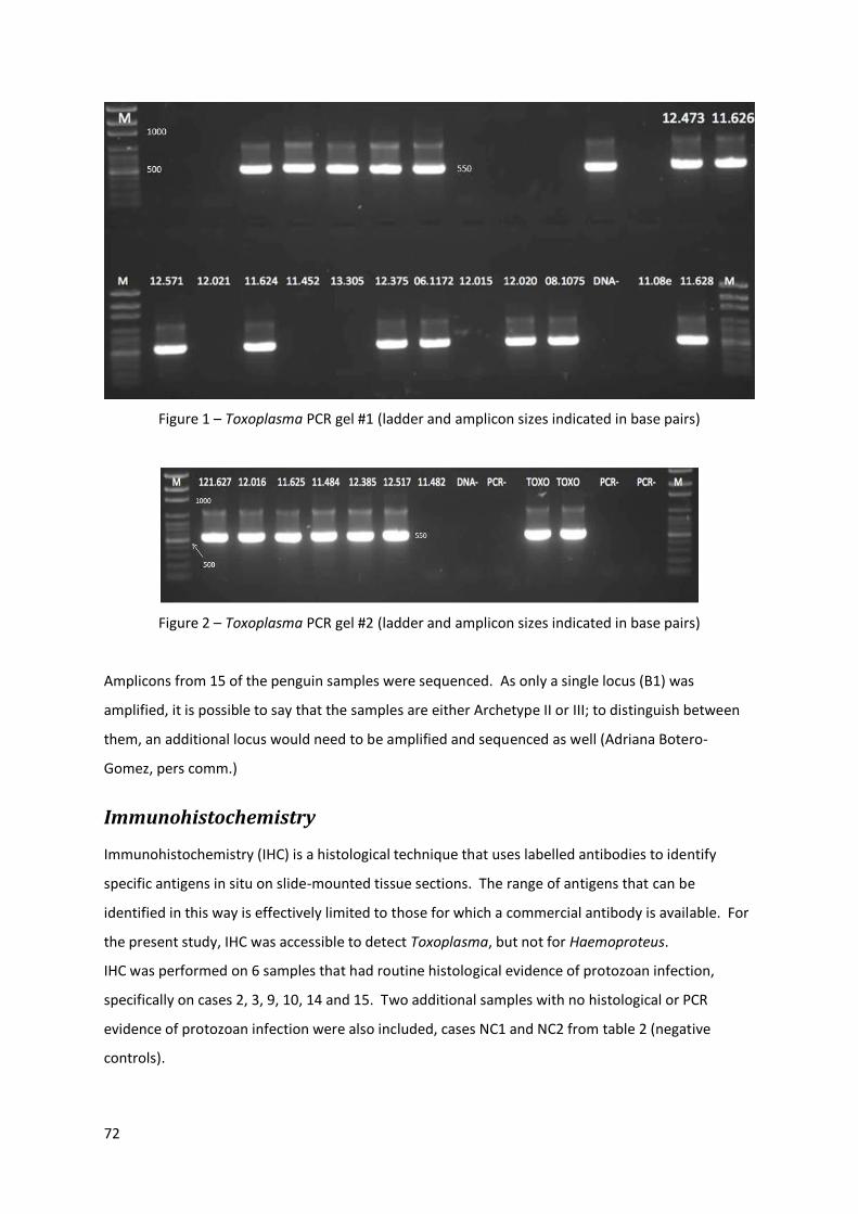
72
Figure 1 – Toxoplasma PCR gel #1 (ladder and amplicon sizes indicated in base pairs)
Figure 2 – Toxoplasma PCR gel #2 (ladder and amplicon sizes indicated in base pairs)
Amplicons from 15 of the penguin samples were sequenced. As only a single locus (B1) was
amplified, it is possible to say that the samples are either Archetype II or III; to distinguish between
them, an additional locus would need to be amplified and sequenced as well (Adriana Botero-
Gomez, pers comm.)
Immunohistochemistry
Immunohistochemistry (IHC) is a histological technique that uses labelled antibodies to identify
specific antigens in situ on slide-mounted tissue sections. The range of antigens that can be
identified in this way is effectively limited to those for which a commercial antibody is available. For
the present study, IHC was accessible to detect Toxoplasma, but not for Haemoproteus.
IHC was performed on 6 samples that had routine histological evidence of protozoan infection,
specifically on cases 2, 3, 9, 10, 14 and 15. Two additional samples with no histological or PCR
evidence of protozoan infection were also included, cases NC1 and NC2 from table 2 (negative
controls).

73
Toxoplasma IHC was performed by the technical staff of the Murdoch University Veterinary Hospital
histology laboratory (School of Veterinary and Life Sciences) using a polyclonal antibody (Thermo
Scientific Polyclonal (Goat) PA5-16921). Polyclonal antibody was used because it detects multiple
life stages of toxoplasma, and it is thus more sensitive than monoclonal antibody, but without a loss
of specificity; the antibody does not even cross react with the closely related Neospora caninum, so
its specificity is considered to be excellent (Michael Slaven, senior histology technician, MUVH, pers
comm.) Briefly, the protocol was as follows: each slide was washed in water, then treated with 3%
Hydrogen Peroxide for 15 minutes. Slides were washed with distilled water followed by TRIS wash
solution, and then treated with normal goat serum for 10 minutes. The goat serum was poured off
and the antibody applied and then incubated for 45 minutes. Negative control slides were prepared
omitting the antibody application, but washing with the same buffer solution. All slides were then
washed and treated with the secondary antibody reagent for 30 minutes, after which they were
washed and treated with a diaminobenzidine substrate-chromogen solution for 3 minutes. Slides
were counterstained with Harris’ Haematoxylin, washed in Scott’s tap water substitute, then
dehydrated, cleared and coverslipped with DPX.
Results
In each of the cases that had routine light microscopic evidence of tissue necrosis and protozoan
parasites, there was distinct positive staining, evident at low and high power, which was colocalised
with the necrotic regions (Fig 3). In the cases without evidence of parasites, there was no additional
staining beyond the background stain seen in the negative control slides.

74
Figure 3 – Toxoplasma polyclonal antibody IHC. Several foci of brown staining, most notably in the
image centre, indicate Toxoplasma antigen within a necrotic focus, liver, case 15, x100 magnification
At high power, the uptake of stain was clearly associated with the organisms in the lesions (Fig 4).
Figure 4- Toxoplasma polyclonal antibody IHC. Brown staining of several protozoan cysts within a
hepatocyte, liver, case 15, x1000 magnification

75
Discussion
Although many of the same tissue samples were used for DNA extraction, including those which had
tested positive for Haemoproteus, the results of the haemosporidian PCR as published by Cannell et
al, 2013 14 could not be reproduced in the first attempts at PCR. This was an unexpected result given
that the assays had been performed using the same primer sequences and the same protocols.
However, in that paper it was noted that only 4 of 10 penguins were positive by PCR, even though all
10 had histologically identifiable parasites in liver and spleen sections. Also, of those that were
positive, the PCR protocol had been repeated on up to 3 occasions before DNA was detected,
indicating a lack of sensitivity for this assay. Possible reasons for the poor sensitivity include: a) a
low concentration of DNA in the samples, b) interference from a reaction inhibitor, or c) degradation
of reaction reagents. Perhaps significantly, it has previously been reported that nested PCR is in fact
not a sensitive technique for the detection of mixed haemosporidian parasite infections in birds84.
Consequently, and particularly given that we did not have access to a dependable positive control
for these assays (notwithstanding that 4 of the samples had tested positive in another lab), the
failure to detect Haemoproteus DNA could not be confidently interpreted as indicating that the birds
were clear of this parasite.
In the present study, for the first two rounds of PCR using the published haemosporidian primers,
there were numerous instances in which a band was detected on the gel; however, the DNA
sequences were invariably of poor quality with multiple signals at many of the data points. It was
presumed that these bands had arisen from non-specific DNA amplification due to aberrant binding
of the primers to host or contaminant DNA. However, toward the end of the time and funding
allocated to this project, the failure to detect Haemoproteus DNA (and possibly the poor quality of
the bands that were produced) was explained, at least in part, by the revelation that the primer
sequences that had been published were incorrect. New primers were obtained to attempt the PCR,
though with very limited time and remaining funds available only one round of PCR was possible.
From this assay there was a single sample, from case 15, for which a band was detected, though
again no valid sequence could be generated from this sample. Of the four samples that had tested
positive in the Cannell et al, 2013 paper (that is, cases 5, 8, 9 and 10), none were positive, though
case 10 was in fact not included in the final assay as the sample had been mislabelled. In summary,
the Haemoproteus PCR results in this study did not in any way clarify the nature of the infection
affecting these birds.
76
On the other hand, the identification of Sarcocystidae family DNA was unexpected, and was
essentially fortuitous. The penguin samples had been included in a mixed batch of samples from
multiple sources as part of the validation process for a set of newly developed primers designed to
detect piroplasmids. Given that they had previously tested as negative for piroplasmids, they were
expected to be negative again. The positive result from this PCR was the most forthright indication
to this point that apicomplexans from another family than the Haemospororida might be implicated
in the disease process in the penguins (note that this result also indicates that the new primers were
less specific for piroplasmids than the developer had anticipated). Probably the most noteworthy
member of the Sarcocystidae, certainly in terms of its pathogenicity, is Toxoplasma gondii, and it is
notable that fatal toxoplasmosis has been reported in a Little Penguin56 with post mortem findings
similar to those observed in these cases. The suggestion arising from this PCR, and also from the
generic apicomplexan PCR, that the organism might be Toxoplasma led to a more directed effort to
demonstrate that organism.
The results of the first round of Toxoplasma PCR, while uniformly positive, were discarded on the
basis of a positive result from the negative control indicating contamination of the samples.
However, after a second DNA extraction with appropriate control outcomes, Toxoplasma was
detected by PCR in 12 of 13 penguins diagnosed with protozoal infections, and in 3 of 8 penguins
diagnosed with other causes of death (see Table 3) (Note also that the penguin negative for
Toxoplasma by PCR was positive by IHC – see further discussion of this finding below). Using a web-
based relative risk calculator (http://www.medcalc.org/calc/relative_risk.php, accessed 17th
September, 2014), the relative risk of death with evidence of protozoal infection for the penguins
positive for Toxoplasma by PCR is 4.8 with a 95% confidence interval of 0.7879 to 29.2432.
However, the p-value for this calculation is 0.0889, so it is not statistically significant using the
conventional cut off for significance of 0.05. Increased numbers of penguins in these groups,
especially the group without histologically detectable parasites, are required to give a more
statistically reliable result.
On the sum of the molecular evidence available, it is likely that some of the penguins were, or had
been, infected with Haemoproteus parasites. Given the apparent insensitivity of the PCR for this
organism, it cannot be stated with confidence how many birds were truly infected. In general, as
has been discussed in Chapter 1, Haemoproteus infections in birds are quite common and usually
cause mild or no clinical disease, though there are exceptions to this observation, especially when

77
infection occurs in aberrant hosts. Three hypotheses may be advanced to account for the
Haemoproteus findings:
1. The penguins are adapted hosts for the described species of Haemoproteus, and the few
positive cases are essentially incidental
2. The penguins are aberrant hosts newly exposed, the Haemoproteus parasite is responsible
for the severe disease and death observed, and it is only the insensitivity of the PCR that
obscures a clearer relationship between the two
3. Haemoproteus as a co-infection is able to exacerbate another disease state, in this instance
toxoplasmosis, resulting in the observed severe disease and death, but it is neither sufficient
nor necessary for expression of the disease
On the evidence at hand, the contribution of Haemoproteus to the observed pathology is effectively
unknown.
The presence of Toxoplasma, however, is strongly correlated with disease in these birds, and
immunohistochemistry positively identifies the parasites within the lesions of 6 out of 6 of the
affected birds; note that one of these (case 9 - 12/021) was the single case that was positive by light
microscopy but negative by PCR, suggesting that the PCR result may represent a false negative.
Given that the sample was run together with all of the other positive samples, it is clear that the
reagents were in good condition and there were no concerns with the presence of inhibitors, so a
false negative cannot be due to those factors. Most likely, the negative result can be attributed to
an error during the extraction process, or to low DNA concentration in the sample; a repeat
extraction and PCR with appropriate controls would be necessary to investigate this further. Death
from toxoplasmosis has been reported in one Little Penguin previously, with similar post mortem
findings to those in this study56. The gross, histological, haematological, ultrastructural,
immunohistochemical and PCR results are all consistent with fulminant toxoplasmosis being the
cause of death in these penguins.

78
6. Discussion
An investigation of the cause (or causes) of a naturally occurring outbreak of disease starts with
observations of the disease and its manifestations, followed by the generation of a hypothesis as to
the aetiology, and then testing of that hypothesis. The first instances of the disease explored in this
thesis were two isolated cases separated by two years. Under these circumstances, the threat of the
disease to the wellbeing of the penguin population appeared to be minimal, so a provisional
diagnosis was acceptable in each case, and there was little to no urgency to follow up with additional
tests to establish a definitive diagnosis. The sudden increase in cases that was observed in late 2011
and early 2012, especially in light of the recent history of declining numbers of breeding penguins on
Penguin Island, meant that a definitive diagnosis came to be considered important. Understanding
the aetiology is the first step in developing a strategic response, or in determining whether a
response is feasible, so the aims of this project were:
1. To document the post mortem findings that are associated with this protozoan infection
2. To identify the parasite at least to the level of genus, and
3. To commence collection of data intended to assess the prevalence of the disease in the live
penguin population and in potential avian hosts other than the penguins
The provisional diagnosis of Avian malaria, which is to say, infection with one or another species of
Plasmodium, was reasonable given the gross and histological findings at post mortem, and the
knowledge that Avian malaria is a significant cause of morbidity and mortality in penguins, though
the link between infection and disease has been better documented in birds in captivity than in
those in the wild. Thus, the hypothesis for this investigation (with respect to the identity of the
parasite) was that those penguins presenting with the distinctive signs of multifocal hepatic and
splenic necrosis with enlargement of those organs had been infected with Plasmodium parasites.
The very low (near to non-existent) incidence of parasitaemia, as judged from histological
assessment of erythrocytes and occasionally from cytology of tissue impression smears, was argued
to be because the animals presenting for post mortem had died in the acute phase of infection from
extensive tissue necrosis leading to the development of a cytokine-mediated haemodynamic shock.
In terms of pathogenesis, this explanation was, and remains, reasonable. In fact, the pathological
interpretation of the lesions has not changed since the project was undertaken – the only real
unknown was the identity of the protozoa in those lesions.
79
The earliest work done in the project was the collection of blood samples from Little Penguins and
Bridled Terns to establish whether parasitaemia could be detected in either of these species, and, if
it could, to estimate the prevalence of infection. An earlier haematological study published in 1999
did not detect parasitaemia in any penguins from this population46. However, given that we
suspected that the infection was relatively new to these birds (on the basis that it was causing
multiple mortalities, as would be expected in an immunologically naïve population) our assumption
was that overt parasitaemia would now be more common. From the 146 blood smears examined
(94 Little Penguins, 52 Bridled Terns), blood parasites were found in the erythrocytes of one penguin
only. On morphological grounds, this parasite could be identified as an apicomplexan, but not
further. Assuming that this parasite was the same as that causing the fatalities (which might not be
the case) this evidence suggested that the prevalence of infection was most likely low. Also, since
the bird in question was not obviously unwell, and that there was evidence of a regenerative red cell
response and the parasitaemia was only mild, it appeared that the infection was at least not
uniformly fatal.
Subsequent to the blood sampling, transmission electron microscopy was performed using tissue
from one penguin, and from this it was possible to identify characteristic features of apicomplexan
protozoa in the splenic tissue. While this confirmed the light microscopic findings to a much higher
resolution, few specific features that might further narrow down its taxonomy were found. The one
feature that was somewhat helpful was that the parasites appeared to multiply by a process of
endodyogeny, though even this feature has been documented in many apicomplexan genera (not,
however, including Plasmodium).
Thus, in terms of testing the hypothesis, early investigations could not demonstrate that the parasite
was Plasmodium. At around this time, a PCR investigation conducted by an external laboratory
offered the explanation that the parasite was in fact from the genus Haemoproteus, an organism
which is so closely related to Plasmodium that it was detected by PCR primers that were originally
designed to identify Plasmodium. Diagnostically, the similarities between the two parasites are such
that, where morphology of the erythrocyte stages of development are lacking (Plasmodium spp.
undergo shizogony in erythrocytes whereas Haemoproteus spp. do not), they can only be
distinguished by techniques such as DNA sequencing. In terms of pathogenicity, also, the two
parasites can be similar, differing primarily in features such as host range and vector; indeed, some
reports of Avian malaria group Plasmodium and Haemoproteus infections together, though it is
more usual to consider them as distinct disease entities. Consequently, the focus of the project

80
shifted to investigating the hypothesis that this fatal disease was haemoproteosis rather than Avian
malaria.
However, the evidence to implicate Haemoproteus as the infective agent was not without its own
problems. To start with, only 4 of the 10 cases assayed were positive by PCR, though all had clear
and consistent evidence of morphologically identical parasites in the tissues by routine light
microscopy. Additionally, even among the cases that were positive by PCR, they were not positive
all of the time. There was a distinct lack of sensitivity to the assay (assuming that the parasite in
each of the cases was the same) that made confident interpretation difficult, particularly in regard to
those samples that tested negative. The fact that the DNA products had been sequenced to
demonstrate a previously undescribed species of Haemoproteus, however, made it unlikely that
there had been contamination of the penguin samples from another source, so it had to be assumed
that the infection was real. The true prevalence and significance, however, were uncertain.
The picture was not clarified when I tried to replicate the PCR results in our own laboratory without
success. After several attempts at the assay, a colleague assisting the investigation (Dr Andrea
Paparini, Vector and Waterborne Pathogens Research Group, Murdoch University) fortuitously
included the penguin DNA samples when testing some new PCR primers he had developed for
working with the piroplasms Babesia and Theileria, the penguin samples in this context being
intended to function as negative controls as they had already tested as negative for those organisms
in an earlier PCR. Unexpectedly, this PCR generated bands of DNA, and a selection of those bands
were sequenced to reveal that the organisms present were members of the family Sarcocystidae.
The Sarcocystidae belong within the phylum Apicomplexa, dividing from the taxonomic branches
which include Plasmodium, Haemoproteus, Babesia and Theileria at the level of class (Conoidasida
versus Aconoidasida). At the same time as this assay, another PCR of the penguin DNA was
performed using generic primers for apicomplexans. The PCR product for this assay is very large,
and while the two samples tested were positive, a complete sequence could not be generated for
either sample. However, the segment of each that did return a good sequence gave a result that
was most consistent with Toxoplasma. Toxoplasma is a single-species genus (T. gondii is the only
member) in the family Sarcocystidae, and it is a significant pathogen of wildlife, domestic animals
and humans worldwide.
On the basis of these results, new PCR assays were undertaken which were directed specifically at
identifying Toxoplasma. Immediately, every sample tested was strongly positive for Toxoplasma.

81
Unfortunately, the extraction blank negative control (the sample made at the time of DNA extraction
which contained no penguin tissue) was also strongly positive, indicating that there had been
contamination of this tube, and possibly other tubes, at some point in the DNA extraction and/or
PCR process. A second DNA extraction was performed, this time including eight samples from
penguins that did not have microscopic evidence of protozoan infections. The Toxoplasma PCR was
repeated and 12 of the 13 samples with light microscopic evidence of protozoa were also positive by
PCR; the reason for the one negative result is unknown, though repeating the assay to determine
whether the result is consistent would be a worthwhile first step. The negative controls were also
negative.
At around this time, we received notification that the Haemoproteus PCR primers we had been using
in our first attempts to reproduce the Haemoproteus results were based on incorrect published
sequences. With very limited time and resources available, we ordered new primers and repeated
the Haemoproteus PCR once only. A single, very faintly positive band of DNA was found, this being
from a new case that had not been submitted to the New Mexico laboratory. The band was
submitted for sequencing, but no sequence could be generated, possibly reflecting poor quality DNA
or, alternatively, degraded reagents. In the end, no confirmed positives were ever achieved from
the Haemoproteus PCR assays performed at Murdoch University.
Perhaps belatedly, a selection of cases, both positive and negative by histology and PCR, were then
investigated by immunohistochemistry, by this time with the primary intention of showing where in
the tissues the Toxoplasma antigen was located. In all positive cases, Toxoplasma was
demonstrated in the necrotic lesions of liver and spleen and direct staining of the protozoan cysts
was apparent when the slides were assessed at high power.
The most compelling interpretation of the gross and microscopic pathology, the ultrastructure of the
protozoan organism and the PCR and immunohistochemistry findings is that the penguins had been
dying of acute toxoplasmosis. The pathological changes observed are in agreement with a previous
published case of toxoplasmosis in one Little Penguin56, and are also consistent with the pathology
reported in experimental toxoplasmosis in other animal species (such as eastern barred bandicoots7)
though it is worth noting that Toxoplasma tachyzoites and bradyzoites are widely distributed in body
tissues, and the range of lesions which may occur when infections progress beyond the acute stage
and become chronic is extensive10. Toxoplasma is a global parasite, which, it is emerging, is a very
common infection in Australian wildlife species65,67,68,86, and which causes fatal infections in wild
82
birds23,30,41,53. In distinction to the life cycle of apicomplexan haemoparasites, Toxoplasma requires
no arthropod vector. The definitive host is the cat, and transmission of disease may occur by
ingestion of infective oocysts from cat faeces, or by ingestion of tissue cysts in animal tissues (as well
as, in all likelihood, by vertical transmission in utero82.) Because sexual development only occurs in
cats and it is not transmitted by a vector, it might be expected that exposure to Toxoplasma would
be low in marine wildlife. However, it is also emerging that Toxoplasma infection is not uncommon
in many marine mammals, implying that the disease can be transmitted by contamination of
waterways with cat faeces47. Furthermore, it has been shown that filter-feeding fish will take up
Toxoplasma oocysts from contaminated water, and that the infection can be passed to mice when
fed on these fish57. Notably, one species of fish that has been demonstrated to take up Toxoplasma
cysts and transmit the disease is the pilchard (Sardinops sagax), an important prey species for the
Little Penguins of Penguin Island57. Also, it has recently been shown that Toxoplasma can aggregate
on extracellular polymeric substances (EPS) in marine environments, enhancing the efficiency with
which the organism is able to enter the marine food chain in coastal ecosystems78.
Toxoplasma DNA was detected in three out of eight penguins that did not have any of the gross or
microscopic lesions associated with severe protozoal disease. These birds were case number NC7,
which was presumed to have died from starvation, case number NC6, for which no diagnosis could
be made, and case number NC4, which was found to have died from traumatic injuries attributed to
a boat strike. Each of these post mortems was conducted at or after the time of the late 2011 spike
in protozoal disease, so it is unlikely that the disease was present and overlooked. The implication is
that infection with Toxoplasma is not uniformly fatal in the penguins, and that virulence, dosage
and/or comorbidity factors are likely to play a role in determining the outcome of infection in any
given bird. Notably, a feature of fatal disease is that the birds were, by and large, in good body
condition. It is possible that these penguins were able to gather adequate prey in the local
environment without the need to travel far afield, and that the fish they were preying on in that
environment themselves carried a large parasite burden, though this hypothesis would be difficult to
prove. The number of penguins presented to Murdoch University for post mortem in recent years
has been 26 in 2011, 40 in 2012, 22 in 2013 and none as of early October in 2014. Thus, with such
low numbers to judge from, there is insufficient evidence to declare whether the cluster of cases in
2011 was a true outbreak of disease that has subsided, or whether deaths are ongoing. Whatever
the case, the incidence of toxoplasmosis is most likely dependent on the both the overall number
and the disease status of cats contributing to faecal contamination of local waterways. A serological

83
survey of the penguins would provide a useful indication of the prevalence of exposure in the
population.
Ultimately, the evidence from this investigation provides no support for the original hypothesis that
Avian malaria was the cause of death or disease in these penguins. Furthermore, while
haemoparasite DNA has been detected in the tissues of some birds (albeit by testing at an external
laboratory), the role of Haemoproteus in causing disease in these penguins is, unfortunately,
unknown. It is likely enough that Haemoproteus represents an enzootic infection in these birds that
had not previously been detected, at least in part because its pathogenicity was low and no effort
had been made to look for it. One implication of the finding is that there is a species of
ceratopogonid biting flies to which the penguins are exposed that is the definitive host for this
parasite. An entomological survey of Penguin Island would be helpful in investigating this
hypothesis, and demonstration of DNA from the same Haemoproteus parasite in any biting flies
isolated from such a survey would effectively confirm it.
With regard to the other aims, the presentation of acute toxoplasmosis in Little Penguins apparently
invariably includes hepatomegaly and splenomegaly with gross and microscopic evidence of
multifocal areas of necrosis in these tissues. Microscopic evidence of protozoa can be found in liver
and spleen sections, and occasionally in other tissues including the heart, lungs, air sacs, adipose
tissue and arteries. No parasites were detected in the brain of any penguin.
Estimation of the prevalence of Toxoplasma infections in the Little Penguins is difficult from the
evidence of this investigation, not least because blood sampling and haematology is simply an
insensitive test for this purpose. A serological or PCR survey is required to answer that question.
Also, given that Toxoplasma is a parasite of cats, the other avian species of Penguin Island can no
longer be considered culpable for introducing the infection to the Little Penguins. Apologies to the
Bridled Terns.

84
References:
1 BirdLife International 2012. Eudyptula minor IUCN 2013. IUCN Red List of Threatened Species. Version 2013.1; 2013.
2 Endangered Population of Little Penguins (Eudyptula minor) at Manly. In: Service NPaW, ed. New
South Wales; 2003. 3 Shoalwater Islands Marine Park Management Plan 2007-2017. Perth, Western Australia:
Department of Environment and Conservation, Marine Parks and Reserves Authority; 2007. 4 Argilla LS, Howe L, Gartrell BD, Alley MR: High prevalence of Leucocytozoon spp. in the
endangered yellow-eyed penguin (Megadyptes antipodes) in the sub-Antarctic regions of New Zealand. Parasitology 2013:140(5):672-682.
5 Atkinson CT, Dusek RJ, Woods KL, Iko WM: Pathogenicity of avian malaria in experimentally-
infected Hawaii Amakihi. J Wildl Dis 2000:36(2):197-204. 6 Atkinson CT, Thomas NJ, Hunter DB: Parasitic diseases of wild birds. Ames, Iowa: Wiley-Blackwell,
2008. 7 Bettiol SS, Obendorf DL, Nowarkowski M, Goldsmid JM: Pathology of experimental toxoplasmosis
in eastern barred bandicoots in Tasmania. J Wildl Dis 2000:36(1):141-144. 8 Bool NM, Page B, Goldsworthy SD: What is causing the decline of little penguins (Eudyptula minor)
on Granite Island, South Australia, Adelaide: South Australian Research and Development Institute; 2007: 55.
9 Bueno MG, Lopez RP, de Menezes RM, Costa-Nascimento Mde J, Lima GF, Araujo RA, et al.:
Identification of Plasmodium relictum causing mortality in penguins (Spheniscus magellanicus) from Sao Paulo Zoo, Brazil. Vet Parasitol 2010:173(1-2):123-127.
10 Canfield PJ, Hartley WJ, Dubey JP: Lesions of toxoplasmosis in Australian marsupials. J Comp
Pathol 1990:103(2):159-167. 11 Cannell B, Chambers L, Wooller R, Bradley S: Poorer breeding by little penguins near Perth,
Western Australia is correlated with above average sea surface temperatures and a stronger Leeuwin Current. Marine and Freshwater Research 2012:63(10):914-925.
12 Cannell B, Pollock K, Bradley S, Wooller R, Sherwin W, Sinclair J: Augmenting mark-recapture
with beach counts to estimate the abundance of little penguins on Penguin Island, Western Australia. Wildlife Research 2011:38(6):491-500.
13 Cannell BL, Krasnec KV, Campbell K, Jones HI, Miller RD, Stephens N: Corrigendum to “The
pathology and pathogenicity of a novel Haemoproteus spp. infection in wild Little Penguins (Eudyptula minor)” [Vet. Parasitol. 197 (1–2) (2013) 74–84]. Veterinary Parasitology 2014:205(1–2):416.

85
14 Cannell BL, Krasnec KV, Campbell K, Jones HI, Miller RD, Stephens N: The pathology and
pathogenicity of a novel Haemoproteus spp. infection in wild Little Penguins (Eudyptula minor). Vet Parasitol 2013:197(1-2):74-84.
15 Cheville NF: Ultrastructural pathology the comparative cellular basis of disease. 2nd ed. Ames, IA:
Wiley-Blackwell, 2009. 16 Connard MN: Patterns of foraging by little penguins Eudyptula minor from Penguin Island,
Western Australia. Perth: Murdoch University; 1995. 17 Cranfield MR, Graczyk TK, Beall FB, Ialeggio DM, Shaw ML, Skjoldager ML: Subclinical avian
malaria infections in African black-footed penguins (Spheniscus demersus) and induction of parasite recrudescence. J Wildl Dis 1994:30(3):372-376.
18 Dann P, Cullen M, Weir I: National Review of the Conservation Status and Management of
Australian Little Penguin Colonies. Final Report: The Australian Nature Conservation Agency; 1996.
19 Deem SL, Merkel J, Ballweber L, Vargas FH, Cruz MB, Parker PG: Exposure to Toxoplasma gondii
in Galapagos Penguins (Spheniscus mendiculus) and flightless cormorants (Phalacrocorax harrisi) in the Galapagos Islands, Ecuador. J Wildl Dis 2010:46(3):1005-1011.
20 Donovan TA, Schrenzel M, Tucker TA, Pessier AP, Stalis IH: Hepatic hemorrhage, hemocoelom,
and sudden death due to Haemoproteus infection in passerine birds: eleven cases. J Vet Diagn Invest 2008:20(3):304-313.
21 Dubey JP: Life Cycle of Cystoisospora felis (Coccidia: Apicomplexa) in Cats and Mice. J Eukaryot
Microbiol 2014:61(6):637-643. 22 Dubey JP: A review of toxoplasmosis in wild birds. Veterinary Parasitology 2002:106(2):121-153. 23 Dubey JP, Garner MW, Willette MM, Batey KL, Gardiner CH: Disseminated toxoplasmosis in
magpie geese (Anseranas semipalmata) with large numbers of tissue cysts in livers. J Parasitol 2001:87(1):219-223.
24 Dubey JP, Lindsay DS: Development and ultrastructure of Besnoitia oryctofelisi tachyzoites, tissue
cysts, bradyzoites, schizonts and merozoites. Int J Parasitol 2003:33(8):807-819. 25 Duignan PJ: Diseases of penguins. Surveillance 2001:28(4):5-11. 26 Dunlop JNN, Rippey E: The natural history of the bridled tern on Penguin Island, Western
Australia. Peron, W.A.: Naragebup Rockingham Regional Environmental Centre, 2007. 27 Earlé RA, Huchzermeyer FW, Bennett GF, Brossy JJ: Babesia peircei sp. nov. from the Jackass
Penguin, S Afr J Zoology 1993:28:88-90. 28 Ewen JG, Bensch S, Blackburn TM, Bonneaud C, Brown R, Cassey P, et al.: Establishment of exotic
parasites: the origins and characteristics of an avian malaria community in an isolated island avifauna. Ecology Letters 2012:15(10):1112-1119.

86
29 Fallis AM, Bisset SA, Allison FR: Leucocytozoon tawaki n.sp. (Eucoccida: Leucocytozoidae) from the penguin Eudyptes pachyrhynchus, and preliminary observations on its development in Austrosimulium spp. (Diptera: Simuliidae). New Zealand Journal of Zoology 1975:3:11-16.
30 Ferreira FC, Jr., Donatti RV, Marques MV, Ecco R, Preis IS, Shivaprasad HL, et al.: Fatal
toxoplasmosis in a vinaceous Amazon parrot (Amazona vinacea). Avian Dis 2012:56(4):774-777.
31 Ferrell ST, Snowden K, Marlar AB, Garner M, Lung NP: Fatal hemoprotozoal infections in multiple
avian species in a zoological park. J Zoo Wildl Med 2007:38(2):309-316. 32 Fix AS, Waterhouse C, Greiner EC, Stoskopf MK: Plasmodium relictum as a Cause of Avian Malaria
in Wild-Caught Magellanic Penguins (Spheniscus magellanicus). Journal of Wildlife Diseases 1988:24(4):610-619.
33 Graczyk TK, Brossy JJ, Plos A, Stoskopf MK: Avian malaria seroprevalence in Jackass penguins
(Spheniscus demersus) in South Africa. J Parasitol 1995:81(5):703-707. 34 Grigg ME, Boothroyd JC: Rapid identification of virulent type I strains of the protozoan pathogen
Toxoplasma gondii by PCR-restriction fragment length polymorphism analysis at the B1 gene. J Clin Microbiol 2001:39(1):398-400.
35 Grim KC, Van der Merwe E, Sullivan M, Parsons N, McCutchan TF, Cranfield M: Plasmodium
Juxtanucleare associated with mortality in black-footed penguins (Spheniscus demersus) admitted to a rehabilitation center. J Zoo Wildl Med 2003:34(3):250-255.
36 Heber S, Wilson K-J, Molles L: Breeding biology and breeding success of the blue penguin
(Eudyptula minor) on the West Coast of New Zealand's South Island. New Zealand Journal of Zoology 2008:35(1):63-71.
37 Hill A: An investigation of Leucocytozoon in the endangered yellow-eyed penguin (Megadyptes
antipodes) Institute of Veterinary, Animal and Biomedical Sciences. Palmerston North, New Zealand: Massey University; 2008.
38 Hocken AG: Cause of death in blue penguins (Eudyptula m. minor) in North Otago, New Zealand.
New Zealand Journal of Zoology 2000:27(4):305-309. 39 Hocken AG: Internal organ weights of the blue penguin Eudyptula minor. New Zealand Journal of
Zoology 2000:27(4):299-304. 40 Homer MJ, Aguilar-Delfin I, Telford SR, 3rd, Krause PJ, Persing DH: Babesiosis. Clin Microbiol Rev
2000:13(3):451-469. 41 Howe L, Hunter S, Burrows E, Roe W: Four cases of fatal toxoplasmosis in three species of
endemic New Zealand birds. Avian Dis 2014:58(1):171-175. 42 Imura T, Suzuki Y, Ejiri H, Sato Y, Ishida K, Sumiyama D, et al.: Prevalence of avian haematozoa in
wild birds in a high-altitude forest in Japan. Vet Parasitol 2012:183(3-4):244-248. 43 Ishtiaq F, Gering E, Rappole JH, Rahmani AR, Jhala YV, Dove CJ, et al.: Prevalence and diversity of
avian hematozoan parasites in Asia: a regional survey. J Wildl Dis 2007:43(3):382-398.

87
44 Jefferies R, Ryan UM, Irwin PJ: PCR-RFLP for the detection and differentiation of the canine
piroplasm species and its use with filter paper-based technologies. Vet Parasitol 2007:144(1-2):20-27.
45 Jones HI, Shellam GR: Blood parasites in penguins, and their potential impact on conservation.
Marine Ornithology 1999:27:181-184. 46 Jones HI, Shellam GR: The occurrence of blood-inhabiting protozoa in captive and free-living
penguins. Polar Biology 1999:21:5-10. 47 Jones JL, Dubey JP: Waterborne toxoplasmosis--recent developments. Exp Parasitol
2010:124(1):10-25. 48 Kemp A, Dann P: Egg size, incubation periods and hatching success of Little Penguins, Eudyptula
minor. Emu 2001:101(3):249-253. 49 Klomp N, Meathrel C, Wienecke B, Wooller R: Surface Nesting by Little Penguins on Penguin
Island, Western Australia. Emu 1991:91(3):190-193. 50 Klomp N, Wooller R: Diet of little penguins, Eudyptula minor, from Penguin Island, Western
Australia. Marine and Freshwater Research 1988:39(5):633-639. 51 Klomp N, Wooller R: Patterns of Arrival and Departure by Breeding Little Penguins at Penguin
Island, Western Australia. Emu 1991:91(1):32-35. 52 Ko K-N, Kang S-C, Jung J-Y, Bae J-H, Kim J-H: Avian malaria associated with Plasmodium spp.
infection in a penguin in Jeju Island. Korean Journal of Veterinary Research 2008:48(2):197-201.
53 Las RD, Shivaprasad HL: An outbreak of toxoplasmosis in an aviary collection of Nicobar pigeons
(Caloenas nicobaria). J S Afr Vet Assoc 2008:79(3):149-152. 54 Levin IL, Outlaw DC, Hernan Vargas F, Parker PG: Plasmodium blood parasite found in
endangered Galapagos penguins (Spheniscus mendiculus). Biological Conservation 2009:142:3191-3195.
55 Lindsay DS, Quick DP, Steger AM, Toivio-Kinnucan MA, Blagburn BL: Complete development of
the porcine coccidium Isospora suis Biester, 1934 in cell cultures. J Parasitol 1998:84(3):635-637.
56 Mason RW, Hartley WJ, Dubey JP: Lethal toxoplasmosis in a little penguin (Eudyptula minor) from
Tasmania. J Parasitol 1991:77(2):328. 57 Massie GN, Ware MW, Villegas EN, Black MW: Uptake and transmission of Toxoplasma gondii
oocysts by migratory, filter-feeding fish. Vet Parasitol 2010:169(3-4):296-303. 58 Miranda K, de Souza W, Plattner H, Hentschel J, Kawazoe U, Fang J, et al.: Acidocalcisomes in
Apicomplexan parasites. Exp Parasitol 2008:118(1):2-9.

88
59 Nicholson LW: Annual variation in the pre-laying attendance, breeding phenology and behaviour of the Little Penguin, Eudyptula minor Biological Sciences. Perth: Murdoch University; 1994.
60 Nishi M, Hu K, Murray JM, Roos DS: Organellar dynamics during the cell cycle of Toxoplasma
gondii. J Cell Sci 2008:121(Pt 9):1559-1568. 61 Olias P, Wegelin M, Zenker W, Freter S, Gruber AD, Klopfleisch R: Avian malaria deaths in parrots,
Europe. Emerg Infect Dis 2011:17(5):950-952. 62 Orr K, Pobar G: Shoalwater Islands Management Plan 1992-2002. In: Management DoCaL,
Authority NPaNC, eds. Perth; 1992. 63 Palinauskas V, Iezhova TA, Krizanauskiene A, Markovets MY, Bensch S, Valkiunas G: Molecular
characterization and distribution of Haemoproteus minutus (Haemosporida, Haemoproteidae): a pathogenic avian parasite. Parasitol Int 2013:62(4):358-363.
64 Palmer JL, McCutchan TF, Vargas FH, Deem SL, Cruz M, Hartman DA, et al.: Seroprevalence of
Malarial Antibodies in Galapagos Penguins (Spheniscus mendiculus). J Parasitol 2013:99(5):770-776.
65 Pan S, Thompson RC, Grigg ME, Sundar N, Smith A, Lymbery AJ: Western Australian marsupials
are multiply infected with genetically diverse strains of Toxoplasma gondii. PLoS One 2012:7(9):e45147.
66 Paperna I: Ultrastructural study of the development of Sarcocystis singaporensis sarcocysts in the
muscles of its rat host. Parasite 2002:9(2):161-166. 67 Parameswaran N, O'Handley RM, Grigg ME, Fenwick SG, Thompson RC: Seroprevalence of
Toxoplasma gondii in wild kangaroos using an ELISA. Parasitol Int 2009:58(2):161-165. 68 Parameswaran N, Thompson RC, Sundar N, Pan S, Johnson M, Smith NC, et al.: Non-archetypal
Type II-like and atypical strains of Toxoplasma gondii infecting marsupials of Australia. Int J Parasitol 2010:40(6):635-640.
69 Peirce MA, Greenwood AG, Stidworthy MF: Leucocytozoon in captive penguins. Vet Rec
2005:157(25):819-820. 70 Peirce MA, Lederer R, Adlard RD, O'Donoghue PJ: Pathology associated with endogenous
development of haematozoa in birds from southeast Queensland. Avian Pathol 2004:33(4):445-450.
71 Ploeg M, Ultee T, Kik M: Disseminated toxoplasmosis in black-footed penguins (Spheniscus
demersus). Avian Dis 2011:55(4):701-703. 72 Quillfeldt P, Arriero E, Martinez J, Masello JF, Merino S: Prevalence of blood parasites in seabirds
- a review. Front Zool 2011:8(1):26. 73 Ricklefs RE, Fallon SM: Diversification and host switching in avian malaria parasites. Proc Biol Sci
2002:269(1494):885-892.

89
74 Ricklefs RE, Fallon SM, Bermingham E: Evolutionary relationships, cospeciation, and host switching in avian malaria parasites. Syst Biol 2004:53(1):111-119.
75 Ropert-Coudert Y, Cannell B, Kato A: Temperature inside nest boxes of little penguins. Wildlife
Society Bulletin 2004:32(1):177-182. 76 Schmidt GD, Roberts LS: Foundations of parasitology. St. Louis: Times Mirror/Mosby College Pub,
1985. 77 Schnittger L, Rodriguez AE, Florin-Christensen M, Morrison DA: Babesia: a world emerging. Infect
Genet Evol 2012:12(8):1788-1809. 78 Shapiro K, Krusor C, Mazzillo FF, Conrad PA, Largier JL, Mazet JA, et al.: Aquatic polymers can
drive pathogen transmission in coastal ecosystems. Proc Biol Sci 2014:281(1795). 79 Stevenson C, Woehler EJ: Population decreases in Little Penguins Eudyptula minor in south-
eastern Tasmania, Australia, over the past 45 years. Marin Ornithology 2007:35(1):71-76. 80 Sturrock HJW, Tompkins DM: Avian malaria parasites (Plasmodium spp.) in Dunedin and on the
Otago Peninsula, southern New Zealand. New Zealand Journal of Ecology 2008:32(1):98-102. 81 Sundermann CA, Estridge BH: Growth of and competition between Neospora caninum and
Toxoplasma gondii in vitro. Int J Parasitol 1999:29(10):1725-1732. 82 Thompson RC: Parasite zoonoses and wildlife: One Health, spillover and human activity. Int J
Parasitol 2013:43(12-13):1079-1088. 83 Valkiunas G: Avian Malaria Parasites and other Haemosporidia. London: CRC Press; 2004: 1
online resource (946 p.). 84 Valkiunas G, Bensch S, Iezhova TA, Krizanauskiene A, Hellgren O, Bolshakov CV: Nested
cytochrome b polymerase chain reaction diagnostics underestimate mixed infections of avian blood haemosporidian parasites: microscopy is still essential. J Parasitol 2006:92(2):418-422.
85 Waldenstrom J, Bensch S, Hasselquist D, Ostman O: A new nested polymerase chain reaction
method very efficient in detecting Plasmodium and Haemoproteus infections from avian blood. Journal of Parasitology 2004:90(1):191-194.
86 Wendte JM, Gibson AK, Grigg ME: Population genetics of Toxoplasma gondii: new perspectives
from parasite genotypes in wildlife. Vet Parasitol 2011:182(1):96-111. 87 Wienecke B: The Size and Breeding Patterns of Little Penguins Eudyptula minor in Australia: a
Comparative Study. Perth: Murdoch University; 1993. 88 Yang R, Brice B, Ryan U: A new Caryospora coccidian species (Apicomplexa: Eimeriidae) from the
laughing kookaburra (Dacelo novaeguineae). Exp Parasitol 2014:145C:68-73.