An Introduction to the Appendicular...
32
An Introduction to the Appendicular Skeleton
Transcript of An Introduction to the Appendicular...
An Introduction to the Appendicular
Skeleton
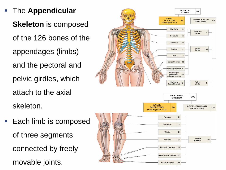
The Appendicular
Skeleton is composed
of the 126 bones of the
appendages (limbs)
and the pectoral and
pelvic girdles, which
attach to the axial
skeleton.
Each limb is composed
of three segments
connected by freely
movable joints.
Identify bone markings of each bone. The
markings will help you to determine
whether a bone is the right or left member
The Pectoral Girdle
Copyright © 2009 Pearson Education, Inc., publishing as Pearson Benjamin Cummings
The pectoral girdle, also called the shoulder
girdle consists of two clavicles two scapula.
The pectoral girdle connects the arms to the
axial skeleton
Provides arm movement and shoulder
movement.

The Clavicles
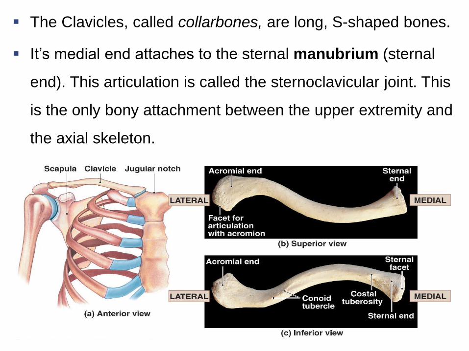
The Clavicles, called collarbones, are long, S-shaped bones.
It’s medial end attaches to the sternal manubrium (sternal
end). This articulation is called the sternoclavicular joint. This
is the only bony attachment between the upper extremity and
the axial skeleton.
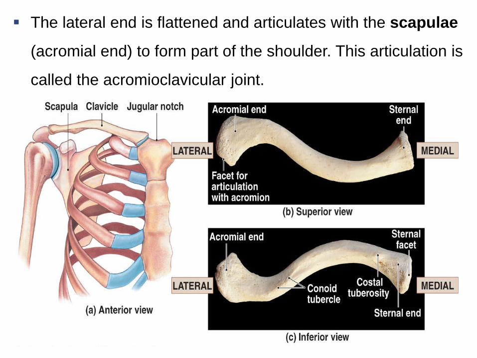
The lateral end is flattened and articulates with the scapulae
(acromial end) to form part of the shoulder. This articulation is
called the acromioclavicular joint.
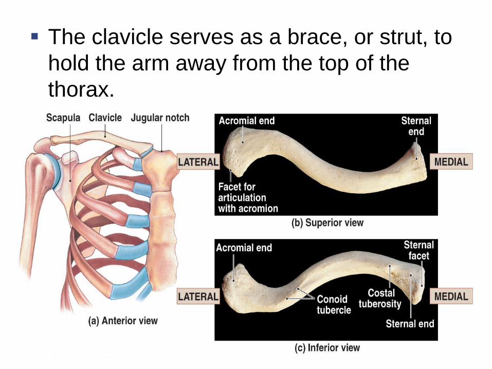
The clavicle serves as a brace, or strut, to
hold the arm away from the top of the
thorax.
The Scapulae
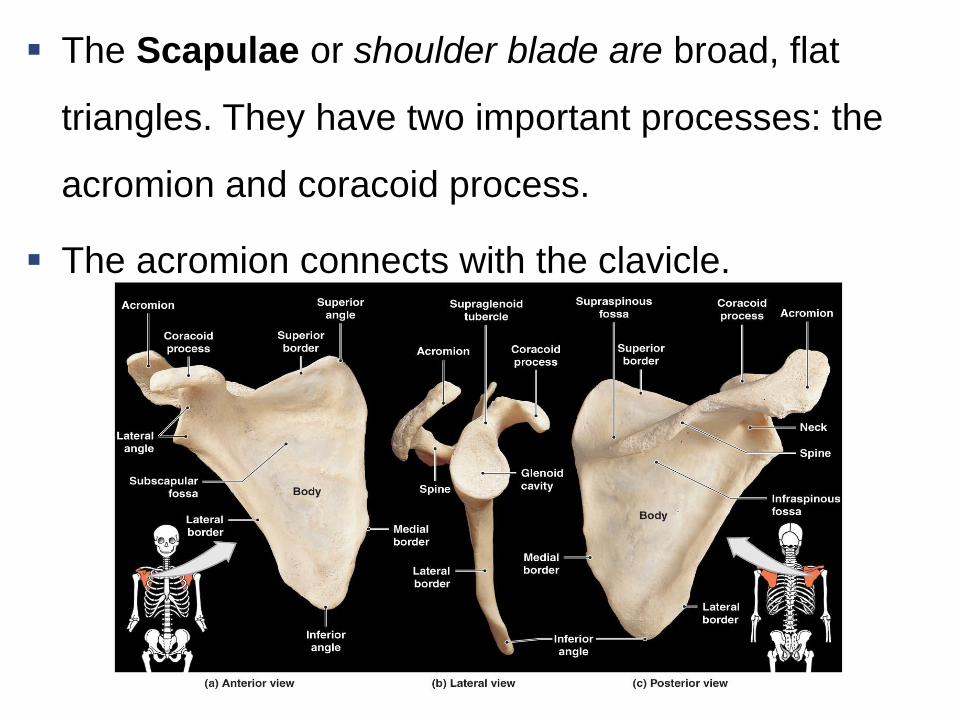
The Scapulae or shoulder blade are broad, flat
triangles. They have two important processes: the
acromion and coracoid process.
The acromion connects with the clavicle.
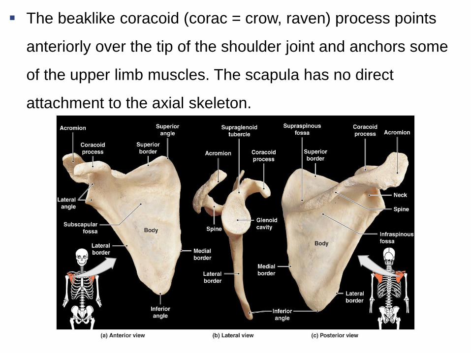
The beaklike coracoid (corac = crow, raven) process points
anteriorly over the tip of the shoulder joint and anchors some
of the upper limb muscles. The scapula has no direct
attachment to the axial skeleton.
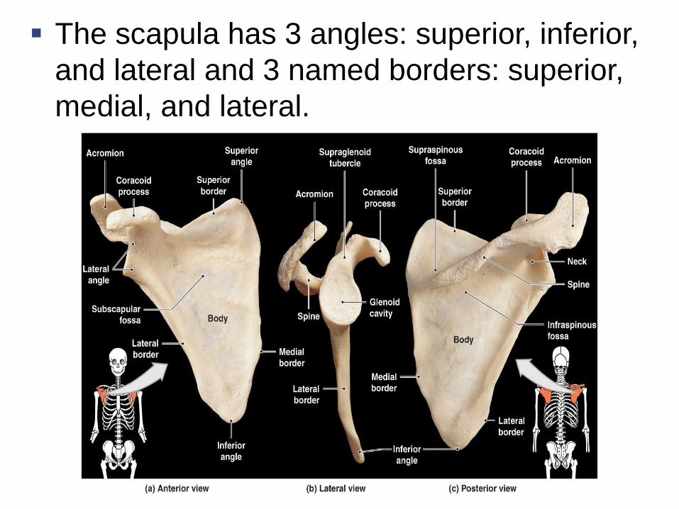
The scapula has 3 angles: superior, inferior,
and lateral and 3 named borders: superior,
medial, and lateral.
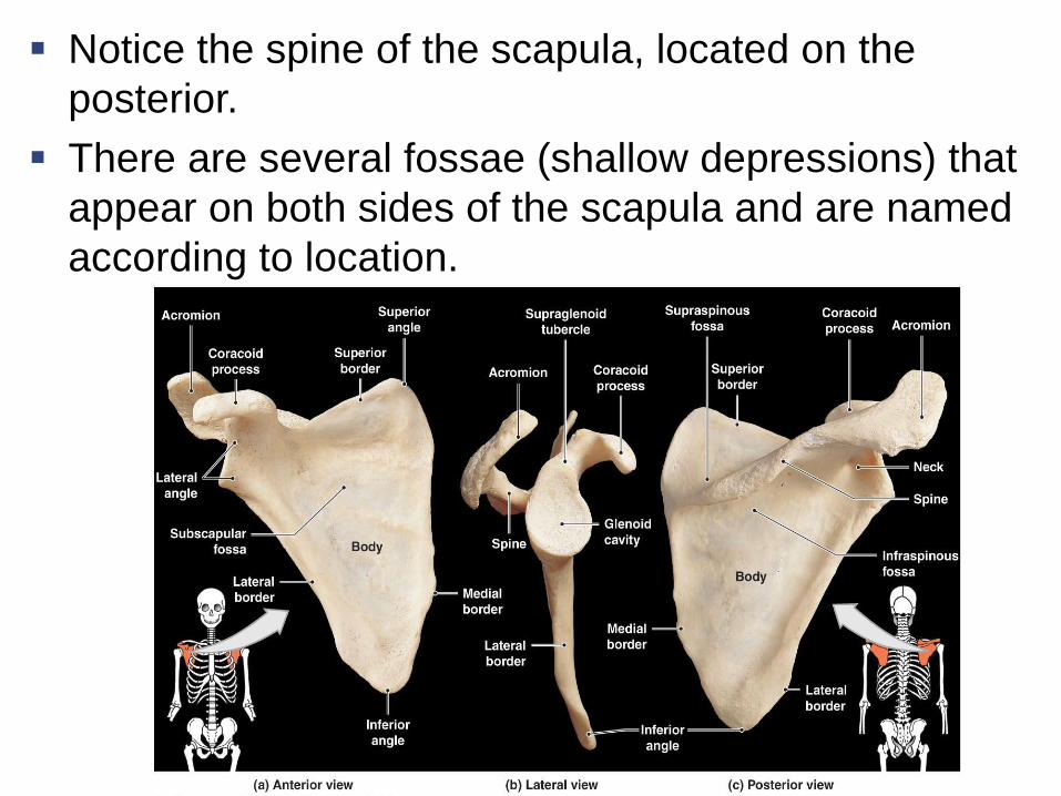
Notice the spine of the scapula, located on the
posterior.
There are several fossae (shallow depressions) that
appear on both sides of the scapula and are named
according to location.
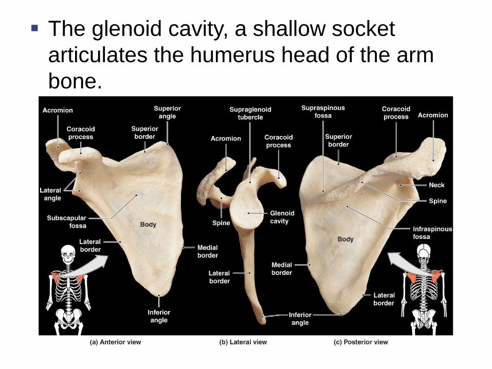
The glenoid cavity, a shallow socket
articulates the humerus head of the arm
bone.

The Arm
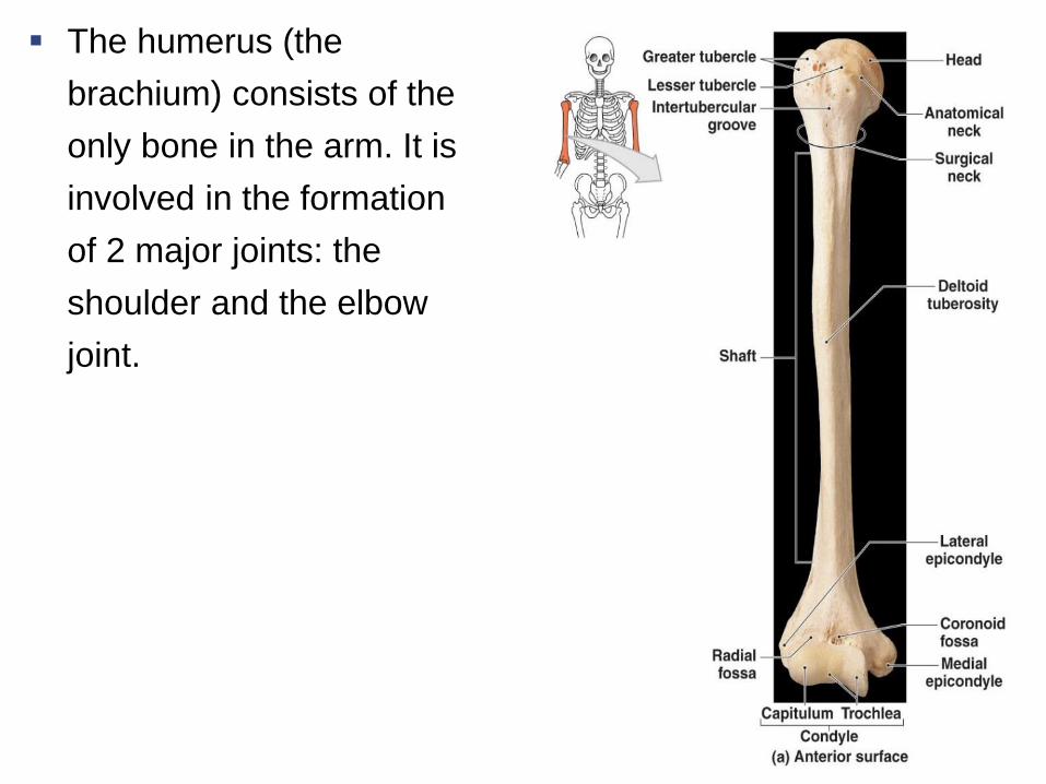
The humerus (the
brachium) consists of the
only bone in the arm. It is
involved in the formation
of 2 major joints: the
shoulder and the elbow
joint.
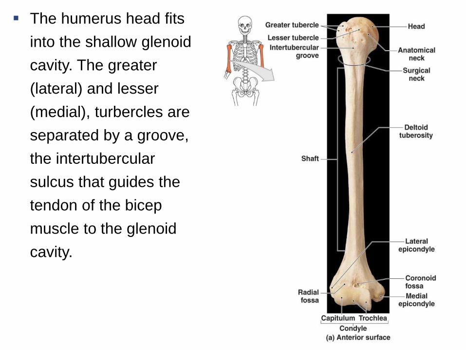
The humerus head fits
into the shallow glenoid
cavity. The greater
(lateral) and lesser
(medial), turbercles are
separated by a groove,
the intertubercular
sulcus that guides the
tendon of the bicep
muscle to the glenoid
cavity.
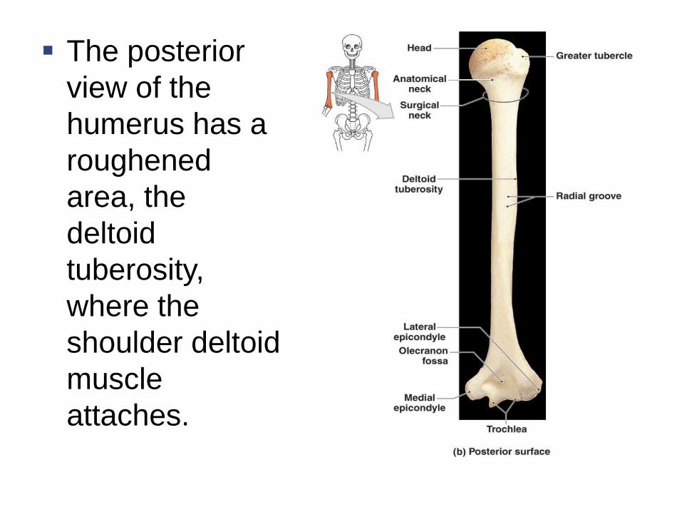
The posterior
view of the
humerus has a
roughened
area, the
deltoid
tuberosity,
where the
shoulder deltoid
muscle
attaches.
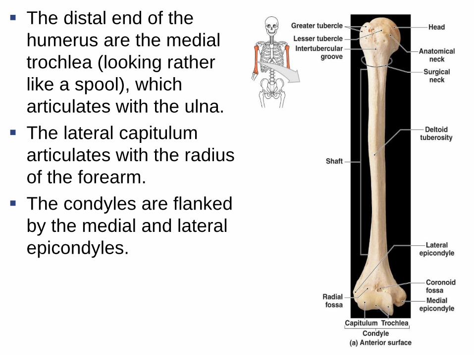
The distal end of the
humerus are the medial
trochlea (looking rather
like a spool), which
articulates with the ulna.
The lateral capitulum
articulates with the radius
of the forearm.
The condyles are flanked
by the medial and lateral
epicondyles.
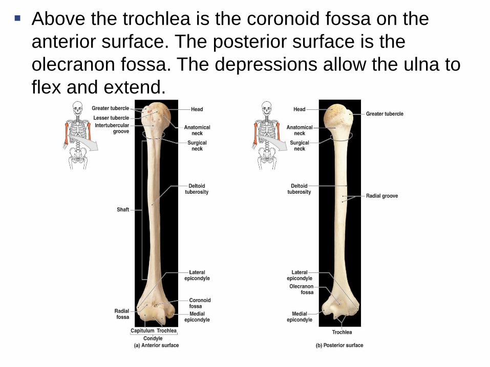
Above the trochlea is the coronoid fossa on the
anterior surface. The posterior surface is the
olecranon fossa. The depressions allow the ulna to
flex and extend.

The Forearm
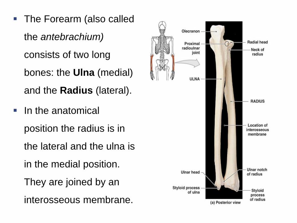
The Forearm (also called
the antebrachium)
consists of two long
bones: the Ulna (medial)
and the Radius (lateral).
In the anatomical
position the radius is in
the lateral and the ulna is
in the medial position.
They are joined by an
interosseous membrane.
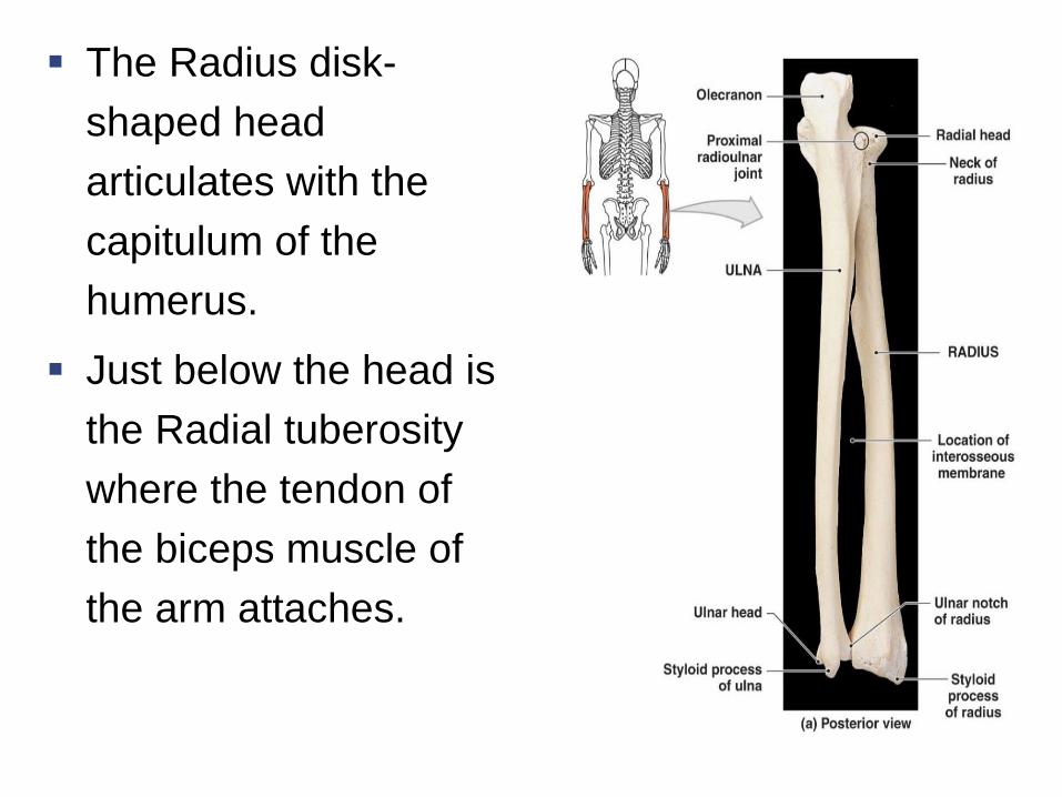
The Radius disk-
shaped head
articulates with the
capitulum of the
humerus.
Just below the head is
the Radial tuberosity
where the tendon of
the biceps muscle of
the arm attaches.
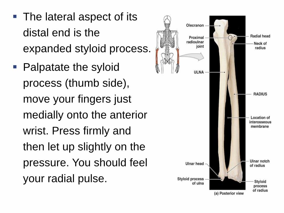
The lateral aspect of its
distal end is the
expanded styloid process.
Palpatate the syloid
process (thumb side),
move your fingers just
medially onto the anterior
wrist. Press firmly and
then let up slightly on the
pressure. You should feel
your radial pulse.
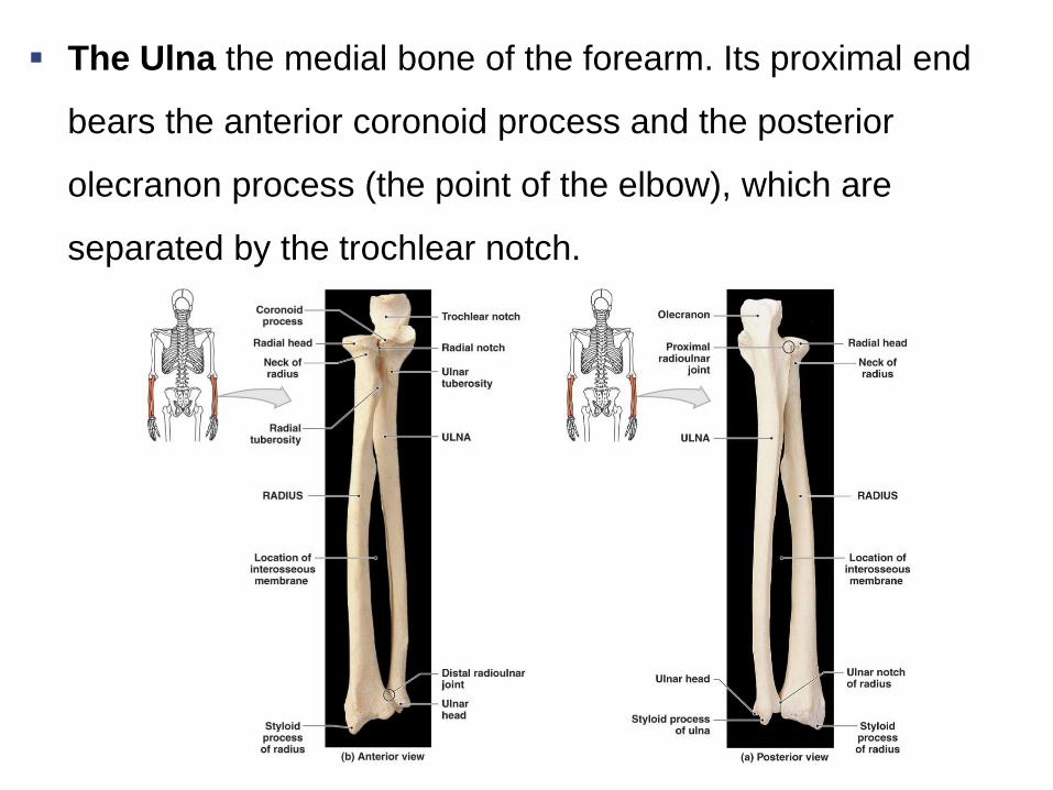
The Ulna the medial bone of the forearm. Its proximal end
bears the anterior coronoid process and the posterior
olecranon process (the point of the elbow), which are
separated by the trochlear notch.

The Hand
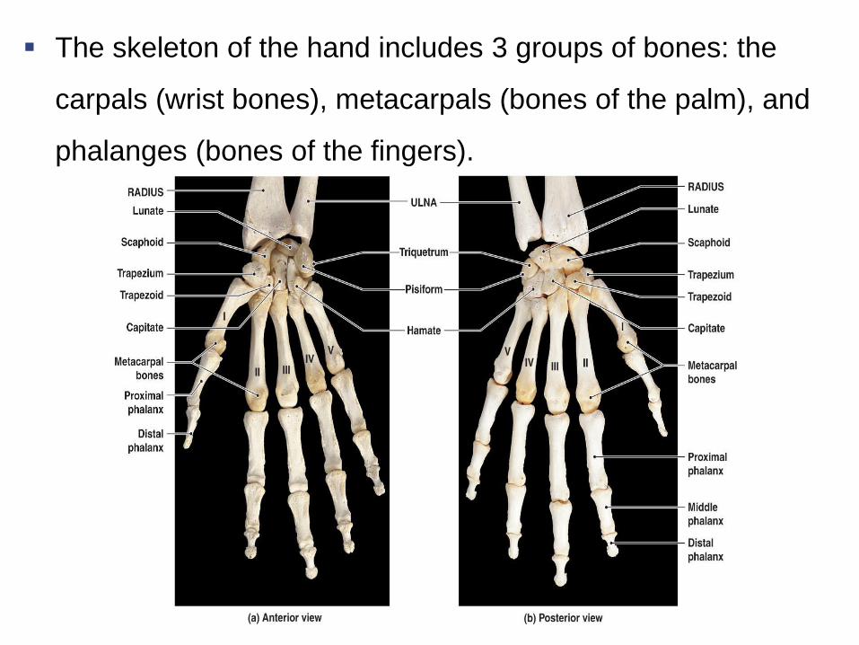
The skeleton of the hand includes 3 groups of bones: the
carpals (wrist bones), metacarpals (bones of the palm), and
phalanges (bones of the fingers).
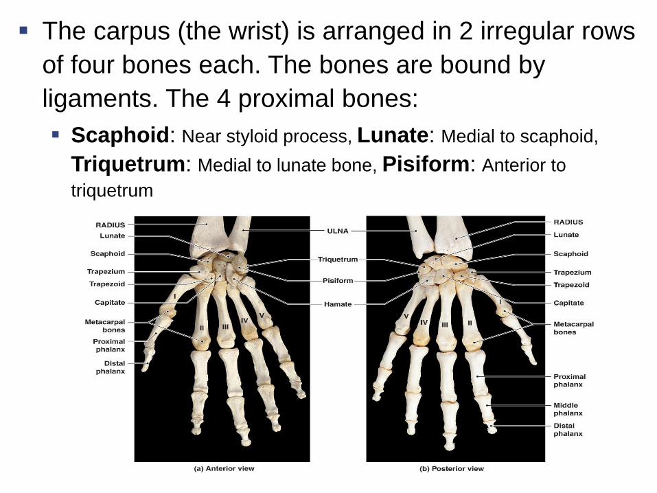
The carpus (the wrist) is arranged in 2 irregular rows
of four bones each. The bones are bound by
ligaments. The 4 proximal bones:
Scaphoid: Near styloid process, Lunate: Medial to scaphoid,
Triquetrum: Medial to lunate bone, Pisiform: Anterior to
triquetrum
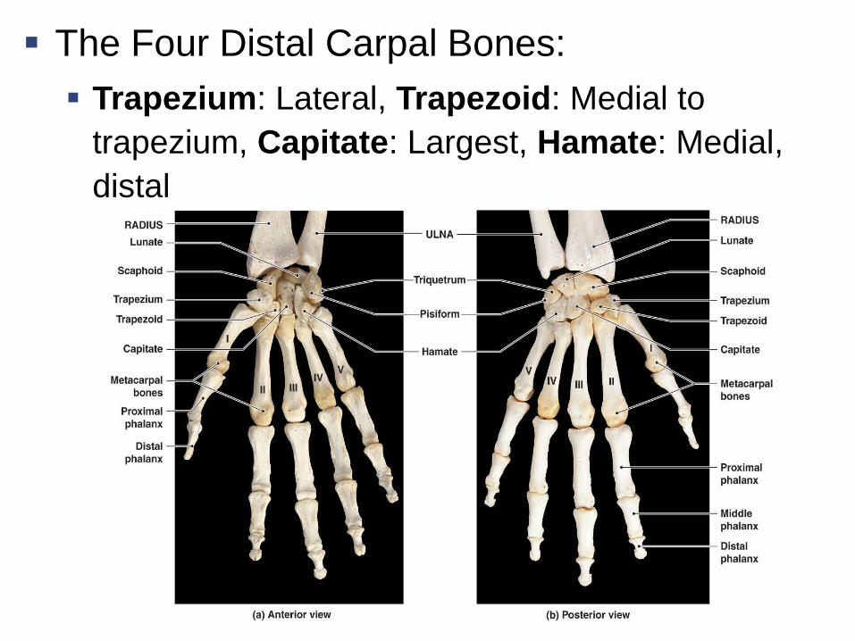
The Four Distal Carpal Bones:
Trapezium: Lateral, Trapezoid: Medial to
trapezium, Capitate: Largest, Hamate: Medial,
distal
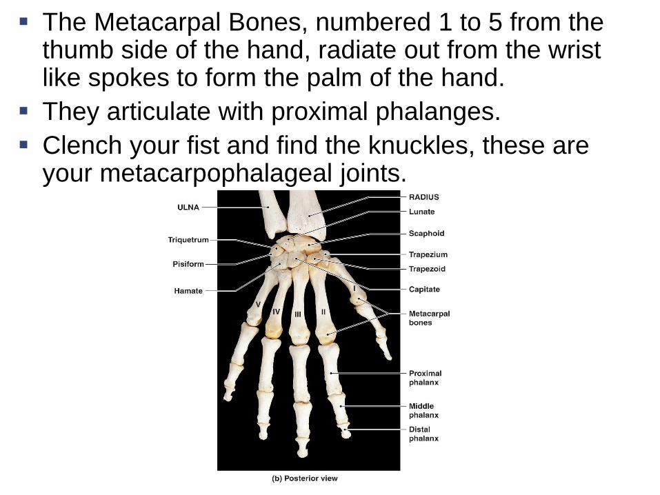
The Metacarpal Bones, numbered 1 to 5 from the thumb side of the hand, radiate out from the wrist like spokes to form the palm of the hand.
They articulate with proximal phalanges.
Clench your fist and find the knuckles, these are your metacarpophalageal joints.
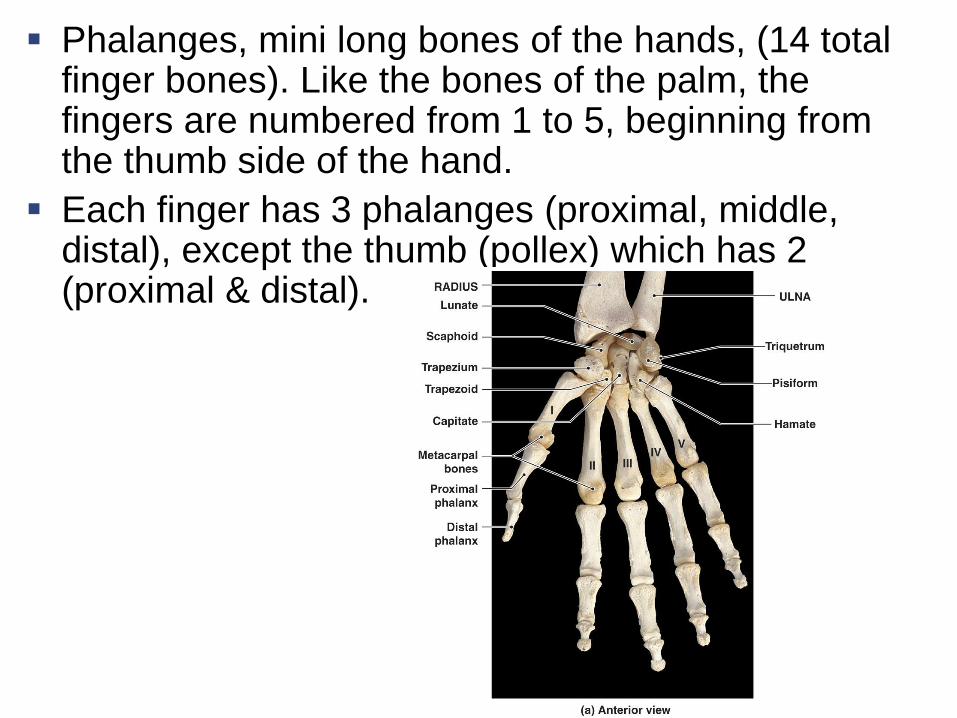
Phalanges, mini long bones of the hands, (14 total finger bones). Like the bones of the palm, the fingers are numbered from 1 to 5, beginning from the thumb side of the hand.
Each finger has 3 phalanges (proximal, middle, distal), except the thumb (pollex) which has 2 (proximal & distal).