An Introduction to EEG Source Analysis with an ...
27
HAL Id: hal-01078589 https://hal.archives-ouvertes.fr/hal-01078589 Submitted on 29 Oct 2014 HAL is a multi-disciplinary open access archive for the deposit and dissemination of sci- entific research documents, whether they are pub- lished or not. The documents may come from teaching and research institutions in France or abroad, or from public or private research centers. L’archive ouverte pluridisciplinaire HAL, est destinée au dépôt et à la diffusion de documents scientifiques de niveau recherche, publiés ou non, émanant des établissements d’enseignement et de recherche français ou étrangers, des laboratoires publics ou privés. An Introduction to EEG Source Analysis with an illustration of a study on Error-Related Potentials Marco Congedo, Sandra Rousseau, Christian Jutten To cite this version: Marco Congedo, Sandra Rousseau, Christian Jutten. An Introduction to EEG Source Analysis with an illustration of a study on Error-Related Potentials. Miranda E and Castet J. Guide to Brain-Computer Music Interfacing, Springer, 2014, ISBN 978-1-4471-6584-2. hal-01078589
Transcript of An Introduction to EEG Source Analysis with an ...
HAL Id: hal-01078589https://hal.archives-ouvertes.fr/hal-01078589
Submitted on 29 Oct 2014
HAL is a multi-disciplinary open accessarchive for the deposit and dissemination of sci-entific research documents, whether they are pub-lished or not. The documents may come fromteaching and research institutions in France orabroad, or from public or private research centers.
L’archive ouverte pluridisciplinaire HAL, estdestinée au dépôt et à la diffusion de documentsscientifiques de niveau recherche, publiés ou non,émanant des établissements d’enseignement et derecherche français ou étrangers, des laboratoirespublics ou privés.
An Introduction to EEG Source Analysis with anillustration of a study on Error-Related Potentials
Marco Congedo, Sandra Rousseau, Christian Jutten
To cite this version:Marco Congedo, Sandra Rousseau, Christian Jutten. An Introduction to EEG Source Analysis with anillustration of a study on Error-Related Potentials. Miranda E and Castet J. Guide to Brain-ComputerMusic Interfacing, Springer, 2014, ISBN 978-1-4471-6584-2. �hal-01078589�

Guide to Brain-Computer Music Interfacing, edited by Eduardo Reck Miranda, Julien
Castet and Benjanim Knapp, Springer, London
CHAPTER 8 - An Introduction to EEG Source Analysis with an
illustration of a study on Error-Related Potentials.
Marco Congedo*, Sandra Rousseau, Christian Jutten
GIPSA-lab, CNRS and Grenoble University, Grenoble, FRANCE
* Corresponding Author
Address :
11 rue des Mathématiques, Domaine universitaire - BP 46 - 38402, Grenoble, France.
E-mail : [email protected]

2
8.1 Introduction
Over the last twenty years blind source separation (BSS) has become a fundamental
signal processing tool in the study of human electroencephalography (EEG), other biological
data, as well as in many other signal processing domains such as speech, images, geophysics
and wireless communication (Comon and Jutten, 2010). Without relying on head modeling
BSS aims at estimating both the waveform and the scalp spatial pattern of the intracranial
dipolar current responsible of the observed EEG, increasing the sensitivity and specificity of
the signal received from the electrodes on the scalp. This chapter begins with a short review
of brain volume conduction theory, demonstrating that BSS modeling is grounded on current
physiological knowledge. We then illustrate a general BSS scheme requiring the estimation of
second-order statistics (SOS) only. A simple and efficient implementation based on the
approximate joint diagonalization of covariance matrices (AJDC) is described. The method
operates in the same way in the time or frequency domain (or both at the same time) and is
capable of modeling explicitly physiological and experimental source of variations with
remarkable flexibility. Finally, we provide a specific example illustrating the analysis of a
new experimental study on error-related potentials.
The AJDC method for EEG data has been reviewed and described in details in
Congedo et al. (2008), based upon theoretical bases to be found in Pham (2002) and Pham
and Cardoso (2001). Typically, it has been used on continuously recorded EEG (spontaneous
activity: e.g., Van der Loo et al., 2007). An extension of the method to treat group EEG data
and normative EEG data has been proposed in Congedo et al. (2010). Such group BSS
approach has been used in a clinical study on obsessive-compulsive disorder in Kopřivová et
al. (2011) and in a cognitive study on spatial navigation in White et al. (2012). The AJDC
method has also been employed for motor imagery-based brain-computer interfaces in Gouy-
Pailler et al. (2010), showing that it can be applied purposefully to event-related
(de)synchronization data (induced activity). Extensions of the method to the analysis of
simultaneous multiple-subject EEG data is a current line of research in our laboratory (Chatel-
Goldman, Congedo and Phlypo, 2013; Congedo, Phlypo and Pham, 2011; Congedo, Phlypo
and Chatel-Goldman, 2012). This chapter contributes demonstrating that the AJDC method
can be used purposefully on event-related potential (ERP) data as well (evoked activity).
8.2 Physiological ground of BSS modeling
It is well established that the generators of brain electric fields recordable from the
scalp are macroscopic post-synaptic potentials created by assemblies of pyramidal cells of the
neocortex (Speckmann and Elger, 2005). Pyramidal cells are aligned and oriented
perpendicularly to the cortical surface. Their synchrony is possible thanks to a dense net of
local horizontal connections (mostly <1mm). At recording distances larger than about
three/four times the diameter of the synchronized assemblies the resulting potential behaves
as if it were produced by electric dipoles; all higher terms of the multipole expansion vanish
and we obtain the often invoked dipole approximation (Lopes Da Silva and Van Rotterdam,

3
2005; Nunez and Srinivasan, 2006, Ch. 3). Three physical phenomena are important for the
arguments we advocate in this study. First, unless dipoles are moving there is no appreciable
delay in the scalp sensor measurement (Lopes da Silva and Van Rotterdam, 2005). Second, in
brain electric fields there is no appreciable electro-magnetic coupling (magnetic induction) in
the frequencies up to about 1MHz, thus the quasi-static approximation of Maxwell equations
holds throughout the spectrum of interest (Nunez and Srinivasan, 2006, p. 535-540). Finally,
for source oscillations below 40Hz it has been verified experimentally that capacitive effects
are also negligible, implying that potential difference is in phase with the corresponding
generator (Nunez and Srinivasan, 2006, p. 61). These phenomena strongly support the
superposition principle, according to which the relation between neocortical dipolar fields and
scalp potentials may be approximated by a system of linear equations (Sarvas, 1987). We can
therefore employ a linear BSS model. Because of these properties of volume conduction, scalp
EEG potentials describe an instantaneous mixture of the fields emitted by several dipoles
extending over large cortical areas. Whether this is a great simplification, we need to keep in
mind that it does not hold true for all cerebral phenomena. Rather, it does at the macroscopic
spatial scale concerned by EEG.
The goal of EEG blind source separation (BSS) is to “isolate” in space and time the
generators of the observed EEG as much as possible, counteracting the mixing caused by
volume conduction and maximizing the signal-to-noise ratio (SNR). First explored in our
laboratory during the first half of the 80’s (Ans et al., 1985; Hérault and Jutten, 1986), BSS
has enjoyed considerable interest worldwide only starting a decade later, inspired by the
seminal papers of Jutten and Hérault (1991), Comon (1994) and Bell and Sejnowski (1995).
Thanks to its flexibility and power BSS has today greatly expanded encompassing a wide
range of applications such as speech enhancement, image processing, geophysical data
analysis, wireless communication and biological signal analysis (Comon and Jutten, 2010).
8.3 The BSS problem for EEG, ERS/ERD and ERP
For N scalp sensors and MN EEG dipolar fields with fixed location and orientation in
the analyzed time interval, the linear BSS model simply states the superposition principle
discussed above, i.e.,
( ) ( ) ( )t t t v As η (8.1)
( ) Nt v is the sensor measurement vector at sample t, N MA is a time-invariant full
column rank mixing matrix, ( ) Mt s holds the time-course of the source components and
( ) Nt η is additive noise, temporally white, possibly uncorrelated to ( )ts and with spatially
uncorrelated components. Equation (8.1) states that each observation ( )tv (EEG) is a linear
combination (mixing) of sources ( )ts , given by the coefficients in the corresponding column
of matrix A . Neither ( )ts nor A are known, that is why the problem is said to be blind. Our
source estimation is given by

4
ˆˆ( ) ( )t ts Bv (8.2)
where M NB is called the demixing or separating matrix. This is what we want to estimate
in order to recover the sources from EEG. Hereafter the hat indicates a statistical estimation.
Although this is the classical BSS model we need a few clarifications for the EEG case: by
( )tη we model instrumental noise only. In the following we drop the ( )tη term because the
instrumental (and quantization) noise of modern EEG equipment is typically low (<1μV). On
the other hand, biological noise (extra-cerebral artifacts such as eye movements and facial
muscle contractions) and environmental noise (external electromagnetic interference) may
obey a mixing process as well, thus they are generally modeled as components of ( )ts , along
with cerebral ones. Notice that while biological and environmental noise can be identified as
separated components of ( )ts , hence removed, source estimation will be affected by the
underlying cerebral background noise propagating with the same coefficients as the signal
(Belouchrani and Amin, 1998).
8.4 A suitable class of solutions to the brain BSS problem
To tackle problem (8.2) assuming knowledge of sensor measurement only we need to
reduce the number of admissible solutions. In this paper we are interested in weak restrictions
converging toward condition
ˆ( ) ( )t ts Gs , (8.3)
where s(t) holds the time-course of the true (unknown) source processes, ˆ( )ts our estimation
and the system matrix
ˆ G BA ΛP (8.4)
approximates a signed scaling (a diagonal matrix Λ) and permutation (P) of the rows of s(t).
Equation (8.3) is obtained by substituting (8.1) in (8.2) ignoring the noise term in the former.
Whether condition (8.3) may be satisfied is a problem of identifiability, which establishes the
theoretical ground of BSS theory (Tong, Inouye and Liu, 1993; Cardoso, 1998; Pham and
Cardoso, 2001; Pham, 2002). We will come back on how identifiability is sought in practice
with the proposed BSS approach. Matching condition (8.3) implies that we can recover
faithfully the source waveform, but only out of a scale (including sign) and permutation
(order) indeterminacy. This limitation is not constraining for EEG, since it is indeed the
waveform that bears meaningful physiological and clinical information. Notice the
correspondence between the mth
source, its separating vector (mth
row of B̂ ) and its scalp
spatial pattern (mixing vector), given by the mth
column of ˆ ˆ A B . Hereafter superscript +
indicates the Moore-Penrose pseudo-inverse. The mono-dimensionality of those vectors and
their sign/energy indeterminacy implies the explicit modeling of the orientation and
localization parameters of the mth
source, but not its moment. This is also the case of inverse
solutions with good source localization performance (Greenblatt et al., 2005). On the other
5
hand, when we estimate current density by EEG inverse solutions we estimate current flowing
in the three orthogonal directions (hence the filter is given by three vectors, not one as here),
resulting in a considerable loss of spatial resolution. Linearity allows switching back from the
source space into the sensor space. Substituting (8.2) into (8.1) and dropping the noise term in
the latter yields BSS filtering
ˆ ˆ ˆˆ'( ) ( ) ( )t t t v ARs ARBv ,
where R is a diagonal matrix with mth
diagonal element equal to 1 if the mth
component is to
be retained and equal to 0 if it is to be removed. BSS filtering is common practice to remove
artifacts from the EEG data.
8.5 An approach for solving the BSS problem based on second-order statistics only
It has been known for a long time that in general the BSS problem cannot be solved
for sources that are Gaussian, independent and identically distributed (iid) (Darmois, 1953).
EEG data is clearly non-iid, thus we may proceed assuming that source components are all
pair-wise uncorrelated and that either (a) within each source component the successive
samples are temporally correlated1, (Molgedey and Schuster, 1994; Belouchrani et al., 1997)
or (b) samples in successive time intervals do not have the same statistical distribution, i.e.,
they are non stationary (Matsuoka et al., 1995; Souloumiac, 1995; Pham and Cardoso, 2001).
Provided that source components have non-proportional spectra or the time courses of their
variance (energy) vary differently, one can show that SOS are sufficient for solving the source
separation problem (Yeredor, 2010 ). Since SOS are sufficient, the method is able to separate
also Gaussian sources, contrary to another well known BSS approach named independent
component analysis (ICA: Comon and Jutten, 2010). If these assumptions are fulfilled the
separating matrix can be identified uniquely, thus source can be recovered regardless the true
mixing process (uniform performance property: see for example Cardoso, 1998) and
regardless the distribution of sources, which is a remarkable theoretical advantage. The
fundamental question is therefore whether or not the above assumptions fits EEG, ERS/ERD
and ERP data.
Sources are uncorrelated: This assumption may be conceived as a working
assumption. In practice, the BSS output is never exactly uncorrelated, but just as
uncorrelated as possible. What we try to estimate is the coherent signal of large
cortical patches, enough separated in space one from the other. BSS may be conceived
as a spatial filter minimizing the correlation of the observed mixtures and recovering
the signal emitted from the most energetic and uncorrelated cortical patches. For EEG
data this is an effective way to counteract the effect of volume conduction. In fact, we
have seen that the brain tissue behaves approximately as a linear conductor, thus
observed potentials (mixtures) must be more correlated than the generating dipolar
fields.
1 Such processes are called colored, in opposition to iid processes, which are called white.

6
Sources are colored and/or their energy varies over time: Observed potentials are
the summation of post-synaptic potentials over large cortical areas caused by trains of
action potentials carried by afferent fibers. The action potentials come in trains/rest
periods, resulting in sinusoidal oscillations of the scalp potentials, with negative shifts
during the train discharges and positive shifts during rest. The periodicity of trains/rest
periods are deemed responsible for high-amplitude EEG rhythms (oscillations) up to
about 12Hz, whereas higher frequency (>12Hz) low-amplitude rhythms may result
from sustained (tonic) afferent discharges (Speckmann and Elegr, 2005). There is no
doubt that an important portion of spontaneous EEG activity is rhythmic, whence
strongly colored (Niedermeyer, 2005 a; Steriade, 2005; Buzsáki, 2006, Ch. 6, 7).
Some rhythmic waves come in more or less short bursts. Typical examples are sleep
spindles (7-14Hz) (Niedermeyer, 2005 b; Steriade, 2005), frontal Theta (4-7Hz) and
Beta (13-35Hz) waves (Niedermeyer, 2005 a). Others are more sustained, as it is the
case for slow Delta (1-2Hz) waves during deep sleep stages III and IV (Niedermeyer,
2005 b), the Rolandic Mu rhythms (around 10Hz and 20Hz) and posterior Alpha
rhythms (8-12Hz) (Niedermeyer, 2005 a). In all cases brain electric oscillations are not
ever-lasting and one can always define time intervals when rhythmic activity is present
and others when it is absent or substantially reduced. Such intervals may be precisely
defined based on known reactivity properties of the rhythms. For example, in event-
related synchronization / desynchronization (ERD/ERS: Pfurtscheller and Lopes da
Silva, 2004), which are time locked, but not phase locked increases and decreases of
the oscillating energy (Steriade, 2005) intervals may be defined before and after event
onset. On the other hand event-related potentials (ERP: Lopes Da Silva, 2005 b),
which are both time-locked and phase-locked can be further partitioned in several
successive intervals comprising the different peaks. Such source energy variation
signatures can be modeled precisely by SOS, as we will show with the ensuing ErrP
study.
8.6 Approximate joint diagonalization of covariance matrices (AJDC)
The SOS BSS method we are considering is consistently solved by approximate joint
diagonalization algorithms (Cardoso and Souloumiac2, 1993; Tichavsky and Yeredor, 2009).
Given a set of covariance matrices {C1, C2,… }, the AJD seeks a matrix B̂ such that the
products 1ˆ ˆ TBC B , 2
ˆ ˆ TBC B , … are as diagonal as possible (subscript “T” indicates matrix
transposition). Given an appropriate choice of the diagonalization set {C1, C2,…} such matrix
B̂ is indeed an estimation of the separating matrix in (8.2) and one obtains an estimate of the
mixing matrix as ˆ ˆ A B . Matrices in {C1, C2,…} are chosen so as to hold in the off-
diagonal entries statistics describing some form of correlation among the sensor measurement
2 This paper does not consider SOS but 4
th order statistics, however, the algorithms is based on approximate joint
diagonalisation of matrices which are the slices of the tensor of 4th order cumulants, thus can be used for SOS
matrices as well.

7
channels; then the AJD will vanish those terms resulting in linear combination vectors (the
rows of B̂ ) extracting uncorrelated components from the observed mixture via (8.2). More
particularly, the joint diagonalization is applied on matrices that change according to the
assumptions about the source. They are those changes, when available, that provide enough
information to solve the BSS problem. Formally, for AJDC the identifiability of sources
discussed above, that is, matching condition (8.3), is described by the fundamental AJD-based
BSS theorem (Afsari, 2008; see also Aïssa-El-Bey et al., 2008): let matrices S1, S2,.. be the K
(unknown) covariance matrices of sources corresponding to the covariance matrices included
in the diagonalization set and sk(ij) their elements. The diagonal elements of these matrices sk(ii)
hold the source variance. The off-diagonal elements sk(ij), ij, are null as sources are assumed
to be uncorrelated. Let
1 11 11
1
1
k
T
M
MM k MM
s s
s s
Y y y
(8.5)
be the matrix formed by stacking one below the other row vectors y1, y2, … yM constructed as
shown in Fig. 8.1. Each vector ym=(s1(mm),…,sK(mm)) holds the energy profile along the
diagonalization set for each source, with m:1…M and M the number of estimated sources.
The fundamental theorem says that the mth
source can be separated as long as its energy
profile vector ym is not collinear 3 with any other vector in Y. Said differently, the wider the
angle between ym and any other vector in Y, the greater the chance to separate the mth
source.
Even if two vectors are collinear, the other sources can still be identified.
Figure 8.1: Graphical illustration of the construction of the source energy profile vectors ym.
Table 8.1 reports useful information to define an appropriate diagonalization set so as
to ensure identifiability of sources.
3 Two vectors are collinear if they are equal out of a scaling factor, that is, the energy profile is proportional.

8
Table 8.1: Criteria to achieve identifiability of sources in BSS methods based on AJD of SOS.
Assumption on
the sources
Covariance Matrices
(CM) Estimation
What is the
energy profile
Sufficient
condition for
Identifiability
Examples of data
Coloration i. Lagged Covariance
matrices,
ii. Fourier Co-spectral
Matrices,
iii. CM estimated with
a filter bank
i. The source
autocorrelation,
ii. The source
power spectrum
iii. as in ii.
The power spectrum
of the source is non-
proportional to the
power spectrum of
any other sources
Spontaneous oscillation with
characteristic power spectrum
such as posterior dominant
rhythms (Alpha), Somatosensory
Mu rhythms, frontal midline
Theta, Beta bursts, etc.
Non-Stationary CM estimated on
j. different time blocks
of data
jj. different
experimental
conditions
The variation of
the energy of the
source along the
(j) blocks or
(jj) experimental
conditions
The variation of the
source energy along
(j) blocks or
(jj) experimental
conditions do not
correlate with the
same variation of
any other sources
- Blocks of data according to
physiological reactivity of EEG
oscillations (e.g., eyes-close vs.
eyes-open)
- CM estimated before and after
the event in ERD/ERS
- CM estimated on different peaks
in ERP (after averaging the ERP)
- Active vs. Control condition,
...
Importantly, the two basic theoretical frameworks for working in a SOS framework
reported in Table 8.1, the coloration and the non-stationary, can be combined in any
reasonable way: one may estimate covariance matrices in different blocks (and/or conditions)
for different frequency band-pass regions, effectively increasing the uniqueness of the source
energy profile. This is for instance the path we have followed for solving the problem of
separating sources generating error potentials, as we will demonstrate here below. In fact,
AJDC method can be applied in different representation spaces; applying to (1) any invertible
and linearity-preserving transform T leads to
( ) ( )t tv A sT T ,
which preserves the mixing model. Then, solving source separation in the transformed space
still provides estimation of the matrix A or of its inverse B, which can be used directly in Eq.
(2) for recovering the source s(t) in the initial space. For example, the transform T may be a
discrete Fourier transform, a time-frequency transform such as the Wigner-Ville transform or
a wavelet transform. AJDC can be easily and conveniently transposed in the frequency
domain, thence in the time-frequency domain, whether we perform the frequency expansion
for several time segments.
It is important to consider that the number of matrices should be high enough to help
non-collinearity of source energy profiles. One may want to have at least as many matrices in
the diagonalization set as sources to be estimated. On the other hand one should not try to
increase the number of matrices indefinitely to the detriment of the goodness of their
estimation, i.e., selecting too many discrete frequencies or blocks of data that are too shorts.
In summary, the key for succeeding with BSS by AJDC is the definition of an adequate size
and content of the diagonalization set; it should include matrices estimated on data as
homogeneous as possible for each matrix, with enough samples to allow a proper estimation,

9
in frequency region and time blocks when the signal-to-noise ratio is high and with an high
probability to uncover unique source energy profiles.
8.7 A study on Error-related Potentials
We now turn to the illustration of the AJDC method by means of a new study on error-
related potentials (ErrP). We show that BSS analysis increases the specificity and sensitivity
that can be obtained working at the sensor level, increasing as a consequence the single-trial
classification rate. ErrPs are a family of event-related potential (ERP) that can be elicited after
the commission of an error, firstly reported in Miltner, Braun and Coles (1997) as associated
to receiving external negative feedback after error commission. This feedback error-related
potential (ErrPf) is characterized by a negative deflection peaking between 250 and 400 ms
with a fronto-central scalp distribution. The authors named it the feedback-related negativity
(FRN) and put it in relation with the response error related negativity (ERN) that had been
previously reported (Felkenstein et al., 1991; Gehring et al., 1993), also characterized by a
negative deflection. Initially the ErrPf has been studied prevalently in the case of gambling
tasks with monetary gain and loss. More recently it has attracted much attention in the brain-
computer interface (BCI) community because its on-line detection provides a unique
opportunity to automatically correct erroneous BCI operations, effectively increasing the
consistency and transfer rate of a BCI system (Farquhar and Hill, 2013). In order to do so
accurate on-line single-trial ErrP detection is necessary. Here we contribute along this
direction in two way: 1) we design a new experimental protocol in order to study single-trial
ErrPf detection in a controlled situation that mimics actual BCI operation and 2), we apply the
AJDC source analysis in order to better characterize this potential, hence increasing the
accuracy of its on-line single-trial detection.
1) New Experimental Protocol
In all previous studies on single-trial detection of ErrP for integration of a control loop
in a BCI system the involvement of the participants is very far from the involvement of
participants during BCI operation, that is, as such they lack ecological validity. In particular,
in previous studies the feedback is the main focus of the subject, while in actual BCI
operations receiving such a feedback is only a small part of a complex cognitive task.
Furthermore, previous studies have mainly returned shame feedback, that is, feedback
completely unrelated to the performance of the subject. Finally, the subject-specific control
capability of a BCI system has not been taken into consideration. Here we study the feedback
related potential in the case of a memory task, with no monetary gain or loss. The feedback is
returned when the subject gives the answer and no reward is given to the subject except a
score, thus our participants have no other interest besides their own performance. Such an
experimental protocol allows to study the ErrPf in a real "error versus correct" condition. The
protocol we use is a memory task inducing a high cognitive load. The subject is continuously

10
engaged in a demanding task (and not only on the feedback presentation), mimicking the
actual conditions of a BCI use, where focus, concentration and attention are essential requisite
for successful BCI operation. Then, in this study the feedback corresponds to the actual
performance achieved in the task, again approximating the actual operation of a BCI. Finally,
the memory task continuously adapts to the ability of the participants during the whole
experiment. This ensures that the cognitive load is approximately constant across the duration
of the experiment, that it is comparable across individuals regardless of their memory span
and that the error rate across subjects is approximately equal. This latter point is particularly
important in ErrP studies since it is known that he error rate affects the ErrP ([8]). In this
study the adaptive algorithm is tuned to engender an error rate of about 20%, which amount
approximately to the reasonable accuracy of a reactive BCI operation in real-world situations.
2) New Multivariate Signal processing Analysis
Some of the previous studies on single trial ErrP classification (correct vs. error) have
reached encouraging results (around 70% of overall accuracy) using only little a-priori
knowledge on this potential. As usual, a more profound knowledge of the electrophysiological
characteristics of the ErrPf can be used to select more relevant and robust features for the
purpose of single-trial on line detection. Previous studies showed that the ErrP can be
characterized both in the temporal domain as an ERP (time and phase-locked event) and as an
event-related synchronization, or ERS (time but non-phase-locked event). The ERP is
characterized by a negative deflection, named Ne, sometimes followed by a positive one
named Pe (Gentsch, Ullsperger and Ullsperger, 2009; Steinhauser and Kiesel, 2011). The
ERS is characterized by an increased oscillatory activity in the theta frequency band-pass
region (4-7.5 Hz) occurring approximately in the same time window and spatial location as
the Ne (Trujillo and Allen, 2007). Source localization of the FRN using dipole analysis has
suggested generators in the anterior cingulate cortex (ACC) and the supplementary motor area
(Gehring and Willoughby, 2002; Miltner, Braun and Coles, 1997). Similar results have been
obtained for the ErrPr. Hereby we propose a sharp spatial filtering approach based on the
blind source separation approach described above with the aim to disentangling the sources
responsible for the ERP and the ERS; if this proves feasible, then the ERP and ERS
components will yield independent features to feed the classifier, hence potentially increasing
the on-line accuracy.
As a first objective we identify the different components of the ErrP along dimensions
time, space and frequency by means of a multivariate analysis both in the sensor space and in
the source space. We jointly estimate the brain sources at the origin of the ERP and ERS
components and assess their different role in error reaction. Then we study the role of these
components on the ErrP with respect to the expectation of participants. Finally, we look at
how these results impact on ErrP single-trial classification, which is the essential step in
integrating ErrPs in BCI systems.

11
8.8 Method
8.8.1 Participants
22 healthy volunteers participated to this experiment. All subjects were BCI-naive at
the time of the experiment and none of them reported neurological or psychiatric disorders in
their lifetime. Due to the presence of excessive artifacts in the EEG data, three subjects were
subsequently excluded from all analyses, leaving 19 participants, of which 9 female and 10
male, with age ranging from 20 to 30 with a mean and a standard deviation of 24 and 2.52,
respectively.
8.8.2 Experimental design
The experiment involved two sessions lasting altogether approximately half an hour.
Each session consisted of six blocks of six trials, for a total of 6x6x2=72 trials. Participants
seated comfortably 80cm in front of a 21-inch computer screen. Nine square boxes were
arranged in circle on the screen. Each trial consisted of the same memory retrieval task: the
trial started with the display of the current score for 3000ms (initialized at zero) followed by a
fixation cross, also displayed for 3000 ms (Fig 8.2a). Then the memorization sequence
started; each memorization comprised a random sequence of two to nine digits appearing
sequentially in random positions, with each digit of the sequence randomly assigned to a
different box for each sequence (Fig 8.2b). Subjects were instructed to retain positions of all
digits. At the end of the sequence the target digit (always contained in the previous sequence)
was displayed (Fig 8.2c) and subjects had to click with the aid of a mouse on the box where it
had appeared. Once the subject had answered, the interface waited for 1500 ms in order to
avoid any contamination of ErrP by beta rebound motor phenomena linked to mouse clicking
(Pfurtscheller, 1981). Then, if the answer was correct, the chosen box background color
turned into green ("correct" feedback), otherwise it turned into red ("error" feedback).
Subjects were then asked to report if the feedback (error/correct) matched their expectation by
a mouse click (“yes”/ “no”) (Fig 8.2d). Following this answer a random break of 1000 to
1500ms preceded the beginning of the new trial.

12
Figure 8.2: Screen shots from the experiment representing different steps of the experiment. a):
Fixation cross. b): One digit appearing in the memorization sequence. c): Target digit appearing. d):
Feedback report question: 'Vous attendiez-vous à ce resultat' = 'Did you expect this result?',
'Oui'='Yes' and 'Non'='No'.
In order to keep the subjects motivated throughout the experiment, the accumulated
score was computed at the beginning of each trial. When subjects localized correctly the
target digits their score increased, otherwise, it remained unchanged. The number of digits in
the sequence was always between two and nine, fixed within blocks and updated, at the
beginning of each block, according to the change in performance from the block just finished
and the previous one, as assessed on-line by means of statistical t-tests. The first block started
always with four digits for all subjects. The parameters of the adaptation were set thanks to a
pilot study and a computer simulation and were chosen to yield about 20% of errors,
regardless the working memory ability. Moreover, our learning approach is capable of
adapting to fatigue as well as other possible nuisance intervening during the experiment. A
random rest break was allowed between blocks, during which the boxes performed a colorful
animation chosen each time at random among four preset animations. Between the two
sessions the screen was shut down to allow a rest break of 2 - 3 minutes.
8.8.3 Data Acquisition
EEG recordings were acquired from 31 silver/chloride electrodes positioned according
to the extended 10/20 system (FP1, FPz, FP2, F7, F3, Fz, F4, F8, FT7, FC3, FCz, FC4, FT8,
T7, C3, Cz, C4, T8, TP7, CP3, CPz, CP4, TP8, P7, P3, Pz, P4, P8, O1, Oz, O2) with the aid
of a standard elastic cap. Both earlobes, digitally linked, were used as electrical reference. The

13
ground sensor was positioned on the forehead. The impedance of each sensor was kept below
5k. The EEG was band-pass filtered in the range 0.1-70 Hz and digitized at 500 Hz using the
Mitsar 202 DC EEG acquisition system (Mitsar Co. Ltd., Saint Petersburg, Russia). During
recording, the stimulation program continuously sent to the Mitsar system triggers to track
precisely all event onsets of each trial. These triggers where received by the Mitsar system as
a logic signal, synchronized with the EEG stream and recorded as a supplementary data
channel.
8.8.4 Preprocessing
Data were filtered in the 1-40Hz band-pass region using an order four Butterworth FIR
filter with linear phase response in the band-pass region. Ocular artifacts were extracted using
the SOBI algorithm (Belouchrani et al., 1993) available in the EEGLAB toolbox (Delorme
and Makeig, 2004). One EOG source corresponding to eye-blinks was suppressed for each
subject. It was manually selected using both the temporal shape of the source and its
topography. All other artifacts were left into the signal, so as to approximate the conditions of
on-line analysis of EEG data acquired during BCI operation.
8.8.5 Analysis in the Sensor Space
The analysis in the sensor space is the traditional analysis of the signal as recorded at
each electrode. We are interested in the analysis of the error vs. correct trials. We performed
both the analysis of the event-related potential (ERP: both time- and phase-locked: Lopes Da
Silva, 2005 b) and of the event-related synchronization (ERS: time-locked, but not necessarily
phase-locked: Pfurtscheller and Lopes da Silva, 2004). ERPs were analyzed contrasting the
average potential obtained from each subject at each electrode and time-sample. ERS were
analyzed contrasting the average time-frequency map obtained on each trial from each subject
at each electrode. In order to compute ERS we employed a multi-tapering Hanning sliding
window (frequency dependent, with the taper equal to 4 cycles for each frequency) over the 2-
32Hz band using a 1Hz step, as implemented in the Fieldtrip software (Oostenveld et al.,
2011). ERS were computed on time window [-0,5s 1,2s] using a time step of 0,03s and a
baseline defined as [-1s 0s] pre-stimulus.
The statistical analysis in the sensor space for contrasting “error” vs. “correct” trials
needs to be performed for each electrode, discrete frequency and time segment in the case of
ERS and for each electrode and time segment for ERP data. In order to account for the
extreme multiple-comparison nature of the test we employed a permutation strategy. The test
chosen is a slight modification of the supra-threshold cluster size permutation test originally
proposed for neuroimaging data by Holmes et al. (1996). Here the statistic is not the supra-
threshold cluster size, but the supra-threshold cluster intensity, defined as the sum of the t-
values within the supra-threshold clusters. As compared to the test described by Holmes et al.
(1996) such a statistic is influenced not only by the spatial extent of the clusters, but also by
the strength of the effect. The test is sensitive to effects that are contiguous in space (adjacent

14
electrodes), frequency and time, in line with physiological considerations. The family-wise
error rate for multiple comparisons was set to 0.05, meaning that the probability of falsely
rejecting even only one hypothesis is less than 0.05. All permutation tests were approximated
by the use of 5000 random permutations.
8.8.6 Analysis in the Source Space
As we have seen a spatial filter computes a weighted sum (linear combination) of the
signal obtained at each electrode, potentially isolating delimited dipolar sources from each
other. We apply here the method introduced above adapting it to ERP data. Our goal is to
separate the source of the Ne (ERP) and the source for the theta ERS. We need to separate
them one from the other, but also from background EEG activity. For our purpose we need to
include in the diagonalization set matrices holding a) the spatial structure of the ERP
component, b) the spatial structure of the ERS component, as well as c) the spatial structure of
the spontaneous EEG oscillations and persistent artifacts such as lateral and horizontal eye
movements, jaw muscle contractions, etc. For (a) and (b) we compute the relevant covariance
matrices both on error trials and correct trials so to exploit variations of source energy
between the two conditions (table 1). We define an exactly determined BSS model, that is to
say, we estimate as many sources (M in the formula above) as electrodes (N=M=31). For the
ERP components (a) we estimate the covariance matrix of the average ERP in the three time
windows were the ERP analysis in the sensor space revealed significant results (see next
section). Covariance matrices were separately computed for error and correct conditions,
providing 3x2=6 matrices. These six matrices provide unique source energy profile about
ERP that have different potential in error vs. correct trials. For the ERS component (b) we
estimate the averaged covariance matrix in the time-frequency region were the sensor space
analysis revealed significant results (see next section). These matrices were computed as the
covariance matrices of the EEG filtered in the frequency band of interest. Again, matrices
were computed separately for error and correct conditions, providing two additional matrices.
These two matrices provide unique source energy profile about ERS that display different
power in the theta band in error vs. correct trials. Notice that matrices for the ERP and the
ERS components are substantially different: for the ERP components EEG trials are averaged
before computing the covariance matrix (thus only both time-locked and phase-locked signals
are preserved), while for the ERS components trials are averaged only after computing
covariance matrices on single-trial data (thus non-phase-locked signal are preserved as long as
they are time-locked). To separate possible sources of ERP and ERS from spontaneous EEG
oscillations and artifacts (c) we include in the set all co-spectral matrices (Bloomfield, 2000)
of the signal during the fixation cross sequence in the frequency range 2-20Hz using a
frequency step of 2Hz, providing 10 additional matrices. These latter 10 matrices provide
unique source energy profile to separate all spontaneous sources having non-proportional
power spectrum (table 1). In summary, our BSS algorithm jointly diagonalizes a total of 18
matrices. For solving the approximate joint diagonalization we employ the iterative algorithm
proposed by Tichavsky and Yeredor (2009), which is fast and in our long-lasting practice has
proven robust.

15
Once estimated the 31 sources, they were inspected analyzing their ERP, ERS,
topographies and the mutual information criterion between the source and the error class
(Grosse-Wentrup and Buss, 2008). Meaningful sources were localized in a standard brain
using the sLORETA inverse solution (Pascual-Marqui, 2002) as implemented in the
LORETA-Key software. This software makes use of revisited realistic electrode coordinates
(Jurcak, Tsuzuki and Dan, 2007) and the head model (and corresponding lead-field matrix)
produced by Fuchs et al. (2002), applying the boundary element method on the MNI-152
(Montreal neurological institute, Canada) template of Mazziotta et al. (2001). The sLORETA-
key anatomical template divides and labels the neocortical (including hippocampus and
anterior cingulate cortex) MNI-152 volume in 6239 voxels of dimension 5 mm3, based on
probabilities returned by the Demon Atlas (Lancaster et al., 2000). The co-registration makes
use of the correct translation from the MNI-152 space into the Talairach and Tournoux (1988)
space (Brett et al., 2002). Source localization was conducted on each participant separately,
normalized to unit global current density (the input of the inverse solution is a vector
estimated by BSS up to a scale indeterminacy) and summed up over participants in the brain
space.
8.8.7 Classification of single trials
For classifying single trials, data were band-pass filtered using an order four
Butterworth FIR filter with linear phase response between 1-10Hz for the ERP component
and 4-8Hz for the ERS component. Data were then spatially filtered using the results of the
BSS analysis. Only samples corresponding to 250-750ms were kept. For the ERP component
we used the temporal signal down-sampled at 32Hz, providing 16 samples (features) for the
classification. For the ERS component we used the square of the temporal signal (power)
dawn-sampled at 32Hz, providing 16 samples (features) for the classification as well. This
procedure assigns to each component equal chance for classification. As a classifier we
employed a LDA (linear discriminant analysis). One hundred random cross-validations were
performed with the classifier trained on a randomly selected set containing 80% of the data
(both errors and corrects) and then tested on the remaining data.
8.9 Results
8.9.1 Behavioral results
All subjects performed the task with a convenient error-rate, with mean (sd) = 22.2
(4)% and a quasi-equal repartition of expected and unexpected errors, with mean (sd) = 10.4
(4.3)% and 11.8(3)%, respectively. Reaction time was higher for error trials as compared to
correct trials in 80% of the subjects (all t-tests with p<0.05). The maximum number of digits
to memorize for each subject was highly variable, ranging from 4 to 10, with mean (sd) = 6.5

16
(1.37). These results demonstrate that our presentation software succeeded in equalizing the
cognitive load across subjects, despite the great inter-subject variability of digit memory span.
8.9.2 Sensor Space Analysis
The ERP in the error trials differed from the correct trials in three time windows with
different timing and/or electrode location (Fig. 8.3). A significant positivity for errors was
found at time window [320ms 400ms] at electrode Cz (p<0.01), a significant negativity for
errors at time window [450ms 550ms] at clustered electrodes Fz, FCz, Cz (p <0.01) and a
significant positivity for errors at time [650ms 775ms] at clustered electrodes Fz, FCz (p =
0.025).
Figure 8.3 : (a): grand average (N=19) ERP for correct (pointed line) and error (solid line) trials.
Time windows where the difference in amplitude between the two conditions is significant (grey
panels) and (b) scalp topographies of t-values computed within the three significant windows. White
disks show the significant clustered electrodes.
An ERS (power increase as compared to baseline) could be seen in the theta band in
both correct and error feedback at fronto-midline locations. This synchronization unfolds
from around 250ms to 600ms post-stimulus. In some subject it goes up to more than 200% of
power increase for error trials. Albeit present in both conditions, this ERS is significantly
more intense for error trials as compared to correct ones (Fig. 8.4) in the frequency band pass
region 5-8Hz and time window [350ms 600ms] post-stimulus over the clustered electrodes Fz
and FCz (p = 0.015).

17
Figure 8.4: Grand average (N=19) ERS averaged at electrodes (Fz, FCz, Cz, CPz) for error (a) and
correct (b) trials. (c): topographic maps of t-values averaged over the theta band and time window
[350ms 600ms]. White disks show the significant clustered electrodes.
8.9.3 Source Analysis
BSS analysis revealed two uncorrelated sources with variable sensitivity and
specificity, however clearly responsible one for the ERP findings and one for the ERS
findings. The source responsible for the ERP differences between error and correct trials, to
which hereafter we will refer to as the “Ne source”, was significantly different in error vs.
correct trials in two time windows, with a first negative peak at time window [460ms 540ms]
(p < 0.01) and a positive peak at time [750ms 830ms] (p = 0.015). The grand-average ERP of
this source computed separately for error and correct trials is displayed in Fig. 8.5a. In Fig.
8.5b it is displayed the same grand average ERP when computed using the spatial filter of the
source responsible of the ERS differences between error and correct trials, to which hereafter
we will refer to as the “theta source”; although differences in amplitude exist also for this
latter source, they are not significant.
Figure 8.5: Grand averaged (N=19) of the ERP generated by the Ne source (a) and by the theta
source (b) for error (solid line) and correct (pointed line) trials. Time windows were the difference in
amplitude between the two conditions is significant are highlighted by grey panels.

18
On the other hand the theta source power increase was significant in frequency band-
pass region [5Hz 8Hz] for time window [300ms 600ms] (p<0.01). The ERS generated by this
source is shown in Fig. 8.6b. In Fig. 8.6a it is displayed the same ERS when computed using
the spatial filter of the Ne source instead; the ERS in this case disappears. These results
suggest that the Ne source and the theta source correspond to separate phenomena generated
by different brain structures with different dynamics. The source responsible for the ERS
(theta source) appears more specific.
We can now illustrate the advantage brought upon from the BSS analysis with these
data. Compare Fig. 8.5a to 8.3 and Fig. 8.6 to 8.4. Although in both cases results in the sensor
space are computed for the optimal cluster of electrodes, in both cases it is clear that working
in the source space allows a better sensitivity and specificity: in both cases the difference
between the error and correct trials is highlighted.
Figure 8.6: Grand average (N=19) of the ERS generated by the Ne source (a) and by the theta source
(b) for error trials.
8.9.4 Source localization
The BSS source responsible for the ERP (Ne source) difference between correct vs.
error trials was localized by sLORETA in the anterior cingulate gyrus (BA 24). The BSS
source responsible for the ERS (theta source) was localized close to the supplementary motor
area (BA 6) (Fig. 8.7). Keeping in mind the approximation of a source localization method
applied on a standard head model, these anatomical results are in line with results reported by
previous studies (Gehring and Willoughby, 2002; Herrmann et al., 2004; Nieuwenhuis et al.,
2003).

19
Figure 8.7: (a) Ne source sLORETA localization. The source is localized in BA 32. (b) Theta source
sLORETA localization. The source is localized in BA 6. For each image, from left to right are the
axial, sagittal and coronal views across the maximum. The images (a) and (b) are scaled to their own
maximum. The activity is color-coded with black representing the maximum and transparent
representing zero. Legend: A=Anterior;P=Posterior; S=Superior; I=Inferior; L=Left; R=Right.
8.9.5 Error Expectation
We then studied the impact of the error expectation on the two identified sources
identified (Ne and ERS). Each trial could outcome one out of four results: unexpected errors
(UE), expected errors (EE), expected corrects (EC), unexpected corrects (UC). Since most
subjects reported no trials from the UC condition we only studied the first three outcomes.
Only subjects providing at least four trials for each condition were kept. Three further subjects
were therefore excluded from this analysis. For each component a one-way repeated-measure
ANOVA with factor “outcome feedback” at three levels was applied i) to the temporal signal
averaged over time window [450ms 520ms] for the Ne source and ii) to the power signal
filtered between 5 and 8Hz and averaged over significant time window [300ms 600ms] for the
theta source. For the Ne source no significant result was found. For the theta source the means
of the three outcomes were not all equal (F = 4.75; p = 0.0138). All pair-wise post-hoc tests
corrected by Bonferroni method showed that the ERS engendered by this source is in this
relationship: ERS(UE)>ERS(EE)>ERS(EC), with both inequality signs indicating a
significant difference (p<0.05).
8.9.6 Classification of single trials
The Ne source alone leads to better accuracy in classifying error trials as compared to
the theta source alone (p < 0.01). The theta source leads to better accuracy for classifying
correct trials (p = 0.028). These corroborate the conclusion that the ERP and ERS represent
20
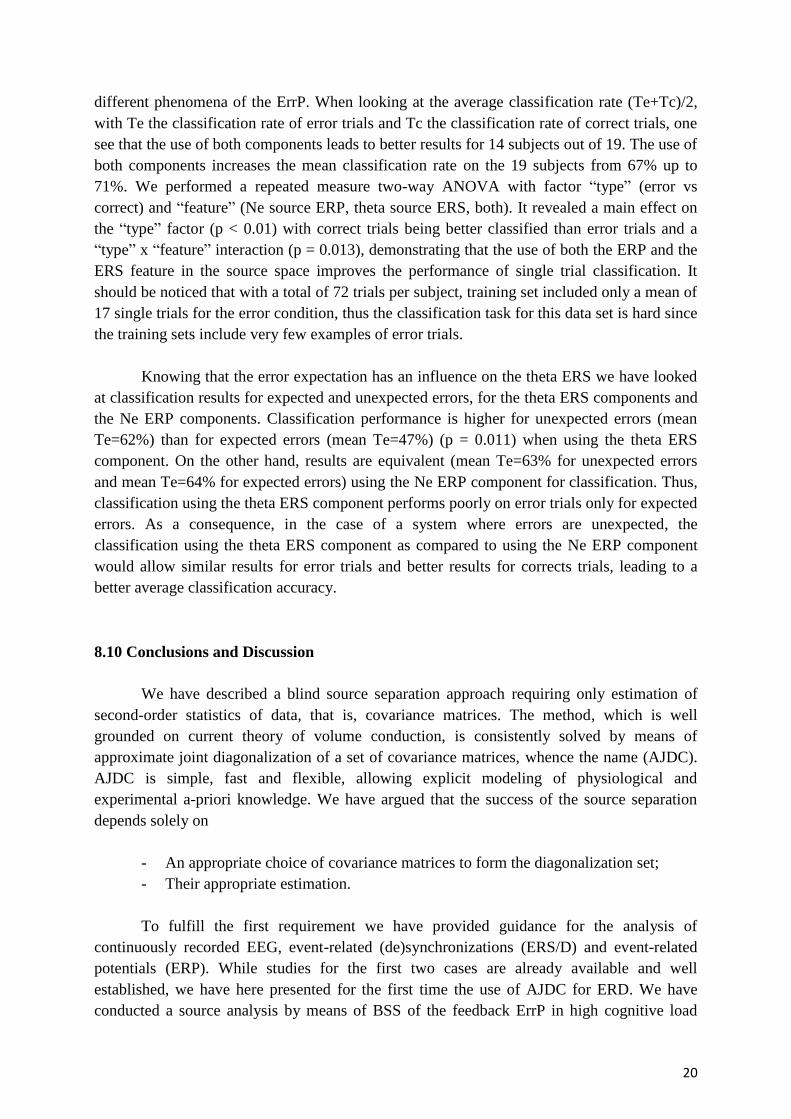
different phenomena of the ErrP. When looking at the average classification rate (Te+Tc)/2,
with Te the classification rate of error trials and Tc the classification rate of correct trials, one
see that the use of both components leads to better results for 14 subjects out of 19. The use of
both components increases the mean classification rate on the 19 subjects from 67% up to
71%. We performed a repeated measure two-way ANOVA with factor “type” (error vs
correct) and “feature” (Ne source ERP, theta source ERS, both). It revealed a main effect on
the “type” factor (p < 0.01) with correct trials being better classified than error trials and a
“type” x “feature” interaction (p = 0.013), demonstrating that the use of both the ERP and the
ERS feature in the source space improves the performance of single trial classification. It
should be noticed that with a total of 72 trials per subject, training set included only a mean of
17 single trials for the error condition, thus the classification task for this data set is hard since
the training sets include very few examples of error trials.
Knowing that the error expectation has an influence on the theta ERS we have looked
at classification results for expected and unexpected errors, for the theta ERS components and
the Ne ERP components. Classification performance is higher for unexpected errors (mean
Te=62%) than for expected errors (mean Te=47%) (p = 0.011) when using the theta ERS
component. On the other hand, results are equivalent (mean Te=63% for unexpected errors
and mean Te=64% for expected errors) using the Ne ERP component for classification. Thus,
classification using the theta ERS component performs poorly on error trials only for expected
errors. As a consequence, in the case of a system where errors are unexpected, the
classification using the theta ERS component as compared to using the Ne ERP component
would allow similar results for error trials and better results for corrects trials, leading to a
better average classification accuracy.
8.10 Conclusions and Discussion
We have described a blind source separation approach requiring only estimation of
second-order statistics of data, that is, covariance matrices. The method, which is well
grounded on current theory of volume conduction, is consistently solved by means of
approximate joint diagonalization of a set of covariance matrices, whence the name (AJDC).
AJDC is simple, fast and flexible, allowing explicit modeling of physiological and
experimental a-priori knowledge. We have argued that the success of the source separation
depends solely on
- An appropriate choice of covariance matrices to form the diagonalization set;
- Their appropriate estimation.
To fulfill the first requirement we have provided guidance for the analysis of
continuously recorded EEG, event-related (de)synchronizations (ERS/D) and event-related
potentials (ERP). While studies for the first two cases are already available and well
established, we have here presented for the first time the use of AJDC for ERD. We have
conducted a source analysis by means of BSS of the feedback ErrP in high cognitive load

21
conditions. In this experiment we have used conditions that resemble those one can find on
real BCI experiments. Our results showed that the feedback-related potential observed here
shares the same characteristics as the FRN observed in gambling tasks and the ERN observed
in reaction-time tasks. Indeed all three error potentials are notably characterized by a negative
deflection generated by the dorsal-ACC, but with different time of activation. A sharp
analysis in the source space by means of approximate diagonalization of covariance matrices
has allowed the identification of three main components accounting for the differentiation
between error and correct trials. Two temporal (ERP) characteristics were identified: a first
sharp negativity (Ne) and a broad positivity (Pe). One frequential (ERS) characteristic was
identified as theta ERS at the same time that the Ne. This observation is in accordance with
previous findings (Luu, Tucker and Makeig, 2004; Trujillo and Allen, 2007) which also
pointed to the implication to oscillations in the theta band as an indicator of response error
related potentials. Luu Tucker and Makeig (2004) reported that the theta band (4-7Hz) is
responsible for most variability of the ERN (57%) meanwhile Trujillo and Allen (2007)
reported a power increase in the theta band at a time course similar to the Ne for erroneous
responses. In this paper we have observed that the ErrPf is characterized by an important ERS
in the theta band. This ERS seems to occur at the same time as the negative evoked potential.
This observation leads to the question of the independence of these two components. Indeed,
even if they occur simultaneously they may represent different manifestation of the same
neuronal process. Blind source separation coupled with source localization (sLORETA) has
allowed the identification of two spatially distinct sources, one accounting for the temporal
component (BA24) and one for the frequency component (BA6). Statistical analysis at source
level validated this separation with a significant temporal activity only for the first source
exhibiting a significant ERP at the time of Ne and Pe and a significant ERS only for the
second source in the theta band. The fact that these two sources are uncorrelated and spatially
segregated suggests that these two phenomena do not reflect the same neuronal process. This
point is of great interest for BCI applications and for the on line detection of the ErrPf since
they may therefore provide independent information for classification. In fact, up to now in
BCI only the negative wave (Ne) has been used as a feature for classifying the ErrPf. Our
results suggest that one could use both the ERP and the ERS component. Indeed our
classification results showed that the theta ERS brings independent information and allows
better classification results (as compared with using the ERP alone). It has to be noted that
while the Ne was clearly identifiable in all subjects, the Pe was not strong enough to be
clearly identified in some subjects. This might explain why our BSS approach has not been
successful in finding separated sources for the Pe peaks (poor signal to noise ratio and/or high
inter-individual variability).
Interestingly, we have found that the expectation of the outcome has a direct impact on
the theta ERS, but not on the Ne; the more the error is expected the weaker is the theta ERS.
To our knowledge no such effect has been reported so far. We conclude that the error-related
potential may depend on two factors: the value of the observation (erroneous or correct) and
the expectation of the outcome. Thus, the error-related potential may be the combination of
two reactions, one to the error and the other to the surprising character of the observation.
Further studies may now try to investigate this new aspect of the error related potential and try

22
to determine if these two components are physiologically separated or interlaced. Within the
frame of a BCI application, the more accurate the BCI is, the more unexpected the error will
be. Classification results showed that, when using theta component, performance is higher for
unexpected errors as compared to expected errors. If the subject is concentrated and performs
well the task, the occurrence of an error will be less expected, since it would result mainly
from a nuisance such as an artifact decreasing the signal-to-noise-ratio. Under these
circumstances the theta ERS component will be more efficient in detecting errors coming
directly from the interface. In order to improve ErrP recognition in a real BCI system the
performance of the system should be maximized, so that the ErrP can be more easily detected.
We conclude that the theta ERS will be stronger for high performance BCIs and therefore that
the error can be more easily detected for high performance BCI. This fact should be taken into
consideration in ensuing attempts to integrate a control loop based on ErrP detection in a BCI.
More in general, the error potential should not be seen as a panacea for correcting BCI
operation errors, since a high number of errors will lead to a poor detection of ErrP.
In conclusion, the AJDC method proves at the same time flexible and powerful. We
hope that it turns out useful for extracting meaningful information to be used in the studies at
the cross road of music and brain electrophysiology.
8.11 Questions
1) What are the physical generators of brain electric fields recordable from the scalp?
2) Why a linear mixing model is a good approximation for the genesis of observable
scalp potentials?
3) What is the relation between the mixing matrix and the demixing matrix?
4) List the main sources of instrumental, biological and environmental noise affecting
EEG recordings
5) What is an error-related potential (ErrP)?
6) The ERS associated to the ErrP is temporally related to a positive or to a negative
evoked potential?
7) What are the advantages of a source-level analysis via blind source separation as
compared to a sensor-level analysis?
8) Why the blind source separation method is said to be “blind”?

23
9) Create an experimental design where a blind source separation method exploiting
source non-stationary would be appropriate for data analysis.
10) Why error-related potentials are of interest in the field of brain-computer interface?
8.12 References
Afsari B (2008) Sensitivity analysis for the problem of matrix joint diagonalization, SIAM Journal on Matrix Analysis and
Applications, 30(3) 1148–1171.
Aïssa-El-Bey A, Abed-Meraim K, Grenier Y, Hua Y (2008) A general framework for second order blind separation of
stationary colored sources. Signal processing, 88(9), 2123-2137.
Ans B, Hérault J. Jutten C. (1985) Adaptive Neural Architectures: Detection of Primitives. In : Proc. COGNITIVA, 593-597.
Bell AJ, Sejnowski TJ. (1995) An Information-Maximization Approach to Blind Separation and Blind Deconvolution. Neural
Comput, 7, 1129-1159.
Belouchrani A, Amin MG. (1998) Blind Source Separation Based on Time-Frequency Signal Representations. IEEE Trans
Signal Process46(11), 2888-2897.
Belouchrani A, Abed-Meraim K, Cardoso JF, Moulines E (1997) A blind source separation technique using second order
statistics', IEEE Trans. on Signal Processing, 45(2), 434-444.
Bloomfield P. Fourier Analysis of Time Series. New York: John Wiley & Sons, 2000.
Bollon JM, Chavarriaga R, Millan J, Bessiere P(2009) EEG error-related potentials detection with a Bayesian, Proceedings of
the 4th International IEEE EMBS Conference on Neural Engineering, 702-705.
Brett M, Anton J-L, Valabregue R, Poline J-B (2002) Region of interest analysis using an SPM toolbox [abstract] presented
at the 8th International Conference on Functional Mapping of the Human Brain, June 2–6, 2002, Sendai, Japan. Available on
CD-ROM in NeuroImage 16(2).
Cardoso J-F (1998) Blind Signal Separation: Statistical Principles. IEEE Proc., 9(10), 2009-2025.
Cardoso JF, Souloumiac A (1993) Blind beamforming for non-Gaussian signals. IEE Proc-F (Radar and Signal Process),
140(6), 362-370.
Chatel-Goldman J, Congedo M, Phlypo R (2013) Joint BSS as a natural analysis framework for EEG-hyperscanning, in press
Comon P (1994) Independent component analysis, A new concept? Signal Processing 36(3), 287–314.
Comon P, Jutten C (2010) Handbook of Blind Source Separation: Independent component analysis and applications.
Academic Press, London.
Congedo C, Gouy-Pailler C, Jutten C (2008) On the blind source separation of human electroencephalogram by approximate
joint diagonalization of second order statistics. Clin Neurophysiol, 119(12), 2677-2686.
Congedo M, John ER, De Ridder D, Prichep L (2010) Group Independent Component Analysis of Resting-State EEG in
Large Normative Samples, International Journal of Psychophysiology, 78, 89-99.

24
Congedo M, Phlypo R, Chatel-Goldman J (2012) Orthogonal and Non-Orthogonal Joint Blind Source Separation in the
Least-Squares Sense, 20th European Signal Processing Conference (EUSIPCO), Aug 27-31, Bucharest, Romania, 1885-9.
Congedo M, Phlypo R, Pham D-T (2011) Approximate Joint Singular Value Decomposition of an Asymmetric Rectangular
Matrix Set, IEEE Transactions on Signal Processing 59(1), 415-424.
Darmois G (1953) Analyse générale des liaisons stochastiques. Rev Inst Inter Stat, 21, 2-8.
Delorme A, Makeig S (2004) EEGLAB: an open source toolbox for analysis of single-trial EEG dynamics including
independent component analysis. J. Neurosci. Methods, 134(1), 9-21.
Falkenstein M, Hohnsbein J, Hoormann J (1991) Effects of crossmodal divided attention on late ERP components. II. Error
processing in choice reaction tasks. Electroencephalography and Clinical Neurophysiology 78, 447–455.
Falkenstein M, Hoormann J, Christ S, Hohnsbein J (2000) ERP components on reaction errors and their functional
significance: a tutorial. Biol Psychol, 51(2-3), 87-107.
Farquhar J, Hill NJ. (2013) Interactions between pre-processing and classification methods for event-related-potential
classification : best-practice guidelines for brain-computer interfacing. Neuroinformatics, 11(2),175-92.
Ferrez PW, Millan JR (2005) You are wrong! Automatic detection of interaction errors from brain waves. In Proceedings of
the 19th International Joint Conference on Artificial Intelligence.
Fuchs M, Kastner J, Wagner M, Hawes S, Ebersole JS (2002) A standardized boundary element method volume conductor
model. Clinical Neurophysiology, 113(5), 702-712.
Gehring WJ, Goss B, Coles MGH, Meyer DE, Donchin E (1993) A neural system for error detection and compensation.
Psychol. Sci., 4(Suppl 6):385{390, 1993.
Gehring WJ, Willoughby AR (2002) The medial frontal cortex and the rapid processing of monetary gains and losses.
Science, 295(5563), 2279-2282.
Gentsch A, Ullsperger P, Ullsperger M (2009) Dissociable medial frontal negativities from a common monitoring system for
self-and externally caused failure of goal achievement. Neuroimage, 47(4), 2023-2030.
Gouy-Pailler C, Congedo M, Brunner C, Jutten C, Pfurtscheller G (2010), Nonstationary brain source separation for
multiclass motor imagery, IEEE Transactions on Biomedical Engineering 57(2), 469-78.
Grosse-Wentrup M, Buss M (2008) Multiclass common spatial patterns and information theoretic feature extraction. IEEE
Trans. Biomed. Eng., 55(8), 1991-2000.
Hérault J, Jutten C. (1986) Space or time adaptive signal processing by neural network models. Proc Int Conf Neural Netw
Computing, Snowbird (Utah), 151, 206-211.
Herrmann M.J. Römmler J, Ehlis AC, Heidrich A, Fallgatter AJ (2004) Source localization (LORETA) of the error-related-
negativity (ERN/Ne) and positivity (Pe). Cognitive Brain Research, 20(2), 294-299.
Holmes AP, Blair R C, Watson JDG, Ford I (1996) Non-Parametric Analysis of Statistic Images From Functional Mapping
Experiments. Journal of Cerebral Blood Flow and Metabolism, 16, 7-22.
Jurcak V, Tsuzuki D, Dan I (2007) 10/20, 10/10, and 10/5 systems revisited: their validity as relative head-surface-based
positioning systems. Neuroimage, 34(4), 1600-1611.
Jutten C, Hérault J. Blind separation of sources, Part 1: an adaptive algorithm based on neuromimetic architecture. Signal
Process 1991; 24(1): 1-10.

25
Kopřivová J, Congedo M, Horáček J, Praško J, Raszka M, Brunovský M, Kohútová B, Höschl C (2011) EEG source analysis
in obsessive–compulsive disorder, Clinical Neurophysiology 122(9), 1735-1743.
Lancaster JL, Woldor MG, Parsons LM, Liotti M, Freitas CS, Rainey L, Kochunov PV, Nickerson D, Mikiten SA, Fox PT
(2000) Automated Talairach atlas labels for functional brain mapping. Human brain mapping, 10(3), 120-131.
Lopes da Silva F (2005) Event Related Potentials: Methodology and Quantification. In: Electroencephalography. Basic
Principles, Clinical Applications, and Related Fields. Niedermeyer E and Lopes da Silva F. (Eds), 5th ed., New York:
Lippincott Williams & Wilkins, 991-1001.
Lopes da Silva F, Van Rotterdam A (2005) Biophysical Aspects of EEG and Magnetoencephalogram Generation. In:
Electroencephalography. Basic Principles, Clinical Applications, and Related Fields. Niedermeyer E and Lopes da Silva F.
(Eds), 5th ed., New York: Lippincott Williams & Wilkins, 107-125.
Luu P, Tucker DM, Makeig S (2004) Frontal midline theta and the error-related negativity: neurophysiological mechanisms
of action regulation. Clin Neurophysiol, 115(8), 1821-1835.
Matsuoka K, Ohya M, Kawamoto M (1995) A neural net for blind separation of nonstationary signals. Neural Netw, 8(3),
411-419.
Mazziotta J, Toga A, Evans A, Fox P, Lancaster J, Zilles K, Woods R, Paus T, Simpson G, Pike B, et al. (2001) A
probabilistic atlas and reference system for the human brain: International consortium for brain mapping (icbm).
Philosophical Transactions of the Royal Society of London. Series B: Biological Sciences, 356(1412), 1293-1322.
Miltner WHR, Braun CH, Coles MGH (1997) Event-related brain potentials following incorrect feedback in a time-
estimation task: Evidence for a generic neural system for error detection. Journal of Cognitive Neuroscience, 9(6), 788-798.
Molgedey L, Schuster, HG Separation of a Mixture of Independent Signals using Time Delayed Correlations. Phys Rev Lett
1994; 72: 3634-3636.
Niedermeyer E (2005 a) The Normal EEG of the waking Adult. In: Electroencephalography. Basic Principles, Clinical
Applications, and Related Fields. Niedermeyer E and Lopes da Silva F. (Eds), 5th ed., New York: Lippincott Williams &
Wilkins, 167-191.
Niedermeyer E (2005 b) Sleep and EEG. In: Electroencephalography. Basic Principles, Clinical Applications, and Related
Fields. Niedermeyer E and Lopes da Silva F. (Eds), 5th ed., New York: Lippincott Williams & Wilkins, 193-207.
Nieuwenhuis S, Yeung N, Van Den Wildenberg W, Ridderinkhof KR (2003) Electrophysiological correlates of anterior
cingulate function in a go/no-go task: E_ects of response conict and trial type frequency. Cognitive, Affective, & Behavioral
Neuroscience, 3(1), 17-26.
Nunez PL, Srinivasan R. (2006) Electric Field of the Brain, 2nd ed., New York: Oxford Univ Press.
Oostenveld R, Fries P, Maris E, Schoffelen JM (2011) Fieldtrip: open source software for advanced analysis of meg, eeg, and
invasive electrophysiological data. Computational Intelligence and Neuroscience, 1, ID 156869.
Pascual-Marqui R D (2002) Standardized Low-Resolution Brain Electromagnetic Tomography (sLORETA): Technical
details, Meth. Find. Exp. Clin. Pharma, 24D, 5-12.
Pham D-T (2002) Exploiting source non stationary and coloration in blind source separation. Digital Signal Process, 1, 151-
154.
Pham D-T, Cardoso J-F. (2001) Blind Separation of Instantaneous Mixtures of Non Stationary Sources. IEEE Trans Signal
Process , 49(9), 1837-1848.
Pfurtscheller G (1981) Central beta rhythm during sensorimotor activities in man. Electroencephalography and clinical
Neurophysiology, 51(3), 253-264.

26
Pfurtscheller G, Lopes da Silva F (1999). Event-related eeg/meg synchronization and desynchronization: basic principles.
Clinical Neurophysiology, 110(11), 1842-1857.
Sarvas J. (1987) Basic Mathematical and Electromagnetic Concepts of the Biomagnetic Inverse Problem. Phys Med Biol,
32(1), 11-22.
Souloumiac A. (1995) Blind Source Detection and separation using second order nonstationarity. In Proc ICASSP, 1912-
1915.
Speckmann E-J, Elger CE (2005) Introduction to the Neurophysiologicalal Basis of the EEG and DC Potentials. In:
Electroencephalography. Basic Principles, Clinical Applications, and Related Fields. Niedermeyer E and Lopes da Silva F.
(Eds), 5th ed., New York: Lippincott Williams & Wilkins, 17-29.
Steinhauser M Kiesel A (2011) Performance monitoring and the causal attribution of errors. Cognitive, Affective, &
Behavioral Neuroscience, 1-12.
Steriade M (2005) Cellular Substrates of Brain Rhythms. In: Electroencephalography. Basic Principles, Clinical
Applications, and Related Fields. Niedermeyer E and Lopes da Silva F. (Eds), 5th ed., New York: Lippincott Williams &
Wilkins, 31-83.
Talairach J, Tournoux P (1988) Co-planar stereotaxic atlas of the human brain: 3-dimensional proportional system: an
approach to cerebral imaging. Thieme.
Tichavsky T, Yeredor A (2009), Fast Approximate Joint Diagonalization Incorporating Weight Matrices, IEEE Tr. on Signal
Processing, 57(3), 878-89.
Tong L, Inouye Y, Liu RW (1993) Waveform-Preserving Blind Estimation of Multiple Independent Sources IEEE Trans
Signal Process, 41(7), 2461-2470.
Trujillo LT, Allen JJB (2007) Theta EEG dynamics of the error-related negativity. Clinical Neurophysiology, 118(3), 645-
668.
Van Der Loo E. Congedo M, Plazier M, Van De Heyning P, De Ridder D (2007) Correlation Between Independent
Components of Scalp EEG and Intra-Cranial EEG (iEEG) Time Series, International Journal of Bioelectromagnetism, 9(4),
270-275.
Yeredor A (2010) Second order Methods based on color. In Comon P and Jutten C (Eds.) Handbook of Blind Source
Separation. Indipendent Component Analysis and Application, Academic Press, Paris.
Westfall P, Young S (1993) Resampling-based Multiple Testing: Examples and methods for p-value adjustment. John Wiley
& Sons, New York.
White D, Congedo M, Ciorciari J, Silberstein R (2012) Brain oscillatory activity during spatial navigation: Theta and gamma
activity link medial temporal and parietal regions, Journal of Cognitive Neuroscience 24(3), 686-697.