AN INTRODUCTION TO ATOMIC...
28
AN INTRODUCTION TO ATOMIC SPECTROSCOPY Atomic spectroscopy deals with the absorption, emission, or fluorescence by atom or elementary ions. Two regions of the spectrum yield atomic information- the UV-visible and the X-ray. Atomic Spectroscopic methods are used for elemental qualitative and quantitative analysis . The elements present in the sample are converted to gaseous free atoms by a process called atomization using either flame or electrical means . Then , these free atoms can be treated in several ways : 1- It can be excited by the flame itself and their ultraviolet/visible emission can be measured . The flame is both ,the atomization and the excitation mean . This technique is called Flame Atomic Emission Spectrophotometry (FAES ) and it is the subject of this unit of this course . 2- It can be atomized and excited by an electrical mean and their ultraviolet/visible emission can be measured . These techniques are termed Induced coupled plasma ( ICP ) and Arc Spark emission and it will be the subject of units 8 and 11 of this course . .
Transcript of AN INTRODUCTION TO ATOMIC...

AN INTRODUCTION TO ATOMIC SPECTROSCOPY
Atomic spectroscopy deals with the absorption, emission, or fluorescence by atom or elementary ions. Two regions of the spectrum yield atomic information- the UV-visible and the X-ray.
Atomic Spectroscopic methods are used for elemental qualitative and quantitative analysis .
The elements present in the sample are converted to gaseous free atoms by a process called atomization using either flame or electrical means . Then , these free atoms can be treated in several ways :
1- It can be excited by the flame itself and their ultraviolet/visible emission can be measured . The flame is both ,the atomization and the excitation mean . This technique is called Flame Atomic Emission Spectrophotometry (FAES ) and it is the subject of this unit of this course .
2- It can be atomized and excited by an electrical mean and their ultraviolet/visible emission can be measured . These techniques are termed Induced coupled plasma ( ICP ) and Arc Spark emission and it will be the subject of units 8 and 11 of this course .
.

3- The free atoms in the flame can be irradiated using ultraviolet/visible source
and their ultraviolet/visible emission ( fluorescence ) can be measured (
atomization by flame or electrical mean while excitation by radiation ) . This
technique is named Atomic Fluorescence Spectroscopy ( AFS ) and it will be
the subject of unit 8 of this course .
4- The analyte is atomized by either the flame or electrical mean and the
absorption of ultraviolet/visible radiation from a radiation source is measured (
atomization by flame or electrical mean , then irradiation using radiation source
) . This method is called Atomic Absorption Spectrophotometry ( AAS) and it
will be the subject of unit 9 of this course .
As atoms have no rotational or vibrational energy, transitions occur
only between electronic levels and bandwidths in atomic spectra are
very narrow ( line spectrum ) .

Atomic spectroscopic methods normally are classified according to the
type of spectral process involved and the method of atomization used.
Atomic spectroscopy is used for the qualitative and quantitative determi
nation of 70 to 80 elements. Detection limits for many of these lie in the
ppm – ppb range.
To summarize : Once atoms are in the gas phase, they can be probed by
any of several spectrometric techniques, including atomic emission
spectrometry (AES), atomic absorption spectrometry (AAS), atomic
fluorescence spectrometry (AFS), atomic mass spectrometry (AMS),,
and several others.


PRINCIPLES OF FAES
In flame emission spectrometry, the sample solution is nebulized
(converted into a fine aerosol) and introduced into the flame where
it is desolvated , vaporized and atomized, all in rapid succession.
Subsequently, atoms and molecules are raised to excited states via
thermal collisions with the constituents of the partially burned
flame gases. Upon their return to a lower or ground electronic state,
the excited atoms and molecules emit radiation characteristic of the
sample components. This means that the flame plays the role of
atomization and excitation processes .If the temperature of the
flame is high enough , the atoms may be converted into ions as we
will explain latter .

The emitted radiation passes through a monochromator that isolates
the specific wavelength for the desired analyte . A photodetector
measures the radiant power of the selected radiation, which is then
amplified and sent to a readout device, meter, recorder, or
microcomputer system .
Pretreatment of Sample :
AFES requires that the analyte be dissolved in a solution in order
to undergo nebulization . The analyst must be aware of substances
that interfere with the emission measurement. When these
substances are in the sample, they must be removed or masked
(complexed). Reagents used to dissolve samples must not contain
substances that lead to interference problems.


INSTRUMENTATION FOR FAES
The basic components of FAES instruments provide the following
functions : (1) deliver the analyte to the flame, (2) atomization
and excitation of the analyte by the flame, (3) isolate the spectral
line required for the analysis using a prism , a grating or a filter ,
(4) measure the intensity of the emission at the isolated line using
a phototube or a photomultiplier tube , and (5) record these
intensity data. Number 3 ( prism or grating ) and 4( phototube or
photomultiplier tube ) have been discussed in unit 3 so here we
will investigate only 1 and 2 .
1
3
5

1
2 3
4 5

1- Sample Delivery :
The device that introduces the sample into the flame plays a major
role in determining the accuracy of the analysis. The most popular
sampling method is nebulization of a liquid sample to provide a
steady flow of aerosol into a flame. An introduction system for
liquid samples consists of three components: (a) a nebulizer that
breaks up the liquid into small' droplets, (b) a spray chamber that
removes large droplets from the stream, allowing only droplets
smaller than a certain size to pass into the flame and atomizer that
converts the anaIyte into free atoms.

Nebulization :
Pneumatic nebulization is the technique used in most atomic
spectroscopy determinations. The sample solution is introduced
through an orifice into a high- velocity gas jet, usually the oxidant.
The sample stream may intersect the gas stream . Liquid is drawn
through the sample capillary by the pressure differential generated
by the high-velocity gas stream passing over the sample orifice. The
liquid stream begins to oscillate, producing filaments. Finally, these
filaments collapse to form a cloud of droplets in the spray chamber.
The final aerosol, now a fine mist, is combined with the oxidant /
fuel mixture and carried into the burner .

2 - Flame Atomizer :
The efficiency with which the flame produces free and excited
analyte atoms is of very importance. Some factors interfere with
the production of free and excited analyte atoms. These factors
include: (1) excitation and emission of radiation by MX(g)
molecules, (2) reaction of M0 atoms with flame components at
high temperatures to produce molecules and ions that also absorb
and emit radiation, and (3) formation of M+ ions, which, in
addition to reducing the efficiency of free and excited atom
production,. complicate the analysis by adding ionic lines to the
spectrum.

Typical optical path
The flame gases ( e.g. hydrogen – oxygen or acetylene – oxygen ) that
their combustion velocity is more than their flow rate can not be used
with the premixed burner otherwise an explosion may occur . Open
oxidant first and close fuel first .

A satisfactory flame source must provide the temperature ( which
depends on the identities of the fuel and oxidant and their ratio and
also the point in the flame where the emission is measured i.e.
burner’s height ) that required for a given analyte . In addition, the
spectrum of the flame itself (flame background emission e.g. from
OH*, C2* , CH* ) should not interfere with the emission line of the
analyte..
Parts of the flame :
1- Primary combustion zone : high background emission .
2- Interzonal region : low background emission and hotest
part of flame , suitable for measuring emission .
3- Secondary combustion zone : high background emission not
suitable for measuring emission .

Properties of flames.
Fuel Oxidant Temperature oC Maximum burning velocity(cm s–1)
Natural gas Air 1800 40
Natural gas Oxygen 2750 400
Hydrogen Air 2000 400
Hydrogen Oxygen 2600 1200
Acetylene Air 2250 200
Acetylene Oxygen 3100 2000
Acetylene Nitrous oxide 2700 300

Only very small portion of the free atoms ( less than 5 % ) in the flame
will be converted into excited atoms because the energy of the flame
is very low . See the following table .
percentage of the excited atoms
( % )
Excitation
energy
( ev )
Analyte
( ʎmax nm )
4000 K 3000 K 2000 K
2,98 0.72 0.04 1.46 Cs ( 852.1 )
0.44 0.06 1X10⁻³ 2.11 Na ( 589.0 )
0.06 1X10⁻³ 4X10⁻⁵ 2.93 Ca ( 422.7 )
1X10⁻⁵ 6X10⁻⁸ 1X10-13 5.80 Zn ( 213.9 )
Table shows the percentage of the excited atoms for some elements
at the resonance line and at various flame temperature .
Only the elements with low excitation energy can be determined by
AFES .

Mechanism of the atomic excitation in the flame
- physical excitation : caused by collision of e- , Ar atoms or H0 radicals …etc.
with analyte free atoms M in the flame where the mechanical energy of e-
..etc. is converted into excitation energy in M thus :
M* , M + Ar M* M + e- M* , M + H0
Note that during collision some of the internal energy of excited atoms or
molecules Z* ( e.g. CH2* , N2
* , H2* …etc. ) in the flame may transferred
into M :
M + Z* M* + Z
The physical excitation is more likely to happen in hot flames .
- Chemical excitation ( chemiluminescence ) : The chemical reaction of
hydrogen radical for example produce energy which can be used to excite
M thus : ( more likely in cool flame )
H0 + H0 H2 + Heat , M + H0 + H0 H2 + M*

INTERFERENCES
Ionization Interference :
At elevated flame temperatures, atoms with low ionization
potentials become ionized. Any ionization reduces the population
of both the ground state and the excited state of neutral free
analyte atoms, thus lowering the intensity of the emission. This
problem is readily overcome either by using cooler flame or by
adding an excess of a more easily ionized element such as K, Cs,
or Sr to suppress ionization in both sample and calibration
solutions. The more easily ionized atoms produce a large
concentration of electrons in the flame. These electrons, by mass
action, suppress the ionization of analyte atoms. The addition of
suppressants is even more important in analysis that require the
hotter acetylene/nitrous oxide flames.

Spectral interferences :
The most important spectral interference is broad, background emission
from the flame gases ( e.g. CH2* , N2
* , H2* …etc. ) . Background
corrections for this flame emission are made by scanning over the
emission line and drawing a baseline (see below Figure ).

Chemical interferences :
Flame emission is subject to chemical interferences where free
analyte atoms form thermally stable compounds e.g. oxides in
the flame due to low flame temperature . This will reduce the
number of excited atoms and hence the intensity of emission
.These interferences are minimized by adjusting the flame’s
composition , using hot flame or adding protecting agents, or
releasing agents,. An additional chemical interference results
from self-absorption.. If an excited state atom in the flame’s
hotest center emits a photon, then a ground state atom in the
cooler, outer regions of the flame may absorb this photon,
decreasing the emission intensity. For more details see unit 9 .
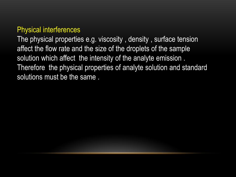
Physical interferences
The physical properties e.g. viscosity , density , surface tension
affect the flow rate and the size of the droplets of the sample
solution which affect the intensity of the analyte emission .
Therefore the physical properties of analyte solution and standard
solutions must be the same .

APPLICATIONS :
To fined the concentration of the analyte we use either the standard
calibration curve method , the standard addition method ( see unit 4 )
or the internal standard method ( see unit 11 ) .
Most applications of FAES have been the determination of trace
metals, especially in liquid samples. It should be remembered that
FAES offers a simple, inexpensive, and sensitive ( down to 0.001 ppm
can be determined ) method for detecting common metals, particularly
the alkali and alkaline earths, as well as several transition metals such
as Fe, Mn, Cu, and Zn. FAES has been extended to include a number
of nonmetals: N, P, and S. FAES detectors for P and S are
commercially available for use in gas chromatography.
FAES has found wide applications in agricultural , environmental,
industrial, and clinical analysis of body fluids ( e.g. blood and urine
analysis ) .

Experiment
Flame Photometric Determination of sodium, Potassium, and
Lithium
Background
FAES is widely used for routine analysis of samples
containing species like Na, K, Li, and Ca. Emission signal at a specific
wavelength is proportional to the concentration of analyte which emits
at that wavelength.
Chemicals and Reagents
1. Standard Na, K, and Li solutions (1000 ppm each).
2. Sample of unknown concentrations of Na, K, and Li.
3. Prepare standard Na, K, and Li solutions that are 1, 5, 10 ,20
,30 ,40 ,50 ,60, 70, 80, 90, and 100 ppm of each metal ion.

Procedure
1. Follow instructions for the correct operation of the flame
photometer available.
2. Adjust the signal, using the Na filter, to zero using distilled
deionized water.
3. Read the signal for the Na set of standards and then that of the
unknown sample.
4. If the signal obtained for the sample is out of range, dilute a
portion of the sample properly till a signal within the range is obtained.
5. Construct a calibration curve for Na in the sample and report
your results in ppm.

6. Repeat steps 2-5 for K and finally for Li and find the
concentration of each species in the sample. Results should also be
reported in ppm analyte.
Note: The sample unknown can be a sample of any drinking
supply. Therefore, each student is asked to bring his own sample
and the class is asked to report an overview of water quality in the
different areas of the city as compared to accepted values

الى محاضرة عن موضوع هذه الوحدة باللغة االستماع على الراغبين
:التالية الروابط العربية الضغط على كل من
: Flame Atomic Emission Spectrometry25 Part
: Flame Atomic Emission Spectrometry26 Part
: Flame Atomic Emission Spectrometry24 Part
: Flame Atomic Emission Spectrometry23 Part
: Flame Atomic Emission Spectrometry27 Part