An interferometric ex vivo study of corneal biomechanics ...
11
RESEARCH Open Access An interferometric ex vivo study of corneal biomechanics under physiologically representative loading, highlighting the role of the limbus in pressure compensation Abby Wilson 1* , John Jones 2 , John R. Tyrer 1,2 and John Marshall 3 Abstract Background: The mechanical properties of the cornea are complex and regionally variable. This paper uses an original method to investigate the biomechanics of the cornea in response to hydrostatic loading over the typical physiological range of intra-ocular pressure (IOP) fluctuations thereby increasing understanding of clinically relevant corneal biomechanical properties and their contributions to the refractive properties of the cornea. Methods: Displacement speckle pattern interferometry (DSPI) was used to measure the total surface displacement of 40 porcine and 6 human corneal-scleral specimens in response to pressure variations up to 1 mmHg from a baseline of 16.5 mmHg. All specimens were mounted in a modified artificial anterior chamber (AAC) and loaded hydrostatically. Areas of high strain in response to loading were identified by comparing the displacements across different regions. Results: The nature of the response of the corneal surface to loading demonstrated high regional topographic variation. Mechanical properties were shown to be asymmetrical, and deformation of the limbal and pre-limbal regions dominated these responses respectively with over 90% (N-T) and 60% (S-I) of the total maximum displacement occurring in these regions indicating high-strain. In contrast, the curvature of the central cornea remained relatively unchanged merely translating in position. Conclusions: The limbal and pre-limbal regions of the cornea appear to be fundamental to the absorption of small pressure fluctuations facilitating the curvature of the central cornea to remain relatively unchanged. The differential mechanical properties of this region could have important implications for the application of corneal surgery and corneal crosslinking, warranting further investigation. Keywords: Cornea, Biomechanics, Interferometry, Limbus, Topography © The Author(s). 2020 Open Access This article is licensed under a Creative Commons Attribution 4.0 International License, which permits use, sharing, adaptation, distribution and reproduction in any medium or format, as long as you give appropriate credit to the original author(s) and the source, provide a link to the Creative Commons licence, and indicate if changes were made. The images or other third party material in this article are included in the article's Creative Commons licence, unless indicated otherwise in a credit line to the material. If material is not included in the article's Creative Commons licence and your intended use is not permitted by statutory regulation or exceeds the permitted use, you will need to obtain permission directly from the copyright holder. To view a copy of this licence, visit http://creativecommons.org/licenses/by/4.0/. The Creative Commons Public Domain Dedication waiver (http://creativecommons.org/publicdomain/zero/1.0/) applies to the data made available in this article, unless otherwise stated in a credit line to the data. * Correspondence: [email protected] 1 Wolfson School of Mechanical, Manufacturing and Electrical Engineering, Loughborough University, Loughborough, UK Full list of author information is available at the end of the article Wilson et al. Eye and Vision (2020) 7:43 https://doi.org/10.1186/s40662-020-00207-1
Transcript of An interferometric ex vivo study of corneal biomechanics ...

RESEARCH Open Access
An interferometric ex vivo study of cornealbiomechanics under physiologicallyrepresentative loading, highlighting therole of the limbus in pressurecompensationAbby Wilson1* , John Jones2, John R. Tyrer1,2 and John Marshall3
Abstract
Background: The mechanical properties of the cornea are complex and regionally variable. This paper uses anoriginal method to investigate the biomechanics of the cornea in response to hydrostatic loading over the typicalphysiological range of intra-ocular pressure (IOP) fluctuations thereby increasing understanding of clinically relevantcorneal biomechanical properties and their contributions to the refractive properties of the cornea.
Methods: Displacement speckle pattern interferometry (DSPI) was used to measure the total surface displacementof 40 porcine and 6 human corneal-scleral specimens in response to pressure variations up to 1 mmHg from abaseline of 16.5 mmHg. All specimens were mounted in a modified artificial anterior chamber (AAC) and loadedhydrostatically. Areas of high strain in response to loading were identified by comparing the displacements acrossdifferent regions.
Results: The nature of the response of the corneal surface to loading demonstrated high regional topographicvariation. Mechanical properties were shown to be asymmetrical, and deformation of the limbal and pre-limbalregions dominated these responses respectively with over 90% (N-T) and 60% (S-I) of the total maximumdisplacement occurring in these regions indicating high-strain. In contrast, the curvature of the central cornearemained relatively unchanged merely translating in position.
Conclusions: The limbal and pre-limbal regions of the cornea appear to be fundamental to the absorption of smallpressure fluctuations facilitating the curvature of the central cornea to remain relatively unchanged. The differentialmechanical properties of this region could have important implications for the application of corneal surgery andcorneal crosslinking, warranting further investigation.
Keywords: Cornea, Biomechanics, Interferometry, Limbus, Topography
© The Author(s). 2020 Open Access This article is licensed under a Creative Commons Attribution 4.0 International License,which permits use, sharing, adaptation, distribution and reproduction in any medium or format, as long as you giveappropriate credit to the original author(s) and the source, provide a link to the Creative Commons licence, and indicate ifchanges were made. The images or other third party material in this article are included in the article's Creative Commonslicence, unless indicated otherwise in a credit line to the material. If material is not included in the article's Creative Commonslicence and your intended use is not permitted by statutory regulation or exceeds the permitted use, you will need to obtainpermission directly from the copyright holder. To view a copy of this licence, visit http://creativecommons.org/licenses/by/4.0/.The Creative Commons Public Domain Dedication waiver (http://creativecommons.org/publicdomain/zero/1.0/) applies to thedata made available in this article, unless otherwise stated in a credit line to the data.
* Correspondence: [email protected] School of Mechanical, Manufacturing and Electrical Engineering,Loughborough University, Loughborough, UKFull list of author information is available at the end of the article
Wilson et al. Eye and Vision (2020) 7:43 https://doi.org/10.1186/s40662-020-00207-1

IntroductionThe biomechanics of the cornea govern its shape andhence its refractive power. Understanding how the mech-anical properties of the cornea contribute to its shape hasbecome of increasing importance since the advent andrapid dissemination of refractive surgery in the 1990’s, asthe introduction of surgical incisions to the cornea resultsin modifications to its mechanical properties [1, 2]. It isnow even more pertinent given the possibility of directtopographic manipulation of corneal stiffness via cornealcrosslinking (CXL) [3], which has demonstrated potentialas a minimally-invasive means to provide small refractivecorrection [4]. However, there have been major limitationsassociated with the techniques employed for biomechan-ical assessment in basic and clinical studies, and to date,there is no established method for evaluating the mechan-ics of the cornea and generating spatially resolved infor-mation that is relevant to the clinician.The cornea is a structurally complex, viscoelastic
membrane with regionally variable, strain dependentbiomechanical properties; hence, many factors such asthe strain rate, hydration and direction of the appliedforce, can lead to variations in response [5]. To gain auseful understanding of how biomechanics govern cor-neal shape and behaviour and predict how the biomech-anical properties of the tissue may change followingdisease, trauma or surgical intervention, it is necessaryto consider the mechanics of the entire cornea in re-sponse to physiologically-relevant forces.However, up to the present time, the majority of stud-
ies on corneal biomechanics have been performedex vivo, using strip extensometry to evaluate cornealelasticity expressed in terms of the elastic moduli(Young’s, tangent, secant) of excised strips of corneal tis-sue [6–8]. Whilst this technique has advantages in termsof its simplicity, low cost, and ability to provide a quanti-tative measure of stiffness, it also has many limitations.Principally, strips are isolated from the dome of the cor-nea, compromising the integrity of its microstructure,and removing the natural physiological state of tensionand shape that plays an important role in governing thebiomechanics of the tissue in vivo. This, in addition tothe difficulties associated with maintaining a strip of tis-sue at physiological hydration during the measuring pro-cedure; and the fact strains generally far exceed thoseexperienced physiologically contributes to the substantialvariation in elastic moduli that have been reported inthe literature, with values ranging from 1.3MPa [9] to57MPa [10]. Hence, despite extensometry testing pro-viding a useful and simplistic measure of changes to tis-sue elasticity in the case of interventions such as CXL, itwould be erroneous to assume that stiffness and tensileproperties measured via this method are truly applicableto whole-tissue models of corneal biomechanics.
Acknowledging the limitations of strip testing, a var-iety of methods have now been investigated to probe thebiomechanics of intact corneal tissue under physiologic-ally relevant loading conditions, these methods include;Brillouin spectroscopy (BrS), optical coherence elasto-graphy (OCE), high frequency ultrasound (HFU), digitalimage correlation (DIC) and interferometry. Of these,BrS and OCE have demonstrated potential to acquiredata in a clinical setting [11, 12]. BrS evaluates longitu-dinal modulus, which is related to compressibility (bulkmodulus) in isotropic materials. However, in hydratedmaterials such as the cornea, BrS signals represent avolume-weighted aggregate longitudinal modulus of thefluid and solid components of a tissue, often dominatedby the fluid component (which for the cornea is spatiallyand temporally variable). As a result, Brillouin spectrado not correlate directly with elastic properties, recentlyleading to one group cautioning against its use as an op-tical elastography tool for biological materials [13].Furthermore, both BrS and OCE are scanning based
methods, so there is an inherent trade-off betweenspatial resolution and acquisition time, with BrS requir-ing several minutes to generate a relatively low-resolution, limited area, 40-point scan [11]; and OCE re-quiring around 5min to obtain high-resolution 3D data[14]. A more prominent limitation of OCE is a loss ofspeckle contrast at the corneal periphery limiting dataacquisition to the central ~ 8mm [14], unless the corneais rotated with respect to the image system, leading tolonger acquisition times and complexities with imagestitching during post-processing. HFU has recently dem-onstrated potential to measure corneal strain in responseto changes in IOP over the cardiac cycle [15], howeverthis method also requires scanning, with data only pre-sented so far for the central region of 2D cross-sections[15, 16]. If clinical translation is to be realised using ei-ther OCE or HFU, further work is required to extendthe region of measurement and to account for eye move-ment over the measurement time.In contrast to the above methods, DIC and Interferom-
etry are snapshot methods where whole-field data can becaptured over a single measurement, requiring only milli-seconds. DIC has successfully been used ex vivo to exam-ine regional differences in surface displacement inresponse to loading [17, 18], however displacement sensi-tivity is limited relative to interferometric techniques.Using speckle interferometric methods, it is possible togenerate high spatial resolution maps of corneal andscleral surface deformation, with a displacement sensitivityof 10’s of nanometres, over a single measurement takingonly milliseconds. Displacement speckle pattern interfer-ometry (DSPI) has previously been used for the examin-ation of ovine corneas [1], where it was used to quantifythe effects of refractive surgery on biomechanics. Radial
Wilson et al. Eye and Vision (2020) 7:43 Page 2 of 11

speckle pattern shearing interferometry has also beenemployed to examine age related stiffening [19] and thebiomechanical effects of CXL [3], however this particularmethod is inherently limited by a lack of sensitivity at thecentral cornea. Despite significant advantages in terms ofacquisition time, both DIC and interferometry measurethe surface movement, hence the bulk movement of thecornea; they cannot provide detail on any compressionthat may occur through the thickness of the tissue.Clinical assessment of corneal biomechanics currently
relies on evaluation of the response of the cornea to anair-puff directed at its centre, either via the ocular re-sponse analyser (ORA) [20] or the dynamic scheimpflugtonometer (DST) [21]. Although DST has recently dem-onstrated efficacy for the early detection of biomechan-ical abnormality [22], neither of these methods directlyasses corneal stiffness and cannot realistically predictmovement parameters outside the location of the air-puff.In the present study, DSPI was used to obtain non-
contact, non-destructive measurement of full-cornealsurface deformation in response to hydrostatic pressurechanges designed to simulate small changes in IOP, suchas those that occur during the cardiac cycle [23]. Build-ing upon previous work [1], where only a single compo-nent of deformation was analysed, here DSPI wascombined with subtractive cross-sectional imaging enab-ling the total set of deformation to be estimated.
Methods and materialsMeasurement and loading systemThe working principles of DSPI are diagrammaticallysummarised in Supplementary Figure 1. During DSPI,the surface of the target is illuminated with coherentlight. In the presence of surface height variations greaterthan the wavelength of the illumination source, lightscattered from the surface of interest has a speckled ap-pearance. The light scattered from this surface is inter-fered with that of a reference and imaged on a detectorto create an interferogram. The speckle pattern formedis unique to that object at the time of measurement,should the surface move, this speckle pattern willchange. Capturing a speckle pattern of the object in itsreference state and then digitally subtracting subsequentspeckle patterns captured as the object moves results inthe formation of interference patterns consisting of lightand dark fringes, which, with knowledge of the opticalset-up, can be mathematically analysed to quantify thesurface displacement [24], which when combined withphase-shifting provides sensitivity to displacement on ascale of 10’s of nanometres [25].The layout of the interferometer that was used in this
study for the measurement of surface deformation, andthe artificial anterior chamber (AAC) onto which
corneas were mounted are shown in Fig. 1a and b, re-spectively. For the interferometry system, illumination wasvia a diode pumped single-mode solid-state laser (λ =532 nm) (06-DPL, Cobolt AB, Solna, SE). The laser wasexpanded and collimated to a diameter of 25mm. The il-lumination beam was passed through a 50:50 beamsplitterwith half directed towards the corneal surface and half to-wards a planar mirror attached to a piezo-electric trans-ducer which was used to generate a phase-steppedreference beam. The beams from the object and the refer-ence were imaged using a CMOS camera with a resolutionof 1296 by 972 pixels (CMOS Aptina MT9P031, BaslerAG, Ahrensburg, DE) through a 12.5mm – 75mm zoomlens (C31204, Pentax, Tokyo, JP).The configuration of the interferometric system, with
illumination and imaging directed normal to the surfaceof the target dictates that the sensitivity is to out-of-plane deformation only (Fig. 1c) and this is constantacross the surface of the object. However, due to thecurved nature of the cornea, the vector of total displace-ment is likely to vary across the surface. Therefore, tocompletely define the deformation of the corneal sur-face, it was necessary to define the out-of-plane andhorizontal and vertical in-plane components of deform-ation. This was impractical to achieve using interferom-etry on a curved surface, due to the requirement for off-axis illumination and imaging, hence, problems withshading and uneven illumination. Consequently, estima-tions for the in-plane component of deformation weremade using subtractive imaging of the central cross-section of a cornea deforming under a larger change ofpressure (< 20 mmHg) and tracking the angle of surfacemovement via tracking the movement of recognisableregions on the corneal surface, identifiable due to theroughness of the applied surface coating.Pressure in the AAC was controlled via a reservoir
supplying fluid to the closed chamber. During testing,the reservoir and the chamber were filled with Phos-phate Buffered Saline (PBS) solution (Sigma-Aldrich,UK, ρ = 0.995 g/ml at 25 °C). The reservoir was raised225 mm above the surface of the cornea to provide abaseline pressure of 16.5 mmHg, within the representa-tive range of normal IOP in both human and porcineeyes [26–28]. To raise and lower the pressure from thisbaseline, the height of the chamber was adjusted elec-tronically via attachment of the reservoir to a motorisedvertical translation stage capable of pressure variationsup to a maximum of 4 mmHg above the baselinepressure.
Validation experimentsTo establish that the interferometer was functioning cor-rectly, prior to corneal measurement, two initial experi-ments were conducted. The first experiment was on a
Wilson et al. Eye and Vision (2020) 7:43 Page 3 of 11

circularly clamped steel plate, centrally point-loadedwith a micrometre screw. A pre-load was applied, and areference image was acquired, the plate was then dis-placed by 1 μm at the centre via movement of the micro-metre screw and data was acquired, 10 repeatedmeasurements were taken. Following this, measurementswere conducted on a curved silicone hemisphericalmembrane of uniform thickness (Mini Semispheremould, Silikomart, IT, rc = 15mm) mounted in the AACto check that the AAC did not induce any abnormalboundary effects. In this case the membrane was setunder an initial pressure of 16.50 mmHg and hydrostaticpressure was increased by 1 mmHg over the course ofmeasurement.
Corneal preparation and testingFresh porcine eyes (< 12 h post-mortem) with a clearcornea and intact epithelium were obtained from a localabattoir (Joseph Morris Butchers, South Kilworth, UK).The eyes were stored in a moist, sealed container and re-frigerated at 4 °C until measurement. All porcine eyeswere used within 3 days of slaughter. Prior to measure-ment, the corneas were isolated from the posteriorglobe, leaving a 3 mm scleral border for mounting. Forthe human eye experiments, 6 human corneal-scleralspecimens from 3 donors were obtained from Moor-field’s Biobank (UCL Institute of Ophthalmology,London, UK), with ethical approval granted by Moor-fields Biobank internal ethics committee (Ref: 10/H0106/57-2015ETR45). The human corneas were sur-plus for donor purposes and had been collected withpermission for research use. The corneal specimens had
been in storage for over 8 weeks prior to being re-leased for research use. The human corneas were sus-pended in organ donor culture (80 ml Eagle’sminimum essential medium with HEPES buffer, 26mmol/l NaHCO3, 2% foetal bovine serum, 2 mmol/lL-glutamine, penicillin, streptomycin and amphoteri-cin B). All corneas remained in the solution until re-quired for measurement.All corneas were de-epithelialized prior to measure-
ment. The epithelium has previously been shown to havea negligible contribution to stiffness [29], and its removalprevented any changes to the epithelium, that may haveoccurred during storage, from affecting the measured re-sponse. The corneal-scleral specimens were thenmounted within the AAC, which imposed a fixedboundary on the scleral region 1.5 mm to 3.0 mm out-side the edge of the cornea. As the cornea is designed totransmit optical radiation, surface reflection was smalland had to be enhanced by means of a scattering agent.The corneas were coated with a thin layer of hollowglass microspheres - Sphericel 110P8 (Potters Ind. LLC,PA, USA), to amplify the scatter from the surface. Priorto loading and measurement, the corneas were set underthe baseline pressure of 16.5 mmHg and maintainedunder this constant pressure for 30-min to allow forstress-relaxation. During testing, each cornea underwent3 loading cycles during which the pressure was raised by0.25, 0.5, 0.75 and 1mmHg in turn. At the end of eachloading increment, the movement of the reservoir waspaused for 0.5 s during which data was captured andprocessed, and after which the pressure was restored tobaseline.
Fig. 1 Schematics showing the layout of the DSPI system (BS – Beamsplitter, PZT – Piezoelectric Transducer, M – Mirror) (a), the AAC onto whichcorneas were mounted during testing (b), and the direction of measured displacement across the corneal surface (c). Out-of-plane displacement(w) corresponds to displacement along the direction of the z-axis, horizontal in-plane (u) and vertical in-plane (v) correspond to displacementalong the direction of the x and y axes, respectively
Wilson et al. Eye and Vision (2020) 7:43 Page 4 of 11

Data analysisThe out-of-plane displacement (w) was calculated fromthe measured phase change due to deformation (Δϕdef
(radians)) using eq. 1 [24], where the wavelength of theillumination source (λ) was 532 nm.
w ¼ Δϕdef :λ4π
ð1Þ
The horizontal in-plane (u) and vertical in-plane (v)displacement components were estimated using themeasured out-of-plane displacement component (w) andthe angle of deformation estimated from subtractivecross-sectional imaging as described by eqs. 2 and 3,where θx and θy are the angles of deformation with re-spect to the horizontal and vertical axes, respectively.
u ¼ w tanθx ð2Þ
v ¼ w tanθy ð3Þ
ResultsLoading plate and response of uniform membraneThe average central displacement of the clamped steelplate loaded via the micrometre screw was as expectedat 0.98 μm (SD ± 0.02 μm). The deformation of thecurved homogenous membrane (Fig. 2a) also showedthe expected distribution, with zero deformation at thefixed boundary, maximum displacement at the centralpoint furthest from the fixed boundary (Fig. 2b) and arelatively constant gradient to the rate-of-change of dis-placement (Fig. 2c).
Regional differences in corneal responseIn contrast to the homogenous membrane (Figs. 2 and 3a),corneas were found to demonstrate high regional variabilityin response to pressure changes (Figs. 3b and 4). In boththe human and porcine corneas examined, there was a ten-dency for deformation to be concentrated in the limbal re-gion. This was evident due to the high fringe concentrationobserved in this region, relative to other regions duringloading (Fig. 3b) which was in clear contrast to the responseof the homogenous membrane, which when mounted inthe AAC, showed relatively even fringe spacing (Fig. 3a).Furthermore, a common trend was observed with 23/40(57%) porcine corneas and 4/6 (67%) of human corneasshowing similarities in the pattern of regional displacement.A typical response of a human cornea is shown in
Fig. 4 (responses of all human corneas are provided inSupplementary Figures 2 and 4). In these specimens, theresponse was asymmetrical; the rate of change of dis-placement across the central 8 to 10 mm of the corneawas close to zero (Table 1), especially across the nasal-temporal axis (Fig. 4d) indicating minimal changes incurvature across this region; and the highest rate ofchange of displacement was at the edge of the cornea inthe limbal region (Fig. 4d and f), where the cornea joinsthe sclera. Taken together, these indicate a high strainconcentration at the limbal/prelimbal region, confirmedby the relatively higher values for δw
δx and δwδy here when
compared to the central 8 mm of the N-T and S-I axes(Table 1). It was also observed that while the magnitudeof δw
δx and δwδy increased significantly in the limbal region
in response to larger pressure variations (0.5 to 1.0
Fig. 2 Out-of-plane surface displacement of a curved silicone hemispherical membrane in response to hydrostatic pressure increase from 16.5mmHg to 17.5 mmHg; whole surface displacement map (a); cross-section of out-of-plane displacement along dashed line (b); cross-section ofgradient of out-of-plane displacement along dashed line (c)
Wilson et al. Eye and Vision (2020) 7:43 Page 5 of 11

mmHg), across the centre δwδx and δw
δy remained almost
constant, showing minimal changes to the curvature ofthis region across the full pressure range. In general,over 90% (N-T axis) and 60% (S-I) of the maximum out-of-plane displacement occurred in the outer 2 mm of thehuman corneas incorporating the limbal region (Table1). A typical response for a porcine cornea is shown inSupplementary Figure 3. As with human corneas, themajority of strain was concentrated at the limbus how-ever, in porcine corneas, deformation was greater in theperipheral cornea at the superior and inferior poleswhen compared to the nasal temporal.
The corneas that did not show this typical distributionof displacement can be grouped into two main categor-ies. A few porcine corneas (5/40) showed a similar dis-placement distribution to the one previously described,but rotated through 90 °, suggesting that this could haveoccurred due to errors in determining initial cornealorientation. The majority of the other corneas tested(12/40 porcine corneas and 2/6 human corneas), showeda distribution where the maximum rate of change of dis-placement remained at the limbal region, but instead ofmaintaining an overall relatively constant level of dis-placement across the central region, the displacement
Fig. 3 Fringe distributions obtained from a curved silicone hemispherical membrane (a) and a human cornea (b) in response to a pressureincrease from 16.5 mmHg to 17.0 mmHg
Fig. 4 Typical response of a human cornea to a pressure increase from 16.5 mmHg to 16.75 mmHg. Photograph of cornea in AAC for positionalreference (a), full-surface map of out-of-plane displacement (b); out-of-plane displacement across section A-A (c); gradient of out-of-planedisplacement across section A-A (d); out-of-plane displacement across section B-B (e); gradient of out-of-plane displacement across section B-B (f)
Wilson et al. Eye and Vision (2020) 7:43 Page 6 of 11

tended to be greater in one particular region, indicatinglocalised regions of biomechanical weakness. The pos-ition of this region varied between corneas. In one hu-man cornea, this area of weakness was clearlyidentifiable as damage introduced from the insertion ofthe surgical thread used to suspend the cornea in thetransplant solution (Supplementary Figure 4). Discount-ing this cornea, the magnitude of out-of-plane displace-ment at the central cornea for the remaining humancorneas tested in response to a 0.25mmHg change inhydrostatic pressure are shown in Table 1 and ranged
from 0.29 μm to 0.71 μm (median 0.50 μm). Assuming lin-earity in response, this corresponds to apical displacementof 3.6 μm to 10.4 μm over a normal range of ocular pulseamplitudes (1.8–4.3mmHg) [23] consistent with a recentstudy examining the deformation of a whole eye-globe overa physiological range of pressure variations [18].
Directional aspects of responseTo examine the directional aspects of the response, thedeformation of the central-cross-section of the corneasin response to a larger pressure change (< 20 mmHg)
Table 1 Comparison of out-of-plane displacement of human corneas at the central cornea and at the limbus in response to a 0.25mmHg increase in hydrostatic pressure
HumancorneaNo.
w at centralcornea, mean ±SD (μm)
w at limbal/prelimbal region(N-T axis) (μm)
w at limbal/prelimbal region(S-I axis) (μm)
Mean dw/dx(central 8mm N-Taxis) ×1.0e-06
Mean dw/dx(limbus N-T axis)×1.0e-06
Mean dw/dy(central 8mm S-Iaxis) ×1.0e-06
Mean dw/dy(limbus S-I axis)×1.0e-06
1 0.46 ± 0.058 0.46 0.44 0.66 3.39 0.74 3.59
2 0.29 ± 0.036 0.27 0.20 0.31 4.21 0.57 2.88
3 0.35 ± 0.028 0.37 0.27 0.26 4.39 0.57 1.91
4 0.68 ± 0.050 0.46 0.42 1.22 3.87 1.16 3.91
5 0.71 ± 0.068 0.66 0.63 0.58 5.70 0.81 5.13
Fig. 5 Subtractive video recording of the central cross-section of a cornea deforming in response to a pressure increase (a), comparison ofmeasured out-of-plane and estimated horizontal in-plane displacement along central N-T cross section of a human cornea (b)
Wilson et al. Eye and Vision (2020) 7:43 Page 7 of 11
was recorded (Fig. 5). From this, it was identified thatdisplacement tended to occur predominantly out-of-plane in comparison to that which would be predictedfor an isotropic hemisphere where it would be expectedto occur in a direction normal to the surface. The esti-mated in-plane component calculated using the angle ofdeformation and the measured out-of-plane componentis shown in Fig. 5b. The in-plane contribution to overalldisplacement was relatively small, especially over thecentral 3 mm of the cornea and was approximately uni-form either side of this over the central ~ 8 mm of thecornea. Hence, δu
δx ≈ 0 over this region, and thus themeasured differences in the out-of-plane displacementcomponent may be considered predominately represen-tative of changes in central curvature.
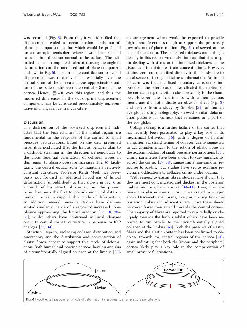
DiscussionThe distribution of the observed displacement indi-cates that the biomechanics of the limbal region arefundamental to the response of the cornea to smallpressure perturbations. Based on the data presentedhere, it is postulated that the limbus behaves akin toa dashpot, straining in the direction perpendicular tothe circumferential orientation of collagen fibres inthis region to absorb pressure increases (Fig. 6), facili-tating the central optical zone to maintain a relativelyconstant curvature. Professor Keith Meek has previ-ously put forward an identical hypothesis of limbaldeformation (unpublished) to that shown in Fig. 6 asa result of his structural studies, but the presentpaper has been the first to provide empirical data onhuman cornea to support this mode of deformation.In addition, several previous studies have demon-strated similar evidence of a region of increased com-pliance approaching the limbal junction [17, 18, 30–32], whilst others have confirmed minimal changesoccur to central corneal curvature in response to IOPchanges [33, 34].Structural aspects, including collagen distribution and
orientation; and the distribution and concentration ofelastin fibres, appear to support this mode of deform-ation. Both human and porcine corneas have an annulusof circumferentially aligned collagen at the limbus [35],
an arrangement which would be expected to providehigh circumferential strength to support the propensitytowards out-of-plane motion (Fig. 5a) observed at theedge of the cornea. The increased thickness and collagendensity in this region would also indicate that it is adeptfor dealing with stress, as the increased thickness of thetissue acts to minimise strain concentrations. However,strains were not quantified directly in this study due toan absence of through thickness information. An initialconcern was that the fixed boundary constraints im-posed on the sclera could have affected the motion ofthe cornea in regions within close proximity to the cham-ber. However, the experiments with a homogenousmembrane did not indicate an obvious effect (Fig. 2)and results from a study by Smolek [31] on humaneye globes using holography, showed similar deform-ation patterns for corneas that remained as a part ofthe eye globe.Collagen crimp is a further feature of the cornea that
has recently been postulated to play a key role in itsmechanical behaviour [36], with a degree of fibrillarelongation via straightening of collagen crimp suggestedto act complementary to the action of elastic fibres inthe accommodation of small pressure perturbations [36].Crimp parameters have been shown to vary significantlyacross the cornea [37, 38], suggesting a non-uniform re-sponse to loading, but studies have yet to examine re-gional modifications to collagen crimp under loading.With respect to elastin fibres, studies have shown that
they are most concentrated and thickest in the posteriorlimbus and peripheral cornea [39–41]. Here, they arepresent as elastin sheets, most concentrated in a layerabove Descemet’s membrane, likely originating from theposterior limbus and adjacent sclera. From these sheetsnarrower fibres then extend towards the central cornea.The majority of fibres are reported to run radially or ob-liquely towards the limbus whilst others have been re-ported to run parallel to the circumferentially alignedcollagen at the limbus [40]. Both the presence of elastinfibres and the elastin content has been confirmed to de-crease towards the central regions of the cornea [41],again indicating that both the limbus and the peripheralcornea likely play a key role in the compensation ofsmall pressure fluctuations.
Fig. 6 Hypothesised predominant mode of deformation in response to small pressure perturbations
Wilson et al. Eye and Vision (2020) 7:43 Page 8 of 11

Two additional aspects of the response that werehighlighted from the displacement maps obtained on hu-man and porcine corneas were differences in the behav-iour of the superior-inferior (S-I) axis and nasal-temporal (N-T) axis, and a lack of symmetry in the re-sponse with respect to each axis. For human corneas,the magnitude of displacement across the N-T axischanged very little inside the limbus, whereas for the S-Iaxis, the magnitude of displacement very gradually in-creased towards the centre, suggesting that this axis wasslightly less resistant to changes in curvature. Structuralstudies have identified differences between these twoaxes. Visually, it can be seen that there is an extensionof limbal tissue onto the cornea at the top and bottomof the S-I axis, making it appear shorter than the N-T[42]. Differences have also been identified in the transi-tioning of collagen at the limbus in human corneas [43].The N-T axis, having a 1.5 mm transition zone wherethe collagen running parallel to this axis, bifurcates togradually become circumferentially aligned, whereas inthe S-I axis, the collagen runs parallel to this axis rightup until the limbus. Porcine corneas, have been reportedin one study to lack the orthogonal arrangement of col-lagen across the central cornea [35], favouring a circum-ferential arrangement across the tissue. This couldpartially account for some of the differences seen in theresponse between human and porcine corneas. Based onthe previous discussion, it would be of interest to exam-ine differences, if any, in the elastic fibre network at thelimbal junction with respect to these axes.In terms of axial symmetry, it was evident, in most of
the human corneas, that greater deformation tended tooccur towards one side of the N-T axis, the specific sideremains ambiguous as the position of the specific poleswith respect to each axis could not be confirmed. Simi-larly, previous structural studies have shown that left andright eyes are structurally distinct [44], indicating that thebiomechanics are unlikely to be axially symmetrical.The specific features of the corneal response to small
pressure changes that have been described here arehighly important and require further investigation acrossa larger number of human samples. The informationthat has been demonstrated from this study and couldbe obtained from future studies using these imagingtechniques, has the potential to enhance the accuracyand validity of computational models of corneal bio-mechanics aimed at predicting the outcomes of surgicalprocedures and clinical therapies such as CXL.The non-destructive nature and high sensitivity of the
measurement technique used in this study makes it ahighly effective method for ex vivo examination of theregional changes to corneal biomechanics that occur dueto invasive procedures such as refractive surgery, anddue to treatments aimed at introducing biomechanical
changes such as CXL. The latter could benefit greatlyfrom spatial quantification of changes to biomechanicsas this could enable the development of optimised treat-ment algorithms to deliver accurate and predictable re-fractive modifications.A limitation of the method used is that it only mea-
sures surface displacements and therefore does not pro-vide a full assessment of biomechanics. Othertechniques have demonstrated compressive strainsthrough the thickness of the cornea in response tophysiological loading [15]. Hence, to gain a complete un-derstanding of corneal biomechanics, it may be most ef-fective to use this technique in combination with others,such as OCE or HFU, capable of measuring through-thickness displacements, while maximising the advan-tages of fast whole-field image acquisition times pro-vided by interferometry.A further limitation of this study, common with the
majority of ex vivo studies, is the challenge in replicatingand maintaining physiological levels of hydration in thetissue. Generally, human corneal samples cannot be ob-tained fresh and storage results in corneal swelling. Sincehydration contributes to tissue biomechanics, swellingcould lead to changes to the response of the tissue.However, a study had demonstrated only slight differ-ences in deformation patterns with IOP despite signifi-cant differences in hydration state [45], and it would beexpected that even if changes in the magnitude of de-formation changed, the overall mechanism of deform-ation, with high strain concentrations at the limbus,would remain the same.Although this study has been ex vivo in nature, it
ultimately is necessary for information of this kind,regarding spatial variation in biomechanics, to bemeasured in vivo. This would facilitate the early de-tection and optimised treatment of diseases, such askeratoconus, prior to significant changes in topog-raphy; and enable individuals that may be at risk ofpost-LASIK ectasia to be identified, preventing themfrom undertaking potentially damaging elective treat-ments. Further, if an individual’s corneal biomechan-ics could be topographically mapped, it would offerthe opportunity for delivery of optimised and custo-mised procedures such as laser refractive surgery forastigmatism, and CXL together with adjustmentspost-cataract astigmatism. Significant efforts are goinginto the development of a clinical device based onsimilar principles.
ConclusionsThe stiffness of the cornea is regionally variable. Thelimbal and pre-limbal regions of the cornea appear toplay a key role in the absorption of small pressure fluc-tuations, facilitating the central cornea to maintain a
Wilson et al. Eye and Vision (2020) 7:43 Page 9 of 11

relatively constant curvature. These findings could haveimportant implications for corneal surgery and for thedelivery of targetted-CXL, hence further investigationsare required on a larger number of human samples.DSPI has been demonstrated here as an effective tech-
nique to examine corneal surface deformation in re-sponse to physiological scale pressure variations, andcan be used alongside other methods to map cornealbiomechanics.
Supplementary informationSupplementary information accompanies this paper at https://doi.org/10.1186/s40662-020-00207-1.
Additional file 1: Supplementary Figure 1. Diagrammatic summaryof the working principles of displacement speckle pattern interferometry(DSPI).
Additional file 2: Supplementary Figure 2. Surface plots showingout-of-plane displacement of human corneas in response to a pressureincrease from 16.50 mmHg to 16.75 mmHg. Positions marked with X re-late to locations at which limbal and central displacements were com-pared in Table 1.
Additional file 3: Supplementary Figure 3. Typical response of aporcine cornea to a pressure increase from 16.50 mmHg to 17.00 mmHg.Full surface map of out-of-plane displacement (a), out-of-plane displace-ment along section A-A (b), out-of-plane displacement along section B-B(c).
Additional file 4: Supplementary Figure 4. Measurement regiondamaged due to insertion of surgical thread; photograph of corneawhilst suspended in transplant solution (a); map of out-of-plane surfacedisplacement in response to pressure increase from 16.5 mmHg to 17.0mmHg (b), area of damage clearly evident as region of increaseddisplacement (dark red region).
AcknowledgmentsProfessor Keith Meek for discussions regarding the relationship of cornealstructure to biomechanics.Dr. Sally Hayes for assistance with manuscript preparation.
Authors’ contributionsAbby Wilson designed the study, was responsible for system design andoperation; data acquisition and analysis; interpretation of data; andpreparation of the manuscript. John Jones contributed to system design, thecreation of software to run the interferometry system and process the dataand revision of the manuscript. John R Tyrer contributed lab facilities andequipment and contributed to the design of the study. John Marshallcontributed to the supply of materials, lab facilities, interpretation of thedata, design of the study and preparation of the manuscript. The author(s)read and approved the final manuscript.
FundingDuring the course of this study, Dr. Abby Wilson was enrolled on a PhD, andwas funded by EPSRC and Fight for Sight.
Availability of data and materialsAll relevant data generated or analysed during this study are included in thissubmitted article and its supplementary files. Full imaging datasets areavailable from the corresponding author on reasonable request.
Ethics approval and consent to participateAll human tissue used in this study was obtained from Moorfields Biobankwith ethical approval granted by Moorfields Biobank internal ethicscommittee (Ref: 10/H0106/57-2015ETR45).
Consent for publicationNot applicable.
Competing interestsThe authors declare they have no competing interests.
Author details1Wolfson School of Mechanical, Manufacturing and Electrical Engineering,Loughborough University, Loughborough, UK. 2Laser Optical EngineeringLtd., Derbyshire, UK. 3Institute of Ophthalmology, UCL, London, UK.
Received: 13 February 2020 Accepted: 25 July 2020
References1. Jaycock PD, Lobo L, Ibrahim J, Tyrer J, Marshall J. Interferometric technique
to measure biomechanical changes in the cornea induced by refractivesurgery. J Cataract Refract Surg. 2005;31(1):175–84.
2. Wilson A, Marshall J, Tyrer JR. The role of light in measuring ocularbiomechanics. Eye (Lond). 2016;30(2):234–40.
3. Knox Cartwright N, Tyrer JR, Marshall J. In vitro quantification of thestiffening effect of corneal cross-linking in the human cornea using radialshearing speckle pattern interferometry. J Refract Surg. 2012;28(7):503–8.
4. Seiler TG, Fischinger I, Koller T, Zapp D, Frueh BE, Seiler T. Customizedcorneal cross-linking: one-year results. Am J Ophthalmol. 2016;166:14–21.
5. Elsheikh A, Wang D, Pye D. Determination of the modulus of elasticity ofthe human cornea. J Refract Surg. 2007;23(8):808–18.
6. Hoeltzel D, Altman P, Buzard K, Choe K. Strip extensiometry for comparisonof the mechanical response of bovine, rabbit, and human corneas. TransASME. 1992;114(2):202–15.
7. Liu X, Wang L, Ji J, Yao W, Wei W, Fan J, et al. A mechanical model of thecornea considering the crimping morphology of collagen fibrils. InvestOpthalmol Vis Sci. 2014;55(4):2739–46.
8. Elsheikh A, Brown M, Alhasso D, Rama P, Campanelli M, Garway-Heath D.Experimental assessment of corneal anisotropy. J Refract Surg. 2008;24(2):178–87.
9. Andreassen TT, Hjorth Simonsen A, Oxlund H. Biomechanical properties ofkeratoconus and normal corneas. Exp Eye Res. 1980;31(4):435–41.
10. Wollensak G, Spoerl E, Seiler T. Stress-strain measurements of human andporcine corneas after riboflavin–ultraviolet-A-induced cross-linking. JCataract Refract Surg. 2003;29(9):1780–5.
11. Shao P, Eltony AM, Seiler TG, Tavakol B, Pineda R, Koller T, et al. Spatially-resolved Brillouin spectroscopy reveals biomechanical abnormalities in mildto advanced keratoconus in vivo. Sci Rep. 2019;9(1):7467.
12. De Stefano VS, Ford MR, Seven I, Dupps WJ. Live human assessment ofdepth-dependent corneal displacements with swept-source opticalcoherence elastography. PLoS One. 2018;13(12):e0209480.
13. Wu P-J, Kabakova IV, Ruberti JW, Sherwood JM, Dunlop IE, Paterson C, et al.Water content, not stiffness, dominates Brillouin spectroscopymeasurements in hydrated materials. Nat Methods. 2018;15(8):561–2.
14. Fu J, Haghighi-Abayneh M, Pierron F, Ruiz PD. Depth-resolved full-fieldmeasurement of corneal deformation by optical coherence tomographyand digital volume correlation. Exp Mech. 2016;56(7):1203–17.
15. Pavlatos E, Chen H, Clayson K, Pan X, Liu J. Imaging corneal biomechanicalresponses to ocular pulse using high-frequency ultrasound. IEEE Trans MedImaging. 2018;37(2):663–70.
16. Clayson K, Pavlatos E, Pan X, Sandwisch T, Ma Y, Liu J. Ocular pulseelastography: imaging corneal biomechanical responses to simulated ocularpulse using ultrasound. Transl Vis Sci Technol. 2020;9(1):5. https://doi.org/10.1167/tvst.9.1.5.
17. Boyce BL, Grazier JM, Jones RE, Nguyen TD. Full-field deformation of bovinecornea under constrained inflation conditions. Biomaterials. 2008;29(28):3896–904.
18. Whitford C, Joda A, Jones S, Bao F, Rama P, Elsheikh A. Ex vivo testing ofintact eye globes under inflation conditions to determine regional variationof mechanical stiffness. Eye Vis (Lond). 2016;3:21.
19. Knox Cartwright NE, Tyrer JR, Marshall J. Age-related differences in theelasticity of the human cornea. Invest Ophthalmol Vis Sci. 2011;52(7):4324–9.
20. Kotecha A, Russell RA, Sinapis A, Pourjavan S, Sinapis D, Garway-Heath DF.Biomechanical parameters of the cornea measured with the ocularresponse analyzer in normal eyes. BMC Ophthalmol. 2014;14:11.
21. Ambrósio R Jr, Ramos I, Luz A, Faria FC, Steinmueller A, Krug M, et al.Dynamic ultra high speed Scheimpflug imaging for assessing cornealbiomechanical properties. Rev Bras Oftalmol. 2013;72(2):99–102.
Wilson et al. Eye and Vision (2020) 7:43 Page 10 of 11

22. Vinciguerra R, Ambrósio R Jr, Roberts CJ, Azzolini C, Vinciguerra P. Biomechanicalcharacterization of subclinical keratoconus without topographic or tomographicabnormalitites. J Refract Surg. 2017;33(6):399–407.
23. Kaufmann C, Bachmann LM, Robert YC, Thiel MA. Ocular pulse amplitude inhealthy subjects as measured by dynamic contour tonometry. ArchOphthalmol. 2006;124(8):1104–8.
24. Petzing JN, Tyrer JR. Recent developments and applications in electronicspeckle pattern interferometry. J Strain Anal Eng Des. 1998;33(2):153–69.
25. Xie X, Yang L, Chen X, Xu N, Wang Y. Review and comparison of temporal-and spatial-phase shift speckle pattern interferometry for 3D deformationmeasurement. Sixth Int Symp Precis Mech Meas. 2013;8916:89160D.
26. Murgatroyd H, Bembridge J. Intraocular pressure. Contin Educ AnaesthesiaCrit Care Pain. 2008;8(3):100–3.
27. Ruiz-Ederra J, García M, Hernández M, Urcola H, Hernández-Barbáchano E,Araiz J, et al. The pig eye as a novel model of glaucoma. Exp Eye Res. 2005;81(5):561–9.
28. Schneider E, Grehn F. Intraocular pressure measurement-comparison ofdynamic contour tonometry and goldmann applanation tonometry. JGlaucoma. 2006;15(1):2–6.
29. Elsheikh A, Alhasso D, Rama P. Assessment of the epithelium’s contributionto corneal biomechanics. Exp Eye Res. 2008;86(2):445–51.
30. Hjortdal JO. Regional elastic performance of the human cornea. J Biomech.1996;29(7):931–42.
31. Smolek MK. Holographic interferometry of intact and radially incised humaneye-bank corneas. J Cataract Refract Surg. 1994;20(3):277–86.
32. Elsheikh A, McMonnies CW, Whitford C, Boneham GC. In vivo study ofcorneal responses to increased intraocular pressure loading. Eye Vis (Lond).2015;2:20.
33. Pierscionek BK, Asejczyk-Widlicka M, Schachar RA. The effect of changingintraocular pressure on the corneal and scleral curvatures in the freshporcine eye. Br J Ophthalmol. 2007;91(6):801–3.
34. McMonnies CW, Boneham GC. Corneal curvature stability with increasedintraocular pressure. Eye Contact Lens. 2007;33(3):130–7.
35. Hayes S, Boote C, Lewis J, Sheppard J, Abahussin M, Quantock AJ, et al.Comparative study of fibrillar collagen arrangement in the corneas ofprimates and other mammals. Vis Biol. 2007;290(12):1542–50.
36. Bell JS, Hayes S, Whitford C, Sanchez-weatherby J, Shebanova O, Vergari C,et al. The hierarchical response of human corneal collagen to load. ActaBiomater. 2018;65:216–25.
37. Gogola A, Jan NJ, Brazile B, Lam P, Lathrop KL, Chan KC, et al. Spatialpatterns and age-related changes of the collagen crimp in the humancornea and sclera. Invest Ophthalmol Vis Sci. 2018;59(7):2987–98.
38. Jan NJ, Brazile BL, Hu D, Grube G, Wallace J, Gogola A, et al. Crimp aroundthe globe; patterns of collagen crimp across the corneoscleral shell. Exp EyeRes. 2018;172:159–70.
39. Kamma-Lorger CS, Boote C, Hayes S, Moger J, Burghammer M, Knupp C,et al. Collagen and mature elastic fibre organisation as a function of depthin the human cornea and limbus. J Struct Biol. 2010;169(3):424–30.
40. Lewis PN, White TL, Young RD, Bell JS, Winlove CP, Meek KM. Three-dimensional arrangement of elastic fibers in the human corneal stroma. ExpEye Res. 2016;146:43–53.
41. White TL, Lewis PN, Young RD, Kitazawa K, Inatomi T, Kinoshita S, et al.Elastic microfibril distribution in the cornea: differences between normaland keratoconic stroma. Exp Eye Res. 2017;159:40–8.
42. Hogan MJ, Alvarado JA, Wedell JE. Histology of the human eye.Philadelphia: W.B. Saunders; 1971.
43. Newton RH, Meek KM. The integration of the corneal and limbal fibrils inthe human eye. Biophys J. 1998;75(5):2508–12.
44. Boote C, Hayes S, Abahussin M, Meek KM. Mapping collagen organization inthe human cornea: left and right eyes are structurally distinct. InvestOphthalmol Vis Sci. 2006;47(3):901–8.
45. Kling S, Marcos S. Effect of hydration state and storage media on cornealbiomechanical response from in vitro inflation tests. J Refract Surg. 2013;29(7):490–7.
Wilson et al. Eye and Vision (2020) 7:43 Page 11 of 11