An Imaging Review Article on Pleura Radiology Section Pulmonary …H)_PF1(PH)_PFA… · ·...
6
International Journal of Anatomy, Radiology and Surgery, 2013 April, Vol-2(1): 29-34 29 ID: IJARS/2013/4482:0007 An Imaging Review Article on Pleura Pulmonary Calcification and its Impact INTRODUCTION In quest of answers to find the cause of calcification processes involving the lung and the pleura, an approach based on the etiological factors appears rational, informative and satisfac- tory. The lung parenchymal shows calcifications as sequelae of prior infections. These infections could be due to viruses or due to bacteria like mycobacterium tuberculosis. Fungal infections too may manifest with calcifications. Primary tu- mors of the lungs like adenocarcinoma and small cell tumors too show calcifications. Lung metastases from primaries in breast, testes, thyroid and peripheral osteosarcoma too dem- onstrate calcifications. Lung parenchymal calcifications may also be seen in occupational lung diseases like coal workers pneumoconiosis. Commonest causes of pleural calcification are tuberculosis, asbestosis, empyema and old hemothorax. Primary pleural malignancy like mesothelioma and pleural me- tastases from carcinoma of breast, testes, thyroid and osteo- sarcoma can also calcify. Imaging modalities like plain radio- graphs and High Resolution Computed Tomography [HRCT], demonstrate the pulmonary as well as pleural calcifications satisfactorily. THE PROCESSES AND DETERMINANTS OF PLEURO-PULMONARY CALCIFICA- TIONS Pleural fibro-hyaline calcification and plaques are noted in about 1% of chest radiographs carried out as a routine in adults [1]. CT scan however demonstrates these calcifica- tions in many more conditions like hamartoma [2], chondroma [3], carcinoid [4], lung carcinoma [5], metastatic calcifications following cardiac surgery in children [6], calcinosis following liver transplant [7], pulmonary alveolar microlithiasis [8], Amio- darone pulmonary toxicity [9] to name a few. Pleural Calcification is commonly seen in Empyema, Tuber- culosis, Hemothorax, Exudative pleural collections, Asbes- tos Exposure, Silicosis and Talc Exposure. Common causes of Empyema are Pneumonia, Septic emboli, Lung abscess, Bacterial Pneumonia like Staphylococcus aureus and Gram negative pneumonias. Pleural calcification is seen as radio- opacity along the pleural contour [Table/Fig-1]. The stages in pleural calcifications in Para pneumonic Ef- fusion are: Stage 1 is an Exudative stage in which the capillary permeabil- ity is increased. Initially the accumulated pleural fluid is sterile. Most resolve with proper antibiotic therapy. Stage 2 is characterised by fibro-purulent fluid containing bac- teria and neutrophils. The fibrin deposition prevents resorption of fluid and leads to loculated collections. Radiology Section SUSHIL. G. KACHEWAR, DEVIDAS. S. KULKARNI Key Words: Tumoural calcinosis, Imaging, Radiology, MRI, X-Ray ABSTRACT The lungs as well as the pleura are common sites of cal- cification. Important causes of calcification of lung paren- chymal are prior infections (like viral pneumonia, miliary Koch`s), primary lung tumours and calcifying metastases to lung from carcinoma of breast, testes, thyroid and os- teosarcoma. Lung parenchymal calcifications may also be seen in occupational lung diseases like coal workers pneu- moconiosis. Common causes of pleural calcification are tuberculosis, asbestosis, empyema and old hemothorax. Primary pleural malignancy like mesothelioma and pleural metastases from carcinoma of breast, testes, thyroid and osteosarcoma can also calcify. While tiny calcifications are seen only on high resolution computed tomography [HRCT], larger calcifica- tions are visible on routine chest radiographs as well. Ul- trasound of the thorax does not demonstrate them satis- factorily. As the calcifications involving the Lung and the Pleura can have grave significance especially in the setting of neoplas- tic conditions, it is important to know how they appear on some common imaging modalities. Review Article
Transcript of An Imaging Review Article on Pleura Radiology Section Pulmonary …H)_PF1(PH)_PFA… · ·...
International Journal of Anatomy, Radiology and Surgery, 2013 April, Vol-2(1): 29-34 29
ID: IJARS/2013/4482:0007
An Imaging Review Article on Pleura Pulmonary Calcification and its Impact
IntroductIonIn quest of answers to find the cause of calcification processes involving the lung and the pleura, an approach based on the etiological factors appears rational, informative and satisfac-tory. The lung parenchymal shows calcifications as sequelae of prior infections. These infections could be due to viruses or due to bacteria like mycobacterium tuberculosis. Fungal infections too may manifest with calcifications. Primary tu-mors of the lungs like adenocarcinoma and small cell tumors too show calcifications. Lung metastases from primaries in breast, testes, thyroid and peripheral osteosarcoma too dem-onstrate calcifications. Lung parenchymal calcifications may also be seen in occupational lung diseases like coal workers pneumoconiosis. Commonest causes of pleural calcification are tuberculosis, asbestosis, empyema and old hemothorax. Primary pleural malignancy like mesothelioma and pleural me-tastases from carcinoma of breast, testes, thyroid and osteo-sarcoma can also calcify. Imaging modalities like plain radio-graphs and High Resolution Computed Tomography [HRCT], demonstrate the pulmonary as well as pleural calcifications satisfactorily.
the Processes and determInants of Pleuro-Pulmonary calcIfIca-tIons
Pleural fibro-hyaline calcification and plaques are noted in about 1% of chest radiographs carried out as a routine in adults [1]. CT scan however demonstrates these calcifica-tions in many more conditions like hamartoma [2], chondroma [3], carcinoid [4], lung carcinoma [5], metastatic calcifications following cardiac surgery in children [6], calcinosis following liver transplant [7], pulmonary alveolar microlithiasis [8], Amio-darone pulmonary toxicity [9] to name a few.
Pleural Calcification is commonly seen in Empyema, Tuber-culosis, Hemothorax, Exudative pleural collections, Asbes-tos Exposure, Silicosis and Talc Exposure. Common causes of Empyema are Pneumonia, Septic emboli, Lung abscess, Bacterial Pneumonia like Staphylococcus aureus and Gram negative pneumonias. Pleural calcification is seen as radio-opacity along the pleural contour [Table/Fig-1].
The stages in pleural calcifications in Para pneumonic Ef-fusion are:Stage 1 is an Exudative stage in which the capillary permeabil-ity is increased. Initially the accumulated pleural fluid is sterile. Most resolve with proper antibiotic therapy.
Stage 2 is characterised by fibro-purulent fluid containing bac-teria and neutrophils. The fibrin deposition prevents resorption of fluid and leads to loculated collections.
Rad
iolo
gy
Sec
tion
SuShil. G. KachEwaR, DEviDaS. S. KulKaRni
Key words: Tumoural calcinosis, Imaging, Radiology, MRI, X-Ray
aBstractThe lungs as well as the pleura are common sites of cal-cification. Important causes of calcification of lung paren-chymal are prior infections (like viral pneumonia, miliary Koch`s), primary lung tumours and calcifying metastases to lung from carcinoma of breast, testes, thyroid and os-teosarcoma. Lung parenchymal calcifications may also be seen in occupational lung diseases like coal workers pneu-moconiosis.
Common causes of pleural calcification are tuberculosis, asbestosis, empyema and old hemothorax. Primary pleural
malignancy like mesothelioma and pleural metastases from carcinoma of breast, testes, thyroid and osteosarcoma can also calcify. While tiny calcifications are seen only on high resolution computed tomography [HRCT], larger calcifica-tions are visible on routine chest radiographs as well. Ul-trasound of the thorax does not demonstrate them satis-factorily.
As the calcifications involving the Lung and the Pleura can have grave significance especially in the setting of neoplas-tic conditions, it is important to know how they appear on some common imaging modalities.
review article
Sushil. G. Kachewar and Devidas. S. Kulkarni, A Review Article on Pleura Pulmonary Calcification and its Impact www.ijars.jcdr.net
30 International Journal of Anatomy, Radiology and Surgery, 2013 April, Vol-2(1): 29-34
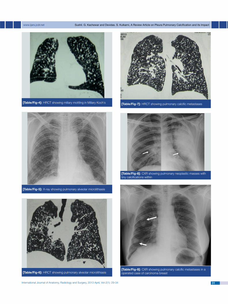
cification can be (a) metastatic, in which the calcium deposits occur in normal tissues, or (b) dystrophic, in which calcifica-tion occurs on injured lung tissue. The pathogenesis of these abnormalities is not fully understood, but hypercalcemia, hy-perphosphatemia, alkalosis, and lung damage predispose to calcification and ossification. The physiological requirements for metastatic calcium deposition are the release of exces-sive calcium salts from bone and its transport through the circulation. Metastatic calcification may occur with normal or even low serum calcium levels; it is the elevated calcium-phosphate product that favors the process. In patients with metastatic calcification with a normal calcium-phosphate product, the presence of azotemia, an elevated parathyroid hormone, and exogenous vitamin D play a role. Metastatic calcification commonly occurs in chronic renal failure. Dystro-phic classification frequently follows granulomatous infections (tuberculosis, histoplasmosis, coccidioidomycosis), viral in-fections (chicken pox and small pox), parasites (Paragonimus westermanii and Pneumocystis jiroveci), amyloidosis, coal and silica induced lung damage, pulmonary hypertension and vascular diseases. In sarcoidosis calcification can be either dystrophic or metastatic. Miliary mottling in Miliary Koch`s is demonstrated in [Table/Fig-3] (CXR) and [Table/Fig-4] (HRCT). The pulmonary alveolar microlithiasis is well demosntrated in [Table/Fig-5] (CXR) and [Table/Fig-6] (HRCT). [Table/Fig-7] is an HRCT image showing calcific pulmonary metastases. At times pulmonary neoplastic masses can present with tiny cal-cifications within as shown in [Table/Fig-8].
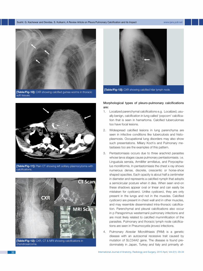
Metastases of carcinoma breast can be seen in lungs even after mastectomy [Table/Fig-9] and at times they too may calcify. Occasionally, calcified guinea worms can be seen in thoracic soft tissues as in [Table/Fig-10]. Masses involving thoracic wall like plasmocytoma [Table/Fig-11] and chondro-sarcoma [Table/Fig-12] also show numerous calcifications. At times even the calcified hilar lymph node might be seen on routine screening [Table/Fig-13].
Stage 3 occurs 2-3 weeks post initial effusion. There develops pleural fibrosis with entrapped lung. Dystrophic calcification finally develops after resolution of effusion.
In tuberculous effusion, there is rupture of sub-pleural ca-seating granulomas that gradually heal if adequately treated or if patient has good immunity; with fibrosis and ultimately develops dystrophic calcifications. As per newer research, it has been realized that lymphocytic alveolitis and pleural calcifications in non-occupational asbestos exposure confer protection against neoplasia [10]. Pulmonary calcifications are caused mainly by two mechanisms: the dystrophic form and the metastatic form [11, 12]. Despite the different aetiologies, it has been found that the pulmonary function and clinical manifestations are quite similar in both forms. It appears as well defined round or irregular radio-opacity within the con-fines of lung parenchyma [Table/Fig-2].
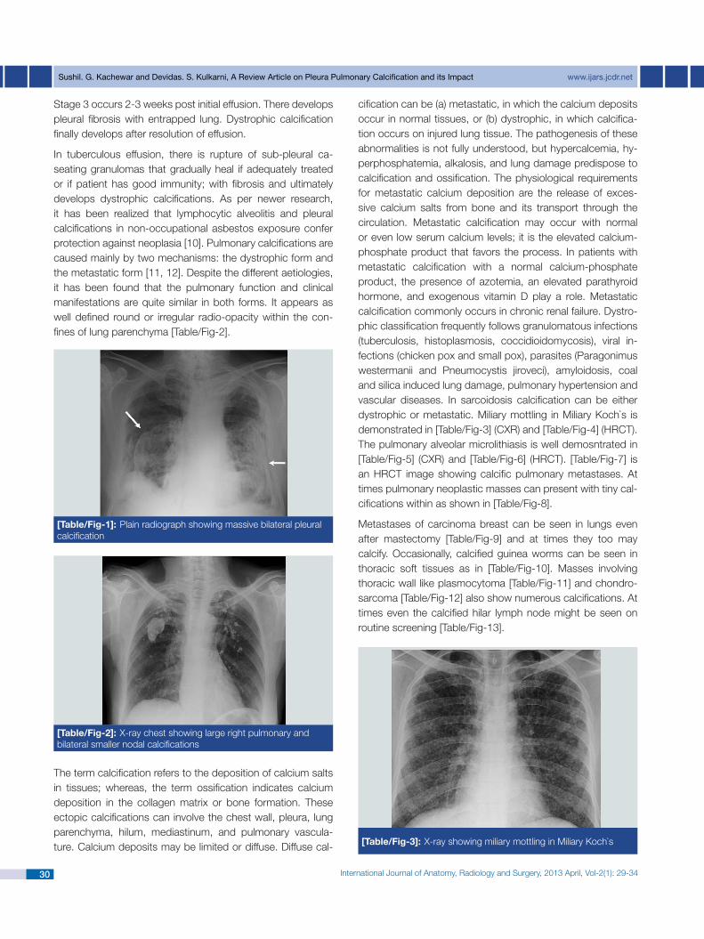
[table/fig-1]: Plain radiograph showing massive bilateral pleuralcalcification
[table/fig-2]: X-ray chest showing large right pulmonary andbilateral smaller nodal calcifications
The term calcification refers to the deposition of calcium salts in tissues; whereas, the term ossification indicates calcium deposition in the collagen matrix or bone formation. These ectopic calcifications can involve the chest wall, pleura, lung parenchyma, hilum, mediastinum, and pulmonary vascula-ture. Calcium deposits may be limited or diffuse. Diffuse cal- [table/fig-3]: X-ray showing miliary mottling in Miliary Koch`s
www.ijars.jcdr.net Sushil. G. Kachewar and Devidas. S. Kulkarni, A Review Article on Pleura Pulmonary Calcification and its Impact
31International Journal of Anatomy, Radiology and Surgery, 2013 April, Vol-2(1): 29-34
[table/fig-6]: HRCT showing pulmonary alveolar microlithiasis
[table/fig-5]: X-ray showing pulmonary alveolar microlithiasis
[table/fig-4]: HRCT showing miliary mottling in Miliary Koch`s [table/fig-7]: HRCT showing pulmonary calcific metastases
[table/fig-8]: CXR showing pulmonary neoplastic masses withtiny calcifications within
[table/fig-9]: CXR showing pulmonary calcific metastases in aoperated case of carcinoma breast
Sushil. G. Kachewar and Devidas. S. Kulkarni, A Review Article on Pleura Pulmonary Calcification and its Impact www.ijars.jcdr.net
32 International Journal of Anatomy, Radiology and Surgery, 2013 April, Vol-2(1): 29-34
Morphological types of pleuro-pulmonary calcifications are:
Localized parenchymal calcifications e.g. Localized, usu-1. ally benign, calcification in lung called ‘popcorn’ calcifica-tion that is seen in hamartoma. Calcified tuberculomas too have focal lesions.
Widespread calcified lesions in lung parenchyma are 2. seen in infective conditions like tuberculosis and histo-plasmosis. Occupational lung disorders may also show such presentations. Miliary Koch`s and Pulmonary me-tastases too are the examples of this pattern.
Pentastomiasis occurs due to three arachnid parasites 3. whose larva stages cause pulmonary pentastomiasis. i.e. Linguatula serrata, Armillifer armillatus, and Porpcepha-lus moniliformis. In pentastomiasis the chest x-ray shows numerous dense, discrete, crescentic or horse-shoe shaped opacities. Each opacity is about half a centimeter in diameter and represents a calcified nymph that adopts a semicircular posture when it dies. When seen end-on these shadows appear oval or linear and can easily be mistaken for cysticerci. Unlike cysticerci, they are only present in the lungs and not in the muscles. Calcified cysticerci are present in chest wall and in other muscles, and may resemble disseminated intra-thoracic calcifica-tion. Parenchymal and pleural calcifications also occur in p Paragonimus westermanii pulmonary infections and are most likely related to calcified mummification of the parasites. Pulmonary and thoracic lymph node calcifica-tions are seen in Pneumocystis jiroveci infections.
Pulmonary Alveolar Microlithiasis (PAM) is a genetic 4. disease with an autosomal recessive trait caused by mutation of SLC34A2 gene. The disease is found pre-dominately in Japan, Turkey and Italy and primarily af-
[table/fig-10]: CXR showing calcified guinea worms in thoracicsoft tissues.
[table/fig-11]: Plain CT showing left solitary plasmocytoma withcalcifications.
[table/fig-12]: CXR, CT & MRI showing calcifications in chondrosarcoma.
[table/fig-13]: CXR showing calcified hilar lymph node.
www.ijars.jcdr.net Sushil. G. Kachewar and Devidas. S. Kulkarni, A Review Article on Pleura Pulmonary Calcification and its Impact
33International Journal of Anatomy, Radiology and Surgery, 2013 April, Vol-2(1): 29-34
fects young people. The lower third of the lung fields are obscured by a granular sandstorm pattern of increased density caused by countless intra-alveolar microliths or calcospherites, which contain calcium and phosphorus. The appearance may be confused with alveolar proteino-sis, but the sand-like granular opacity is diagnostic [12]. The long-term prognosis is poor even if the disease is diagnosed at the asymptomatic stage. No effective treat-ment exists.
Diffuse pulmonary ossification (DPO) is a rare disease 5. characterized by diffuse small bone fragments in the lung tissue. It can be idiopathic or associated with chronic heart disease. The first case of this syndrome was de-scribed by Luschka in 1856. Now we recognize two types of DPO: dendritic and nodular [12]. Dendritic form is associated with history of pulmonary inflammation and fibrosis; whereas, nodular form is associated with chronic cardiac disease and pulmonary congestion. Once sus-pected the diagnosis can be confirmed by HRCT of the chest showing characteristic multiple punctuate and branching bone density lesions. Ossification can also be detected by using technetium-methylene diphosphate (Tc-MDP) bone scintigraphy. Bronchoalveolar lavage may reveal lymphocytes, eosinophils, macrophages, and gi-ant cells. A lung biopsy showing characteristic intersti-tial branching bone spicules establishes the diagnosis of dendritic ossification. The nodular form usually displays intra-alveolar bone deposits without marrow elements. The pathogenesis is not known. Serum calcium, phos-phorus, and alkaline phosphatase are normal. Transform-ing growth factor-B and bone morphogenic protein may play a role. Inter Leukin-4 and monocyte colony stimulat-ing factor can transform human alveolar macrophage to osteoclasts, a cell important in bone remodeling. The dis-ease mostly affects the elderly above the age of 60. Thus, it differs from alveolar microlithiasis which is a disease of the younger set. Regardless, most of the patients are as-ymptomatic; the diagnosis is suspected on routine chest radiography and confirmed by a lung biopsy. The disease is indolent and progresses slowly. There is no specific treatment. Early diagnosis will enable us to understand the disease and develop an appropriate treatment.
Calcification of hilar and mediastinal lymph nodes- It is 6. usually irregular and amorphous. When caused by tuber-culosis it is often associated with a small parenchymal nidus of calcium in the peripheral lung field; the combina-tion is called Ghon complex. Rarely, only the rim of the hilar node calcifies; this is called eggshell calcification and is characteristic of silicosis, but may also occur in tuberculosis, sarcoidosis, and Hodgkin’s disease after
radiation. It should not be confused with the crescentic calcium deposits seen in the dilated pulmonary arteries of chronic pulmonary hypertension. It may consist of a heavy central nidus with a lighter halo, giving rise to the characteristic target nodule. The calcified nodules that result from varicella pneumonia do not usually appear un-til two years after the infection, and are indistinguishable from histoplasmosis, pneumoconiosis and mitral steno-sis. Pulmonary calcification from smallpox is now history and from herpes simplex virus a rarity. Occasionally, sar-coidosis may produce multiple nodular calcifications with or without hilar node calcification. The radiological picture may resemble alveolar microlithiasis. In sarcoidosis how-ever, the calcification is confined to non-caseating granu-lomas, whereas, in pulmonary microlithiasis, the calcified stones are intra-alveolar.
health ImPacts of Pleuro-Pulmo-nary calcIfIcatIonsThe possible complications are functional disturbance, sec-ondary infection, fistula formation and malignant change [1]. Medico-legally the calcified plaques of the parietal pleura rep-resent an excellent sign of exposure to asbestos in a popula-tion [1]. In a study [10] conducted to see whether lympho-cytic alveolitis and pleural calcifications in non-occupational asbestos exposure confers protection against neoplasia, it was found that none of those with neoplasia had alveolitis. The lymphocytes were mainly helper T-cells, and activation markers were more frequent among those with pleural calcifi-cations. There was relative absence of pleural calcifications in cases with malignant pleural mesothelioma. This suggest that lymphocytic alveolitis correlates with pleural calcifications, whereas both are rarely present in patients with neoplasia.
Pleuro-Pulmonary Calcifications are thus mysterious strang-ers amongst us [12]. The availability of computerized tomog-raphy has expanded the differential diagnosis of calcifications involving the lung and pleural. Nevertheless, these still remain infrequent radiological oddities and are easily recognized dur-ing routine radiological examination. The conditions that cause thoracic calcification and ossification are usually asymptom-atic and relatively benign. They are detected by chance on a routine chest x-ray examination or advanced imaging tech-niques. Bone scintigraphy and HRCT scan of with mediastinal images are superior in detecting calcification and ossification. A lung biopsy is needed to establish the diagnosis. Although, not much is known about their pathogenesis and treatment, they remain a source of clinical curiosity and stimulus to fur-ther research.
It must therefore be remembered that pleuro-pulmonary cal-cifications are a frequent finding in CT examinations of the

Sushil. G. Kachewar and Devidas. S. Kulkarni, A Review Article on Pleura Pulmonary Calcification and its Impact www.ijars.jcdr.net
34 International Journal of Anatomy, Radiology and Surgery, 2013 April, Vol-2(1): 29-34
auThOR(S):1. Dr. Sushil. G. Kachewar2. Dr. Devidas. S. Kulkarni
PaRTiculaRS OF cOnTRiBuTORS:1. Associate Professor, Rural Medical College (RMC),
PIMS, Loni, India.2. Professor, Rural Medical College (RMC), PIMS, Loni,
India.
naME, aDDRESS, E-Mail iD OF ThE cORRESPOnDinG auThOR:Dr. Sushil Kachewar,Associate Professor, Rural Medical College,PIMS, Loni, IndiaEmail: [email protected]: 9921160357
Financial OR OThER cOMPETinG inTERESTS: None.
Date of Submission: May 05, 2012 Date of Peer Review: Oct 30, 2012 Date of Acceptance: Feb 08, 2013
Date of Publishing: apr 01, 2013
chest. In many cases, characteristic CT morphology and distribution of pulmonary and mediastinal calcifications may lead to a straightforward specific diagnosis of the underlying disease. In that respect, calcifications are often the residual finding of previous infections. Less often, they may be due to neoplasms, metabolic disorders, occupational exposure or previous therapy. Knowledge of the technical aspects of CT imaging is required to verify calcifications and avoid pitfalls [13]. The high attenuation on CT scan is most often caused by calcification, but may also be due to iodine, barium, or radiopaque foreign bodies [14].
Although, realms such as pathogenesis and correct manage-ment of this condition are still indistinct, they remain a source of clinical curiosity and stimulus to further research [15]. Tradi-tional imaging modalities such as chest radiography and com-puted tomography are quite helpful in identifying as well as quantifying advanced calcification. Ultrasound can also dem-onstrate the superficial calcifications, but is of limited value for deeper lesions. Magnetic Resonance Imaging as well as Posi-tron Emission Tomography is costly. But none of the imaging modalities can detect the early stages of this disorder and offer limited insight into the mechanisms of mineral dysregula-tion. Advances in optical molecular imaging are emerging as a promising tool that simultaneously detects pathobiological processes associated with inflammation and early stages of calcification in vivo at the (sub) cellular levels [16].
references [1] Sastremartin R, Polo J, Alvarez D. Pleural calcifications. Annales
De Medecine Interne. 1975; 126:151-60. [2] Siegelman SS, Khouri NF, Scott WW, et al. Pulmonary hamar-
toma: CT findings. Radiology. 1986;160:313-17 [3] Camey JA. The triad of gastric epithelioid leiomyosarcoma, func-
tioning extra adrenal paraganglioma and pulmonary chondroma. Cancer. l979; 43:374-82.
[4] Zweibel BR, Austin JHM, Grimes MM. Bronchial carcinoid tu-mors: assessment with CT of location and intratumoral calcifica-tion in 31 patients. Radiology. 1991; 179:483-86.
[5] Mahoney MC, Shipley AT, Corcoran HL, Dickson BA. CT demonstration of calcification in carcinoma of the lung. AJR. 1990;154:255-58
[6] Mani TM, Lallemand D, Corone S, Mauriat P. Metastatic pulmo-nary calcifications after cardiac surgery in children. Radiology. 1990;174:463-67
[7] Libson E, Wechsler AJ, Steiner AM. Pulmonary calcinosis fol-lowing orthotopic liver transplantation. J Thorac Imaging. 1993;8:305-08
[8] Cluzel P, Grenier P, Bernadac P, et al. Pulmonary alveolar micro-lithiasis: CT findings. J Comput Assist Tomogr 199115:938-42
[9] Kuhlman JE, Teigen C, Aen H, et al., Amiodarone pulmo-nary toxicity: CT findings in symptomatic patients. Radiology. 1990;177:121-25
[10] Constantopoulos SH, Dalavanga YA, Sakellariou K, Goudevenos J and Kotoulas OB. Lymphocytic Alveolitis and Pleural Calcifica-tions in Nonoccupational Asbestos Exposure: Protection against Neoplasia? Am. J. Respir. Crit. Care Med. 1992; 146: 1565-70.
[11] Bendayan D, Barziv Y, Kramer MR. Pulmonary calcifications: a review. Respir Med. 2000; 94(3):190-93.
[12] Huizar, Isham; Kavuru, Mani S. Alveolar proteinosis syndrome: pathogenesis, diagnosis, and management. Current Opinion in Pulmonary Medicine. 2009; 15(5):491-98.
[13] Henk CB, Liskutin J, Fleischmann D, Mostbeck GH. Calcium in the lung: it might not always be tuberculosis. Radiologe. 1996; 36(7): 534-42.
[14] Chai 1 JL and Patz Jr. EF. CT of the Lung: Patterns of Calcifi-cation and Other High- Attenuation Abnormalities. AJR. 1994; 162:1063-66.
[15] Fraser RS, Pare JA, Fraser RG, Pare PD. Synopsis of Diseases of the Chest. 2nd Edition. W.B. Saunders Company, 1994: 218.
[16] New SEP and Aikawa E. Molecular Imaging Insights into Early Inflammatory Stages of Arterial and Aortic Valve Calcification. Circulation Research. 2011; 108: 1381-91.