An extracorporeal carbon dioxide removal (ECCO2R) device ...
12
RESEARCH Open Access An extracorporeal carbon dioxide removal (ECCO 2 R) device operating at hemodialysis blood flow rates R. Garrett Jeffries 1,2 , Laura Lund 3 , Brian Frankowski 2 and William J. Federspiel 1,2,4,5* * Correspondence: [email protected] 1 Department of Bioengineering, University of Pittsburgh, Pittsburgh, PA, USA 2 McGowan Institute for Regenerative Medicine, University of Pittsburgh, 3025 E Carson St, Suite 226, Pittsburgh, PA 15203, USA Full list of author information is available at the end of the article Abstract Background: Extracorporeal carbon dioxide removal (ECCO 2 R) systems have gained clinical appeal as supplemental therapy in the treatment of acute and chronic respiratory injuries with low tidal volume or non-invasive ventilation. We have developed an ultra-low-flow ECCO 2 R device (ULFED) capable of operating at blood flows comparable to renal hemodialysis (250 mL/min). Comparable operating conditions allow use of minimally invasive dialysis cannulation strategies with potential for direct integration to existing dialysis circuitry. Methods: A carbon dioxide (CO 2 ) removal device was fabricated with rotating impellers inside an annular hollow fiber membrane bundle to disrupt blood flow patterns and enhance gas exchange. In vitro gas exchange and hemolysis testing was conducted at hemodialysis blood flows (250 mL/min). Results: In vitro carbon dioxide removal rates up to 75 mL/min were achieved in blood at normocapnia (pCO 2 = 45 mmHg). In vitro hemolysis (including cannula and blood pump) was comparable to a Medtronic Minimax oxygenator control loop using a time-of-therapy normalized index of hemolysis (0.19 ± 0.04 g/100 min versus 0.12 ± 0.01 g/100 min, p = 0.169). Conclusions: In vitro performance suggests a new ultra-low-flow extracorporeal CO 2 removal device could be utilized for safe and effective CO 2 removal at hemodialysis flow rates using simplified and minimally invasive connection strategies. Keywords: Extracorporeal carbon dioxide removal, CO 2 removal, Artificial lung, Gas exchange, Chronic obstructive pulmonary disease, Acute respiratory distress syndrome Background Mechanical ventilation has long been the standard of care for severe lung failure. A major and paradoxical complication of mechanical ventilation is direct trauma to already ailing lungs caused by over-distention and damage of alveolar tissue due to excessive positive pressures or volumes in the lung [1]. Extracorporeal membrane oxy- genation, or ECMO, has more recently become recognized as a last resort option for severe lung failure when mechanical ventilation is failing or is not an alternative. Unlike mechanical ventilation, ECMO performs the function of blood oxygenation and carbon dioxide (CO 2 ) removal independently of the lungs, allowing injured tissue to rest and heal [2]. ECMO is associated, however, with a higher risk of severe complications compared Intensive Care Medicine Experimental © The Author(s). 2017 Open Access This article is distributed under the terms of the Creative Commons Attribution 4.0 International License (http://creativecommons.org/licenses/by/4.0/), which permits unrestricted use, distribution, and reproduction in any medium, provided you give appropriate credit to the original author(s) and the source, provide a link to the Creative Commons license, and indicate if changes were made. Jeffries et al. Intensive Care Medicine Experimental (2017) 5:41 DOI 10.1186/s40635-017-0154-1
Transcript of An extracorporeal carbon dioxide removal (ECCO2R) device ...

RESEARCH Open Access
An extracorporeal carbon dioxide removal(ECCO2R) device operating at hemodialysisblood flow ratesR. Garrett Jeffries1,2, Laura Lund3, Brian Frankowski2 and William J. Federspiel1,2,4,5*
* Correspondence:[email protected] of Bioengineering,University of Pittsburgh, Pittsburgh,PA, USA2McGowan Institute forRegenerative Medicine, University ofPittsburgh, 3025 E Carson St, Suite226, Pittsburgh, PA 15203, USAFull list of author information isavailable at the end of the article
Abstract
Background: Extracorporeal carbon dioxide removal (ECCO2R) systems have gainedclinical appeal as supplemental therapy in the treatment of acute and chronicrespiratory injuries with low tidal volume or non-invasive ventilation. We havedeveloped an ultra-low-flow ECCO2R device (ULFED) capable of operating at bloodflows comparable to renal hemodialysis (250 mL/min). Comparable operatingconditions allow use of minimally invasive dialysis cannulation strategies withpotential for direct integration to existing dialysis circuitry.
Methods: A carbon dioxide (CO2) removal device was fabricated with rotatingimpellers inside an annular hollow fiber membrane bundle to disrupt blood flowpatterns and enhance gas exchange. In vitro gas exchange and hemolysis testingwas conducted at hemodialysis blood flows (250 mL/min).
Results: In vitro carbon dioxide removal rates up to 75 mL/min were achieved inblood at normocapnia (pCO2 = 45 mmHg). In vitro hemolysis (including cannula andblood pump) was comparable to a Medtronic Minimax oxygenator control loopusing a time-of-therapy normalized index of hemolysis (0.19 ± 0.04 g/100 min versus0.12 ± 0.01 g/100 min, p = 0.169).
Conclusions: In vitro performance suggests a new ultra-low-flow extracorporeal CO2
removal device could be utilized for safe and effective CO2 removal at hemodialysis flowrates using simplified and minimally invasive connection strategies.
Keywords: Extracorporeal carbon dioxide removal, CO2 removal, Artificial lung, Gasexchange, Chronic obstructive pulmonary disease, Acute respiratory distress syndrome
BackgroundMechanical ventilation has long been the standard of care for severe lung failure. A
major and paradoxical complication of mechanical ventilation is direct trauma to
already ailing lungs caused by over-distention and damage of alveolar tissue due to
excessive positive pressures or volumes in the lung [1]. Extracorporeal membrane oxy-
genation, or ECMO, has more recently become recognized as a last resort option for
severe lung failure when mechanical ventilation is failing or is not an alternative. Unlike
mechanical ventilation, ECMO performs the function of blood oxygenation and carbon
dioxide (CO2) removal independently of the lungs, allowing injured tissue to rest and heal
[2]. ECMO is associated, however, with a higher risk of severe complications compared
Intensive Care MedicineExperimental
© The Author(s). 2017 Open Access This article is distributed under the terms of the Creative Commons Attribution 4.0 InternationalLicense (http://creativecommons.org/licenses/by/4.0/), which permits unrestricted use, distribution, and reproduction in any medium,provided you give appropriate credit to the original author(s) and the source, provide a link to the Creative Commons license, andindicate if changes were made.
Jeffries et al. Intensive Care Medicine Experimental (2017) 5:41 DOI 10.1186/s40635-017-0154-1

to mechanical ventilation because it requires full circulatory diversion of venous blood to
achieve its intended function [3].
The primary complication risks of ECMO are associated with cannulation, exposure of
blood to foreign materials, the concomitant requirement for systemic anticoagulation, and
the stresses induced by mechanical pumping [4]. The degree of risk associated with these
factors correlates with the extracorporeal blood flow rate necessary for treatment [4–6].
To provide full extracorporeal oxygenation of venous blood requires circuit flows up to
the full cardiac output (4000–7000 mL/min) [5, 7]. In contrast, full metabolic CO2 re-
moval can be achieved at much lower extracorporeal blood flows. CO2 is predominantly
carried in the form of highly soluble bicarbonate ion that rapidly restores depleting CO2
as it is eliminated, and the CO2 dissociation curve is essentially linear and does not satur-
ate like the oxyhemoglobin dissociation curve [8–10]. These differences also provide the
opportunity to augment CO2 removal efficiency with gas exchanger design features aimed
at reducing the thickness of the diffusive boundary layer at the gas exchange surface,
where gas transport through blood is limited to diffusion [11].
The degree of risk associated with extracorporeal lung support is reduced when lower
blood flows are needed to provide clinically meaningful benefit [5]. The ability to effi-
ciently remove CO2 at lower blood flows has motivated use of extracorporeal CO2
removal, or ECCO2R, as an alternative or supplement to mechanical ventilation. The
two primary clinical indications where this objective is feasible are acute exacerbations
of chronic obstructive pulmonary disease (ae-COPD) and moderate to severe ARDS,
where lung protective ventilation strategies are necessary but are unable to maintain
safe levels of CO2 removal [12, 13]. ECCO2R was shown to reduce intubation rates in
ae-COPD patients failing less-invasive ventilation and assisted in weaning from ventila-
tion [14–19]. Hypercapnia was also managed in moderate ARDS patients using
ECCO2R to facilitate more protective ventilation strategies by enabling reduction of
tidal volumes to ≤ 4 mL/kg without complications [20, 21]. Associated risk remains the
primary obstacle of ECCO2R adoption in these indications however. Currently
approved ECCO2R systems can operate at blood flows around 500 mL/min, but still
require cannula with size greater than 15 Fr [4]. The ability to provide the same levels
of CO2 removal at even lower flows will enable the use of smaller catheters that are
similar in size to commonly used dialysis catheters that are 9–14 Fr.
We are developing a next-generation ECCO2R device that operates at lower
blood flows (250 mL/min) with minimal surface area and clinically significant CO2
removal rates. CO2 removal of 70–160 mL/min has been shown to benefit patients
with hypercapnia, which translates to a target CO2 removal rate of ≥ 25–35% meta-
bolic CO2 production (~ 200–250 mL/min) at normocapnia (pCO2 = 45 mmHg)
[22, 23]. The device also should maintain an optimal degree of fluid washing
around the hollow fiber membranes to eliminate regions of stagnation but without
causing unacceptable levels of blood cell trauma. We have adapted technology from
our intravenous respiratory assist catheter to accomplish these objectives [24, 25]. Using
an array of rotating impellers within an annular hollow fiber membrane bundle,
high fluid velocities, and improved blood flow distribution enhances gas exchange.
This paper reports on the design and bench testing of an ultra-low-flow ECCO2R
device (ULFED) utilizing the rotating impeller concept. In vitro gas exchange and
hemolysis were evaluated.
Jeffries et al. Intensive Care Medicine Experimental (2017) 5:41 Page 2 of 12

MethodsDevice description
The ultra-low-flow ECCO2R device (ULFED) (Fig. 1) contains six rotating impellers
fixed on a rigid stainless steel driveshaft (3/16 in. diameter [4.76 mm]). A stainless steel
safety coil surrounding impellers (diameter 14.4 mm) protects a 30-cm-long polypro-
pylene (PP) fiber bundle (300 μm diameter, x30-240; Membrana Celgard, Wuppertal,
Germany) with total surface area 0.42 m2. The bundle/impeller assembly is housed in
cylindrical acrylic tubing (inner diameter 1.375 in. [34.9 mm]) with 1/4 in [6.35 mm]
inflow/outflow ports for a total priming volume of 240 mL. Impellers (Fig. 2) were fab-
ricated from a hydrophobic epoxy resin (Watershed XC11122; DSM Somos, Sittard)
using stereolithography (SLA). Impellers measured 4 mm in length with a maximum
outer diameter of 11.7 mm. Impellers are designed to only generate flow radially in/out
of the surrounding fiber bundle to avoid perturbing circuit blood flow rates.
The impeller drive shaft extends out of the blood pathway and is sealed (400054; SKF,
Gothenburg, Sweden) and supported by bearings (Ceramic R3; Ortech, Inc., Sacramento,
CA). An external DC brushless servomotor (4490 H 048B; MicroMo Electronics, Inc.,
Clearwater, FL) drives shaft rotation. Saline is continuously infused along the shaft at
30 mL/h to lubricate and protect the seal and bearing from blood backflow up the drive-
shaft. Heparin was added to the saline infusion (20 U/mL) to maintain anticoagulation
levels in the blood for consistency across test circuits. The shaft is supported distally using
a custom pivot bearing (ceramic pin (MSC Industrial Supply, Melville, NY) nested in an
ultra-high molecular weight polyethylene cup (UHMWPE; Orthoplastics, Lancashire,
UK)) shown in Fig. 2.
Gas exchange
CO2 removal performance of the ULFED prototype was evaluated in a single-pass flow
loop (Fig. 3) at a hemodialysis blood flow rate of 250 mL/min. The evaluations followed
ISO 7199:2009 standards for gas exchange testing in blood oxygenators [26]. Filtered and
heparinized bovine blood (20 U/mL) was collected fresh from the slaughterhouse the day
of testing. The fluid circuit consisted of a centrifugal blood pump (BPX-80; Medtronic,
Minneapolis, MN), a commercial oxygenator (Affinity NT; Medtronic, Minneapolis,
MN), two blood reservoirs connected in parallel, and the ULFED. Blood continuously
recirculated at 4500–5500 mL/min while gas tensions were balanced by the commercial
Acrylic Housing
Impeller
Safety coil Fiber bundle
Blood inlet Gas outlet
Gas inlet
Blood outlet Saline infusion port
Driveshaft Shaft bearings and seal
Fig. 1 ULFED prototype and cross-sectional schematic showing arrangement of impellers surrounded bysafety coil and annular fiber bundle
Jeffries et al. Intensive Care Medicine Experimental (2017) 5:41 Page 3 of 12
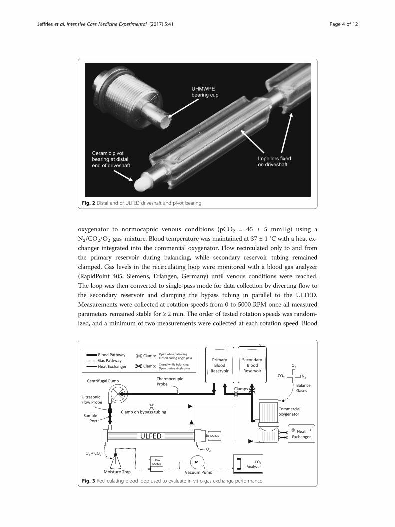
oxygenator to normocapnic venous conditions (pCO2 = 45 ± 5 mmHg) using a
N2/CO2/O2 gas mixture. Blood temperature was maintained at 37 ± 1 °C with a heat ex-
changer integrated into the commercial oxygenator. Flow recirculated only to and from
the primary reservoir during balancing, while secondary reservoir tubing remained
clamped. Gas levels in the recirculating loop were monitored with a blood gas analyzer
(RapidPoint 405; Siemens, Erlangen, Germany) until venous conditions were reached.
The loop was then converted to single-pass mode for data collection by diverting flow to
the secondary reservoir and clamping the bypass tubing in parallel to the ULFED.
Measurements were collected at rotation speeds from 0 to 5000 RPM once all measured
parameters remained stable for ≥ 2 min. The order of tested rotation speeds was random-
ized, and a minimum of two measurements were collected at each rotation speed. Blood
Fig. 2 Distal end of ULFED driveshaft and pivot bearing
Fig. 3 Recirculating blood loop used to evaluate in vitro gas exchange performance
Jeffries et al. Intensive Care Medicine Experimental (2017) 5:41 Page 4 of 12

flow rate was continuously monitored with an ultrasonic flow probe (Transonic Systems,
Ithaca, NY).
Pure O2 sweep gas was pulled through fibers counter-current to blood flow at 8.0 L/min
by a sealed vacuum pump (N811 KV.45P; KNF Neuberger, Trenton, NJ) and was regu-
lated with a thermal mass flow controller (GR-116-1-A-PV-O2; Fathom Technologies,
Georgetown, TX). The fraction of CO2 in outlet sweep gas FCO2ð Þ was measured by a
gaseous CO2 analyzer (WMA-4; PP Systems, Amesbury, MA) and was used to calculate
total CO2 removal (VCO2 ) together with the STP-corrected sweep gas flow rate QSTPOUT
� �
according to Eq. 1.
VCO2 ¼ QSTPOUTFCO2 ð1Þ
VCO2 was normalized to our target inlet pCO2 of 45 mmHg to reduce variability in
measurements associated with small fluctuations in gas inlet conditions (± 5 mmHg)
according to Eq. 2.
V �CO2
¼ VCO2 �45mmHg
pCOINLET2
ð2Þ
pCOINLET2 was measured from a fresh blood sample immediately prior to each data
point. Three identical ULFED prototypes were fabricated for repeatability testing, but
gas pathway failure in one device limited gas exchange testing to two devices. Total
ULFED gas exchange is reported as average and standard deviation of the CO2 removal
rates of both prototypes at each rotation speed.
In vitro hemolysis testing
Filtered and heparinized bovine blood (20 U/mL) was collected fresh from the slaughter-
house the day of testing per ASTM standards (F1841–97) [27]. The gas exchange loop
was modified for hemolysis testing by removing the bypass tubing parallel to the ULFED,
the commercial oxygenator, the secondary reservoir, and the ULFED gas pathway compo-
nents. The ULFED was evaluated in two circuits so that overall hemolysis reflected that of
clinical setups. A reasonable cannula for ECCO2R at the target 250 mL/min of blood flow
(13 Fr Avalon Elite DLC 10013; Maquet, Rastatt, Germany) and a pediatric centrifugal
pump (PediMag; Thoratec, Pleasanton, CA) were selected for the ULFED “standard
circuit”. Blood (1000 mL) was continuously recirculated for 3 h. The reservoir was sub-
merged in a heated water bath to maintain a circuit temperature of 37° ± 1 °C. ULFED
rotation was set to the minimum speed necessary where CO2 removal did not differ
significantly from the maximum rate achieved. The second ULFED circuit (“dialysis con-
figuration”) evaluated performance using a hemodialysis controller roller pump (Prisma;
Baxter, Deerfield, IL) and cannula. A larger bore 14-Fr, 15-cm dialysis cannula
(AK-22142-F; Teleflex, Morrisville, NC) was used in the second circuit due to
availability of parts recommended for the target blood flows.
A control circuit (“Minimax”) was tested to evaluate ULFED hemolysis against an ap-
proved low-flow blood oxygenator (Minimax Plus; Medtronic, Minneapolis, MN). Blood
flow in the control loop was maintained at the minimum rate necessary (1250 mL/min)
to match ULFED CO2 removal performance according to the manufacturer [28]. Pump
(BP-50; Medtronic, Minneapolis, MN) rotation speed in the loop was maintained at
2100–2200 RPM against 180 mmHg to simulate inclusion of cannula recommended for
Jeffries et al. Intensive Care Medicine Experimental (2017) 5:41 Page 5 of 12

use at the target blood flows (14 Fr Biomedicus 96820-014 venous, 12 Fr Biomedicus
96820-012 arterial) [29, 30]. Pressure against the pump was adjusted using a Hoffman
clamp on ULFED outlet tubing and was continuously monitored with a differential fluid
pressure transducer (PX771-025DI; Omega Engineering, Inc., Stamford, CT) across the
pump. All other components and conditions were consistent between circuits. All three
ULFED prototypes fabricated for gas exchange testing were evaluated for hemolysis in
both circuit configurations, as the gas pathway failure observed in one prototype did not
interfere with hemolysis testing.
Samples were drawn every 30 min to measure hematocrit (HCT) and plasma-free
hemoglobin (pfHb). Plasma was isolated from whole blood in two centrifuge spins
(15 min at 0.8g, 10 min at 7.2g), and absorbance at 540 nm was measured spectrophoto-
metrically (Genesys 10S UV-Vis; Thermo Scientific, Waltham, MA). PfHb concentration
was calculated from absorbance using a standard curve developed from a linear-fit of
serially diluted whole blood with 100% hemolysis versus absorbance [31].
The normalized index of hemolysis (NIH) was calculated for circuit comparisons:
NIH g=100Lð Þ ¼ ΔpfHb� V � 100−HCT100
� 100Δt � Q
ð3Þ
Where NIH = normalized index of hemolysis in grams of hemoglobin released into
the blood per 100 L of flow through the circuit (g/100 L); ΔpfHb = increase in pfHb
over the sampling time interval (g/L); V = circuit volume (L); HCT = hematocrit (%);
Δt = sampling time interval (min); Q = average blood flow rate (L/min). A time-of-
therapy normalized index was also calculated, since the NIH equation does not reflect
total hemolysis returned to a patient in the context of treatment duration. Flow rate
normalization in the NIH equation is eliminated in the new therapeutic index of
hemolysis (TIH) calculation to indicate the total grams of hemoglobin released to the
blood per 100 min of therapy (g/100 min):
TIH g=100minð Þ ¼ ΔpfHb� V � 100−HCT100
� 100Δt
ð4Þ
Statistics
All statistical comparisons were conducted in SPSS (IBM, Armonk, NY). A one-way
ANOVA with Tukey HSD post hoc testing was used to compare removal rates at each
RPM after data satisfied assumptions of homogeneity of variance, normality, and inde-
pendence. Comparisons were used to identify the minimum speed necessary to achieve
statistically equivalent performance to the maximum CO2 removal rate. The determined
rotation speed was used for subsequent hemolysis testing. Mean NIH values were com-
pared using a one-way ANOVA with Tukey HSD post hoc test after satisfying relevant as-
sumptions. TIH data violated the assumption of homogeneity of variance via Levene’s
test, and means were compared with Welch’s F test. Subsequent Games-Howell post hoc
tests were used for between-group comparisons of means. All comparisons of means were
considered significant at the level p < 0.05.
Jeffries et al. Intensive Care Medicine Experimental (2017) 5:41 Page 6 of 12

ResultsIn vitro gas exchange
Figure 4 shows the raw and normalized CO2 removal rates of the ultra-low-flow CO2
removal device (ULFED) as a function of impeller rotational speed. A sharp increase in
CO2 removal occurred between 0 and 2000 RPM before subsequently leveling off at
higher rotation speeds. A maximum normalized CO2 removal rate of 75.1 ± 1.1 mL/min
was achieved at 5000 RPM. Normalized performance at 4000 RPM did not differ signifi-
cantly from the maximum rate however (74.1 ± 3.3 mL/min, p = 0.99).
In vitro hemolysis
Measured rates of pfHb accumulation were highly linear over testing periods as shown in
Fig. 5 (ΔpfHb versus elapsed time R2 > 0.95 in all tests). Table 1 shows the calculated
hemolysis indices for the ULFED (at 4000 PRM) and the control device. NIH values for
the standard-ULFED circuit (0.78 ± 0.19 g/100 L), dialysis-ULFED circuit (1.55 ± 0.03 g/
100 L), and the control circuit (0.11 ± 0.01 g/100 L) each differed significantly from one
another (ANOVA p < 0.001, all group-wise comparisons p < 0.001). The TIH value of the
standard-ULFED (0.190 ± 0.041 g/100 min) did not differ significantly from the control
circuit (0.123 ± 0.013 g/100 min; Welch’s test p < 0.001, group-wise p = 0.169). The
hemolysis using dialysis circuit components (0.386 ± 0.010 g/100 min) was significantly
greater than both other test groups (each p < 0.05). Average hematocrit at each sampling
interval is shown in Fig. 5 (right). Hematocrit decreased by ~ 1.5–2.5% from baseline over
test periods, which is consistent with dilution due to saline infusion.
DiscussionSupplementing respiration by removing CO2 independent of the lungs can improve
outcomes for patients at risk of requiring or already receiving invasive mechanical
ventilation. We developed the ultra-low-flow ECCO2R device (ULFED) to operate
at blood flow rates consistent with renal hemodialysis to simplify circuit manage-
ment and minimize invasiveness of CO2 removal. In vitro CO2 removal rates up to
74 mL/min at 4000 RPM were achieved by the ULFED with minimal cell trauma
Fig. 4 Raw (VCO2 ) and normalized (V�CO2
) CO2 removal rates versus rotation speed. Error bars represent onestandard deviation of measured removal rates
Jeffries et al. Intensive Care Medicine Experimental (2017) 5:41 Page 7 of 12

(therapeutic index of hemolysis, TIH = 0.19 g/100 min) at blood flows consistent
with dialysis (250 mL/min).
CO2 removal systems used in conjunction with non-invasive or protective ventilation
strategies have been shown to correct pCO2 and pH in hypercapnic patients with
removal rates equivalent to ~ 25–35% of the metabolic CO2 production (~ 200–
250 mL/min) [22, 23]. CO2 removal at these levels prevented intubation in patients
with ae-COPD failing or unresponsive to non-invasive ventilation [14, 16, 17, 32].
Partial respiratory assistance has also aided weaning from ventilation [14, 33, 34] and allows
reduction of ventilator tidal volumes to ultra-protective levels (3–4 mL/kg) [21, 35, 36]. The
ULFED exceeded these CO2 removal rates by eliminating ~ 30–37% of the metabolic CO2
production at normocapnic test conditions (inlet pCO2 = 45 mmHg). Gas exchange will
also increase proportionally with pCO2 in hypercapnic patients, where CO2 removal up to
50% or more of metabolic production can be required.
Efficient gas exchange in the ULFED minimizes necessary fiber surface area and enables
clinically significant CO2 removal rates at hemodialysis blood flows. Pump-less arterioven-
ous CO2 removal (AVCO2R) requires dual cannulation (13–19 Fr) for circuit flows of
600–2000 mL/min that is shunted between the femoral artery and vein through a 1.3-m2
oxygenator [36–38]. A newer integrated pump-oxygenator system uses a rotating core to
generate active mixing to improve gas exchange up to 60% with a 0.59-m2 bundle [39].
Comparatively lower flows (350–500 mL/min) are possible in the simplified veno-venous
circuit, but CO2 removal decreases with blood flow (~ 50 mL/min at 300 mL/min blood
flow) and connection requires 15.5-Fr cannulation [4, 39]. Developing systems
combine existing oxygenators with dialysis controllers targeting even lower flows
(200–300 mL/min) to minimize cannulation invasiveness (≤ 14 Fr). These systems
utilize larger surface area gas exchangers (≥ 1 m2) to improve performance [32, 40] or tar-
get lower CO2 removal using smaller pediatric oxygenators (40–55 mL/min with a 0.3-m2
Fig. 5 Average change in total plasma free hemoglobin (pfHb) versus baseline (left) and average hematocrit(right) at 30 min sampling intervals during in vitro hemolysis tests. Error bars represent one standard deviationat each time-point between tests for each circuit
Table 1 Summary of in vitro hemolysis testing
Test device Blood flow TIH NIH
(mL/min) (g/100 min) (g/100 L)
ULFED (standard circuit) 250 0.190 ±0.041 0.775 ±0.186*
ULFED (dialysis configuration) 250 0.386 ±0.010* 1.551 ±0.025*
Minimax 1250 0.123 ±0.013 0.105 ±0.012*
*Significant at p < 0.05 versus all other devices. TIH therapeutic index of hemolysis. NIH normalized index of hemolysis
Jeffries et al. Intensive Care Medicine Experimental (2017) 5:41 Page 8 of 12

bundle in pigs with PaCO2 > 80 mmHg) [41]. Approaches to enhance CO2 removal such
as bicarbonate dialysis [42], blood acidification [43], electrodialysis [44], plasma recircula-
tion [45], and fiber enzyme coatings [46] are also being explored to reduce necessary
blood flows for treatment.
The rotating impellers in the ULFED enhance gas transfer by generating an “active
mixing” effect in the fiber bundle that improves convective mixing at gas exchange
surfaces [24, 25, 47, 48]. Computational simulations have indicated development of
continuously recirculating flow pathways in/out of the fiber bundle with impeller mix-
ing [25]. Blood is pumped radially outward through the bundle by impellers, then
pulled back into the bundle toward low-pressure regions in the gaps between impellers
before converging onto the impeller blade and cycling through the bundle again. In-
creasing flow velocity past gas exchange surfaces is a well-established mechanism for
improving transfer efficiency by diminishing the thickness of the surface diffusive
boundary layer [49]. This facilitates replenishment of gases to the membrane surface to
maximize the concentration gradients spanning fiber walls. Recirculating flow also
maintains a high level of washing in the bundle that eliminates regions of stagnation
where thrombus formation may otherwise occur at low blood flows.
The measured rate of hemolysis in the standard-ULFED circuit was comparable to a
clinically approved oxygenator circuit. Two indices of red cell trauma are reported here
that indicate the rate of pfHb accumulation over time, the key difference being how
time is reported. A major limitation of the NIH calculation is that hemolysis is normal-
ized for blood flow rate, but operating flow rate is ultimately irrelevant. Two systems
intended for use at 5000 mL/min versus 250 mL/min that cause equivalent rates of
total cell damage would differ in NIH by a factor of 20, despite returning an equal
number of pfHb species to a patient. As a result the NIH calculation is bias against
low-flow devices. The TIH calculation removes the flow rate normalization and pro-
vides a clinically relevant time-of-therapy rate of hemolysis. The limitation of both indi-
ces however is that no reliable benchmark threshold values have been validated for
low-flow devices against in vivo performance to our knowledge. More information or
in vivo testing is therefore necessary to make conclusions regarding acceptability of the
dialysis-ULFED performance. No difference in hemolysis was observed between the
control and standard-ULFED circuits, so we expect in vivo hemolysis to be acceptable
in this configuration.
Anticoagulation of saline infused to the ULFED may have clinical implications with
extended use. Heparin levels in the saline infusion line were chosen primarily to avoid di-
lution of circuit anticoagulation for consistency between tests. Approved blood pumping
devices utilizing saline-lubricated seals anticoagulate infusion lines at higher rates [50],
while others do not require anticoagulation [51]. Elimination or minimization of local
anticoagulation from the ULFED will be investigated in future prototypes utilizing a saline
infusion line.
ConclusionsEvidence continues to grow that ECCO2R can effectively prevent intubation, facilitate
earlier extubation, or allow reduction of ventilator settings in hypercapnic respiratory fail-
ure. The ULFED eliminates clinically significant levels of CO2 from blood with acceptable
hemolysis at hemodialysis blood flows, making minimally invasive dialysis connection
Jeffries et al. Intensive Care Medicine Experimental (2017) 5:41 Page 9 of 12
strategies and simplified management possible for ECCO2R. Future work may focus on in
vivo validation of benchtop performance or improvements to the ULFED aimed at simpli-
fying the design, such as sealing the blood compartment with a magnetically coupled
driveshaft that would obviate the driveshaft seal and saline infusion.
AbbreviationsECCO2R: Extracorporeal carbon dioxide removal; ECMO: Extracorporeal membrane oxygenation; NIH: Normalized index ofhemolysis; pfHb: Plasma-free hemoglobin; TIH: Therapeutic index of hemolysis; ULFED: Ultra-low-flow ECCO2R device
AcknowledgementsThis work was supported by the grants 5R01HL070051-08 and 5R01HL117637-05 from the National Institutes of Health,and the National Heart, Lung, and Blood Institute. Its contents are solely the responsibility of the authors anddo not necessarily represent the official views of the NIH or NHLB. This work was also supported by theUniversity of Pittsburgh’s McGowan Institute for Regenerative Medicine. Funding for R. Garrett Jeffries was partially providedby an NIH training grant (T32-HL076124) for the University of Pittsburgh Cardiovascular Bioengineering TrainingProgram (CBTP).
FundingRJ, BF, and WF received research support from the NIH and NHLBI (5R01HL070051-08 and 5R01HL117637-05), and theMcGowan Institute for Regenerative Medicine. RJ was partially funded by an NIH training grant (T32-HL076124).
Availability of data and materialsAll relevant data is presented within the text to support findings. No additional data was submitted with the article.
Authors’ contributionsRJ, BF, and WF contributed to the conception and design of the study and analysis and interpretation of data. RJ, LL,and WF contributed to the drafting the manuscript for important intellectual content. All authors read and approvedthe final manuscript.
Ethics approval and consent to participateNot applicable.
Consent for publicationNot applicable.
Competing interestsWF is an equity holder and Head of the Scientific Advisory Board, for which he receives compensation, at ALungTechnologies, which is commercializing an artificial lung device independent of the device described in this article. LLis a full-time employee at ALung Technologies. RJ and BF have no relevant competing interests to report.
Publisher’s NoteSpringer Nature remains neutral with regard to jurisdictional claims in published maps and institutional affiliations.
Author details1Department of Bioengineering, University of Pittsburgh, Pittsburgh, PA, USA. 2McGowan Institute for RegenerativeMedicine, University of Pittsburgh, 3025 E Carson St, Suite 226, Pittsburgh, PA 15203, USA. 3ALung Technologies, Inc.,2500 Jane Street, Suite 1, Pittsburgh, PA 15203, USA. 4Department of Chemical Engineering, University of Pittsburgh,Pittsburgh, PA, USA. 5Department of Critical Care Medicine, University of Pittsburgh Medical Center, Pittsburgh, PA,USA.
Received: 8 May 2017 Accepted: 21 August 2017
References1. Ricard J-D, Dreyfuss D, Saumon G (2003) Ventilator-induced lung injury. Eur Respir J 22:2s–9s. https://doi.org/10.
1183/09031936.03.004201032. Peek GJ, Mugford M, Tiruvoipati R et al (2009) Efficacy and economic assessment of conventional ventilatory
support versus extracorporeal membrane oxygenation for severe adult respiratory failure (CESAR): a multicentrerandomised controlled trial. Lancet 374:1351–1363. https://doi.org/10.1016/S0140-6736(09)61069-2
3. Terragni PP, Maiolo G, Tenaglia T et al (2011) Extracorporeal CO2 removal and O2 transfer: a review of theconcept, improvements and future development. Trends Anaesth Crit Care 1:123–127. https://doi.org/10.1016/j.tacc.2011.03.002
4. Lund LW, Federspiel WJ (2013) Removing extra CO2 in COPD patients. Curr Respir Care Rep 2:131–138. https://doi.org/10.1007/s13665-013-0057-x
5. Schmidt M, Tachon G, Devilliers C et al (2013) Blood oxygenation and decarboxylation determinants duringvenovenous ECMO for respiratory failure in adults. Intensive Care Med 39:838–846. https://doi.org/10.1007/s00134-012-2785-8
Jeffries et al. Intensive Care Medicine Experimental (2017) 5:41 Page 10 of 12

6. Oliver WC (2009) Anticoagulation and coagulation management for ECMO. Semin Cardiothorac Vasc Anesth 13:154–175. https://doi.org/10.1177/1089253209347384
7. MacLaren G, Combes A, Bartlett R (2012) Contemporary extracorporeal membrane oxygenation for adult respiratoryfailure: life support in the new era. Intensive Care Med 38:210–220. https://doi.org/10.1007/s00134-011-2439-2
8. Silverthorn DU (2007) Human physiology: an integrated approach, 4th edn. Pearson Education, Inc., San Francisco9. Kolobow T, Gattinoni L, Tomlinson T et al (1977) The carbon dioxide membrane lung (CDML): a new concept.
ASAIO J 23:17–2110. Loeppky JA, Luft UC, Fletcher ER (1983) Quantitative description of whole blood CO2 dissociation curve and
Haldane effect. Respir Physiol 51:167–181. https://doi.org/10.1016/0034-5687(83)90038-511. Cove M, MacLaren G, Federspiel W, Kellum J (2012) Bench to bedside review: extracorporeal carbon dioxide
removal, past present and future. Crit Care 16:232–230. https://doi.org/10.1186/cc1135612. Brower RG, Matthay MA, Morris A et al (2000) Ventilation with lower tidal volumes as compared with traditional
tidal volumes for acute lung injury and the acute respiratory distress syndrome. N Engl J Med 342:1301–130813. Lightowler JV, Wedzicha JA, Elliott MW, Ram FSF (2003) Non-invasive positive pressure ventilation to treat
respiratory failure resulting from exacerbations of chronic obstructive pulmonary disease: Cochrane systematicreview and meta-analysis. BMJ 326:185
14. Burki NK, Mani RK, Schmidt W et al (2013) A novel extracorporeal CO2 removal system: results of a pilot study inCOPD patients with hypercapnic respiratory failure. Chest 143:678–686. https://doi.org/10.1378/chest.12-0228
15. Bonin F, Sommerwerck U, Lund LW, Teschler H (2013) Avoidance of intubation during acute exacerbation ofchronic obstructive pulmonary disease for a lung transplant candidate using extracorporeal carbon dioxideremoval with the Hemolung. J Thorac Cardiovasc Surg 145:e43–e44. https://doi.org/10.1016/j.jtcvs.2013.01.040
16. Kluge S, Braune SA, Engel M et al (2012) Avoiding invasive mechanical ventilation by extracorporeal carbondioxide removal in patients failing noninvasive ventilation. Intensive Care Med 38:1632–1639. https://doi.org/10.1007/s00134-012-2649-2
17. Braune S, Sieweke A, Brettner F et al (2016) The feasibility and safety of extracorporeal carbon dioxide removal toavoid intubation in patients with COPD unresponsive to noninvasive ventilation for acute hypercapnic respiratoryfailure (ECLAIR study): multicentre case-control study. Intensive Care Med 42(9):1437–1444. https://doi.org/10.1007/s00134-016-4452-y
18. Mani RK, Schmidt W, Lund LW, Herth FJF (2013) Respiratory dialysis for avoidance of intubation in acuteexacerbation of COPD. J Novemb 59:675–678. https://doi.org/10.1097/MAT.0000000000000004
19. Cole S, Barrett N, Glover G et al (2014) Extracorporeal carbon dioxide removal as an alternative to endotrachealintubation for non-invasive ventilation failure in acute exacerbation of COPD. J Intensive Care Soc 15:1–3
20. Terragni PP, Del Sorbo L, Mascia L et al (2009) Tidal volume lower than 6 ml/kg enhances lung protection: role ofextracorporeal carbon dioxide removal. Anesthesiology 111:826–835. https://doi.org/10.1097/ALN.0b013e3181b764d2
21. Fanelli V, Ranieri MV, Mancebo J et al (2016) Feasibility and safety of low-flow extracorporeal carbon dioxideremoval to facilitate ultra-protective ventilation in patients with moderate acute respiratory distress sindrome. CritCare 20:36. https://doi.org/10.1186/s13054-016-1211-y
22. Trahanas JM, Lynch WR, Bartlett RH (2016) Extracorporeal support for chronic obstructive pulmonarydisease: a bright future. J Intensive Care Med. https://doi.org/10.1177/0885066616663119
23. Morelli A, Del Sorbo L, Pesenti A et al (2017) Extracorporeal carbon dioxide removal (ECCO2R) in patients withacute respiratory failure. Intensive Care Med. https://doi.org/10.1007/s00134-016-4673-0
24. Mihelc KM, Frankowski BJ, Lieber SC et al (2009) Evaluation of a respiratory assist catheter that uses an impellerwithin a hollow fiber membrane bundle. ASAIO J 55:569–574
25. Jeffries RG, Frankowski BJ, Burgreen GW, Federspiel WJ (2014) Effect of impeller design and spacing on gas exchangein a Percutaneous respiratory assist catheter. Artif Organs 38:1007–1017. https://doi.org/10.1111/aor.12308
26. (2009) ANSI/AAMI/ISO 7199:2009 – Cardiovascular implants and artificial organs—blood-gas exchangers (oxygenators)27. ASTM F1841–97 (2005). Standard practice for assessment of hemolysis in continuous flow blood pumps28. (2008) Medtronic Minimax Plus, Instructions For Use: M932349A001 Rev. 1.029. Svitek RG, Smith DE, Magovern JA (2007) In vitro evaluation of the TandemHeart pediatric centrifugal pump.
ASAIO J 53:747–753. https://doi.org/10.1097/MAT.0b013e318154ca7430. Paulsen MJ, Orizondo R, Le D et al (2013) A simple, standard method to characterize pressure/flow performance
of vascular access Cannulas. ASAIO J 59:24–29. https://doi.org/10.1097/MAT.0b013e318274640131. Dobrovolskaia MA, Clogston JD, Neun BW et al (2008) Method for analysis of Nanoparticle hemolytic properties in
vitro. Nano Lett 8:2180–2187. https://doi.org/10.1021/nl080561532. Del Sorbo L, Pisani L, Filippini C et al (2015) Extracorporeal CO2 removal in hypercapnic patients at risk of
noninvasive ventilation failure: a matched cohort study with historical control. Crit Care Med 43:120–127. https://doi.org/10.1097/CCM.0000000000000607
33. Abrams DC, Brenner K, Burkart KM et al (2013) Pilot study of extracorporeal carbon dioxide removal to facilitateextubation and ambulation in exacerbations of chronic obstructive pulmonary disease. Ann Am Thorac Soc 10:307–314. https://doi.org/10.1513/AnnalsATS.201301-021OC
34. Hermann A, Staudinger T, Bojic A et al (2014) First experience with a new miniaturized pump-driven venovenousextracorporeal CO2 removal system (iLA Activve): a retrospective data analysis. ASAIO J 60:342–347. https://doi.org/10.1097/MAT.0000000000000073
35. Moss CE, Galtrey EJ, Camporota L, et al (2016) A retrospective observational case series of low flow veno-venousextracorporeal carbon dioxide removal use in patients with respiratory failure: ASAIO J 1. doi: https://doi.org/10.1097/MAT.0000000000000386
36. Bein T, Weber-Carstens S, Goldmann A et al (2013) Lower tidal volume strategy (≈3 ml/kg) combined withextracorporeal CO2 removal versus “conventional” protective ventilation (6 ml/kg) in severe ARDS. Intensive CareMed 39:847–856. https://doi.org/10.1007/s00134-012-2787-6
37. Zimmermann M, Bein T, Arlt M et al (2009) Pumpless extracorporeal interventional lung assist in patients withacute respiratory distress syndrome: a prospective pilot study. Crit Care 13:R10
Jeffries et al. Intensive Care Medicine Experimental (2017) 5:41 Page 11 of 12

38. Bein T, Weber F, Philipp A et al (2006) A new pumpless extracorporeal interventional lung assist in criticalhypoxemia/hypercapnia. Crit Care Med 34:1372–1377. https://doi.org/10.1097/01.CCM.0000215111.85483.BD
39. Jeffries RG (2014) In vitro and seven-day chronic in vivo evaluation of the hemolung adult CO2 removal systemfor pediatric respiratory support
40. Karagiannidis C, Kampe KA, Sipmann FS et al (2014) Veno-venous extracorporeal CO2 removal for the treatmentof severe respiratory acidosis: pathophysiological and technical considerations. Crit Care 18:R124. https://doi.org/10.1186/cc13928
41. Godet T, Combes A, Zogheib E et al (2015) Novel CO2 removal device driven by a renal-replacement systemwithout hemofilter. A first step experimental validation. Anaesth Crit Care Pain Med 34:135–140. https://doi.org/10.1016/j.accpm.2014.08.006
42. Cressoni M, Zanella A, Epp M et al (2009) Decreasing pulmonary ventilation through bicarbonate ultrafiltration: anexperimental study. Crit Care Med 37:2612–2618. https://doi.org/10.1097/CCM.0b013e3181a5668a
43. Zanella A, Mangili P, Redaelli S et al (2014) Regional blood acidification enhances extracorporeal carbon dioxideremoval: a 48-hour animal study. Anesthesiol Febr 120:416–424. https://doi.org/10.1097/ALN.0000000000000099
44. Zanella A, Castagna L, Salerno D et al (2015) Respiratory electrodialysis: a novel, highly efficient, extracorporealCO2 removal technique. Am J Respir Crit Care Med 192:719–726. https://doi.org/10.1164/rccm.201502-0289OC
45. Gramaticopolo S, Chronopoulos A, Piccinni P et al (2010) Extracorporeal CO2 removal—a way to achieveultraprotective mechanical ventilation and lung support: the missing piece of multiple organ support therapy.Contrib Nephrol 165:174–184. https://doi.org/10.1159/000313757.
46. Arazawa DT, Kimmel JD, Federspiel WJ (2015) Kinetics of CO2 exchange with carbonic anhydrase immobilized onfiber membranes in artificial lungs. J Mater Sci Mater Med 26:5525. https://doi.org/10.1007/s10856-015-5525-0
47. Hewitt TJ, Hattler BG, Federspiel WJ (1998) A mathematical model of gas exchange in an intravenous membraneoxygenator. Ann Biomed Eng 26:166–178
48. Makarewicz AJ, Mockros LF, Anderson RW (1993) A pumping intravascular artificial lung with active mixing. ASAIOJ 39:M466–M469
49. Wickramasinghe SR, Semmens MJ, Cussler EL (1992) Mass transfer in various hollow fiber geometries. J Membr Sci69:235–250. https://doi.org/10.1016/0376-7388(92)80042-I
50. Lee Y, Weeks PA (2015) Effectiveness of protocol guided heparin anticoagulation in patients with theTandemHeart percutaneous ventricular assist device. ASAIO J Am Soc Artif Intern Organs 61:207–208. https://doi.org/10.1097/MAT.0000000000000176
51. Jeffries RG, Mussin Y, Bulanin DS et al (2014) Pre-clinical evaluation of an adult extracorporeal carbon dioxideremoval system with active mixing for pediatric respiratory support. Int J Artif Organs 37:888–899. https://doi.org/10.5301/ijao.5000372
Jeffries et al. Intensive Care Medicine Experimental (2017) 5:41 Page 12 of 12