An Essential Role for the Plasmodium Nek-2 Nima-Related
11
An Essential Ro le for the Plasmodium Nek- 2 Nima-r elated Protein Kinase in the Sexual Development of Malaria Parasites * Receiv ed forpubl ication,Februar y 17,2009, andin revised for m, May8, 2009 Publ ishe d, JBC Pape rs in Press, June2, 2009, DOI 10.1 074/ jbc. M109 .017 988 Luc Rein inge r ‡1 , Ri ta Tewari §¶1,2 , Cla re Fen nel l ‡3 , Zoe Holl and ‡3 , Dean Gol dr ing , Lisa Ranf ord-C artwr ight**, Oliv er Billker ¶‡‡ , and Chr istianDoerig ‡§§4 From the ‡ INSER M U609-We llcome Centre for Molecu lar Paras itolog y, Biome dical Resea rch Centr e, and **Divisio n of Infe ction and Immuni ty, Fac ult y of Biomed ical and Lif e Sci enc es, Uni ver sit y of Glasgow, 120 Uni ver sit y Pla ce, Glas gow G12 8TA, Scotl and, Unite d Kingd om, the § Ins tit ute of Gen eti cs, Sch ool of Biol ogy , Uni ver sit y of Not tingha m, Not tingha m NG7 2UH, Uni ted Kingdo m, the Depart ment of Bioche mistry , Scho ol of Bioche mistry , Genet ics Microbi ology and Plant Patho logy, Unive rsity of Kwa Zul u-Nata l, Scotts vill e 3209 , Sou th Afr ica, the ‡‡ Wel lcome Trust San ger Ins tit ute , Hin xto n, Cambrid ge CB1 0 1SA, Uni ted Kingd om, the ¶ Div isio n of Cel l and Mole cul ar Biology, Imp eri al Col lege Lon don , Lon don SW7 2AZ, Uni ted Kingdo m, and §§ INSERM U609, Global Healt h Insti tute, Ecole Polyt echn ique Fe ´de ´rale de Lausa nne (EPFL ), Stati on 19, CH-1015 Lausann e, Switz erlan d The molecular control of cell division and development in malaria par asi tes is far fro m und ers too d. We pre vio usl y showed that a Plasmodium gametocyte-sp ecific NIMA-related protein kinase, nek-4, is required for completion of meiosis in the ooki- nete, the motile form that develops from the zygote in the mos- quito vector. Here, we show that another NIMA-related kinase, Pfnek-2, is also predominantly expressed in gametocytes, and tha t Pfnek-2 is an act ive enz yme dis pla yin g an in vitro substrate preference distinct from that of Pfnek-4. A functional nek-2 gene is required for transmission of both Plasmodium falcipa- rum and the rodentmalaria par asi te Plasmodium berghei to the mosquitovecto r, which is exp lai ned by the obser vat ion that dis- ruption of the nek-2 gene in P. berghei causes dysregulation of DNA replication during meiosis and blocks ookinete develop- ment. This has implications (i) in our understanding of sexual development of malaria parasites and (ii) in the context of con- trol strategies aimed at interfering with malaria transmission. Malari a, cause d by infection with intracellu lar protoz oan parasites of the genus Plasmodium, is a major public health proble m in the developin g world (1). The species responsi ble for the vast majority of lethal cases is Plasmodium falciparum. The life cycle of malaria parasites consists of a succession of develo pment al stage s: asexual multip lication occurs in the human host (first in a single round of schizogony in a hepato- cyte infected by a sporozoite injected by the mosquito vector, and the n mul tiple roun ds of schizogony in ery thr ocy tes ), whereas the sexual cycle is initiated by the formation of cell cycle-arrested gametocytes in infected erythrocytes and pro- ceeds, in the midgut of the mosquito vector, to gametogenesis, fertilization, and formation of a motile ookinete. The ookinete crosses the midgut epithelium and establishes an oocyst at the basal lamina, in which sporogony occurs, generating sporozo- ites tha t renderthe vec tor inf ect ious once the y reach its sal iva ry glands. The alternation of proliferative and non-proliferative phases implies that the control of cell cycle progression is of pri me import anc e for comple tio n of the life cyc le of the parasite. The NIMA-related protein kinases (Neks) 5 constitute an exten ded family of eukary otic mitot ic serine /threo nine kinase s. The best characterized members of the Nek family include NIMA (never in mitosis/ Aspergillus), the founding member from the fungus Aspergillus nidulans (2), and its closest homo- logue in mammals, Nek2 (3, 4). Initially identified as a kinase essential for mitotic entry in Aspergillus, NIMA has been also shown to participate in nuclear membrane fission (5). Eleven members of the NI MA ki nase family (Nek1–11) ha ve now been identified in various human tissues, and together fulfill a num- ber of cell cycle-related functions in centrosome separation, mitosis, meiosis, and checkpoint control (reviewed in Ref. 6). It has been proposed that expansion of the Nek family accompa- nied the evolution of a complex system for the coordination of progression through the cell cycle with the replication of cellu- lar components such as cilia, basal bodies, and centrioles. Sev- * Work at the Plasmodium Genome Consortium was supported by the Bur- roughs Wellcome Fund, the Wellcome Trust, the National Institutes of Health (NIAID), and the U.S. Department of Defense, Military Infectious DiseasesResearch Prog ram. Finan cial suppo rt for PlasmoDB was prov ided by theBurro ugh s Wel lco me Fun d. Wor k in thelabor ato ry of C. D. wassup- ported by the Frenc h Instit ut National de la Sante ´ et de la Recherche Me ´di- cale (Inserm), FP6 (SIGMAL and ANTIMAL projects, BioMalPar Network of Exc ell ence) andFP7 (MALSIG pro jec t) pro gra ms of theEurop eanCommis- sion, andby a gra nt fro m theNova rti s Instit utefor Tro pic al Dis eas es (NI TD, Singa pore ). The workon P. berg hei wassupport edby gra ntsfromthe Wel l- come Trust (to R. T. and O.B.) and the Medical Research Council (to O. B.). Author’s Choice—Fina l vers ion full acce ss. 1 Both authors contributed equally to this work. 2 To whom correspondence may be addressed: Institute of Genetics, School of Biology, University of Nottingham, Nottingham NG7 2UH, United King- dom. Tel.: 44-115 -8230362; Fax: 44-115 -8230338; E-ma il: rita. tewa ri@ nottingham.ac.uk. 3 Recipients of Ph.D. studentships awarded by the Wellcome Trust. 4 To whomcorresp onde nce may be add resse d: INSER M U609,Wellcome Cent re for MolecularParasitology, Global Health Institute, Ecole Polytechniq ue Fe ´ de ´ - rale de Lausanne (EPFL), Station 19, CH-1015 Lausanne, Switzerland. Tel.: 41-21-693-0983; Fax: 41-21-693-9538; E-mail: [email protected]. 5 The abbreviations used are: Nek, NIMA-related protein kinases; RT, reverse tran scrip tase;MAPK,mitogen- activ atedproteinkinase;GFP, gree n fluor es- cent protein; RT, reverse transcriptase; DHFR, dihydrofolate reductase; TRITC, tetramethylrhodamin e isothiocyanate; UTR, untranslated region; GST, glutathione S-transferase; MBP, myelin basic protein. THE JOURNAL OF BIOLOGICAL CHEMISTRY VOL. 284, NO. 31, pp. 2 0858 –20868, July 31, 2009 Author’s Choice © 2009 by The American Society for Bioc hemistry and Mole cular Biology , Inc. Printed in the U.S.A . 20858 JOURNAL OF BIOLOGICAL CHEMISTRY VOLUME 284• NUMB ER 31• JULY 31, 2009 b y g u e s t , o n S e p t e b e r 5 , 2 0 1 1 w w w j b c . o r g D o w l o a d e d f r o
-
Upload
rebecca-potter -
Category
Documents
-
view
219 -
download
0
Transcript of An Essential Role for the Plasmodium Nek-2 Nima-Related

8/2/2019 An Essential Role for the Plasmodium Nek-2 Nima-Related
http://slidepdf.com/reader/full/an-essential-role-for-the-plasmodium-nek-2-nima-related 1/11
An Essential Role for the PlasmodiumNek-2 Nima-relatedProtein Kinase in the Sexual Development of Malaria Parasites*Received forpublication,February 17,2009, andin revised form, May8, 2009 Published, JBC Papers in Press, June2, 2009, DOI 10.1074/jbc.M109.017988
Luc Reininger‡1, Rita Tewari§¶1,2, Clare Fennell‡3, Zoe Holland‡3, Dean Goldring, LisaRanford-Cartwright**,Oliver Billker¶‡‡, andChristianDoerig‡§§4
From the‡INSERM U609-Wellcome Centre for Molecular Parasitology, Biomedical Research Centre, and **Division of Infectionand Immunity, Faculty of Biomedical and Life Sciences, University of Glasgow, 120 University Place, Glasgow G12 8TA,Scotland, United Kingdom, the §Institute of Genetics, School of Biology, University of Nottingham, Nottingham NG72UH,United Kingdom, the Department of Biochemistry, School of Biochemistry, Genetics Microbiology and Plant Pathology, University of KwaZulu-Natal, Scottsville 3209, South Africa, the‡‡Wellcome Trust Sanger Institute, Hinxton, Cambridge CB10 1SA, United Kingdom, the ¶Division of Cell and Molecular Biology, Imperial College London, London SW7 2AZ, United Kingdom, and §§INSERMU609, Global Health Institute, Ecole Polytechnique Federale de Lausanne (EPFL), Station 19, CH-1015 Lausanne, Switzerland
The molecular control of cell division and development in
malaria parasites is far from understood. We previously showed
that a Plasmodium gametocyte-specific NIMA-related proteinkinase, nek-4, is required for completion of meiosis in the ooki-
nete, the motile form that develops from the zygote in the mos-
quito vector. Here, we show that another NIMA-related kinase,
Pfnek-2, is also predominantly expressed in gametocytes, and
that Pfnek-2 is an active enzyme displaying an in vitro substrate
preference distinct from that of Pfnek-4. A functional nek-2
gene is required for transmission of both Plasmodium falcipa-
rum and the rodent malaria parasite Plasmodium berghei to the
mosquitovector, which is explained by the observation that dis-
ruption of the nek-2 gene in P. berghei causes dysregulation of
DNA replication during meiosis and blocks ookinete develop-
ment. This has implications (i) in our understanding of sexualdevelopment of malaria parasites and (ii) in the context of con-
trol strategies aimed at interfering with malaria transmission.
Malaria, caused by infection with intracellular protozoanparasites of the genus Plasmodium, is a major public health
problem in the developing world (1). The species responsiblefor the vast majority of lethal cases is Plasmodium falciparum.
The life cycle of malaria parasites consists of a succession of developmental stages: asexual multiplication occurs in thehuman host (first in a single round of schizogony in a hepato-cyte infected by a sporozoite injected by the mosquito vector,and then multiple rounds of schizogony in erythrocytes),
whereas the sexual cycle is initiated by the formation of cellcycle-arrested gametocytes in infected erythrocytes and pro-ceeds, in the midgut of the mosquito vector, to gametogenesis,fertilization, and formation of a motile ookinete. The ookinetecrosses the midgut epithelium and establishes an oocyst at the
basal lamina, in which sporogony occurs, generating sporozo-ites that render the vector infectious once they reach its salivary
glands. The alternation of proliferative and non-proliferativephases implies that the control of cell cycle progression is of prime importance for completion of the life cycle of the
parasite.The NIMA-related protein kinases (Neks)5 constitute an
extended family of eukaryotic mitotic serine/threonine kinases.The best characterized members of the Nek family include
NIMA (never in mitosis/ Aspergillus), the founding memberfrom the fungus Aspergillus nidulans (2), and its closest homo-logue in mammals, Nek2 (3, 4). Initially identified as a kinaseessential for mitotic entry in Aspergillus, NIMA has been alsoshown to participate in nuclear membrane fission (5). Eleven
members of the NIMA kinase family (Nek1–11) have now been
identified in various human tissues, and together fulfill a num-ber of cell cycle-related functions in centrosome separation,mitosis, meiosis, and checkpoint control (reviewed in Ref. 6). Ithas been proposed that expansion of the Nek family accompa-
nied the evolution of a complex system for the coordination of progression through the cell cycle with the replication of cellu-lar components such as cilia, basal bodies, and centrioles. Sev-
* Work at the Plasmodium Genome Consortium was supported by the Bur-roughs Wellcome Fund, the Wellcome Trust, the National Institutes of Health (NIAID), and the U.S. Department of Defense, Military InfectiousDiseases Research Program. Financial support for PlasmoDB was providedby theBurroughs Wellcome Fund. Work in thelaboratory of C. D. wassup-ported by the French Institut National de la Sante et de la Recherche Medi-cale (Inserm), FP6 (SIGMAL and ANTIMAL projects, BioMalPar Network of Excellence) andFP7 (MALSIG project) programs of theEuropeanCommis-sion, andby a grant from theNovartis Institutefor Tropical Diseases (NITD,Singapore). The workon P. berghei wassupportedby grantsfromthe Well-come Trust (to R. T. and O.B.) and the Medical Research Council (to O. B.).
Author’s Choice—Final version full access.1 Both authors contributed equally to this work.2 To whom correspondence may be addressed: Institute of Genetics, School
of Biology, University of Nottingham, Nottingham NG7 2UH, United King-dom. Tel.: 44-115-8230362; Fax: 44-115-8230338; E-mail: [email protected].
3 Recipients of Ph.D. studentships awarded by the Wellcome Trust.4 To whomcorrespondence may be addressed: INSERM U609,Wellcome Centre
for MolecularParasitology, GlobalHealth Institute, Ecole Polytechnique Fede-rale de Lausanne (EPFL), Station 19, CH-1015 Lausanne, Switzerland. Tel.:41-21-693-0983; Fax: 41-21-693-9538; E-mail: [email protected].
5 The abbreviations used are: Nek, NIMA-related protein kinases; RT, reversetranscriptase;MAPK,mitogen-activatedproteinkinase;GFP, green fluores-cent protein; RT, reverse transcriptase; DHFR, dihydrofolate reductase;
TRITC, tetramethylrhodamine isothiocyanate; UTR, untranslated region;GST, glutathione S-transferase; MBP, myelin basic protein.
THE JOURNAL OF BIOLOGICAL CHEMISTRY VOL. 284, NO. 31, pp. 2 0858 –20868, July 31, 2009 Author’s Choice © 2009 by The American Society for Biochemistry and Molecular Biology, Inc. Printed in the U.S.A.
20858 JOURNAL OF BIOLOGICAL CHEMISTRY VOLUME 284• NUMBER 31• JULY 31, 2009

8/2/2019 An Essential Role for the Plasmodium Nek-2 Nima-Related
http://slidepdf.com/reader/full/an-essential-role-for-the-plasmodium-nek-2-nima-related 2/11
8/2/2019 An Essential Role for the Plasmodium Nek-2 Nima-Related
http://slidepdf.com/reader/full/an-essential-role-for-the-plasmodium-nek-2-nima-related 3/11
Generation of P. falciparum Transgenic Parasites—ThePfnek-2 disruption plasmid (pCAM-Pfnek-2) was generated by
inserting a PCR product corresponding to a central portionof the catalytic domain of the enzyme into the pCAM-BSD vector (a gift from David Fidock), which contains a cassetteconferring resistance to blasticidin. The insert was obtainedusing 3D7 genomic DNA as template and the following oli-
gonucleotides: forward, OL42 GGGGGGATCCTCGTTTG-GAATTGTAACTGC; reverse, OL43 GGGGCGGCCGCT-GGTGCCATATATCCTA, which contain BamHI and NotIsites (underlined), respectively.
The Pfnek-2-GFP plasmid (pCHD-Pfnek-2) was generated
by using the pHGB and pCHD-1/2 transfection vectors basedon Gateway TM recombinational cloning established by Tonkinet al. (16). The1-kb 5-flanking region of Pfnek-2 was ampli-fied from 3D7 genomic DNA using the forward (GGGTCGAC-
CTATTAGGAAATATGAAG) and reverse (CCAGATCTAC-TAATATGATTATTCATAC) oligonucleotides containingSalI and BglII sites (underlined), respectively, and inserted intothe SalI/BglII sites of pHGB to produce the plasmid pHGB-
Pfnek-2 5. We next amplified the Pfnek-2 open reading framefrom the gametocyte cDNA library indicated above and theoligonucleotides forward, CCCAGATCTATGTCTAAAC-CCAAAATGATAG and reverse, CCCCCTAGGAATTTG-GCTATTCCTTTCTTGC, containing BglII and AvrII sites(underlined), respectively. The digested product was ligated
into the BglII/AvrII sites of plasmid pHGB-Pfnek-2 5 to pro-duce the pHGB-Pfnek-2 entry clone. This plasmid was used in arecombination reaction with the pCHD-1/2 destination vectorcontaining the cassette responsible for expression of hDHFR,
the gene mediating resistance to WR99210 treatment, to pro-duce the final transfection vector pCHD-Pfnek-2. pCHD-Pfnek-2 contains the full-length Pfnek-2 coding sequence in-frame with the downstream fluorescent reporter GFP gene,driven by its own 5 promoter region and terminated by the P.
berghei DHFR-TS 3 terminator.Transfections were carried out by electroporation of ring
stage 3D7parasiteswith 50–100g of plasmid DNA, accordingto Sidhu et al. (17). Blasticidin (Calbiochem) or WR99210(Jacobus Pharmaceutical Co., Inc., Princeton, NJ)were added to
a final concentration of 2.5 g/ml and 5 nM, respectively, 48 hafter transfection to select for transformed parasites. Resistantparasites appeared after 3–4 weeks and were maintained underdrug selection. Subsequent to genotyping indicating that inte-
gration occurred at the target locus, Pfnek-2 knock-out para-
sites were cloned by limiting dilution in 96-well plates for fur-ther genotypic and phenotypic analyses.
Genotype Analysis of pfnek-2 Parasites—Genotypes of Pfnek-2 knock-out parasites were analyzed by PCR and South-
ern blotting, using standard procedures. Genomic DNA fromtransfectants and 3D7 control parasites were extracted fromfrozen saponin lysis pellets by standard proteinase K digestionin the presence of SDS, phenol/chloroform/isoamyl alcohol(24:24:1) extraction, and ethanol precipitation. Genomic DNA
pellets were resuspended in 10 mM Tris-HCl, pH 8.0, 1 mM
EDTA (TE) buffer prior to analysis. Disruption of the Pfnek-2locus was analyzed by diagnostic PCR using various primerpairs. The primer pair OL10/OL9 produced a 1.8-kb fragment
corresponding to the undisrupted Pfnek-2 locus only with tem-plate from wild-type 3D7. The primer pair OL167 (TATTC-
CTAATCATGTAAATCTTAAA) and OL168 (CAATTAAC-CCTCACTAAAG) specific for the pCam-BSD vector,produced a 1.4-kb fragment corresponding to the episome only in pCAM-Pfnek-2 transfectants. Primer pairs OL10/OL168and OL167/OL9 amplified across the 5 and 3 ends of the inte-
gration site, giving rise to 1.3- and 1.9-kb products only in thedisruptedlocus,respectively. For Southern blot analysis, 5g of genomic DNA was digested with EcoRI, separated on a 0.7%agarose gel and transferred to Hybond Nmembrane accord-ing to the manufacturer’s procedures (Amersham Biosciences).
The blot was probed with the fluorescein isothiocyanate-labeled Pfnek-2 sequence amplified from 3D7 genomic DNAwith the primer pair OL42/OL43 (used to produce thePfnek-2 knock-out construct), and incubated with an anti-
fluorescein isothiocyanate monoclonal antibody conjugatedto horseradish peroxidase purchased from Amersham Bio-sciences. Chemoluminescence detection was performedusing the Western Lightning Chemoluminescence Reagent
Plus detection kit (PerkinElmer) and exposure to imagingHyperfilm MP (Amersham Biosciences).
Microscopy—Parasites expressing GFP were fixed withmethanol. Images were captured using a Delta vision deconvo-lution fluorescence microscope (100 objective OlympusIX-70) after counterstaining (i) with a rat anti-Pfg377 antibody
(a kind gift from Pietro Alano), using a red Alexa Fluor 594-conjugated rabbit anti-rat IgG (HL) secondary antibody and(ii) with 4,6-diamidino-2-phenylindole. The pattern of micro-tubules in gametocytes was determined on methanol-fixed par-
asites stained with a mouse monoclonal antibody specific forchick brain -tubulin (clone DM1A, purchased from Sigma)and a secondary TRITC-labeled goat anti-mouse IgG (South-ern Biotech, Birmingham, AL).
Western Blotting —Western blot analysis was performed on
cell-free extracts prepared by resuspending parasite pellets inphosphate-buffered saline containing 0.1% SDS, 0.05% sodiumdeoxycholate, 1 mM phenylmethylsulfonyl fluoride, and Com-plexTM mixture protease inhibitor tablet from Roche AppliedScience. 10 g of each extract were boiled in Laemmli sample
buffer, separated on 12% SDS-polyacrylamide gels, and subse-quently electrotransferred to nitrocellulose membranes (Bio-Rad). Membranes were blocked in 5% skim milk and Tris-buff-ered saline containing 0.05% Tween 20 overnight at 4 °C.
Immunoblotting was performed using mouse monoclonal anti-
GFP antibody (1:5000 dilution) (Roche) and horseradish perox-idase-conjugated sheep anti-mouse antiserum (1/10 000 dilu-tion) (Sigma). Bound antibodies were detected using WesternLightning Chemiluminescence Reagent Plus detection kit
(PerkinElmer) and exposure to imaging hyperfilm MP (Amer-sham Biosciences).
Mosquito Infection with P. falciparum—Gametocytes of eachparasite clone were grown in vitro and fed to Anopheles gam-biae mosquitoes through membrane feeders as described pre-
viously (15), using medium containing 10% human seruminstead of Albumax. Mosquitoes were dissected at 48 h or 10days post-infection for microscopic analysis of ookinete forma-tion and oocyst infection of the midgut, respectively.
A PlasmodiumKinase Required forMeiosis
20860 JOURNAL OF BIOLOGICAL CHEMISTRY VOLUME 284• NUMBER 31• JULY 31, 2009
8/2/2019 An Essential Role for the Plasmodium Nek-2 Nima-Related
http://slidepdf.com/reader/full/an-essential-role-for-the-plasmodium-nek-2-nima-related 4/11
Generation of pbnek-2 Parasite Clones— pbnek-2 P.berghei (ANKA) parasites were generated by double homolo-gous recombination using the targeting vector pBSDHFR, in
which the Toxoplasma gondii dihydrofolate reductase/thymi-dylatesynthasegene (DHFR/TS) is flankedby theupstream anddownstream control elements from P. berghei DHFR/TS. A776-bp fragment of the 5UTR of the pbnek-2 gene was ampli-
fied from genomic DNA using the following primers (restric-tion sites are underlined): forward (K0071), GGGGGGTACC-TTGGTTCAAAATCATACATAATG; reverse (K0072), GGG-GGGGCCCTGCCATTCTTCAATGACTTAT. The ampliconwasinserted into pBSDHFR as a KpnI/ApaI fragment upstream
of the DHFR/TS cassette. 550 bp of the pbnek-2 3 UTR werethen amplified from genomic DNAusing thefollowing primers:forward (K0073), GGGGGGATCCGCCTGATCCACTTC-CTAGTA; reverse (K0074), GGGGCCGCGGATTCAATG-GACGGACGC. The amplicon was inserted as a BamHI/SacII
fragment downstream of the DHFR/TS cassette. The final con-struct was digested with KpnI and SacII to excise the fragmentprior to transfection into P. berghei. A second independent pbnek-2 clone was generated in P. berghei ANKA clone 507
expressing GFP (18). The pyrimethamine-resistant parasiteswere then cloned by limiting dilution and two independentclones (one from each transfection) were genotyped.
Genotype Analysis of pbnek-2 Mutant Parasites—Forpulsed field gel electrophoresis, chromosomes of wild-type and
pbnek-2 clones were separated on an LKB 2015 Pulsaphorsystem using a linear ramp of 60–500 s for 72 h at 4 V/cm. Thegel was blotted and hybridized with a probe that binds to the 3UTR of DHFR/TS detecting both the endogenous dhfr locus
(chromosome 7) and the modified pbnek-2 locus (chromosome 10).
For Southern analysis, genomicDNA from wild-type mutant para-sites was digested with EcoRI. Thefragments were separated on a 0.8%agarose gel, blotted onto a nylon
membrane, and probed with thefragment of the pbnek-2 3 UTRused in the knock-out vector (seeabove).
Phenotypic Analysis of pbnek-2
Parasites—We followed proceduressimilar to those described earlier (9,19). In short, gamete activation wastriggered by treating the parasite-
infected blood with 50 M xan-thurenic acid. Zygote formation andookinete conversion rates weremonitored in in vitro cultures by
immunolabeling the macrogamete/zygote/ookinete marker P28 asreported earlier (9, 19); Hoechst33342 was used for nuclear staining.The stained cells were analyzed on aLeica DMR microscope fitted with
an Axiovision digital camera. Formosquito transmission triplicate
sets of 50–70 Anopheles stephensi mosquitoes were allowed tofeed on anesthetized infected mice on days 3–4 of blood infec-
tion for 30 min at 20 °C.
RESULTS
pfnek-2 Gene Structure—Gene prediction algorithms onPlasmoDB proposed conflictinggene structures for Pfnek-2. To
resolve this issue, we amplified, cloned, and sequenced thePfnek-2 open reading frame from a 3D7 gametocyte cDNAlibrary, using primers hybridizing to the most distal of the pre-dicted START and STOP codons. Sequences obtained from 12clones concurred to show that the 864-bp Pfnek-2 open reading
frame comprises eight exons, a gene structure that differs fromall gene predictions proposed in PlasmoDB: exons 1 and 2 fol-low the “Glimmer” prediction, whereas exons 3–8 are as pro-posed by the Pf annotation. Translation of the Pfnek-2 open
reading frame would generate a 286-amino acid, 33.3-kDa pro-
tein witha pI of 7.8 (Fig. 1). The 11 subdomains characteristicof serine/threonine protein kinases, as well as most of the key residues that are largely invariant in this family, are conservedin Pfnek-2. In contrast to most Nek family members, Pfnek-2
has a very short (19 amino acids) C-terminal extension with noidentified motifs or domains. However, a possible PEST motif (PESTfind score 7.63), targeting proteins to proteolytic deg-radation, is found within the catalytic domain. Although someNeks have been reported to oligomerize via a coiled-coil
domain present in their C-terminal extension, no suchsequence is found within Pfnek-2.
Kinase Activity of Recombinant GST-Pfnek-2—The Pfnek-2protein was expressed in E. coli with an N-terminal GST tag.
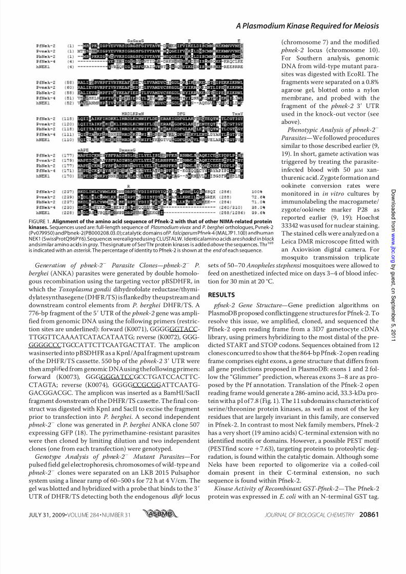
FIGURE 1. Alignment of the amino acid sequence of Pfnek-2 with that of other NIMA-related proteinkinases. Sequences used are: full-length sequence of Plasmodium vivax and P. berghei orthologues, Pvnek-2(Pv079950) andPbnek-2(PB000208.03.0);catalytic domains of P. falciparumPfnek-4 (MAL7P1.100) andhumanNEK1 (SwissProtQ96PY6).Sequences werealignedusing CLUSTALW. Identicalamino acids areshaded in black andsimilar amino acids in gray . Thesignature of Ser/Thr protein kinases is added above the sequences. Thr169
is indicated with an asterisk . The percentage of identity to Pfnek-2 is shown at the end of each sequence.
A PlasmodiumKinase Required forMeiosis
JULY 31, 2009• VOLUME 284 • NUMBER 31 JOURNAL OF BIOLOGICAL CHEMISTRY 20861

8/2/2019 An Essential Role for the Plasmodium Nek-2 Nima-Related
http://slidepdf.com/reader/full/an-essential-role-for-the-plasmodium-nek-2-nima-related 5/11
Purified recombinant GST-Pfnek-2 possessed kinase activity,as demonstrated by its ability to autophosphorylate (Fig. 2 A)and to phosphorylate exogenous substrates such as myelinbasic protein (MBP), histone H1 (Fig. 2 B), and -casein (data
not shown). To verify whether the activity was indeed due toGST-Pfnek-2 rather than to a co-purifying contaminant, weshowed that a catalytically inactive mutant enzyme (Lys38 3
Met) did not yield any signal in the phosphorylation assay.
FIGURE 2. Kinase activityof recombinant Pfnek-2. A, GST-Pfnek-2 autophosphorylation. A kinase assaycontaining [ -32P]ATPand 1g of wild-typePfnek-2,without exogenous substrates, was run on an SDS-PAGE, stained with Coomassie Blue, dried, and exposed for autoradiography. Left , autoradiogram; right ,CoomassieBlue-stained gel. B, kinase activity ofGST-Pfnek-2towardexogenous substrates. Kinase assays containing [ -32P]ATP, 1g of wild-typeGST-Pfnek-2,K38M GST-Pfnek-2 mutant, or T169A GST-Pfnek-2 mutant. MBP and histone H1 (H1) were used as substrates. Top, Coomassie Blue-stained gel; bottom,autoradiogram. C , absence of cross-phosphorylation/activation between GST-Pfnek-2 and GST-Pfnek-4. Autoradiograms for kinase assays containing[ -32P]ATP, 2.5 g of wild-type GST-Pfnek-2, K38M GST-Pfnek-2 mutant, wild type GST-Pfnek-4, K32M GST-Pfnek-4 mutant, and combinations of GST-recom-binant fusion proteins toward exogenous substrates. MBP and H1 (left panel ) and dephosphorylated caseine (CAS) (right panel ) were used as substrates.GST-Pfnek-2 autophosphorylation is indicated.
A PlasmodiumKinase Required forMeiosis
20862 JOURNAL OF BIOLOGICAL CHEMISTRY VOLUME 284• NUMBER 31• JULY 31, 2009
8/2/2019 An Essential Role for the Plasmodium Nek-2 Nima-Related
http://slidepdf.com/reader/full/an-essential-role-for-the-plasmodium-nek-2-nima-related 6/11
Thus, Pfnek-2 is a genuine protein kinase like Pfnek-4, but dis-plays a different substrate preference (Pfnek-4 is unable to
phosphorylate MBP or histone H1 (Fig. 2C and Ref. 9). Accord-ingly, kinase assays using heat-inactivated parasite extracts assubstrates consistently display different patterns of phospho-rylated proteins depending on whether GST-Pfnek-2 or GST-Pfnek-4 is used in the reaction (data not shown). Thr169 is con-
served in many protein kinases (including Neks) as the site foractivating phosphorylation, and mutant human NEK2 lackingthis residue display altered kinase activity (20). Likewise,replacement of this threonine with an alanine ablated Pfnek-2kinase activity, indicating that the amino acid at this position is
crucial to enzyme function and suggesting possible regulationby autophosphorylation; the Thr169 residue may also be a targetfor other upstream kinase(s) in vivo; the corresponding residuein Nek6 (Ser206) has been shown to be the target of phospho-
rylation by Nek9 (7). We did not observe any synergy between
the activities of GST-Pfnek-2 and GST-Pfmap-2, as had beenobserved for Pfnek-1 (12) and Pfnek-3 (13) (data not shown).Kinase assays using wild-type and kinase-dead enzymes per-formed to detect a possible phosphorylation/activation of
recombinant Pfnek-2 by Pfnek-4 (or vice versa) did not provideany evidence that such cross-activation occurs, at least in vitro(Fig. 2C ).
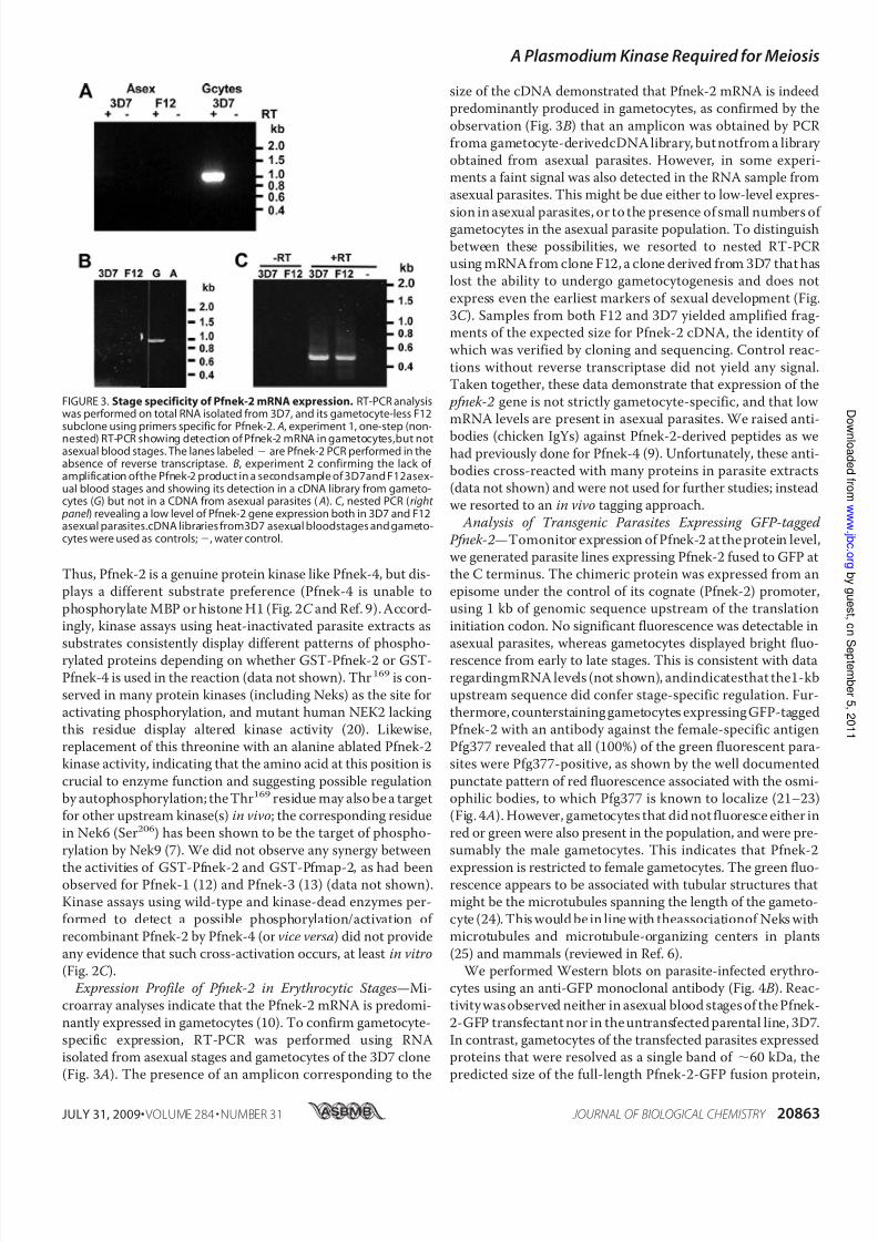
Expression Profile of Pfnek-2 in Erythrocytic Stages—Mi-croarray analyses indicate that the Pfnek-2 mRNA is predomi-
nantly expressed in gametocytes (10). To confirm gametocyte-specific expression, RT-PCR was performed using RNAisolated from asexual stages and gametocytes of the 3D7 clone(Fig. 3 A). The presence of an amplicon corresponding to the
size of the cDNA demonstrated that Pfnek-2 mRNA is indeedpredominantly produced in gametocytes, as confirmed by the
observation (Fig. 3 B) that an amplicon was obtained by PCRfroma gametocyte-derivedcDNA library, but notfrom a library obtained from asexual parasites. However, in some experi-ments a faint signal was also detected in the RNA sample fromasexual parasites. This might be due either to low-level expres-
sion in asexual parasites, or to the presence of small numbers of gametocytes in the asexual parasite population. To distinguishbetween these possibilities, we resorted to nested RT-PCRusing mRNA from clone F12, a clone derived from 3D7 that haslost the ability to undergo gametocytogenesis and does not
express even the earliest markers of sexual development (Fig.3C ). Samples from both F12 and 3D7 yielded amplified frag-ments of the expected size for Pfnek-2 cDNA, the identity of which was verified by cloning and sequencing. Control reac-
tions without reverse transcriptase did not yield any signal.Taken together, these data demonstrate that expression of the pfnek-2 gene is not strictly gametocyte-specific, and that low mRNA levels are present in asexual parasites. We raised anti-
bodies (chicken IgYs) against Pfnek-2-derived peptides as wehad previously done for Pfnek-4 (9). Unfortunately, these anti-bodies cross-reacted with many proteins in parasite extracts(data not shown) and were not used for further studies; insteadwe resorted to an in vivo tagging approach.
Analysis of Transgenic Parasites Expressing GFP-tagged
Pfnek-2—Tomonitor expression of Pfnek-2 at the protein level,we generated parasite lines expressing Pfnek-2 fused to GFP atthe C terminus. The chimeric protein was expressed from anepisome under the control of its cognate (Pfnek-2) promoter,using 1 kb of genomic sequence upstream of the translation
initiation codon. No significant fluorescence was detectable inasexual parasites, whereas gametocytes displayed bright fluo-rescence from early to late stages. This is consistent with dataregardingmRNA levels (not shown), andindicatesthat the1-kb
upstream sequence did confer stage-specific regulation. Fur-thermore, counterstaining gametocytes expressing GFP-taggedPfnek-2 with an antibody against the female-specific antigenPfg377 revealed that all (100%) of the green fluorescent para-sites were Pfg377-positive, as shown by the well documented
punctate pattern of red fluorescence associated with the osmi-ophilic bodies, to which Pfg377 is known to localize (21–23)(Fig. 4 A). However, gametocytes that did not fluoresce either inred or green were also present in the population, and were pre-sumably the male gametocytes. This indicates that Pfnek-2
expression is restricted to female gametocytes. The green fluo-rescence appears to be associated with tubular structures thatmight be the microtubules spanning the length of the gameto-cyte (24). This would be in line with theassociationof Neks withmicrotubules and microtubule-organizing centers in plants
(25) and mammals (reviewed in Ref. 6).We performed Western blots on parasite-infected erythro-
cytes using an anti-GFP monoclonal antibody (Fig. 4 B). Reac-tivity was observed neither in asexual blood stages of the Pfnek-
2-GFP transfectant nor in the untransfected parental line, 3D7.In contrast, gametocytes of the transfected parasites expressedproteins that were resolved as a single band of 60 kDa, thepredicted size of the full-length Pfnek-2-GFP fusion protein,
FIGURE 3. Stage specificity of Pfnek-2 mRNA expression. RT-PCR analysiswas performed on total RNA isolated from 3D7, and its gametocyte-less F12subclone using primers specific for Pfnek-2. A, experiment 1, one-step (non-nested) RT-PCR showing detection of Pfnek-2 mRNA in gametocytes,but notasexual blood stages. The lanes labeled are Pfnek-2 PCR performed in theabsence of reverse transcriptase. B, experiment 2 confirming the lack of amplification ofthe Pfnek-2 product in a secondsample of 3D7and F12asex-ual blood stages and showing its detection in a cDNA library from gameto-cytes (G) but not in a CDNA from asexual parasites ( A). C , nested PCR (right
panel ) revealing a low level of Pfnek-2 gene expression both in 3D7 and F12asexual parasites.cDNA libraries from3D7 asexual bloodstages and gameto-cytes were used as controls;, water control.
A PlasmodiumKinase Required forMeiosis
JULY 31, 2009• VOLUME 284 • NUMBER 31 JOURNAL OF BIOLOGICAL CHEMISTRY 20863

8/2/2019 An Essential Role for the Plasmodium Nek-2 Nima-Related
http://slidepdf.com/reader/full/an-essential-role-for-the-plasmodium-nek-2-nima-related 7/11
showing that the fusion protein is not proteolytically processedand thus validating the live fluorescence imaging.
Nek-2 Is Dispensable for Completion of the Erythrocytic Asex-
ual Cycle and Gametocyte Formation in P. falciparum and P.berghei—To investigate the function of Pfnek-2, we used areverse genetics approach in which parasite clones with a dis-rupted pfnek-2 gene were generated. To this aim, a plasmidbased on the pCAM-BSD vector (17), in which the central
region of the Pfnek-2 catalytic domain had been inserted nextto a cassette conferring resistance to blasticidin, was trans-ferred by electroporation into asexual parasites of the 3D7clone of P. falciparum. After single crossover homologous
recombination, neither of the two truncated copies of the pseu-
do-diploid locus will encode a functional kinase (Fig. 5 A). Mon-itoring the blasticidin-resistant population by PCR allowed usto readily detect integration-specific amplicons. To confirmthat parasites would be viable despite a disrupted pfnek-2 gene,
we proceeded to clone the transfected population by limitingdilution and to characterize the genotype of individual clones.Two clones, cl.8 and cl.9, obtained from independent transfec-tion experiments were selected for further characterization.PCR analysis showed (i) that these parasites had lost the wild-
type gene, (ii) that the episome (or an integrated concatemer)waspresent in cl.8 but not cl.9, and (iii) that amplicons diagnos-tic for the 5 and 3 boundaries of plasmid integration wereproduced (Fig. 5 B). To independently ascertain integration of
the pCAM-Pfnek-2 construct at thetarget locus, we performed a South-
ern blot (Fig. 5C ) using EcoRI-di-gested genomic DNA from wild-type 3D7, cl.8 and cl.9 transfectants,and a Pfnek-2 probe, which gavebands of the expected sizes: the
band corresponding to the wild-type locus disappears in the pfnek-2 clones, and is replaced by the two expected bands resultingfrom integration. In clone c9, the
episomal band is readily detectable,indicating that in this clone the epi-some is retained, or that more thanone copy of theknock-out construct
is integrated in the locus. Takentogether, the PCR and Southernblot analyses show that the Pfnek-2locus has been disrupted in cl.8 and
cl.9. Neither of the pfnek-2
cloneswas affected in their asexual growthrate in vitro (data not shown), andthe number, appearance, and sexratio of gametocytes in the pfnek-2
clones were undistinguishable from
those in wild-type parasites, asassessed by examination of Giemsa-stained slides. The association of GFP-Pfnek-2 with microtubular
structures (Fig. 4 A) prompted us to
investigate the morphology of
microtubules in pfnek-2 gametocytes. Clearly, the mutant
gametocytes display normal morphology (Fig. 5 D). Thus,
Pfnek-2 is required neither for erythrocytic schizogony nor for
the early stages of the sexual cycle in P. falciparum.
To investigate a possible phenotype caused by the absence of
a functional Nek-2 enzyme in subsequent stages of the sexual
cycle, we resorted to using the P. berghei/mouse system, as we
did previously with the Nek-4 enzyme (this system is consider-
ably more amenable than P. falciparum to this type of studies).
The Pbnek-2 protein (PlasmoDB identifier PB000208.03.0) dis-
plays 71% identity and 86% similarity to Pfnek-2. The gene was
replaced by a DHFR expression cassette conferring resistance
to pyrimethamine, using a double cross-over approach (Fig.
6 A). The pyrimethamine-resistant population was cloned by
limiting dilution. Genotype analysis of one of the resulting
clones by PCR (not shown), Southern blotting, and pulse-field
gel analysis (Fig. 6 B) indicated that gene replacement had
indeed occurred. Two independent clones were generated, one
in the wild-type ANKA strain background and the other in an
ANKA strain(cl 507)that expressesGFP (18). Theasexual cycle
of these two clones was then analyzed during infection in mice.
No difference in growth rate was observed when compared
with wild-type parasites. Furthermore, the pbnek-2 clones
produced normal numbers of gametocytes (data not shown).
Hence, the nek-2 gene is dispensable for erythrocytic schizog-
FIGURE 4. Analysis of GFP in P. falciparum gametocytes expressing Pfnek-2-GFP fusion protein. A, fluo-rescence imaging of two female gametocytes. Images from left to right in each set represent: (i) the fluores-cence signal from the GFP protein; (ii) the staining obtained with an anti-Pfg377 antibody (the secondaryantibody is conjugated to theAlexa Fluor 594 (red )); (iii) 4,6-diamidino-2-phenylindole (DAPI ) staining; (iv) anoverlay ofthe formertwo images; and (v) the DIC image ofthe cells inthe rightpanel . DIC , differential interfer-encecontrast. B, Western blot. 10g of parasite cell lysatefrom asexual stagesand stage III–V gametocytes of the3D7 parentalline or Pfnek-2-GFP transfectantwere subjectedto SDS-PAGE(12% acrylamide),transferredtonitrocellulose membrane, and probed with an anti-GFP antibody (Roche Molecular Biochemicals; 1:5000 dilu-tion). Leftpanel , Western blot;right panel , CoomassieBlue-stainedSDS-PAGE, stained aftertransfer. Lane1,3D7parental line; lane 2, Pfnek-2-GFP transfectant.
A PlasmodiumKinase Required forMeiosis
20864 JOURNAL OF BIOLOGICAL CHEMISTRY VOLUME 284• NUMBER 31• JULY 31, 2009
8/2/2019 An Essential Role for the Plasmodium Nek-2 Nima-Related
http://slidepdf.com/reader/full/an-essential-role-for-the-plasmodium-nek-2-nima-related 8/11
ony and gametocytogenesis in both P. berghei and P. falciparum.
pbnek-2 Parasites Do Not Develop into Ookinetes and Dis-
play Dysregulation of Premeiotic DNA Replication—The acti- vation of pbnek-2 gametocytes after exposure to conditionsthat trigger gamete formation in vitro (26) was then monitored.Microgametocytes were able to exflagellate as efficiently as
wild-type parasites (data not shown) and macrogametocytes
emerged and rounded normally, as shown by expression of theP28 antigen (Fig. 7 A). P28 mRNA accumulates in macrogame-tocytes, but its translation is repressed until gametogenesisoccurs, and the protein remains at the cell surface throughout
the macrogamete, zygote, and ookinete stages; conversion fromzygote to elongated ookinete can be monitored by microscopi-cally assessing cell shape using an anti-P28 antibody. We foundthat although the total number of P28-expressing parasites wascomparable in both pbnek-2 clones and wild-type parasites
20–24 h post-activation, the conversion of parasites intobanana-shaped ookinetes was abolished in both pbnek-2 lines(Fig. 7 B). Genetic crosses were then used as described previ-ously (9, 19) to determine whether the defect leading to the
block in ookinete development wascarried by the male or female game-
tocytes. Crossing pbnek-2 with pbcdpk4 parasites, in which malegametocyte exflagellation is abol-ished and which therefore can pro-duce only female gametes (27),
restored the ookinete development.This indicates that pbnek-2 malegametes are fully functional and cancross-fertilize the pbcdpk4 femalegametes. In the cross the percentage
of females transforming into ooki-netes was about half that of wild-type, consistent with pbnek-2
female gametes being sterile. In
contrast, crossing the pbnek-2
clone with a clone lacking Pbnek-4(in which female gametocytes carry a defect preventing ookinete devel-
opment (9)) did not restore ooki-nete conversion capacity (Fig. 8).Thus, absence of Pbnek-2 causesfemale gametocytes to be unable tosupport normal development fromzygote to ookinete, similar to the
developmental phenotype wereported previously for pbnek-4
parasites.To investigate in more detail the
stage at which the block in ookinetedevelopment occurs, we then quan-tified the amount of DNA in indi- vidual cells using a fluorescent dye.In wild-type parasites, fertilization
is followed by fusion of gametenuclei and one round of replication,
increasing the nuclear DNA content of the zygote to 4C prior tomeiosis (Fig. 8). Following meiosis four sets of chromosomesare maintained within the nucleus of the ookinete, making this
stage tetraploid. pbnek-2 parasites appeared to undergo acti- vation and fertilization like wild-type parasites, but fusion of thetwo nuclei was impaired in a large proportion of the parasites,and the DNA content remained at just above 2C (Fig. 9),a value
consistent with the sum of the two gamete nuclei. In all round
cells (i.e. female gametes, zygotes, and “failed” ookinetes), theDNA content remained belowthe 4C value expected in normalookinetes, indicating that the DNA replication process thatprecedes meiosis is affected in the mutant parasites. This is
strikingly similar to the phenotype observed in pbnek4 para-sites (9). Interestingly, we also observed that about 5% of the pbnek-2 P28-positive cells contained an abnormally largeamount of DNA, on average 30-fold the haploid amount (Fig.9), a feature that had not been seen in pbnek4 parasites.
The nek-2 Gene Is Essentialfor Transmission of P. berghei and P. falciparum to the Mosquito—The block in ookinete develop-ment in vitro suggests that pbnek-2 parasites may be unable toestablish an infection in the mosquito vector. To verify this, the
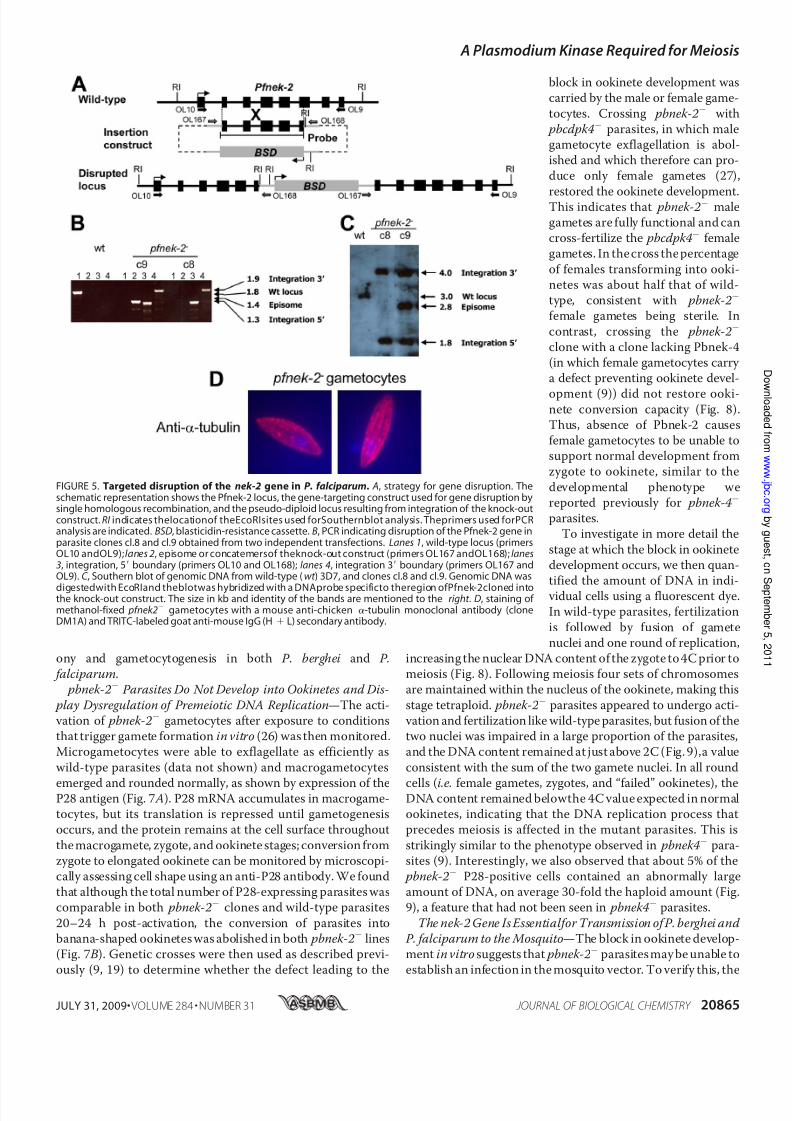
FIGURE 5. Targeted disruption of the nek-2 gene in P. falciparum. A, strategy for gene disruption. Theschematic representation shows the Pfnek-2 locus, the gene-targeting construct used for gene disruption bysingle homologous recombination, and the pseudo-diploid locus resulting from integration of the knock-outconstruct. RI indicates thelocationof theEcoRIsites used forSouthernblot analysis. Theprimers used forPCRanalysis are indicated. BSD, blasticidin-resistance cassette. B, PCR indicating disruption of the Pfnek-2 gene inparasite clones cl.8 and cl.9 obtained from two independent transfections. Lanes 1, wild-type locus (primersOL10 andOL9);lanes 2, episome or concatemersof theknock-out construct (primers OL167 andOL168);lanes3, integration, 5 boundary (primers OL10 and OL168); lanes 4, integration 3 boundary (primers OL167 andOL9). C , Southern blot of genomic DNA from wild-type (wt ) 3D7, and clones cl.8 and cl.9. Genomic DNA was
digestedwith EcoRIand theblotwas hybridized with a DNAprobe specificto theregion ofPfnek-2cloned intothe knock-out construct. The size in kb and identity of the bands are mentioned to the right . D, staining of methanol-fixed pfnek2 gametocytes with a mouse anti-chicken -tubulin monoclonal antibody (cloneDM1A) and TRITC-labeled goat anti-mouse IgG (H L) secondary antibody.
A PlasmodiumKinase Required forMeiosis
JULY 31, 2009• VOLUME 284 • NUMBER 31 JOURNAL OF BIOLOGICAL CHEMISTRY 20865

8/2/2019 An Essential Role for the Plasmodium Nek-2 Nima-Related
http://slidepdf.com/reader/full/an-essential-role-for-the-plasmodium-nek-2-nima-related 9/11
midguts of A. stephensi mosquitoes were examined 10–12 daysafter the insects had been fed with pbnek-2 or wild-type game-tocytes. As shown in Table 1 no oocysts were seen in mosqui-
toes infected with the knock-outparasite clones, whereas the mid-
guts of mosquitoes infected withwild-type parasite contained80–100 oocysts. Because disruptionof orthologous protein kinase genescan result in different phenotypes in
P. falciparum and P. berghei as seenin case of Pfmap-2 (28), we also pro-ceeded to a similar transmissionassay using the P. falciparum pfnek-2 clones. Like the P. bergheimutants, the pfnek-2 parasiteswere unable to produce oocysts inthe mosquito (Table 1). This clearly showed that the nek-2 gene is essen-
tial for transmission to the insect vector in both P. falciparum and P.berghei.
DISCUSSIONTaken together, the phenotypic
investigations reported here indi-cate that parasites lacking Pbnek-2are unable to properly control pre-
meiotic DNA replication. MurineNek2 mRNA is present in meiotictissues with particularly highamounts in testes (29), and the pro-
tein associates with meiotic chro-mosomes (30). In mammals it hasbeen shown that many Nek family members are involved in mitoticprogression (31), and human Nek2
is overexpressed in a variety of tumors and may be responsible fordefects in chromosome segregation(32). The phenotype of pbnek2
zygotes is thus consistent with
Pbnek-2 being involved in functionsrelated to genome replication, simi-lar to Neks in other eukaryotes.
What might be the molecular
basis for the meiotic phenotypecaused by the lack of Pbnek-2? Our
observation (Fig. 4 A) that theenzyme localizes with what lookslike microtubules in female gameto-
cytes is consistent with the estab-lished role of Neks in the regulationof microtubule dynamics. Anattractive hypothesis to explain thefemale-carried phenotype in the
events that precede meiosis might
be that the Nek-2 enzyme, which is
present only in female cells, is required for the ontogenesis of
the spindles functioning in premeiotic nuclear division. In most
animals including humans (33, 34), and in at least some plants
FIGURE 6. Disruption of the nek-2 gene in P. berghei. A, strategy to disrupt thewild-type (WT ) pbnek-2 locusby double crossover homologous recombination.The targeting constructcontained a cassette expressing theselectable marker T. gondii DHFR-TS (hatched box ), flanked by the 5 and 3 UTRs of the pbnek-2 gene. RI indicatesthe location ofthe EcoRI sites used forSouthern blot analysis. B, genotypeanalysis.A pbnek-2 clonewas analyzed by pulsed field gel electrophoresis using P. berghei dhfr 3 UTR probes that detect both theendogenous pbdhfr locus on chromosome 7 and the disrupted pbnek-2 locus on chromosome 10 (arrow ).
The transgenic clone was also analyzed by Southern blot after digestion of genomic DNA with EcoRI. A probespecific for the pbnek-2 3 UTR (white box in panel A) was used to detect the expected 3.1-kb fragment in the
wild-type and 565-bp fragment in the transgenic parasites.
FIGURE 7. Pbnek-2 is essentialfor ookinete developmentand DNAreplicationin thezygote. A, live para-sites from 20–24-h cultures were immunostained with a monoclonal antibody against the female gamete/zygote/ookinete marker P28 (red ) and counterstained with the nuclear marker Hoechst ( blue). No ookineteswere detected in the pbnek-2 parasites (N2). The presence of 2 nuclei in many cells suggests impairment innuclear fusion. A small proportion of pbnek-2 cells (arrow ) showed abnormally elevated DNA content.B, quantified ookinete conversion rates for wild-type (WT ) and the pbnek-2 parasites (N2). The histogramdisplays the percentage of round P28-positive cells adopting the elongated shape of ookinetes. Arithmeticmean S.D. from three experiments with parasites from different infected mice are shown.
A PlasmodiumKinase Required forMeiosis
20866 JOURNAL OF BIOLOGICAL CHEMISTRY VOLUME 284• NUMBER 31• JULY 31, 2009
8/2/2019 An Essential Role for the Plasmodium Nek-2 Nima-Related
http://slidepdf.com/reader/full/an-essential-role-for-the-plasmodium-nek-2-nima-related 10/11
(35), the centrioles are paternally inherited, and the same may
be truefor the centriolar-like structures and microtubule-orga-nizing centers in Plasmodium. A possible scenario might thusbe that the male-inherited microtubule-organizing centers donot find, in the nek-2 females, the molecular tool that isneeded for their development into the meiotic spindle. This
could result in a failure of pronuclear fusion and hence thereplication step that precedes meiosis, leading to the reducedDNA content we observed. In a minority of nek-2 P. bergheizygotes (or possibly unfertilized macrogametes) DNA content
appeared to increase unchecked to high levels, a phenomenon
not seen in wild-type zygotes and indicating a breakdown of normal cell cycle control in these cells.
The similarity in the phenotypes caused by the loss of Pbnek-2 and Pbnek-4, and the observation (Fig. 2C ) that the
two enzymes do not trans-phosphorylate or -activate in vitro,suggest that they function in parallel pathways involved in thesame cell development process, but are not able to complementfor each other (contrary to what has been observed for the P. falciparum MAPKs (28)). This is consistent with our observa-
tion that Pfnek-2 and Pfnek-4 display distinct substrate speci-ficity in vitro when classical exogenous substrates such as MBP,histone H1, or casein are used (Fig. 2C ). Similarly, kinase assaysusing heat-inactivated parasite extracts as substrates consis-
tently display different patterns of phosphorylated proteinsdepending on whether GST-Pfnek-2 or GST-Pfnek-4 is used inthe reaction (data not shown). Thus, biochemical and reverse
genetics data (absence of cross-complementation) concur tosuggest that nek-2 and nek-4 fulfill non-redundant functionsduring meiosis. This, together with the fact that both enzymesare (i) active in vitro when expressed in E. coli as recombinantproteins, and (ii) essential for infection of the mosquito vector,
is of considerable interest in thecontext of thesearch for targetsfor transmission-blocking intervention. The advantages of tar-geting more than one single target are well established (36), andthe availability of enzymatic assays makes high throughputscreening of chemical libraries on Pbnek-2 and Pbnek-4 a real-
istic possibility. The developmental effect of any hits can betested in transmission experiments.
Acknowledgments—We are grateful to Pietro Alano for the gift of the
anti-Pfg377 antiserum, and David Fidock and Geoff MacFadden for
the pCAM BSD and pHGB/pCHD vectors, respectively. This work is
based on gene identification made possible by the availability of the
genome sequences of P. falciparum and P. berghei, and the PlasmoDB
data base. We thank Dr. J. Chevalier (Service Scientifique de
l’Ambassade de France a Londres) for continuing interest and
support.
REFERENCES
1. Breman, J. G., Alilio, M. S., and Mills, A. (2004) Am. J. Trop. Med. Hyg. 71,
1–15
2. Oakley, B. R., and Morris, N. R. (1983) J. Cell Biol. 96, 1155–1158
3. Schultz, S. J., Fry, A. M., Sutterlin, C., Ried, T., and Nigg, E. A. (1994) Cell
Growth & Differ. 5, 625–635
4. Fry, A. M., Meraldi, P., and Nigg, E. A. (1998) EMBO J. 17, 470–481
5. Davies, J. R., Osmani, A. H., De Souza, C. P., Bachewich, C., and Osmani,
S. A. (2004) Eukaryot. Cell 3, 1433–1444
6. O’Regan, L., Blot, J., and Fry, A. M. (2007) Cell Div. 2, 25
TABLE 1
nek-2 is essential for transmissionto themosquitovector A. gambiae mosquitoes werefed on blood containing P. falciparum wild-type (3D7)or pfnek-2 (clone c9) gametocytes, and A. stephensi mosquitoes were fed on miceinfected with wild-type and pbnek-2 P. berghei parasites. Midgut oocyst numberswere determined at days 10–12 after infection. Shown is the prevalence (mosqui-toes with oocysts/total mosquitoes) and (in parentheses) the range of numbers of oocysts per midgut for the positive mosquitoes.
Oocysts
P. falciparumExperiment 1
3D7 wild-type 21/24 (1–7) pfnek-2 1/36 (2)a
Experiment 23D7 wild-type 15/22 (1–42)
pfnek-2 0/20 (0)Experiment 3
3D7 wild-type 10/20 (1–9) pfnek-2 0/24 (0)
P. bergheiExperiment 1
Wild-type 20/20 (83–200) pbnek-2 0/20
Experiment 2Wild-type 18/20 (52–200)
pbnek-2 0/20Experiment 3
Wild-type 20/20 (61–200) pbnek-2 0/20a The single mosquito infected with pfnek-2 gametocytes in experiment 1 con-
tained 2 oocysts.
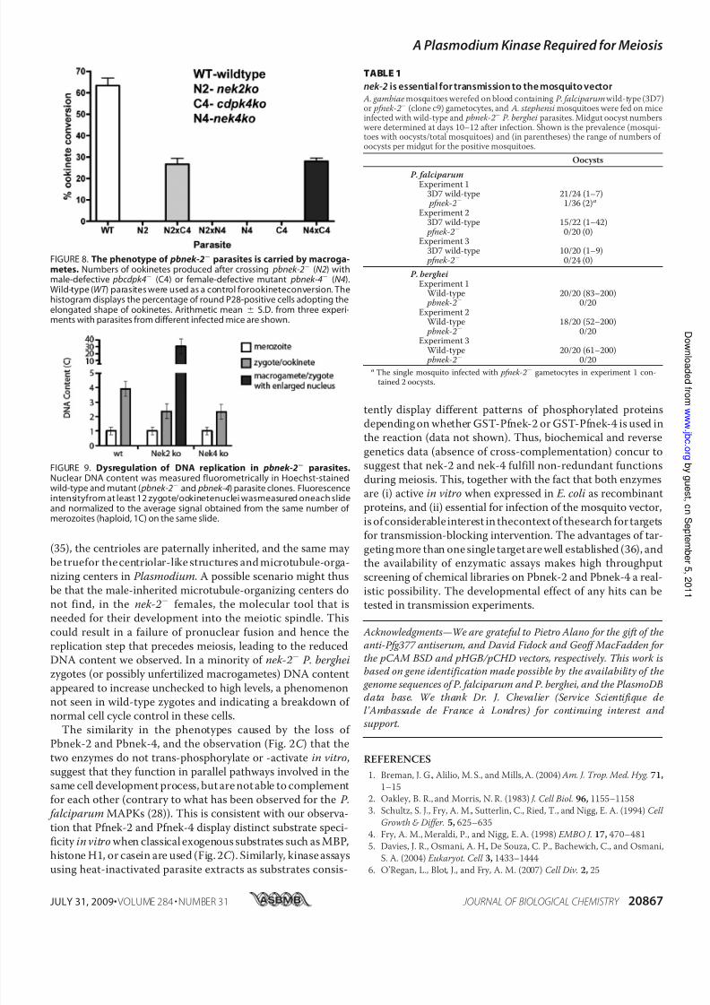
FIGURE 8. The phenotype of pbnek-2 parasites is carried by macroga-metes. Numbers of ookinetes produced after crossing pbnek-2 (N2) withmale-defective pbcdpk4 (C4) or female-defective mutant pbnek-4 (N4).Wild-type (WT ) parasites were used as a control forookineteconversion. Thehistogram displays the percentage of round P28-positive cells adopting theelongated shape of ookinetes. Arithmetic mean S.D. from three experi-ments with parasites from different infected mice are shown.
FIGURE 9. Dysregulation of DNA replication in pbnek-2 parasites.Nuclear DNA content was measured fluorometrically in Hoechst-stainedwild-type and mutant ( pbnek-2 and pbnek-4) parasite clones. Fluorescenceintensityfrom at least 12 zygote/ookinetenuclei wasmeasured oneach slideand normalized to the average signal obtained from the same number of merozoites (haploid, 1C) on the same slide.
A PlasmodiumKinase Required forMeiosis
JULY 31, 2009• VOLUME 284 • NUMBER 31 JOURNAL OF BIOLOGICAL CHEMISTRY 20867
8/2/2019 An Essential Role for the Plasmodium Nek-2 Nima-Related
http://slidepdf.com/reader/full/an-essential-role-for-the-plasmodium-nek-2-nima-related 11/11
7. Belham, C., Roig, J., Caldwell, J. A., Aoyama, Y., Kemp, B. E., Comb, M.,
and Avruch, J. (2003) J. Biol. Chem. 278, 34897–34909
8. Ward, P., Equinet, L., Packer, J., and Doerig, C. (2004) BMC Genomics 5,
79
9. Reininger, L., Billker, O., Tewari, R., Mukhopadhyay, A., Fennell, C.,
Dorin-Semblat, D., Doerig, C., Goldring, D., Harmse, L., Ranford-Cart-
wright, L., Packer, J., and Doerig, C. (2005) J. Biol. Chem. 280,
31957–31964
10. Le Roch, K. G., Zhou, Y., Blair, P. L., Grainger, M., Moch, J. K., Haynes,
J.D., De La Vega, P., Holder, A.A., Batalov,S., Carucci, D.J., andWinzeler,
E. A. (2003) Science 301, 1503–1508
11. Bahl, A., Brunk, B., Crabtree, J., Fraunholz, M. J., Gajria, B., Grant, G. R.,
Ginsburg, H., Gupta, D., Kissinger, J. C., Labo, P., Li, L., Mailman, M. D.,
Milgram, A. J.,Pearson, D. S., Roos, D. S.,Schug, J.,Stoeckert, C. J.,Jr., and
Whetzel, P. (2003) Nucleic Acids Res. 31, 212–215
12. Dorin, D., Le Roch, K., Sallicandro, P., Alano, P., Parzy, D., Poullet, P.,
Meijer, L., and Doerig, C. (2001) Eur. J. Biochem. 268, 2600–2608
13. Lye, Y. M., Chan, M., and Sim, T. S. (2006) FEBS Lett. 580, 6083–6092
14. Ringwald, P., Meche, F. S., Bickii, J., and Basco, L. K. (1999) J. Clin. Micro-
biol. 37, 700–705
15. Carter, R., Ranford-Cartwright, L., and Alano, P. (1993) Methods Mol.
Biol. 21, 67–88
16. Tonkin, C. J., van Dooren, G. G., Spurck, T. P., Struck, N. S., Good, R. T.,
Handman, E., Cowman, A. F., and McFadden, G. I. (2004) Mol. Biochem.
Parasitol. 137, 13–21
17. Sidhu, A. B., Valderramos, S. G., and Fidock, D. A. (2005) Mol. Microbiol.
57, 913–926
18. Mair, G. R., Braks, J. A., Garver, L. S., Wiegant, J. C., Hall, N., Dirks, R. W.,
Khan, S. M., Dimopoulos, G., Janse, C. J., and Waters, A. P. (2006) Science
313, 667–669
19. Liu, J., Gluzman, I. Y., Drew, M. E., and Goldberg, D. E. (2005) J. Biol.
Chem. 280, 1432–1437
20. Rellos, P.,Ivins,F. J.,Baxter,J. E.,Pike, A.,Nott, T.J., Parkinson, D.M., Das,
S.,Howell,S., Fedorov,O., Shen,Q. Y.,Fry,A. M.,Knapp,S., andSmerdon,
S. J. (2007) J. Biol. Chem. 282, 6833–6842
21. de Koning-Ward, T. F.,Olivieri, A.,Bertuccini,L., Hood,A., Silvestrini, F.,
Charvalias, K., Berzosa Díaz, P., Camarda, G., McElwain, T. F., Papenfuss,
T., Healer, J., Baldassarri, L., Crabb, B. S., Alano, P., and Ranford-Cart-
wright, L. C. (2008) Mol. Microbiol. 67, 278–290
22. Alano, P., Read, D., Bruce, M., Aikawa, M., Kaido, T., Tegoshi, T., Bhatti,
S., Smith, D. K., Luo, C., Hansra, S., Carter, R., and Elliott, J. F. (1995) Mol.
Biochem. Parasitol. 74, 143–156
23. Silvestrini, F., Alano, P., and Williams, J. L. (2000) Parasitology 121,465–471
24. Sinden, R. E., Canning, E. U., Bray, R. S., and Smalley, M. E. (1978) Proc. R.
Soc. Lond. B Biol. Sci. 201, 375–399
25. Motose, H., Tominaga, R., Wada, T., Sugiyama, M., and Watanabe, Y.
(2008) Plant J. 54, 829–844
26. Billker, O., Lindo, V., Panico, M., Etienne, A. E., Paxton, T., Dell, A., Rog-
ers, M., Sinden, R. E., and Morris, H. R. (1998) Nature 392, 289–292
27. Billker, O., Dechamps, S., Tewari, R., Wenig, G., Franke-Fayard, B., and
Brinkmann, V. (2004) Cell 117, 503–514
28. Dorin-Semblat, D., Quashie, N., Halbert, J., Sicard, A., Doerig, C., Peat, E.,
Ranford-Cartwright, L., and Doerig, C. (2007) Mol. Microbiol. 65,
1170–1180
29. Arama, E., Yanai, A., Kilfin, G., Bernstein, A., and Motro, B. (1998) Onco-
gene 16, 1813–1823
30. Rhee, K., and Wolgemuth, D. J. (1997) Development 124, 2167–217731. Malumbres, M., and Barbacid, M. (2007) Curr. Opin. Genet. Dev. 17,
60–65
32. Hayward, D. G., and Fry, A. M. (2006) Cancer Lett. 237, 155–166
33. Palermo, G., Munne, S., and Cohen, J. (1994) Hum. Reprod. 9, 1220–1225
34. Sathananthan, A. H.,Kola,I., Osborne, J.,Trounson, A.,Ng, S. C.,Bongso,
A., and Ratnam, S. S. (1991) Proc. Natl. Acad. Sci. U.S.A. 88, 4806–4810
35. Nagasato, C. (2005) J. Plant Res. 118, 361–369
36. Zimmermann, G. R., Lehar, J., and Keith, C. T. (2007) Drug Discov. Today
12, 34–42
A PlasmodiumKinase Required forMeiosis
20868 JOURNAL OF BIOLOGICAL CHEMISTRY VOLUME 284• NUMBER 31• JULY 31 2009