An Coláiste Ollscoile Baile Átha Cliath · University College Dublin An Coláiste Ollscoile Baile...
28
University College Dublin An Coláiste Ollscoile Baile Átha Cliath National University of Ireland, Dublin Ollscoil na hÉireann, Baile Átha Cliath Radiography Session 2004/05
Transcript of An Coláiste Ollscoile Baile Átha Cliath · University College Dublin An Coláiste Ollscoile Baile...

University College Dublin An Coláiste Ollscoile Baile Átha Cliath
National University of Ireland, Dublin Ollscoil na hÉireann, Baile Átha Cliath
Radiography
Session 2004/05

University College Dublin
2
Note
Where the tuition in a subject is completed in the first semester, the University Examination in that subject may be held during the prescribed period at the end of the semester. Supplemental
examinations, if required, will take place in the Autumn.

Radiography
3
Contents Degree of Bachelor of Science (Radiography) .............................................................4 Introductory Information ..................................................................................................................... 4 Summary of Courses and Examination Subjects ............................................................................. 6 Regulations ........................................................................................................................................... 7 Syllabus of Courses.............................................................................................................................. 8
First Year ...................................................................................................................................................8 Second Year............................................................................................................................................10 Third Year................................................................................................................................................11 Fourth Year..............................................................................................................................................13
Postgraduate Courses .................................................................................................14 Taught MSc (BI, CT, MR, RNI, or US)..............................................................................................14 Modular Structure..............................................................................................................................17
Section A: Professional Practice And Departmental Operation .....................................................17 Section B: Practice..................................................................................................................................17 Section C: Technology............................................................................................................................18 Section D: Clinical Practice ...................................................................................................................18 Section E: Research ................................................................................................................................18
Syllabus of Modules...........................................................................................................................19 Section A: Professional Practice and Departmental Operation......................................................19 Section B: Practice..................................................................................................................................20 Section C: Technology............................................................................................................................21 Section D: Clinical Practice ...................................................................................................................22 Section E: Research ................................................................................................................................25
Research MSc (Diagnostic Imaging Subject) .................................................................................26 Doctor of Philosophy (PhD) ..............................................................................................................28
University College Dublin
4
Degree of Bachelor of Science (Radiography)
Introductory Information The full-time degree course leads to the examination for the Degree of Bachelor of Science (Radiography) of the National University of Ireland and to professional recognition by the Irish Institute of Radiography. The degree is internationally recognised for the practise of Radiography.
Application and Admission
In session 2004/2005 not more than forty places will be available in the first year. Admission is competitive and is based on the points system for students taking Leaving Certificate examinations.
Information on the application procedure may be obtained from the Central Applications Office, Tower House, Eglinton Street, Galway, (telephone: +353-91-509 800). Information on admission requirements may be obtained from the Admissions Office, University College Dublin, Belfield, Dublin 4 (telephone: +353-1-716 1425).
Note: Prospective students must have a laboratory science subject, and either Chemistry or Physics is recommended.
Prospective students are strongly advised to spend some time in an X-ray department before completing the CAO form. Experience gained in an X-ray department will be of great benefit to candidates considering Radiography as a career.
Induction
Advisory meetings are held in the week before term begins. Attendance is obligatory for first year students. Details are forwarded to students early in autumn.
Fees
Additional expenses arise from uniforms, books, and travel to clinical centres and costs associated with the professional recognition of the Irish Institute of Radiography.
Hepatitis B Vaccination:
In line with currently accepted policies for health care workers, Hepatitis B vaccination is arranged for all undergraduate radiographers during the first year.

Radiography
5
Dates of the Academic Session 2004/2005
The dates of the academic session 2004/2005 for Radiography students are as follows: First Semester: (Michaelmas term) 16th September 2004 – 8th December 2004 Second Semester: (Hilary/Trinity terms)*10th January 2005 – 23rd April 2005 Note: Clinical instruction and hospital placements continue beyond the above dates in
all four years of the course. In years one to three, summer clinical placements are from mid May until mid July. The length of the summer recess is thus approximately eight weeks.
Attendance at lectures and clinicals
Students attend lectures in the School of Diagnostic Imaging, Earlsfort Terrace Medical School and Belfield. In 2005, the School will relocate completely to the Belfield campus. Clinical experience within the course is undertaken mainly in St. Vincent's, the Mater Misericordiae, Beaumont, Tallaght and St. James' Hospitals, as well as in other Dublin hospitals.
* Dates of mid-term break vary depending on year of the course.

University College Dublin
6
Summary of Courses and Examination Subjects
First Year Anatomy I
Biochemistry
Chemistry
Functional Histology and Physiology I
Physics
Psychology I
Radiography I
Clinical Skills I
Third Year Diagnostic Imaging I: • Radiography • Ultrasound • Radionuclide Imaging
Digital Imaging I
Epidemiology, Statistics and Research Methods
Equipment II
Imaging Technology II
Management and Health Service Structure
Mechanisms of Disease
Clinical Skills III
Second Year Anatomy II
Equipment I
Hospital Studies
Imaging Technology I
Interpersonal Skills
Healthcare Informatics
Medical Sociology
Functional histology and Physiology II
Radiography II
Radiation and Dosimetry
Clinical Skills II
Fourth Year Diagnostic Imaging II: • Radiography • Computed Tomography • Magnetic Resonance Imaging
Digital Imaging II
Legal Medicine
Systematic Pathology
Research Project
Clinical Skills IV

Radiography
7
Regulations Students must abide by the Student Code and University Regulations as presented in the Student Information Handbook.
Attendance at Courses Students proceeding to the Degree of Bachelor of Science in Radiography must satisfactorily attend courses for four years and pass each year's examination.
Clinical placements are an elemental part of each year's course and full attendance is mandatory. Unless there are extenuating circumstances, students will be required to make up clinical non-attendance before presenting for Clinical Assessment, or progressing to the next course year. Students taking repeat examinations may be required to repeat clinical placements from the previous academic session.
Examinations Most examinations are held in the summer of each year of the course. Supplemental examinations (if required) will be held in the autumn. Where a course of study is delivered completely in the first semester, examinations are normally held in the winter examination period, with repeat examinations (if required) in the following autumn.
Candidates for admission to any examination must have satisfactorily attended the prescribed course of instruction. A candidate must have satisfactory attendance at all clinical placements before presenting for assessment of clinical skills. All examinations must be passed as a whole before proceeding to the course of the following year. Students who do not pass an examination may be required to re-attend the respective courses in the following session before re-entering for the examination. A proportion of marks in some subjects may be allocated to the year's work.
The University Examinations of the first and second year of Radiography must be passed within two years of entering the respective year.
The Degree will be awarded on the basis of the results of the Third University Examination in Radiography and the Final University Examination in Radiography.
University College Dublin
8
Syllabus of Courses
First Year
Anatomy I (ANAT 1003) Lectures, dissection and applied anatomy/functional anatomy tutorials during semesters I and II.The course in Anatomy is continued over the first and second year. The topics covered are: osteology and arthrology; myology; anatomy of the limbs and thorax; surface and functional anatomy on the living model; abdomen, pelvis and perineum; nervous system; anatomy of the head and neck; embryological development of the human with emphasis on the risks of radiation.
Chemistry (CHEM 1605) Lectures and practicals during semesters I and IIThe elements; their electronic structures and properties. Ionic and covalent bonding. Water, solutions and colloids, dialysis. Acids, bases and ionic compounds, including buffers. Structures and properties of important organic compounds, including hydrocarbons, alcohol, amines, carboxylic acids, amides, amino acids, peptides. Topics of special relevance to radiography focusing on radiochemistry, the chemistry of photographic processes and contrast agents.
Biochemistry (BIOC 1601) Lectures: Twenty hours during the second semester.Cell Biology and Biochemistry. The building blocks of the cell – proteins, lipids and carbohydrates. Structure and function of enzymes. Structure and function of biological membranes DNA and RNA, molecules of heredity. Heredity and the cell. The structure of nucleic acids. RNA directed protein synthesis. Hereditary diseases and genetic engineering. Generation and storage of metabolic energy. Glycolysis. Glycogen, Gluconeogenesis and Glucose homeostasis. Storage and mobilisation of lipids. Oxidation of fatty acids. Metabolism of nitrogenous compounds: aspects of nutrition. Synthesis and catabolism of amino acids. Nitrogen balance and protein requirements. Formation of urea. Vitamins.
Experimental Physics (EXPH 1604) Lectures and laboratory sessions during semesters I and II.Mechanics, atomic theory of matter, wave phenomena, light and sound, thermal physics, electricity and magnetism, X-rays, nuclear physics, properties of fluid and matter, current electricity.

Radiography
9
Physiology I (PHYS 1003) Lectures and practical classes during semesters I and IIThe course in Physiology over the first year is designed to give the student an in-depth knowledge of fundamental reactions of living organisms, particularly in the human body. The major topics covered include the following: the cell; primary tissue; connective tissue; skin; muscle; nervous tissue; blood; lymphoid tissues. Laboratory classes are concerned with the microscopic structure of tissues, organs and systems and particular emphasis is placed on relationship of structure and function.
Radiography I (RDGY 1001) Lectures and practical classes during semesters I and II.Terminology. Role of the radiographer. Departmental protocols and aspects of practice. Hygiene in the hospital. Ergonomics Principles of radiation protection. Recognition, resuscitation and stabilisation of the injured patient, with emphasis on practical application. Photographic principles, film materials, intensifying screens, film cassettes, principles of processing. Principles of exposure factor selection. Image quality and appraisal. Simple introductions relating theoretical physics to radiographic practice. Radiographic techniques of the following: Upper limb; lower limb; thorax and shoulder girdle; pelvic girdle; vertebral column; respiratory system; simple consideration of abdominal and pelvic contents; macro radiography.
Psychology (RDGY 1002) Lectures in semester I and semester IIThis section of the course is designed to give a basic knowledge of the psychological function of man in health and disease, and to outline the processes of interaction between organism and environment. The main subjects for study will be: perception, learning, emotion and motivation, measurement and individual differences, personality, social psychology.
Clinical Skills I RDGY1101 Students will attend hospital departments throughout the year, and will undertake general radiographic examinations under the supervision of clinical staff. All clinical placements are organised and scheduled by School staff.
Throughout the course, the attendance of students for clinical experience in the general and specialist hospitals affiliated to the University must be certified by School staff before the student may proceed to the relevant examinations.

University College Dublin
10
Second Year
Anatomy II (ANAT 2003) Lectures, dissection and applied anatomy/functional anatomy tutorials during semesters I and II.See First Year Curriculum.
Imaging Technology I (RDGY 2001) Lectures and practical classes during semesters I and II.Photographic principles. Sensitometry. Film materials. Intensifying screens. Cassettes. Storage of photographic materials. Dry processing techniques. The radiographic image. Principles and practice of processing. Silver conservation and recovery. Automated film handling. Film presentation and archival.
Physiology II (PHYS 2003) Lectures during semesters I and IIThe course in Physiology over the second year is designed to continue on from the course in first year giving the student an in-depth knowledge of fundamental reactions of living organisms, particularly in the human body. The major topics covered include the following: respiration; blood vessels; circulation; cardiac cycle; systemic circulation; sensory receptors; special senses; motor unit; spinal cord; control of movement; hypothalamic functions; gastro-intestinal tract; kidneys; uterus; urinary tract; pregnancy; endocrine system.
Radiography II (RDGY 2002) Lectures and practical classes during semesters I and II.Manipulation of exposure factors and image quality. Radiographic examinations to cover: Skull. Dentition and orthodontic practice; abdominal and pelvic contents, foreign body localisation; gastro-intestinal tract; urinary system; lacrimal system; salivary system. Conventional tomography.
Equipment I (RDGY 2003) Lectures and practicals in semester I and IIThe X-ray tube. AC supply. Circuitry for X-ray generation. Generators. Microprocessor control. Control and destabilisation equipment. Physics of X-ray generators, tubes and intensifiers, Quality assurance and Quality control.
Radiation and Dosimetry (RDGY 2004) Lectures in semester IElectromagnetic radiation. Interaction with matter. Radiation detection. Dosimetry. Biological effects of radiation. Radiation protection.

Radiography
11
Healthcare Informatics (HCIN 1002) Lectures and PracticalsBasics of computer technology; computer architecture; hardware and software; operating systems. Information systems; database; knowledge-based systems. Communications and networks. Applications of computing in medicine. Generic software packages. Laboratory: Practical exercises designed to develop familiarity with generic software packages.
Hospital Studies (RDGY 2005) Lectures in semester I and IIHealth and safety. Patients with special needs. Trauma immobilisation. General observations of the patient. Principles of nursing care. Surgical procedures. Critical care. Infection control. Pharmacology. Contrast agents, applied pharmacology, storage, stock control and disposal.
Interpersonal Skills (RDGY 2006) Lectures in semester IIEffective communication. Related factors. Initiating and responding skills. Counselling and communication.
Medical Sociology (SOC 2701) Lectures in semester IDistribution of health and illness in society. The roles and settings of medical practice. Public Health Policy. Social impact of advances in medical technology.
Clinical Skills II (RDGY 2007) Students will attend a range of hospital departments throughout the year, and will undertake general and more specialised radiographic examinations under the supervision of clinical staff. All clinical placements are organised and scheduled by School staff.
Third Year
Imaging Technology II (RDGY 3001) Lectures and practical classes during semesters I and IIDuplication and subtraction. Photofluorography. Monitor photography. Special Imaging techniques. Quality Assurance.
Mechanisms of Disease (PATH 3002) Lectures during semester IIntroduction. Molecular pathology. The immune system. Genetics. Environmental and nutritional pathology.

University College Dublin
12
Diagnostic Imaging I (RDGY 3002) Lectures and practical classes during semesters I and IIRadiography Hepato-biliary-pancreatic systems. Arthrography. Lymphatic system. Mammography. Cardio-vascular system. Paediatric radiography. Interventional techniques or alternate ERASMUS topic.
Ultrasound Physics and principles. Scanning protocols. Clinical applications.
Radionuclide Imaging Physics and principles. Scanning protocols. Clinical applications, Radiopharmacy.
Digital Imaging I (RDGY 3008) Lectures and practical classes in semester ITerminology, computer fundamentals, hardware and software, operating systems, Quantitisation, sampling, analogue to digital conversion, image quality, image acquisition, digitisers, video capture, direct and computed radiography, fluoroscopic systems, transmission, archival, image display.
Equipment II (RDGY 3004) Lectures and practicals in semester I and IIAdaptations of equipment design. Equipment for mobile radiography, tomography, skull and dental radiography, mammography. Fluoroscopic equipment. Accident and Emergency equipment. Angiographic equipment. Care and maintenance. Design specifications. Quality assurance.
Epidemiology, Statistics and Research Methods (PHME 3002) Lectures in semester INatural history of diseases and prevention. Concepts and models. Descriptive and analytical epidemiology. Epidemiology of major chronic diseases.
Types and areas of research. Research design. Methodology. Statistics. Population. Presentation of research.
Management and Health Service Structure (RDGY 3005) Concepts of management with particular reference to the needs of health care professionals in a changing health care environment.
Clinical Skills III (RDGY 3006) Students will attend a range of hospital departments, undertaking most radiographic examinations under the supervision of clinical staff. Students will undertake limited clinical placements at the weekend and in the evenings. Some students may be allowed to attend a European Imaging department. All clinical placements are organised and scheduled by School staff.

Radiography
13
Fourth Year
Diagnostic Imaging II (RDGY 4005) Lectures and practical classes during semesters I and II.Radiography Accident and Emergency techniques. Skeletal survey and bone densitometry. Central nervous system. Radiography of the elderly. Gynaecological and obstetric examinations. Mobile and operating theatre radiography.
Computed Tomography Physics and principles. Scanning protocols. Clinical applications.
Magnetic Resonance Imaging Physics and principles. Scanning protocols. Clinical applications.
Digital Imaging II (RDGY 4004) Image processing, compression, analysis and synthesis. Local and wide area networks. Topology. Industry standards. Quality Assurance.
Legal Medicine (FMED 4001) Medical law and the radiographer. Tort. Medical negligence. Consent. Registration and professional organisations. Ethical issues. Contracts. Acts of the Oireachtas.
Systematic Pathology (PATH 4005) Lectures and tutorials during semester II.Pathology relevant to diagnostic imaging in each of the following: blood; cardiovascular system including congenital heart defects; respiratory system including mediastinum and pleura; musculo-skeletal system; gastrointestinal tract; hepato-biliary pancreatic systems; genito-urinary systems; reproductive systems and female breast; central nervous system; endocrine disorders; other miscellaneous pathology.
Dissertation (RDGY 4101) Students are required to undertake an individual project that is related to Diagnostic Imaging under the supervision of a member of staff. Three copies of the project must be submitted at a specified time before the fourth year examinations. Candidates may be required to take an oral examination in the subject matter of the project.
Clinical Skills IV (RDGY 4002) Students will attend a range of hospital departments, undertaking most radiographic examinations under the supervision of clinical staff. Students will undertake limited clinical placements at the weekend and in the evenings. All clinical placements are organised and scheduled by School staff.

University College Dublin
14
Postgraduate Courses
Taught MSc (BI, CT, MR, RNI, or US)
Course Aims
1. To provide flexible and accessible programmes of postgraduate education that are appropriate for Radiographers and other health care professionals at various levels.
2. To equip candidates with the conceptual knowledge and necessary skills to:
• Competently perform examinations in the chosen field to an expert level.
• Develop greater professional autonomy and the ability to adapt to the needs of a changing health care sector.
• Develop an analytical approach relevant to the professional practice of Diagnostic Imaging and associated disciplines.
Course Overview and Credits
There are five pathways available: Computed Tomography; Magnetic Resonance Imaging; Breast Imaging; Radionuclide Imaging; Ultrasound.
Certificates are available for any module in any pathway.
Each pathway is delivered via a modular structure, and may be followed on a full or part time basis, to the end point of choice, i.e.: Certificate, Diploma (Mammography only), Higher Diploma or MSc. All modules are assigned ECTS credits. Courses commence in January each year.
A degree of MSc is awarded following successful completion of appropriate modules to a total of 120 credits. Full time students may complete the MSc programme in five semesters.
A Higher Diploma is awarded following successful completion of appropriate modules to a total of 60 credits. Full time students may complete the HDip programme in three semesters.
N.B. Candidates cannot be awarded an MSc and a Higher Diploma arising from the same course of study. All candidates register initially for the MSc.
A Diploma is awarded following successful completion of appropriate modules to a total of 26 credits. The Diploma option is only available for the Breast Imaging pathway. Full

Radiography
15
time students may complete the Dip programme in one semester plus clinical practice extending to the following Autumn.
A Certificate is awarded following successful completion of any course module that is offered on a stand-alone basis. The credits and time for completion of individual modules vary depending on the subject.
For all awards, part time students may extend completion of taught modules over a maximum of five semesters and research modules over a maximum of four semesters.
Admission Requirements
Applicants should hold an honours degree in Radiography, or other qualification deemed equivalent, plus appropriate clinical experience in Diagnostic Imaging. Some courses are open to health care professionals other than radiographers, provided they have an appropriate qualification and relevant clinical experience. Candidates will not normally be accepted with less than one year of general post-qualification experience. Applicants holding awards from other third level institutions may be granted exemptions on the basis of ECTS credits.
N.B. All applicants registering for any pathway must present a written commitment from a suitable clinical department confirming availability of at least the minimum amount of clinical experience.
Application Programmes are widely advertised prior to the course. Application forms are distributed nation-wide to Clinical Imaging departments during recruitment. In addition, on-line information is available at http://www.ucd.ie/diagnosticimaging
Places are limited and applicants may be required to undertake an assessment procedure before being offered a place.
Course Structure
Postgraduate students complete a combination of clinical and academic modules over the first two semesters (full time), or four semesters (part time). Attendance for lectures and tutorials is grouped into blocks per module over the calendar year, to facilitate the nation-wide student cohort, as well as students studying part-time or by independent module. Clinical experience is usually in the student's own hospital, and also sometimes in specialist centres. Clinical instruction will be given by course lecturers and specific trained staff within the clinical departments. Each student must spend substantial time in the specialist field to achieve the required experience.
In the next two semesters (full time students) or four semesters (part-time students) students complete the final taught module in Research Methodology and Statistics (by either attendance or distance learning), and undertake a supervised research project focussed in Diagnostic Imaging, which is presented in the MSc thesis.
The Degree of MSc (Diagnostic Imaging subject) is awarded on the basis of all examination results and the thesis. An oral examination in the subject matter of the thesis
University College Dublin
16
may be required by the examiners. Students may exit relevant programmes at Certificate, Diploma, or Higher Diploma level.
Course Assessment
Each programme pathway is assessed by a combination of coursework, clinical assessment and written examination.
Coursework is scheduled throughout the programme and is aimed at developing practical and professional skills appropriate at postgraduate level. Coursework may include production of evaluative reports, commentaries on clinical practice and presentation to peers.
For clinical assessment, each student compiles a Record of Clinical Practice that documents given case studies and examination records. In addition, appropriate objective clinical assessment is staged throughout the duration of each taught clinical programme.
Written examinations, in the form of Practice and Technology papers for each modality, are scheduled twice yearly in May and December. Students failing at first presentation may present for repeat examinations in the following semester. Research Methodology is assessed on completion of the taught module.
Candidates may be required to undertake an oral examination associated with either coursework, clinical assessment or written examination.
Candidates failing coursework or clinical assessment at the first presentation will normally be given the opportunity to repeat the assessment twice within prescribed time periods. Repeated coursework or clinical assessment will record a maximum mark of 50%.
Written examinations must normally be passed by the fourth examination sitting for which the candidate is eligible.
Course Progression
Where a student fails a module assessment at the first attempt, progression to the modules of the next semester will normally be permitted. Where a student fails a module assessment at the second or further attempt, progression to the modules of the next semester will not normally be permitted until the assessment has been passed.
Suitable applicants may be accepted to study any module as a stand-alone unit, in which case an appropriate Certificate will be awarded on successful completion of all associated assessments. Similar Certificates as above may be awarded to postgraduates who fail to complete an entire Diploma/Higher Diploma programme as a result of extenuating circumstances.

Radiography
17
Modular Structure
Each student selects modules appropriate to the pathway from each section, to a total of 120 credits for MSC, 60 credits for Higher Diploma and 26 credits for Diploma (Mammography only). Students may not mix pathway specific modules. Suitable candidates may be accepted to study individual modules for the award of Certificate.
Section A: Professional Practice And Departmental Operation Higher Diploma and MSc students require 7 credits in this section. Diploma students require 5 credits. Certificates may be awarded for individual modules.
RDGY P040 Current Imaging* (3 credits) RDGY P041 Counselling & Communication** (2 credits) RDGY P042 Equipment Management (2 credits) RDGY P043 Health Screening*** (2 credits) RDGY P044 Psychology (2 credits) RDGY P045 Quality in Healthcare (2 credits) RDGY P046 Quality in Imaging (2 credits)
Section B: Practice Higher Diploma and MSc students require 15 pathway specific credits in this section (except Ultrasound pathway requires 16 credits). Diploma students require 6 credits. Certificates may be awarded for individual modules.
RDGY P121 Practice of Breast Imaging I (6 credits) RDGY P122 Practice of Breast Imaging II (3 credits) RDGY P123 Practice of Breast Imaging III (6 credits) RDGY P124 Practice of Computed Tomography (15 credits) RDGY P125 Practice of Magnetic Resonance Imaging (15 credits) RDGY P126 Practice of Radionuclide Imaging (15 credits) RDGY P127 Practice of Abdominal Ultrasound (8 credits) RDGY P128 Practice of Vascular Ultrasound (8 credits) RDGY P129 Practice of Obstetric Ultrasound (8 credits) RDGY P130 Practice of Small Parts Ultrasound (4 credits) RDGY P131 Practice of Gynaecological Ultrasound (4 credits) RDGY P132 Practice of Obstetric and Gynaecological Ultrasound (16 credits)
* Compulsory module ** Advised with Obstetric Ultrasound *** Compulsory with Breast Imaging
University College Dublin
18
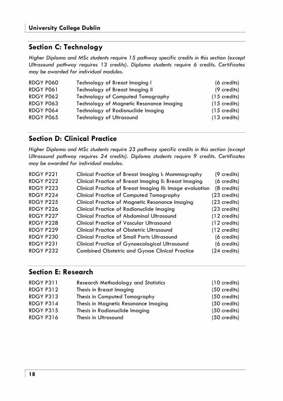
Section C: Technology Higher Diploma and MSc students require 15 pathway specific credits in this section (except Ultrasound pathway requires 13 credits). Diploma students require 6 credits. Certificates may be awarded for individual modules.
RDGY P060 Technology of Breast Imaging I (6 credits) RDGY P061 Technology of Breast Imaging II (9 credits) RDGY P062 Technology of Computed Tomography (15 credits) RDGY P063 Technology of Magnetic Resonance Imaging (15 credits) RDGY P064 Technology of Radionuclide Imaging (15 credits) RDGY P065 Technology of Ultrasound (13 credits)
Section D: Clinical Practice Higher Diploma and MSc students require 23 pathway specific credits in this section (except Ultrasound pathway requires 24 credits). Diploma students require 9 credits. Certificates may be awarded for individual modules.
RDGY P221 Clinical Practice of Breast Imaging I: Mammography (9 credits) RDGY P222 Clinical Practice of Breast Imaging II: Breast Imaging (6 credits) RDGY P223 Clinical Practice of Breast Imaging III: Image evaluation (8 credits) RDGY P224 Clinical Practice of Computed Tomography (23 credits) RDGY P225 Clinical Practice of Magnetic Resonance Imaging (23 credits) RDGY P226 Clinical Practice of Radionuclide Imaging (23 credits) RDGY P227 Clinical Practice of Abdominal Ultrasound (12 credits) RDGY P228 Clinical Practice of Vascular Ultrasound (12 credits) RDGY P229 Clinical Practice of Obstetric Ultrasound (12 credits) RDGY P230 Clinical Practice of Small Parts Ultrasound (6 credits) RDGY P231 Clinical Practice of Gynaecological Ultrasound (6 credits) RDGY P232 Combined Obstetric and Gynae Clinical Practice (24 credits)
Section E: Research RDGY P311 Research Methodology and Statistics (10 credits) RDGY P312 Thesis in Breast Imaging (50 credits) RDGY P313 Thesis in Computed Tomography (50 credits) RDGY P314 Thesis in Magnetic Resonance Imaging (50 credits) RDGY P315 Thesis in Radionuclide Imaging (50 credits) RDGY P316 Thesis in Ultrasound (50 credits)

Radiography
19
Syllabus of Modules
Section A: Professional Practice and Departmental Operation
Current Imaging RDGY P040 Communication between digital systems: essential factors, compatibility and incompatibility. DICOM III. Transmission methods: Ethernet, fibre optics, Network topologies. Data transmission rates. Impact of signal to noise ratios. Picture Archiving and Communications. Telemedicine. Send and receive stations: Correlation of modalities: clinically useful mergers. Clinical impact of image transmission. Cost effectiveness and impact on patient management.
Counselling and Communication RDGY P041 Counselling theories. Listening and reflecting. Bereavement and loss. Care of the councillor. Includes group work.
Equipment Management RDGY P042 Report Analysis. Specifications. Facility Design. Commissioning and operation.
Health Screening RDGY P043 WHO principles. Preventative medicine. The screening approach. Requirements of a screening test. Screening methods. Screening population. Cost – benefit analyses. Management of positive screening. Ethical issues.
Psychology RDGY P044 Current issues in sick patient psychology. Group dynamics. Stress management.
Quality in Healthcare RDGY P045 Concepts of quality in healthcare. Quality management, service industry, models of healthcare quality. Quality in healthcare: management, professional and patient perspectives. Measuring quality. Accreditation: theory and practice. Risk management. Freedom of Information.
Quality in Imaging RDGY P046 Theoretical concepts of quality: multiprofessional team, multiprofessional audit. Management of change. Accreditation. Medical/clinical: Audit, analysis and feedback.
University College Dublin
20
Section B: Practice
Practice of Breast Imaging I RDGY P121 Breast anatomy, physiology and development. Breast pathology. Operating principles. Breast examinations: clinical examination; mammography; biopsy; stereotaxis. Normal and abnormal radiographic appearances.
Practice of Breast Imaging II RDGYP122 Disease process and disease management. Screening referral. The asymptomatic patient. Alternate modalities: ultrasound; magnetic resonance; scintimammography; sentinel node imaging. Functional imaging. Normal and abnormal radiographic appearances. Image appraisal and evaluation. Audit of practice.
Practice of Breast Imaging III RDGY P123 Indication and presentation. Concepts of normality and abnormality. Analysis of diagnostic efficacy. Issues in observer variation.
Image quality evaluation. Quality criteria. Pattern recognition. Normal and abnormal radiographic appearances. Documenting and transmitting results.
Practice of Computed Tomography RDGY P124 Operating principles. Scanning techniques. Clinical applications, to include consideration of examination techniques on both a systemic and regional basis, to include 3D and image analysis. Radiation Protection and Dosimetry: Predicted and recorded bioeffects. Dose audit. Dose minimisation techniques.
Practice of Magnetic Resonance Imaging RDGY P125 Operating principles, scanning techniques. Consideration of examination techniques on both a systemic and regional basis. 3D techniques and image analysis.
Practice of Radionuclide Imaging RDGY P126 Operating principles. Scanning techniques. Clinical applications to include consideration of examination techniques on both a systemic and regional basis, encompassing principles and applications image analysis. Radiopharmacy design and operation. Radiopharmaceuticals. Radiation Protection and Dosimetry: General dosimetry and dose measurement. Dose calculations. Legislation: general and specific. Hazard control.
Practice of Abdominal ultrasound RDGY P127 Consideration of the indications; scanning techniques; normal and abnormal appearances; invasive, interventional and endoscopic procedures; and paediatric examinations of the liver, biliary system, pancreas, stomach, spleen, diaphragm, small and large bowel, kidneys, ureters, bladder, suprarenals, aorta and branches, inferior vena cava, mesentery and fluid collections.

Radiography
21
Practice of Obstetric ultrasound RDGY P129 Consideration of the indications; transabdominal and transvaginal scanning techniques; normal and abnormal appearances; and invasive and interventional procedures for: first trimester scans; gestational age assessment; foetal anatomy scans; foetal abnormality scans; foetal growth and weight estimation; foetal biophysical profiles; multiple pregnancy scans; Doppler ultrasound in pregnancy; investigation of placenta, cord and liquor; investigation of maternal conditions; examination for chromosomal abnormalities and syndromes; post partum scans.
Practice of Vascular ultrasound RDGY P128 Consideration of the indications; Doppler, duplex and colour flow ultrasound techniques; Doppler signal processing and spectral analysis; invasive and interventional and endoscopic procedures; normal and abnormal appearances and plethysmographic examinations for: cerebrovascular circulation; intra-abdominal vasculature; arterial and venous circulation of upper and lower extremities.
Practice of Gynaecological ultrasound RDGY P131 Consideration of the indications; transabdominal and transvaginal scanning techniques; normal and abnormal appearances; and invasive, interventional and endoscopic procedures for uterus, ovaries and adnexa; infertility.
Practice of Small parts ultrasound RDGY P130 Consideration of the indications; scanning techniques; normal and abnormal appearances; invasive, interventional and endoscopic procedures; and paediatric examinations for the breast; eye, musculo-skeletal system; neonatal intracranial structures; scrotum; thyroid and parathyroid glands.
Combined Obs/gynae practice RDGY P132 The obstetric and gynaecological syllabi are covered. In addition, specialist expertise in the field is developed by detailed consideration of an aspect of obstetric or gynaecological practice, which is presented in a small research project.
Section C: Technology
Technology of Breast Imaging I RDGY P060 Mammography units: generator; tube; filtration; console; image recording systems, conventional and digital. Compression. Exposure factors. Dosimetry and safety. Legislation. Processing: dedicated and shared units. Routine quality assurance.
Technology of Breast Imaging II RDGY P061 Mammography units: comparative evaluation of units available. Advanced consideration of filtration and target material. Examination optimisation: exposure and dose, variables with digital units. Advanced quality assurance.
University College Dublin
22
Technology of Computed Tomography RDGY P062 X-ray generation. Tubes, detectors, collimators. Hardware and software. Data acquisition, processing and presentation. Radiation Protection and Dosimetry: Predicted and recorded bioeffects. Dose audit. Dose minimisation techniques.
Technology of Magnetic Resonance Imaging RDGY P063 Magnets and magnetism, signal generation, gradients, pulse sequences, image formation, sequence parameters, flow phenomena, safety. Bioeffects, artefacts, quality assurance.
Technology of Radionuclide Imaging RDGY P064 Radiation detection. Computers and electronics. Gamma camera. Calibrators, monitors, counters and dosimeters. Quality assurance. SPECT and PET. Radiopharmacy. Radionuclides. Cyclotron. Isotope generator.
Technology of Ultrasound RDGY P065 Ultrasound physics: continuous waves, pulsed waves, propagation in tissues, bioeffects. Ultrasound production: beam shapes and transducers. A, M, B and Doppler mode scanners, combination scanners. Mensuration. Quality assurance. Dosimetry and safety.
Section D: Clinical Practice
Clinical Practice of Breast Imaging I (Mammography) RDGY P221 Each student will have 30 hours of practical tuition in the technique of mammography, followed by at least 400 hours of clinical practice in mammography. This should encompass performing mammograms, attending reporting sessions and involvement in administration and operation of the department.
Clinical Practice of Breast Imaging II (Breast Imaging) RDGY P222 Each student will undertake a range of mammographic procedures, and be involved in breast imaging using other modalities. Critical review of practice is implicit.
Clinical Practice of Breast Imaging III (Image Evaluation) RDGY P223 Each student will have 45 hours of supervised mammographic image evaluation, followed by extensive involvement in image evaluation. Three performance audits drawn from clinical practice will be compiled.
Clinical Practice of Computed Tomography RDGY P224 Each student undertakes an extensive range of CT examinations during approximately 1000 hours of clinical practice in a department with a reasonable case load. This should encompass performing scans, attending reporting and review sessions and involvement in administration and operation of the department.
The range of techniques learned should at least include common scanning sequences and approaches to image reconstruction for head, thorax, abdomen and limbs. Experience in sequential and spiral scanning is essential; experience in multi-slice scanning is desirable.

Radiography
23
Clinical Practice of Magnetic Resonance Imaging RDGY P225 Each student undertakes an extensive range of MR examinations during approximately 1000 hours of clinical practice in a department with a reasonable case load. This should encompass performing scans, attending reporting and review sessions and involvement in administration and operation of the department. The range of techniques learned should at least include common scanning sequences and approaches to image reconstruction for head, thorax, abdomen and limbs. Experience in vascular imaging is essential; experience in functional imaging, diffusion weighted imaging and spectroscopy is desirable.
Clinical Practice of Radionuclide Imaging RDGY P226 Each student undertakes an extensive range of RNI examinations during approximately 1000 hours of clinical practice in a department with a reasonable case load. This should encompass radiopharmacy preparation, performing scans, attending reporting and review sessions and involvement in administration and operation of the department. The range of techniques learned should at least include common scanning sequences and post processing techniques for skeletal, respiratory, nephro-urological, endocrine, GI, cardiovascular and central nervous systems. Experience in SPECT is desirable.
Clinical Practice of Abdominal ultrasound RDGY P227 Each student undertakes an extensive range of abdominal US examinations during approximately 500 hours of clinical practice in a department with a reasonable case load. This should encompass performing scans, attending reporting and review sessions and involvement in administration and operation of the department. The range of techniques learned should at least include scanning of the liver, biliary system, pancreas, stomach, spleen, diaphragm, small and large bowel, kidneys, ureters, bladder, suprarenals, aorta and branches, inferior vena cava, mesentery and fluid collections. Experience in invasive and interventional procedures is essential, experience in endoscopic procedures and paediatric examinations is desirable.
Clinical Practice of Obstetric ultrasound RDGY P229 Each student undertakes an extensive range of obstetric US examinations during approximately 500 hours of clinical practice in a department with a reasonable case load. This should encompass performing scans, attending reporting and review sessions and involvement in administration and operation of the department. The range of techniques learned should at least include first trimester scans; gestational age assessment; foetal anatomy scans; foetal abnormality scans; foetal growth and weight estimation; foetal biophysical profiles; multiple pregnancy scans; Doppler ultrasound in pregnancy; investigation of placenta, cord and liquor; investigation of maternal conditions; examination for chromosomal abnormalities and syndromes; and post partum scans. Experience in transabdominal and transvaginal scanning is essential.

University College Dublin
24
Clinical Practice of Vascular ultrasound RDGY P228 Each student undertakes an extensive range of vascular US examinations during approximately 500 hours of clinical practice in a department with a reasonable case load. This should encompass performing scans, attending reporting and review sessions and involvement in administration and operation of the department. The range of techniques learned should at least include scanning of cerebrovascular circulation; intra-abdominal vasculature; arterial and venous circulation of upper and lower extremities. Experience in Doppler, duplex and colour flow ultrasound techniques; Doppler signal processing and spectral analysis is essential, experience in plethysmographic examinations is desirable.
Clinical Practice of Gynaecological ultrasound RDGY P231 Each student undertakes an extensive range of gynaecological US examinations during approximately 250 hours of clinical practice in a department with a reasonable case load. This should encompass performing scans, attending reporting and review sessions and involvement in administration and operation of the department. The range of techniques learned should at least include transabdominal and transvaginal scanning techniques for uterus, ovaries and adnexa. Experience in infertility scanning is desirable.
Clinical Practice of Small parts ultrasound RDGY P230 Each student undertakes an extensive range of small parts US examinations during approximately 250 hours of clinical practice in a department with a reasonable case load. This should encompass performing scans, attending reporting and review sessions and involvement in administration and operation of the department. The range of techniques learned should at least include scanning of the following: the breast; eye, musculo-skeletal system; neonatal intracranial structures; scrotum; thyroid and parathyroid glands.
Clinical Practice of Obstetric and Gynae Ultrasound RDGY P232 Each student undertakes an extensive range of obstetric and gynaecological US examinations during approximately 1000 hours of clinical practice in a department with a reasonable case load. This should encompass performing scans, attending reporting and review sessions and involvement in administration and operation of the department. The syllabus for Clinical Practice in obstetric and gynaecological scanning should be followed. In addition, specialist expertise in the field should be supported by further scanning and case studies associated with a particular aspect of obstetric or gynaecological practice.
Radiography
25
Section E: Research
Research Methodology and Statistics RDGY P311 Introduction to research. Types of research. Survey design. Research management. Data collection. Data analysis. Presenting data and drawing conclusions.
Thesis Breast Imaging II RDGY P312 Computed Tomography RDGY P313 Magnetic Resonance Imaging RDGY P314 Radionuclide Imaging RDGY P315 Ultrasound RDGY P316
Each student undertakes a supervised research project that must be based in or related to radiographic practise in the particular speciality. The research may be undertaken in any clinical department or facility that is approved by the university. The findings of the research are presented in a thesis.

University College Dublin
26
Research MSc (Diagnostic Imaging Subject) The aims of the research MSc are to equip postgraduate radiographers with the capability to meet the challenge of major advances within Diagnostic Imaging, and to promote evidence based practice in the profession.
Admission
A candidate who has obtained the Degree of Bachelor of Science (Radiography) from the National University of Ireland, or other primary degree, or other qualification deemed equivalent by the Faculty of Medicine and Health Sciences, and who wishes to obtain further postgraduate training, with particular reference to academic and research aspects, shall be eligible to enter for the Degree of Master of Science in Radiography or other Diagnostic Imaging subject.
Application Individuals with a research interest are invited to contact +353-1-2094424 to discuss the possibilities. If the field of research is acceptable, the applicant is asked to submit an outline proposal and a CV. This application is considered individually by a Board of Studies. If deemed suitable, a supervisor is appointed and the applicant registers as an MSc student.
Induction Advisory meetings are held in the week before term begins. Attendance is obligatory for all postgraduates. Candidates meet their supervisor to discuss the proposed project in detail.
Outline structure and Progression
The Mode I MSc can be followed full or part time. Candidates must carry out research under the direction of the professor or university lecturer in the subject concerned. The thesis presented by the candidate is to embody the results of this research. The Faculty may approve of the work being carried out elsewhere under the direction of the professor or university lecturer in the subject concerned.
Running concurrently with the development of the research proposal, candidates attend a course in Research Methodology and Statistics and take an assessment, with credit accruing from the examination towards the MSc degree.
The allocated supervisor will give ongoing advice and guidance, and will engage in dialogue with the student at least monthly. Whilst the supervisor will direct the project, completion of the work remains the ultimate responsibility of the candidate.
Postgraduate research meetings are held in the School once a month during term time. Attendance is obligatory for all MSc students.

Radiography
27
Students are encouraged to publish their work and to present at international meetings. Generally, a Mode I MSc project should generate at least two presentations and at least two publications.
Examination
The Degree of MSc is awarded on the basis of all examination results and the thesis.
Three copies of the thesis must be lodged with the Supervisor of Examinations, University College Dublin, on or before the date fixed by the University.
Candidates give a twenty-minute presentation of their thesis to the examiners, and take questions. This is followed by an oral on the subject matter of the thesis, of not more than thirty minutes duration.

University College Dublin
28
Doctor of Philosophy (PhD) Candidates for this degree are required to be admitted by the Faculty on the recommendation of the head of department; admission must then be confirmed by the Academic Council and the Senate of the University. Candidates who have not graduated in this college may be admitted if suitably qualified. No candidate can be allowed to enter on a course of study and research for a PhD unless he/she has reached a high honours standard at the examination for the primary degree, or has presented such other evidence as will satisfy the Faculty of his/her fitness.
Requirements The candidate shall pursue research for a period of nine terms but the Senate may accept a period of six terms on the recommendation of the General Board of Studies in the case of a graduate whose attainments justify such shorter course.
The thesis must normally be prepared under the supervision of the Professor, but the Faculty may, on the recommendation of the Professor, assign another member of staff to supervise the candidate's research, under the professor's direction.
Examinations Candidates must carry out research under the direction of the professor or other supervisor in the subject concerned. The thesis presented by the candidate is to embody the results of this research. Three copies of the thesis must be lodged with the Supervisor of Examinations, University College Dublin, on or before the date fixed by the University. Candidates will be required to take an oral examination on the subject matter of their thesis
The Degree will not be awarded unless the examiners report the work is worthy of publication as a whole or in part.
Candidates for the PhD Degree will be allowed six years from the date of registration in which complete their Degree. If they have not done so within this time period, they must re-apply for registration.
Application Procedure Particulars and application forms may be obtained from the
School of Diagnostic Imaging, St. Anthony's, Herbert Avenue, Dublin 4, Ireland. Telephone: +353-1-209 4288