An Atypical Case of Poland Syndrome with Bilateral ...
5
Introduction Poland syndrome was first described in 1841 by Alfred Poland [1]. The pathophysiology is not yet fully under- stood. The diagnosis is often made clinically, but a thorough evaluation using different imaging modalities is required to fully depict the extent of the anomalies. Case Report A girl was born at gestational age of 36 weeks as a member of dizygotic twins. On the first postnatal clinical exami- nation, a focal, parasternal bulging of the left lung was noted, accentuated during crying, with impression of missing ribs 4 and 5 on the left side and missing muscle at those levels (Figure 1). She had bilateral gynaecomastia, normal extremities and no dysmorphic features. Her neurological examination was normal. A chest X-ray (Figure 2) showed 11 ribs on the right side and 11 ribs plus an apparent cervical rib on the left side. The medial end of the left clavicle had an abnormal low position compared to the right clavicle, and the cardiac silhouette was positioned on the right side. Based on the clinical findings and chest X-ray, the diagnosis of Poland syndrome was made. Abdominal ultrasound showed no associated abnor- malities nor signs of situs inversus. Transthoracic cardiac ultrasound showed dextroposition of the heart with nor- mal morphology and functionality. A chest computed tomography (CT) scan (Figures 3–6) using a low-dose pediatric protocol with a CTDI of 0.33 mGy showed a dysplastic left sternal half with abnormal orientation of the left clavicle. There were 12 ribs present bilaterally, with a hypoplastic first rib bilaterally. On the left side Geeroms, D, et al. An Atypical Case of Poland Syndrome with Bilateral Features and Dextroposition of the Heart. Journal of the Belgian Society of Radiology. 2019; 103(1): 45, 1–5. DOI: https://doi.org/10.5334/jbsr.1860 UZ Leuven, BE Corresponding author: Barbara Geeroms ([email protected]) CASE REPORT An Atypical Case of Poland Syndrome with Bilateral Features and Dextroposition of the Heart In the work-up of Poland syndrome, different imaging modalities are necessary to depict the full extent of the anomalies Barbara Geeroms, Luc Breysem and Michaël Aertsen Poland syndrome is defined by the unilateral aplasia or hypoplasia of the sternocostal head of the major pectoral muscle and is associated with variable ipsilateral thoracic and upper limb anomalies. Most frequently, the abnormalities are unilateral and on the right side. We present an atypical case of Poland syndrome in a baby girl with bilateral chest abnormalities, mainly on the left side, and second- ary dextroposition of the heart. As required in almost all cases of Poland syndrome, different imaging modalities were used to evaluate the extent of our patient’s anomalies. Keywords: Case report; Poland syndrome; dextrocardia; chest wall; chest deformity Figure 1: Photograph of the baby girl at 2 months of age demonstrating the asymmetry in the chest wall with impression of missing muscle and ribs 4 and 5 on the left side (white arrow). The bilateral gynaecomastia is also visible.
Transcript of An Atypical Case of Poland Syndrome with Bilateral ...

IntroductionPoland syndrome was first described in 1841 by Alfred Poland [1]. The pathophysiology is not yet fully under-stood. The diagnosis is often made clinically, but a thorough evaluation using different imaging modalities is required to fully depict the extent of the anomalies.
Case ReportA girl was born at gestational age of 36 weeks as a member of dizygotic twins. On the first postnatal clinical exami-nation, a focal, parasternal bulging of the left lung was noted, accentuated during crying, with impression of missing ribs 4 and 5 on the left side and missing muscle at those levels (Figure 1). She had bilateral gynaecomastia, normal extremities and no dysmorphic features. Her neurological examination was normal.
A chest X-ray (Figure 2) showed 11 ribs on the right side and 11 ribs plus an apparent cervical rib on the left side. The medial end of the left clavicle had an abnormal low position compared to the right clavicle, and the cardiac silhouette was positioned on the right side. Based on the clinical findings and chest X-ray, the diagnosis of Poland syndrome was made.
Abdominal ultrasound showed no associated abnor-malities nor signs of situs inversus. Transthoracic cardiac ultrasound showed dextroposition of the heart with nor-mal morphology and functionality. A chest computed tomography (CT) scan (Figures 3–6) using a low-dose pediatric protocol with a CTDI of 0.33 mGy showed a
dysplastic left sternal half with abnormal orientation of the left clavicle. There were 12 ribs present bilaterally, with a hypoplastic first rib bilaterally. On the left side
Geeroms, D, et al. An Atypical Case of Poland Syndrome with Bilateral Features and Dextroposition of the Heart. Journal of the Belgian Society of Radiology. 2019; 103(1): 45, 1–5. DOI: https://doi.org/10.5334/jbsr.1860
UZ Leuven, BECorresponding author: Barbara Geeroms ([email protected])
CASE REPORT
An Atypical Case of Poland Syndrome with Bilateral Features and Dextroposition of the HeartIn the work-up of Poland syndrome, different imaging modalities are necessary to depict the full extent of the anomalies
Barbara Geeroms, Luc Breysem and Michaël Aertsen
Poland syndrome is defined by the unilateral aplasia or hypoplasia of the sternocostal head of the major pectoral muscle and is associated with variable ipsilateral thoracic and upper limb anomalies. Most frequently, the abnormalities are unilateral and on the right side. We present an atypical case of Poland syndrome in a baby girl with bilateral chest abnormalities, mainly on the left side, and second-ary dextroposition of the heart. As required in almost all cases of Poland syndrome, different imaging modalities were used to evaluate the extent of our patient’s anomalies.
Keywords: Case report; Poland syndrome; dextrocardia; chest wall; chest deformity
Figure 1: Photograph of the baby girl at 2 months of age demonstrating the asymmetry in the chest wall with impression of missing muscle and ribs 4 and 5 on the left side (white arrow). The bilateral gynaecomastia is also visible.
Geeroms et al: An Atypical Case of Poland Syndrome with Bilateral Features and Dextroposition of the Heart
Art. 45, page 2 of 5
there was partial agenesis of the anterior arches of ribs 2, 4, and 5 and, less pronounced, of ribs 6 and 7. On the right side there was partial agenesis of the anterior arch of rib 2. Secondary to these rib defects, the intercostal spaces between left ribs 4 and 8 were narrowed. The sternocos-tal head of the left major pectoral muscle was absent, the clavicular head was intact. Glandular breast tissue was present bilaterally. The heart was positioned in the right hemithorax with normal orientation of the apex: ‘dex-troposition of the heart’. There were no vertebral defects.
The diagnosis of Poland syndrome was confirmed with bilateral, mainly left-sided chest wall abnormalities and secondary dextroposition of the heart.
DiscussionPoland syndrome is seen in 1/20,000 to 1/30,000 of patients, and it is more frequent in males than females [1–3]. Most commonly (60–75% of cases) the anomalies are unilateral and affect the right hemithorax [1–3]. The condition is defined by the unilateral aplasia/hypoplasia of the sterno-costal head of the major pectoral muscle but can be asso-ciated with other thoracic defects, including hypoplasia or aplasia of breasts and ribs, chest wall depression, and sternal anomalies [4]. Upper-limb anomalies can be associ-ated with Poland syndrome; the most frequent is ipsilateral brachysyndactyly [1, 2]. Lung herniation and dextroposi-tion of the heart are rarely associated with it. Cardiac dex-troposition has been described in 5.6 to 11.2% of patients with Poland syndrome [1]. The volume loss of the left
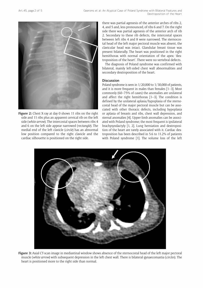
Figure 2: Chest X-ray at day 0 shows 11 ribs on the right side and 11 ribs plus an apparent cervical rib on the left side (white arrow). The intercostal spaces between ribs 4 and 6 on the left side appear narrowed (rectangle). The medial end of the left clavicle (circle) has an abnormal low position compared to the right clavicle and the cardiac silhouette is positioned on the right side.
Figure 3: Axial CT-scan image in mediastinal window shows absence of the sternocostal head of the left major pectoral muscle (white arrow) with subsequent depression in the left chest wall. There is bilateral gynaecomastia (circles). The heart is positioned more to the right side than normal.

Geeroms et al: An Atypical Case of Poland Syndrome with Bilateral Features and Dextroposition of the Heart
Art. 45, page 3 of 5
Figure 4: 3D reconstruction of the chest CT-scan shows a dysplastic left sternal half and a dextroposition of the heart. There is a hypoplastic first rib on the left side (circle).
Figure 5: 3D reconstruction of the chest CT-scan shows partial agenesis of the anterior arches of ribs 2, 4 to 7 on the left side (white arrows).

Geeroms et al: An Atypical Case of Poland Syndrome with Bilateral Features and Dextroposition of the Heart
Art. 45, page 4 of 5
hemithorax is only significant enough to cause displace-ment of the heart to the right hemithorax in patients with left-sided Poland syndrome with partial agenesis of at least two ribs [1, 2]. Dextroposition of the heart differs from dex-trocardia. Dextrocardia is mirroring of the heart with the heart located in the right hemithorax and the apex point-ing to the right side [1] and can be associated with situs inversus. In dextroposition, the heart is positioned in the right hemithorax, but the apex is pointing to the left and there is no association with situs inversus.
The pathogenesis of Poland syndrome is not completely understood. Different hypotheses have been formulated, including teratogenic factors, like smoking and drugs [1]. The most adopted theory is the vascular hypothesis, which proposes a transient decrease in flow during the sixth week of gestation in one or more branches of the subclavian or vertebral artery [1, 4]. The duration and loca-tion of the vascular disruption determines the severity and extent of the abnormalities [2, 5]. This is called the ‘subclavian artery supply disruption sequence’ and also explains other anomalies that are frequently associated with Poland syndrome, such as Klippel-Feil syndrome and Sprengel deformity [1, 4]. A third theory proposes genetic factors [1]. The most frequently proposed inheritance is a paradominant inheritance, in which a mutation does not translate into abnormal phenotype in heterozygous indi-viduals, but only expresses itself when a somatic muta-tion occurs during embryogenesis, which leads to loss of heterozygosity. This concept explains the occasional familial occurrence of usually sporadic traits such as Klippel-Trénaunay syndrome [6]. In such cases, the muta-tions could be transmitted through many generations
in the absence of apparent phenotype [2]. This genetic hypothesis would explain the occurrence of familial and bilateral affected cases, although these could be caused by vascular disruption of multiple arteries at different stages in development [7].
Our patient is a special case in many ways. She is female, has bilateral chest wall abnormalities, and has dextropo-sition of the heart secondary to the partial agenesis of multiple ribs on the left side. Her twin sister does not show any signs of Poland syndrome, although this cannot contradict a genetic hypothesis as dizygotic twins are not genetically identical.
The diagnosis of Poland syndrome is mostly made clini-cally. However, a thorough evaluation using different imaging modalities is required to evaluate the extent of the anomalies. So far, no clear guidelines have been pub-lished regarding the types and timing of imaging modali-ties that should be performed. In practice, a screening chest X-ray is often followed by a chest CT scan to depict the thoracic abnormalities. Other technical examinations are requested based on clinical findings and can include a chest MRI, X-rays of the upper limbs, echocardiography, or abdominal ultrasound. An individual approach and work-up appears to be the best way, as each patient exhibits different abnormalities.
ConclusionPoland syndrome with bilateral features and dextroposi-tion of the heart is an atypical and rare presentation of a rare syndrome. A thorough clinical examination and different imaging modalities are necessary to fully depict the anomalies of patients with Poland syndrome.
Figure 6: 3D reconstruction of the chest CT-scan shows bilateral hypoplastic first ribs (circles) and a partial agenesis of the anterior arches of ribs 2 bilaterally and of ribs 4 to 7 on the left side (white arrows).

Geeroms et al: An Atypical Case of Poland Syndrome with Bilateral Features and Dextroposition of the Heart
Art. 45, page 5 of 5
AcknowledgementsSpecial acknowledgement goes to Prof. Dr. Anne Debeer, neonatologist at UZ Leuven, and Prof. Dr. Marleen Smet, paediatric radiologist at UZ Leuven, for their scientific contribution to this article.
Competing InterestsThe authors have no competing interests to declare.
References 1. Torre, M, Baban, A, Buluggiu, A, et al. Dextrocardia
in patients with Poland syndrome: Phenotypic char-acterization provides insight into the pathogenesis. J Thorac Cardiovasc Surg. 2010; 139(5): 1177–82. DOI: https://doi.org/10.1016/j.jtcvs.2009.08.024
2. Lacorte, D, Marsella, M and Guerrini, P. A case of Poland syndrome associated with dextroposition. Ital J Pediatr. 2010; 36(1): 21–3. DOI: https://doi.org/10.1186/1824-7288-36-21
3. Sepulveda, W. Poland syndrome – A rare cause of car-diac dextroposition in the foetus. Prenat Diagn. 2009; 29: 903–5. DOI: https://doi.org/10.1002/pd.2310
4. Cingel, V, Bohac, M, Mestanova, V, et al. Poland syndrome: From embryological basis to plastic surgery. Surg Radiol Anat. 2013; 35(8): 639–46. DOI: https://doi.org/10.1007/s00276-013-1083-7
5. Ferraro, GA, Perrotta, A, Rossano, F, et al. Poland syndrome: Description of an atypical variant. Aesthetic Plast Surg. 2005; 29(1): 32–3. DOI: https://doi.org/10.1007/s00266-004-0 047-z
6. Steijlen, PM and Van Steensel, MAM. Paradominant inheritance, a hypothesis explain-ing occasional familial occurrence of sporadic syn-dromes. American Journal of Medical Genetics. 1999; 85(4): 359–60. DOI: https://doi.org/10.1002/(SICI)1096-8628(19990806)85:4<359::AID-AJMG10>3.0.CO;2-V
7. Baban, A, Torre, M, Bianca, S, et al. Poland syn-drome with bilateral features: Case description with review of the literature. Am J Med Genet Part A. 2009; 149(7): 1597–602. DOI: https://doi.org/10.1002/ajmg.a.32922
How to cite this article: Geeroms, B, Breysem, L and Aertsen, M. An Atypical Case of Poland Syndrome with Bilateral Features and Dextroposition of the Heart. Journal of the Belgian Society of Radiology. 2019; 103(1): 45, 1–5. DOI: https://doi.org/10.5334/jbsr.1860
Submitted: 06 June 2019 Accepted: 30 June 2019 Published: 11 July 2019
Copyright: © 2019 The Author(s). This is an open-access article distributed under the terms of the Creative Commons Attribution 4.0 International License (CC-BY 4.0), which permits unrestricted use, distribution, and reproduction in any medium, provided the original author and source are credited. See http://creativecommons.org/licenses/by/4.0/.
OPEN ACCESS Journal of the Belgian Society of Radiology is a peer-reviewed open access journal published by Ubiquity Press.