
An attractor hypothesis of obsessive–compulsive disorderLoh+08_OCD.pdf · in OCD, and the...
13
An attractor hypothesis of obsessive–compulsive disorder Edmund T. Rolls, 1 Marco Loh 2 and Gustavo Deco 2,3 1 Oxford Centre for Computational Neuroscience, Oxford, UK 2 Department of Technology, Computational Neuroscience, Universitas Pompeu Fabra, Barcelona, Spain 3 Institucio ´ Catalana de Recerca i Estudis Avanc ¸ ats (ICREA), Barcelona, Spain Keywords: emotion, glutamate, neural network, obsession, OCD, stability Abstract We propose a top-down approach to the symptoms of obsessive–compulsive disorder (OCD) based on a statistical dynamical framework. An increased depth in the basins of attraction of attractor network states in the brain makes each state too stable, so that it tends to remain locked in that state and can not easily be moved on to another state. We suggest that the different symptoms that may be present in OCD could be related to changes of this type in different brain regions. For example, the difficulty in attentional and cognitive set switching could be related to networks operating in this way in the prefrontal cortex. Repetitive actions and a difficulty in moving to new actions could be related to overstability in networks in the higher order motor, including cingulate areas. In integrate- and-fire network simulations, an increase in the N-methyl-d-aspartate (NMDA) and ⁄ or a-amino-3-hydroxy-5-methyl-4-isoxazole- propionic acid (AMPA) receptor conductances, which increases the depth of the attractor basins, increases the stability of attractor networks, and makes them less easily moved on to another state by a new stimulus. Increasing c-aminobutyric acid (GABA)-receptor activated currents can partly reverse this overstability. There is now some evidence for overactivity in glutamate transmitter systems in OCD, and the hypothesis presented here shows how some of the symptoms of OCD could be produced by the increase in the stability of attractor networks that is produced by increased glutamatergic activity. Introduction Obsessive–compulsive disorder (OCD) is a chronically debilitating disorder with a lifetime prevalence of 2–3% (Robins et al., 1984; Karno et al., 1988; Weissman et al., 1994). It is characterized by two sets of symptoms, obsessive and compulsive. Obsessions are unwanted, intrusive, recurrent thoughts or impulses that are often concerned with themes of contamination and ‘germs’, checking household items in case of fire or burglary, order and symmetry of objects, or fears of harming oneself or others. Compulsions are ritualistic, repetitive behaviours or mental acts carried out in relation to these obsessions, e.g. washing, household safety checks, counting, rearrangement of objects in symmetrical array or constant checking of oneself and others to ensure no harm has occurred (Menzies et al., 2008). Patients with OCD experience the persistent intrusion of thoughts that they generally perceive as foreign and irrational but which cannot be dismissed. The anxiety associated with these unwanted and disturbing thoughts can be extremely intense; it is often described as a feeling that something is incomplete or wrong, or that terrible consequences will ensue if specific actions are not taken. Many patients engage in repetitive, compulsive behaviours that aim to discharge the anxieties associ- ated with these obsessional thoughts. Severely affected patients can spend many hours each day in their obsessional thinking and resultant compulsive behaviours, leading to marked disability (Pittenger et al., 2006). While patients with OCD exhibit a wide variety of obsessions and compulsions, the symptoms tend to fall into specific clusters. Common patterns include obsessions of contamination, with accompanying cleaning compulsions; obsessions with symmetry or order, with accompanying ordering behaviours; obsessions of saving, with accompanying hoarding; somatic obsessions; aggressive obsessions with checking compulsions; and sexual and religious obsessions (Pittenger et al., 2006). In this paper we describe a theory of how OCDs arise, and of the different symptoms. The theory is based on the top-down proposal that there is overstability of attractor neuronal networks in cortical and related areas in OCDs. The approach is top-down in that it starts with the set of symptoms and maps them onto the dynamical systems framework, and only after this considers detailed underlying biological mechanisms, of which there could be many, that might produce the effects. (In contrast, a complementary bottom-up approach starts from detailed neurobiological mechanisms, and aims to interpret their implications with a brain-like model for higher level phenomena.) We show by integrate-and-fire neuronal network simulations that the overstability could arise by, for example, overactivity in glutamatergic excitatory neurotransmitter synapses, which produces an increased depth of the basins of attraction, in the presence of which neuronal spiking-related and potentially other noise is insufficient to help the system move out of an attractor basin. We relate this top-down proposal, related to the stochastic dynamics of neuronal networks, to new evidence that there may be overactivity in glutamatergic systems in OCDs, and consider the implications for treatment. There has been interest for some time in the application of complex systems theory to understanding brain function and behaviour Correspondence: Professor E. T. Rolls, as above. E-mail: [email protected]; http://www.oxcns.org Received 8 May 2008, revised 6 June 2008, accepted 23 June 2008 European Journal of Neuroscience, pp. 1–13, 2008 doi:10.1111/j.1460-9568.2008.06379.x ª The Authors (2008). Journal Compilation ª Federation of European Neuroscience Societies and Blackwell Publishing Ltd European Journal of Neuroscience
Transcript of An attractor hypothesis of obsessive–compulsive disorderLoh+08_OCD.pdf · in OCD, and the...
An attractor hypothesis of obsessive–compulsive disorder
Edmund T. Rolls,1 Marco Loh2 and Gustavo Deco2,3
1Oxford Centre for Computational Neuroscience, Oxford, UK2Department of Technology, Computational Neuroscience, Universitas Pompeu Fabra, Barcelona, Spain3Institucio Catalana de Recerca i Estudis Avancats (ICREA), Barcelona, Spain
Keywords: emotion, glutamate, neural network, obsession, OCD, stability
Abstract
We propose a top-down approach to the symptoms of obsessive–compulsive disorder (OCD) based on a statistical dynamicalframework. An increased depth in the basins of attraction of attractor network states in the brain makes each state too stable, so thatit tends to remain locked in that state and can not easily be moved on to another state. We suggest that the different symptoms thatmay be present in OCD could be related to changes of this type in different brain regions. For example, the difficulty in attentional andcognitive set switching could be related to networks operating in this way in the prefrontal cortex. Repetitive actions and a difficulty inmoving to new actions could be related to overstability in networks in the higher order motor, including cingulate areas. In integrate-and-fire network simulations, an increase in the N-methyl-d-aspartate (NMDA) and ⁄ or a-amino-3-hydroxy-5-methyl-4-isoxazole-propionic acid (AMPA) receptor conductances, which increases the depth of the attractor basins, increases the stability of attractornetworks, and makes them less easily moved on to another state by a new stimulus. Increasing c-aminobutyric acid (GABA)-receptoractivated currents can partly reverse this overstability. There is now some evidence for overactivity in glutamate transmitter systemsin OCD, and the hypothesis presented here shows how some of the symptoms of OCD could be produced by the increase in thestability of attractor networks that is produced by increased glutamatergic activity.
Introduction
Obsessive–compulsive disorder (OCD) is a chronically debilitatingdisorder with a lifetime prevalence of 2–3% (Robins et al., 1984;Karno et al., 1988; Weissman et al., 1994). It is characterized bytwo sets of symptoms, obsessive and compulsive. Obsessions areunwanted, intrusive, recurrent thoughts or impulses that are oftenconcerned with themes of contamination and ‘germs’, checkinghousehold items in case of fire or burglary, order and symmetry ofobjects, or fears of harming oneself or others. Compulsions areritualistic, repetitive behaviours or mental acts carried out inrelation to these obsessions, e.g. washing, household safety checks,counting, rearrangement of objects in symmetrical array or constantchecking of oneself and others to ensure no harm has occurred(Menzies et al., 2008). Patients with OCD experience the persistentintrusion of thoughts that they generally perceive as foreign andirrational but which cannot be dismissed. The anxiety associatedwith these unwanted and disturbing thoughts can be extremelyintense; it is often described as a feeling that something isincomplete or wrong, or that terrible consequences will ensue ifspecific actions are not taken. Many patients engage in repetitive,compulsive behaviours that aim to discharge the anxieties associ-ated with these obsessional thoughts. Severely affected patients canspend many hours each day in their obsessional thinking andresultant compulsive behaviours, leading to marked disability(Pittenger et al., 2006).
While patients with OCD exhibit a wide variety of obsessions andcompulsions, the symptoms tend to fall into specific clusters. Commonpatterns include obsessions of contamination, with accompanyingcleaning compulsions; obsessions with symmetry or order, withaccompanying ordering behaviours; obsessions of saving, withaccompanying hoarding; somatic obsessions; aggressive obsessionswith checking compulsions; and sexual and religious obsessions(Pittenger et al., 2006).In this paper we describe a theory of how OCDs arise, and of the
different symptoms. The theory is based on the top-down proposal thatthere is overstability of attractor neuronal networks in cortical andrelated areas in OCDs. The approach is top-down in that it starts withthe set of symptoms and maps them onto the dynamical systemsframework, and only after this considers detailed underlying biologicalmechanisms, of which there could be many, that might produce theeffects. (In contrast, a complementary bottom-up approach starts fromdetailed neurobiological mechanisms, and aims to interpret theirimplications with a brain-like model for higher level phenomena.) Weshow by integrate-and-fire neuronal network simulations that theoverstability could arise by, for example, overactivity in glutamatergicexcitatory neurotransmitter synapses, which produces an increaseddepth of the basins of attraction, in the presence of which neuronalspiking-related and potentially other noise is insufficient to help thesystem move out of an attractor basin. We relate this top-downproposal, related to the stochastic dynamics of neuronal networks, tonew evidence that there may be overactivity in glutamatergic systemsin OCDs, and consider the implications for treatment.There has been interest for some time in the application of
complex systems theory to understanding brain function and behaviour
Correspondence: Professor E. T. Rolls, as above.E-mail: [email protected]; http://www.oxcns.org
Received 8 May 2008, revised 6 June 2008, accepted 23 June 2008
European Journal of Neuroscience, pp. 1–13, 2008 doi:10.1111/j.1460-9568.2008.06379.x
ª The Authors (2008). Journal Compilation ª Federation of European Neuroscience Societies and Blackwell Publishing Ltd
E u r o p e a n J o u r n a l o f N e u r o s c i e n c e
(Blackerby, 1993; Riley & Turvey, 2001; Peled, 2004; Heinrichs, 2005;Lewis, 2005). Dynamically realistic neuronal network models ofworking memory (Wang, 1999, 2001; Compte et al., 2000; Durstewitzet al., 2000a, b; Brunel & Wang, 2001) and decision-making (Wang,2002; Deco & Rolls, 2006), and attention (Rolls & Deco, 2002; Deco &Rolls, 2005), and how they are influenced by neuromodulators such asdopamine (Durstewitz et al., 1999, 2000a; Brunel & Wang, 2001;Durstewitz & Seamans, 2002; Deco & Rolls, 2003) have beenproduced, and it has been suggested that alterations in the attractorlandscape may contribute to schizophrenia (Seamans & Yang, 2004;Winterer & Weinberger, 2004; Rolls, 2005). Indeed we have recentlyconsidered how a decreased depth in the basins of attraction ofprefrontal cortical networks might contribute to, for example, thecognitive symptoms of schizophrenia, by making short-term memorysystems unstable, and thereby contributing to the failure to maintainattention and the disorganization that characterizes the dysexecutivesymptoms that are among the cognitive symptoms of schizophrenia(Loh et al., 2007). This decreased depth in the basins of attraction inschizophrenia is attributed inter alia to decreased currents in N-methyl-d-aspartate (NMDA) receptor-activated channels (Coyle, 2006), andthe associated decrease in firing rates when present in the orbitofrontaland anterior cingulate cortices is related to the negative symptoms, suchas anhedonia, and flattening of affect, motivation and volition, whichcontribute to the psychomotor poverty in schizophrenia according tothis computational approach (Rolls, 2005; Loh et al., 2007).
Attractor networks, and their stability
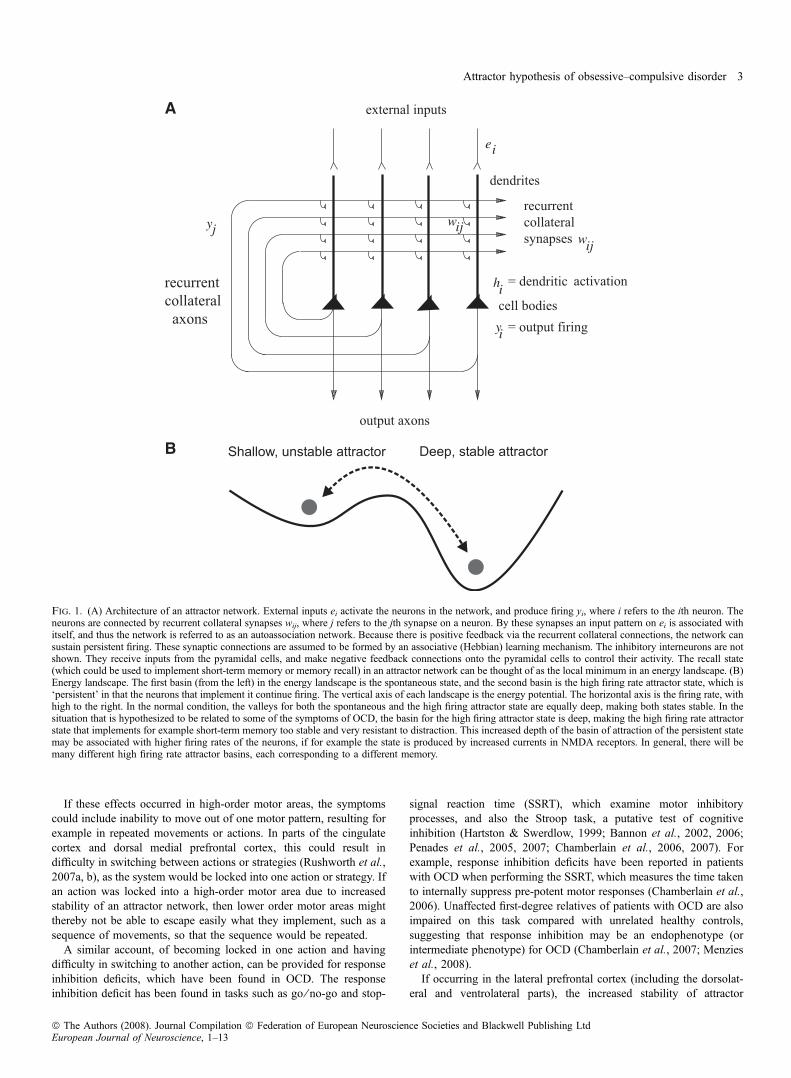
The attractor framework is based on dynamical systems theory. In anetwork of interconnected neurons, a memory pattern (represented bya set of active neurons) can be stored by synaptic modification, andlater recalled by external inputs. Furthermore, a pattern activated by aninput is then stably maintained by the system even after input offset.These patterns could correspond to memories, perceptual representa-tions or thoughts.The architecture of an attractor or autoassociation network is as
follows (Fig. 1A). External inputs ei activate the neurons in thenetwork, and produce firing yi, where i refers to the ith neuron. Theneurons are connected to each other by recurrent collateral synapseswij, where j refers to the jth synapse on a neuron. By these synapses aninput pattern on ei is associated with itself, and thus the network isreferred to as an autoassociation network. Because there is positivefeedback implemented via the recurrent collateral connections, thenetwork can sustain persistent firing. These synaptic connections areassumed to build up by an associative (Hebbian) learning mechanism(Hebb, 1949). The inhibitory interneurons are not shown. Theyreceive inputs from the pyramidal cells, and make inhibitory negativefeedback connections onto the pyramidal cells to keep their activityunder control. Hopfield (1982) showed that the recall state in a simpleattractor network can be thought of as the local minimum in an energylandscape, where the energy would be defined as
E ¼ � 1
2
X
i; j
wijðyi � <y>Þðyj � <y>Þ: ð1Þ
Autoassociation attractor systems have two types of stable fixedpoints: a spontaneous state with a low firing rate, and one or moreattractor states with high firing rates in which the positive feedbackimplemented by the recurrent collateral connections maintains a highfiring rate. We sometimes refer to this latter state as the persistent state.The area in the energy landscape within which the system will move toa stable attractor state is called its basin of attraction.
The attractor dynamics can be pictured by energy landscapes, whichindicate the basin of attraction by valleys, and the attractor states orfixed points by the bottom of the valleys. [Although energy functionsapply to recurrent networks with symmetric connections between theneurons (Hopfield, 1982), as would be the case in a fully connectednetwork with associative synaptic modification, and do not necessarilyapply to more complicated networks with, for example, incompleteconnectivity, nevertheless the properties of these other recurrentnetworks are similar (Treves, 1991; Treves & Rolls, 1991; Rolls &Treves, 1998), and the concept of an energy function and landscape isuseful for discussion purposes. In practice, a Lyapunov function canbe used to prove analytically that there is a stable fixed point such asan attractor basin (Khalil, 1996), and even in systems where this cannot be proved analytically, it may still be possible to show numericallythat there are stable fixed points, to measure the flow towards thosefixed points that describes the depth of the attractor basin as we havedone for this type of network (Loh et al., 2007), and to use the conceptof energy or potential landscapes to help visualize the properties of thesystem.] The stability of an attractor is characterized by the averagetime in which the system stays in the basin of attraction under theinfluence of noise. The noise provokes transitions to other attractorstates. One source of noise results from the interplay between thePoissonian character of the spikes and the finite-size effect due tothe limited number of neurons in the network. Two factors determinethe stability. First, if the depths of the attractors are shallow (as in theleft compared with the right valley in Fig. 1B), then less force isneeded to move a ball from one valley to the next. Second, high noisewill make it more likely that the system will jump over an energyboundary from one state to another. We envision that the brain as adynamical system has characteristics of such an attractor system,including statistical fluctuations. The noise could arise not only fromthe probabilistic spiking of the neurons, which has significant effectsin finite size integrate-and-fire networks (Deco & Rolls, 2006), butalso from any other source of noise in the brain or the environment(Faisal et al., 2008), including the effects of distracting stimuli.
A hypothesis about the increased stability of attractornetworks and the symptoms of OCD
We hypothesize that cortical and related attractor networks become toostable in OCD, so that once in an attractor state, the networks tend toremain there too long. The hypothesis is that the depths of the basinsof attraction become deeper, and that this is what makes the attractornetworks more stable. We further hypothesize that part of themechanism for the increased depth of the basins of attraction isincreased glutamatergic transmission, which increases the depth of thebasins of attraction by increasing the firing rates of the neurons, and byincreasing the effective value of the synaptic weights between theassociatively modified synapses that define the attractor, as is madeevident in Eq. (1) above. The synaptic strength is effectively increasedif more glutamate is released per action potential at the synapse, or ifin other ways the currents injected into the neurons through theNMDA and ⁄ or a-amino-3-hydroxy-5-methyl-4-isoxazolepropionicacid (AMPA) synapses are larger. In addition, if NMDA receptorfunction is increased, this could also increase the stability of thesystem because of the temporal smoothing effect of the long timeconstant of the NMDA receptors (Wang, 1999).This increased stability of cortical and related attractor networks,
and the associated higher neuronal firing rates, could occur indifferent brain regions, and thereby produce different symptoms, asfollows.
2 E. T. Rolls et al.
ª The Authors (2008). Journal Compilation ª Federation of European Neuroscience Societies and Blackwell Publishing LtdEuropean Journal of Neuroscience, 1–13
If these effects occurred in high-order motor areas, the symptomscould include inability to move out of one motor pattern, resulting forexample in repeated movements or actions. In parts of the cingulatecortex and dorsal medial prefrontal cortex, this could result indifficulty in switching between actions or strategies (Rushworth et al.,2007a, b), as the system would be locked into one action or strategy. Ifan action was locked into a high-order motor area due to increasedstability of an attractor network, then lower order motor areas mightthereby not be able to escape easily what they implement, such as asequence of movements, so that the sequence would be repeated.
A similar account, of becoming locked in one action and havingdifficulty in switching to another action, can be provided for responseinhibition deficits, which have been found in OCD. The responseinhibition deficit has been found in tasks such as go ⁄ no-go and stop-
signal reaction time (SSRT), which examine motor inhibitoryprocesses, and also the Stroop task, a putative test of cognitiveinhibition (Hartston & Swerdlow, 1999; Bannon et al., 2002, 2006;Penades et al., 2005, 2007; Chamberlain et al., 2006, 2007). Forexample, response inhibition deficits have been reported in patientswith OCD when performing the SSRT, which measures the time takento internally suppress pre-potent motor responses (Chamberlain et al.,2006). Unaffected first-degree relatives of patients with OCD are alsoimpaired on this task compared with unrelated healthy controls,suggesting that response inhibition may be an endophenotype (orintermediate phenotype) for OCD (Chamberlain et al., 2007; Menzieset al., 2008).If occurring in the lateral prefrontal cortex (including the dorsolat-
eral and ventrolateral parts), the increased stability of attractor
A
B
Fig. 1. (A) Architecture of an attractor network. External inputs ei activate the neurons in the network, and produce firing yi, where i refers to the ith neuron. Theneurons are connected by recurrent collateral synapses wij, where j refers to the jth synapse on a neuron. By these synapses an input pattern on ei is associated withitself, and thus the network is referred to as an autoassociation network. Because there is positive feedback via the recurrent collateral connections, the network cansustain persistent firing. These synaptic connections are assumed to be formed by an associative (Hebbian) learning mechanism. The inhibitory interneurons are notshown. They receive inputs from the pyramidal cells, and make negative feedback connections onto the pyramidal cells to control their activity. The recall state(which could be used to implement short-term memory or memory recall) in an attractor network can be thought of as the local minimum in an energy landscape. (B)Energy landscape. The first basin (from the left) in the energy landscape is the spontaneous state, and the second basin is the high firing rate attractor state, which is‘persistent’ in that the neurons that implement it continue firing. The vertical axis of each landscape is the energy potential. The horizontal axis is the firing rate, withhigh to the right. In the normal condition, the valleys for both the spontaneous and the high firing attractor state are equally deep, making both states stable. In thesituation that is hypothesized to be related to some of the symptoms of OCD, the basin for the high firing attractor state is deep, making the high firing rate attractorstate that implements for example short-term memory too stable and very resistant to distraction. This increased depth of the basin of attraction of the persistent statemay be associated with higher firing rates of the neurons, if for example the state is produced by increased currents in NMDA receptors. In general, there will bemany different high firing rate attractor basins, each corresponding to a different memory.
Attractor hypothesis of obsessive–compulsive disorder 3
ª The Authors (2008). Journal Compilation ª Federation of European Neuroscience Societies and Blackwell Publishing LtdEuropean Journal of Neuroscience, 1–13
networks could produce symptoms that include a difficulty in shiftingattention and in cognitive set shifting. These are in fact importantsymptoms that can be found in OCD (Menzies et al., 2008). Thesehave been concerned with two quite different forms of shift: affectiveset shifting, where the affective or reward value of a stimulus changesover time (e.g. a rewarded stimulus is no longer rewarded; intradi-mensional or ID set shifting); and attentional set shifting, where thestimulus dimension (e.g. shapes or colours) to which the subject mustattend is changed (extradimensional or ED set shifting). Deficits ofattentional set shifting in OCD have been found in several neurocog-nitive studies using the CANTAB ID ⁄ ED set shifting task (Vealeet al., 1996; Watkins et al., 2005; Chamberlain et al., 2006, 2007).This deficit is most consistently reported at the ED stage (in which thestimulus dimension, e.g. shape, colour or number, alters and subjectshave to inhibit their attention to this dimension and attend to a new,previously irrelevant dimension). The ED stage is analogous to thestage in the Wisconsin Card Sorting Task where a previously correctrule for card sorting is changed and the subject has to respond to thenew rule (Berg, 1948). This ED shift impairment in patients with OCDis considered to reflect a lack of cognitive or attentional flexibility andmay be related to the repetitive nature of OCD symptoms andbehaviours. Deficits in attentional set shifting are considered to bemore dependent upon dorsolateral and ventrolateral prefrontal regionsthan the orbital prefrontal regions included in the orbitofronto-striatalmodel of OCD (Pantelis et al., 1999; Rogers et al., 2000; Nagahamaet al., 2001; Hampshire & Owen, 2006), suggesting that cognitivedeficits in OCD may not be underpinned exclusively by orbitofrontalcortex pathology. Indeed, ID or affective set shifting may not beconsistently impaired in OCD (Menzies et al., 2008).Planning may also be impaired in patients with OCD (Menzies
et al., 2008), and this could arise because there is too much stabilityof attractor networks in the dorsolateral prefrontal cortex concernedwith holding in mind the different short-term memory representa-tions that encode the different steps of a plan (Rolls, 2008b).Indeed, there is evidence for dorsolateral prefrontal cortex dysfunc-tion in patients with OCD, in conjunction with impairment on aversion of the Tower of London, a task often used to probe planningaspects of executive function (van den Heuvel et al., 2005).Impairment on the Tower of London task has also been demon-strated in healthy first-degree relatives of patients with OCD(Delorme et al., 2007).An increased firing rate of neurons in the orbitofrontal cortex and
anterior cingulate cortex, produced by hyperactivity of glutamatergictransmitter systems, would increase emotionality, which is frequentlyfound in OCD. Part of the increased anxiety found in OCD could berelated to an inability to complete tasks or actions in which one islocked. But part of our unifying proposal is that part of the increasedemotionality in OCD may be directly related to increased firingproduced by the increased glutamatergic activity in brain areas such asthe orbitofrontal and anterior cingulate cortex. The orbitofrontal cortexand anterior cingulate cortex are involved in emotion, in that they areactivated by primary and secondary reinforcers that produce affectivestates (Rolls, 2004, 2005, 2008a), and in that damage to these regionsalters emotional behaviour and emotional experience (Rolls et al.,1994; Hornak et al., 1996, 2003). Indeed, negative emotions as well aspositive emotions activate the orbitofrontal cortex, with the emotionalstates produced by negative events tending to be represented in thelateral orbitofrontal cortex and dorsal part of the anterior cingulatecortex (Kringelbach & Rolls, 2004; Rolls, 2005, 2008a). We may notethat stimulus-reinforcer reversal tasks (also known as ID shifts oraffective reversal) are not generally impaired in patients with OCD(Menzies et al., 2008), and this is as predicted, for the machinery for
the reversal including the detection of non-reward (Rolls, 2005) ispresent even if the system is hyperglutamatergic.If the increased stability of attractor networks occurred in temporal
lobe semantic memory networks, then this would result in a difficultyin moving from one thought to another, and possibly in stereotypedthoughts, which again may be a symptom of OCD (Menzies et al.,2008).The obsessional states are thus proposed to arise because cortical
areas concerned with cognitive functions have states that become toostable. The compulsive states are proposed to arise partly in responseto the obsessional states, but also partly because cortical areasconcerned with actions have states that become too stable. The theoryprovides a unifying computational account of both the obsessional andcompulsive symptoms, in that both arise due to increased stability ofcortical attractor networks, with the different symptoms related tooverstability in different cortical areas. The theory is also unifying inthat a similar increase in glutamatergic activity in the orbitofrontal andanterior cingulate cortex could increase emotionality, as describedabove.
Alterations to glutamatergic transmitter systems that mayincrease the depth of the basins of attraction of corticaland related attractor networks
To demonstrate how alterations of glutamate as a transmitter forthe connections between the neurons may influence the stabilityof attractor networks, we performed integrate-and-fire simulations.A feature of these simulations is that we simulated the currentsproduced by activation of NMDA and AMPA receptors in therecurrent collateral synapses, and took into account the effects of thespike-related noise, which is an important factor in determiningwhether the attractor stays in a basin of attraction or jumps over anenergy barrier into another basin (Loh et al., 2007). These attractorsare likely to be implemented in many parts of the cerebral cortex bythe recurrent collateral connections between pyramidal cells, and haveshort-term memory properties with basins of attraction that allowsystematic investigation of stability and distractibility. The particularneural network implementation we adopt includes channels activatedby AMPA, NMDA and c-aminobutyric acid (GABA)A receptors, andallows not only the spiking activity to be simulated but also aconsistent mean-field approach to be used (Brunel & Wang, 2001).
Methods
Our aim is to investigate stability and distractibility in a biophys-ically realistic attractor framework, so that the properties ofreceptors, synaptic currents and the statistical effects related to thenoisy probabilistic spiking of the neurons can be part of the model.We use a minimal architecture, a single attractor or autoassociationnetwork (Hopfield, 1982; Amit, 1989; Hertz et al., 1991; Rolls &Treves, 1998; Rolls & Deco, 2002). We chose a recurrent (attractor)integrate-and-fire network model, which includes synaptic channelsfor AMPA, NMDA and GABAA receptors (Brunel & Wang, 2001).The integrate-and-fire model is necessary to characterize and exploitthe effects of the spiking noise produced by the neurons in a finite-sized network. However, to initialize the parameters of the integrate-and-fire model such as the synaptic connection strengths to producestable attractors, and to ensure that the spontaneous activity is in thecorrect range, we used a mean-field approximation consistent withthe integrate-and-fire network, as described in the Supplementarymaterial, Appendix S1.
4 E. T. Rolls et al.
ª The Authors (2008). Journal Compilation ª Federation of European Neuroscience Societies and Blackwell Publishing LtdEuropean Journal of Neuroscience, 1–13
Both excitatory and inhibitory neurons are represented by a leakyintegrate-and-fire model (Tuckwell, 1988). The basic state variable ofa single model neuron is the membrane potential. It decays in timewhen the neurons receive no synaptic input down to a restingpotential. When synaptic input causes the membrane potential toreach a threshold, a spike is emitted and the neuron is set to the resetpotential at which it is kept for the refractory period. The emittedaction potential is propagated to the other neurons in the network. Theexcitatory neurons transmit their action potentials via the AMPA andNMDA glutamatergic receptors, which are both modelled by theireffect in producing exponentially decaying currents in the postsyn-aptic neuron. The rise time of the AMPA current is neglected, becauseit is typically very short. The NMDA channel is modeled with analpha function including both a rise and a long decay term. Inaddition, the synaptic function of the NMDA current includes avoltage dependence controlled by the extracellular magnesiumconcentration (Jahr & Stevens, 1990). The inhibitory postsynapticpotential is mediated by a GABAA receptor model and is described bya decay term.
The single attractor network contains 400 excitatory and 100inhibitory neurons, which is consistent with the observed proportionsof pyramidal cells and interneurons in the cerebral cortex (Abeles,1991; Braitenberg & Schutz, 1991). The connection strengths areadjusted using mean-field analysis (Brunel & Wang, 2001), so that theexcitatory and inhibitory neurons exhibit a spontaneous activity of3 Hz and 9 Hz, respectively (Koch & Fuster, 1989; Wilson et al.,1994). The recurrent excitation mediated by the AMPA and NMDAreceptors is dominated by the long time constant NMDA currents toavoid instabilities during the delay periods (Wang, 1999, 2002).
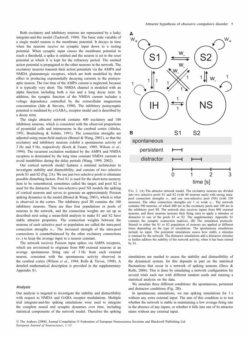
Our cortical network model features a minimal architecture toinvestigate stability and distractibility, and consists of two selectivepools S1 and S2 (Fig. 2A). We use just two selective pools to eliminatepossible disturbing factors. Pool S1 is used for the short-term memoryitem to be remembered, sometimes called the target; and pool S2 isused for the distractor. The non-selective pool NS models the spikingof cortical neurons and serves to generate an approximately Poissonspiking dynamics in the model (Brunel & Wang, 2001), which is whatis observed in the cortex. The inhibitory pool IH contains the 100inhibitory neurons. There are thus four populations or pools ofneurons in the network, and the connection weights are set up asdescribed next using a mean-field analysis to make S1 and S2 havestable attractor properties. The connection weights between theneurons of each selective pool or population are called the intra-poolconnection strengths w+. The increased strength of the intra-poolconnections is counterbalanced by the other excitatory connections(w)) to keep the average input to a neuron constant.
The network receives Poisson input spikes via AMPA receptors,which are envisioned to originate from 800 external neurons at anaverage spontaneous firing rate of 3 Hz from each externalneuron, consistent with the spontaneous activity observed inthe cerebral cortex (Wilson et al., 1994; Rolls & Treves, 1998). Adetailed mathematical description is provided in the supplementaryAppendix S1.
Analysis
Our analysis is targeted to investigate the stability and distractibilitywith respect to NMDA and GABA receptor modulations. Multipletrial integrate-and-fire spiking simulations were used to integratethe complete neural and synaptic dynamics over time, includingstatistical components of the network model. Therefore the spiking
simulations are needed to assess the stability and distractibility ofthe dynamical system, for this depends in part on the statisticalfluctuations that occur in a network of spiking neurons (Deco &Rolls, 2006). This is done by simulating a network configuration forseveral trials each run with different random seeds and running astatistical analysis on the data.We simulate three different conditions: the spontaneous, persistent
and distractor conditions (Fig. 2B).In spontaneous simulations, we run spiking simulations for 3 s
without any extra external input. The aim of this condition is to testwhether the network is stable in maintaining a low average firing ratein the absence of any inputs, or whether it falls into one of its attractorstates without any external input.
A
B
Fig. 2. (A) The attractor network model. The excitatory neurons are dividedinto two selective pools S1 and S2 (with 40 neurons each) with strong intra-pool connection strengths w+ and one non-selective pool (NS) (with 320neurons). The other connection strengths are 1 or weak w). The networkcontains 500 neurons, of which 400 are in the excitatory pools and 100 are inthe inhibitory pool IH. The network also receives inputs from 800 externalneurons, and these neurons increase their firing rates to apply a stimulus ordistractor to one of the pools S1 or S2. The supplementary Appendix S1contains the synaptic connection matrices. (B) The simulation protocols.Stimuli to either the S1 or S2 population of neurons are applied at differenttimes depending on the type of simulations. The spontaneous simulationsinclude no input. The persistent simulations assess how stably a stimulusis retained by the network. The distractor simulations add a distractor stimulusto further address the stability of the network activity, when it has been startedby S1.
Attractor hypothesis of obsessive–compulsive disorder 5
ª The Authors (2008). Journal Compilation ª Federation of European Neuroscience Societies and Blackwell Publishing LtdEuropean Journal of Neuroscience, 1–13
In persistent simulations, an external cue of 120 Hz above thebackground firing rate of 2400 Hz is applied to each neuron in pool S1during the first 500 ms to induce a high activity state and then thesystem is run for another 2.5 s. The 2400 Hz is distributed acrossthe 800 synapses of each S1 neuron for the external inputs, with thespontaneous Poisson spike trains received by each synapse thushaving a mean rate of 3 Hz. The aim of this condition is to investigatewhether once in an attractor short-term memory state the network canmaintain its activity stably, or whether it falls out of its attractor, whichmight correspond to an inability to maintain attention.The distractor simulations start off like the persistent simulations
with a 500 ms input to pool S1 to start the S1 short-term memoryattractor state, but between 1 s and 1.5 s we apply a distracting inputto pool S2 with varying strengths. The aim of this condition is tomeasure how distractible the network is. The degree of distractibility ismeasured parametrically by the strength of the input to S2 required toremove the high activity state of the S1 population. These simulationprotocols serve to assess the generic properties of the dynamicalattractor system rather than to model specific experimental dataobtained in particular paradigms.The mean-field approach (described in the supplementary Appendix
S1) was used to calculate the synaptic weights to set the normalconditions for the operation of the network to be as follows. For thespontaneous state, the conditions for the numerical simulations of themean-field method were set to 3 Hz for all excitatory pools and 9 Hzfor the inhibitory pool. These values correspond to the approximatevalues of the spontaneous attractors when the network is not driven bystimulus-specific inputs. For the persistent state, a selective pool was
set to a higher initial value (30 Hz) to account for the excitation ofthese neurons during the preceding cue period.
Results
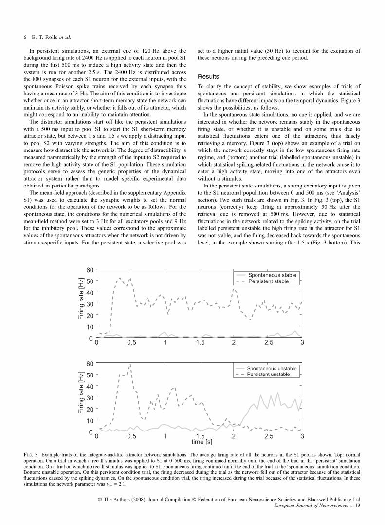
To clarify the concept of stability, we show examples of trials ofspontaneous and persistent simulations in which the statisticalfluctuations have different impacts on the temporal dynamics. Figure 3shows the possibilities, as follows.In the spontaneous state simulations, no cue is applied, and we are
interested in whether the network remains stably in the spontaneousfiring state, or whether it is unstable and on some trials due tostatistical fluctuations enters one of the attractors, thus falselyretrieving a memory. Figure 3 (top) shows an example of a trial onwhich the network correctly stays in the low spontaneous firing rateregime, and (bottom) another trial (labelled spontaneous unstable) inwhich statistical spiking-related fluctuations in the network cause it toenter a high activity state, moving into one of the attractors evenwithout a stimulus.In the persistent state simulations, a strong excitatory input is given
to the S1 neuronal population between 0 and 500 ms (see ‘Analysis’section). Two such trials are shown in Fig. 3. In Fig. 3 (top), the S1neurons (correctly) keep firing at approximately 30 Hz after theretrieval cue is removed at 500 ms. However, due to statisticalfluctuations in the network related to the spiking activity, on the triallabelled persistent unstable the high firing rate in the attractor for S1was not stable, and the firing decreased back towards the spontaneouslevel, in the example shown starting after 1.5 s (Fig. 3 bottom). This
Fig. 3. Example trials of the integrate-and-fire attractor network simulations. The average firing rate of all the neurons in the S1 pool is shown. Top: normaloperation. On a trial in which a recall stimulus was applied to S1 at 0–500 ms, firing continued normally until the end of the trial in the ‘persistent’ simulationcondition. On a trial on which no recall stimulus was applied to S1, spontaneous firing continued until the end of the trial in the ‘spontaneous’ simulation condition.Bottom: unstable operation. On this persistent condition trial, the firing decreased during the trial as the network fell out of the attractor because of the statisticalfluctuations caused by the spiking dynamics. On the spontaneous condition trial, the firing increased during the trial because of the statistical fluctuations. In thesesimulations the network parameter was w+ = 2.1.
6 E. T. Rolls et al.
ª The Authors (2008). Journal Compilation ª Federation of European Neuroscience Societies and Blackwell Publishing LtdEuropean Journal of Neuroscience, 1–13
trial illustrates the failure to maintain a stable short-term memory state,even when no distractor is applied.
In Fig. 3 the transitions to the incorrect activity states are caused bystatistical fluctuations in the spiking activity of the integrate-and-fireneurons. We hypothesize that the stability of the high firing rate‘persistent’ attractor state may be increased in OCD, and that inaddition the spontaneous state may be less likely to remain low, butmay jump into a high firing rate attractor state. We note that there aretwo sources of noise in the spiking networks that cause the statisticalfluctuations: the randomly arriving external Poisson spike trains, andthe statistical fluctuations caused by the spiking of the neurons in thefinite-sized network. The magnitude of these fluctuations increases asthe number of neurons in the network becomes smaller (Mattia & DelGiudice, 2004).
For our investigations, we selected w+ = 2.1, which with the defaultvalues of the NMDA and GABA conductances yielded stabledynamics, that is, a stable spontaneous state if no retrieval cue wasapplied, and a stable state of persistent firing after a retrieval cue hadbeen applied and removed. To investigate the effects of changes(modulations) in the NMDA, AMPA and GABA conductances, wechose for demonstration purposes increases of 3% for the NMDA, and10% for the AMPA and GABA synapses between the neurons in thenetwork shown in Fig. 2A, as these were found to be sufficient to alterthe stability of the attractor network. A strength of our approach is thatwe show that even quite small increases in the synaptic currents can alterthe global behaviour of the network, e.g. the stability of its attractors.
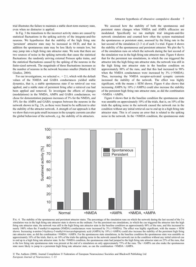
We assessed how the stability of both the spontaneous andpersistent states changes when NMDA and AMPA efficacies aremodulated. Specifically we ran multiple trial integrate-and-firenetwork simulations and counted how often the system maintainedthe spontaneous or persistent state, assessed by the firing rate in thelast second of the simulation (2–3 s) of each 3-s trial. Figure 4 showsthe stability of the spontaneous and persistent attractor. We plot the %of the simulation runs on which the network during the last second ofthe simulation was in the high firing rate attractor state. Figure 4 showsthat for the persistent run simulations, in which the cue triggered theattractor into the high firing rate attractor state, the network was still inthe high firing rate attractor state in the baseline condition onapproximately 88% of the runs, and that this had increased to 98%when the NMDA conductances were increased by 3% (+NMDA).Thus, increasing the NMDA receptor-activated synaptic currentsincreased the stability of the network. The effect was highlysignificant, with the means ± SEM shown. Figure 4 also shows thatincreasing AMPA by 10% (+AMPA) could also increase the stabilityof the persistent high firing rate attractor state, as did the combination+NMDA +AMPA.Figure 4 shows that in the baseline condition the spontaneous state
was unstable on approximately 10% of the trials, that is, on 10% of thetrials the spiking noise in the network caused the network run in thecondition without any initial retrieval cue to end up in a high firing rateattractor state. This is of course an error that is related to the spikingnoise in the network. In the +NMDA condition, the spontaneous state
Fig. 4. The stability of the spontaneous and persistent attractor states. The percentage of the simulation runs on which the network during the last second of the 3-ssimulation was in the high firing rate attractor state is shown on the ordinate. For the persistent run simulations, in which the cue triggered the attractor into the highfiring rate attractor state, the network was still in the high firing rate attractor state in the baseline condition on approximately 88% of the runs, and this increased tonearly 100% when the N-methyl-d-aspartate (NMDA) conductances were increased by 3% (+NMDA). The effect was highly significant, with the means ± SEMshown. Increasing a-amino-3-hydroxy-5-methyl-4-isoxazolepropionic acid (AMPA) by 10% (+AMPA) could also increase the stability of the persistent high firingrate attractor state, as did the combination +NMDA +AMPA. For the spontaneous state simulations, in the baseline condition the spontaneous state was unstable onapproximately 10% of the trials, that is, on 10% of the trials the spiking noise in the network caused the network run in the condition without any initial retrieval cueto end up in a high firing rate attractor state. In the +NMDA condition, the spontaneous state had jumped to the high firing rate attractor state on 25% of the runs, thatis the low firing rate spontaneous state was present at the end of a simulation on only approximately 75% of the runs. The +AMPA can also make the spontaneousstate more likely to jump to a persistent high firing rate attractor state, as can the combination +NMDA +AMPA.
Attractor hypothesis of obsessive–compulsive disorder 7
ª The Authors (2008). Journal Compilation ª Federation of European Neuroscience Societies and Blackwell Publishing LtdEuropean Journal of Neuroscience, 1–13
had jumped to the high firing rate attractor state on approximately 22%of the runs, that is the low firing rate spontaneous state was present atthe end of a simulation on only approximately 78% of the runs. Thus,increasing NMDA receptor-activated currents can contribute to thenetwork jumping from what should be a quiescent state of sponta-neous activity into a high firing rate attractor state. We relate this to thesymptoms of OCDs, in that the system can jump into a state with adominant memory (which might be an idea or concern or action) evenwhen there is no initiating input. Figure 4 also shows that +AMPA canmake the spontaneous state more likely to jump to a persistent highfiring rate attractor state, as can the combination +NMDA +AMPA.We next investigated to what extent alterations of the GABA
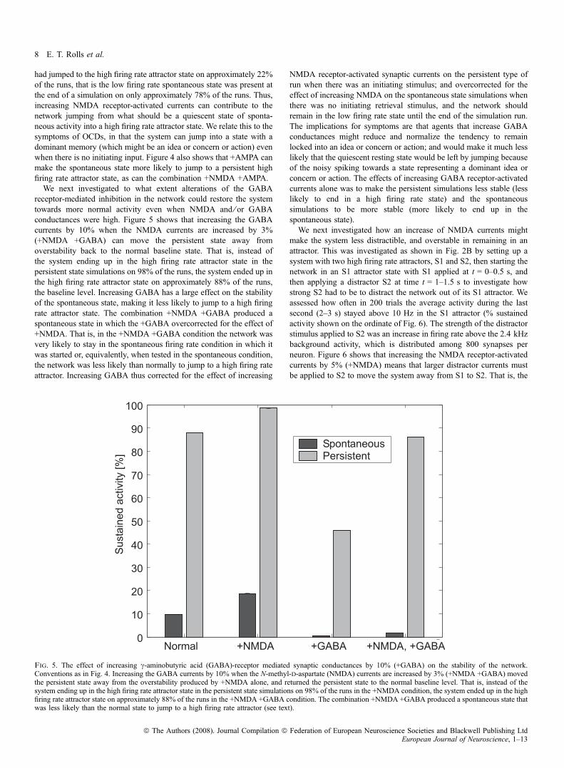
receptor-mediated inhibition in the network could restore the systemtowards more normal activity even when NMDA and ⁄ or GABAconductances were high. Figure 5 shows that increasing the GABAcurrents by 10% when the NMDA currents are increased by 3%(+NMDA +GABA) can move the persistent state away fromoverstability back to the normal baseline state. That is, instead ofthe system ending up in the high firing rate attractor state in thepersistent state simulations on 98% of the runs, the system ended up inthe high firing rate attractor state on approximately 88% of the runs,the baseline level. Increasing GABA has a large effect on the stabilityof the spontaneous state, making it less likely to jump to a high firingrate attractor state. The combination +NMDA +GABA produced aspontaneous state in which the +GABA overcorrected for the effect of+NMDA. That is, in the +NMDA +GABA condition the network wasvery likely to stay in the spontaneous firing rate condition in which itwas started or, equivalently, when tested in the spontaneous condition,the network was less likely than normally to jump to a high firing rateattractor. Increasing GABA thus corrected for the effect of increasing
NMDA receptor-activated synaptic currents on the persistent type ofrun when there was an initiating stimulus; and overcorrected for theeffect of increasing NMDA on the spontaneous state simulations whenthere was no initiating retrieval stimulus, and the network shouldremain in the low firing rate state until the end of the simulation run.The implications for symptoms are that agents that increase GABAconductances might reduce and normalize the tendency to remainlocked into an idea or concern or action; and would make it much lesslikely that the quiescent resting state would be left by jumping becauseof the noisy spiking towards a state representing a dominant idea orconcern or action. The effects of increasing GABA receptor-activatedcurrents alone was to make the persistent simulations less stable (lesslikely to end in a high firing rate state) and the spontaneoussimulations to be more stable (more likely to end up in thespontaneous state).We next investigated how an increase of NMDA currents might
make the system less distractible, and overstable in remaining in anattractor. This was investigated as shown in Fig. 2B by setting up asystem with two high firing rate attractors, S1 and S2, then starting thenetwork in an S1 attractor state with S1 applied at t = 0–0.5 s, andthen applying a distractor S2 at time t = 1–1.5 s to investigate howstrong S2 had to be to distract the network out of its S1 attractor. Weassessed how often in 200 trials the average activity during the lastsecond (2–3 s) stayed above 10 Hz in the S1 attractor (% sustainedactivity shown on the ordinate of Fig. 6). The strength of the distractorstimulus applied to S2 was an increase in firing rate above the 2.4 kHzbackground activity, which is distributed among 800 synapses perneuron. Figure 6 shows that increasing the NMDA receptor-activatedcurrents by 5% (+NMDA) means that larger distractor currents mustbe applied to S2 to move the system away from S1 to S2. That is, the
Fig. 5. The effect of increasing c-aminobutyric acid (GABA)-receptor mediated synaptic conductances by 10% (+GABA) on the stability of the network.Conventions as in Fig. 4. Increasing the GABA currents by 10% when the N-methyl-d-aspartate (NMDA) currents are increased by 3% (+NMDA +GABA) movedthe persistent state away from the overstability produced by +NMDA alone, and returned the persistent state to the normal baseline level. That is, instead of thesystem ending up in the high firing rate attractor state in the persistent state simulations on 98% of the runs in the +NMDA condition, the system ended up in the highfiring rate attractor state on approximately 88% of the runs in the +NMDA +GABA condition. The combination +NMDA +GABA produced a spontaneous state thatwas less likely than the normal state to jump to a high firing rate attractor (see text).
8 E. T. Rolls et al.
ª The Authors (2008). Journal Compilation ª Federation of European Neuroscience Societies and Blackwell Publishing LtdEuropean Journal of Neuroscience, 1–13
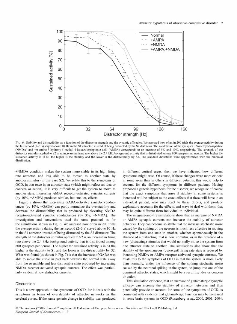
+NMDA condition makes the system more stable in its high firingrate attractor, and less able to be moved to another state byanother stimulus (in this case S2). We relate this to the symptoms ofOCD, in that once in an attractor state (which might reflect an idea orconcern or action), it is very difficult to get the system to move toanother state. Increasing AMPA receptor-activated synaptic currents(by 10%, +AMPA) produces similar, but smaller, effects.
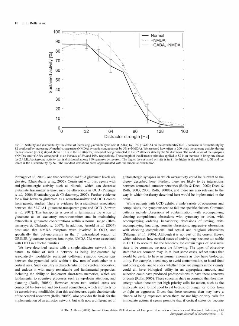
Figure 7 shows that increasing GABA-activated synaptic conduc-tances (by 10%, +GABA) can partly normalize the overstability anddecrease the distractibility that is produced by elevating NMDAreceptor-activated synaptic conductances (by 3%, +NMDA). Theinvestigation and conventions used the same protocol as forthe simulations shown in Fig. 6. We assessed how often in 200 trialsthe average activity during the last second (2–3 s) stayed above 10 Hzin the S1 attractor, instead of being distracted by the S2 distractor. Thestrength of the distractor stimulus applied to S2 is an increase in firingrate above the 2.4 kHz background activity that is distributed among800 synapses per neuron. The higher the sustained activity is in S1 thehigher is the stability to S1 and the lower is the distractibility by S2.What was found (as shown in Fig. 7) is that the increase of GABAwasable to move the curve in part back towards the normal state awayfrom the overstable and less distractible state produced by increasingNMDA receptor-activated synaptic currents. The effect was particu-larly evident at low distractor currents.
Discussion
This is a new approach to the symptoms of OCD, for it deals with thesymptoms in terms of overstability of attractor networks in thecerebral cortex. If the same generic change in stability was produced
in different cortical areas, then we have indicated how differentsymptoms might arise. Of course, if these changes were more evidentin some areas than in others in different patients, this would help toaccount for the different symptoms in different patients. Havingproposed a generic hypothesis for the disorder, we recognize of coursethat the exact symptoms that arise if stability in some systems isincreased will be subject to the exact effects that these will have in anindividual patient, who may react to these effects, and produceexplanatory accounts for the effects, and ways to deal with them, thatmay be quite different from individual to individual.The integrate-and-fire simulations show that an increase of NMDA
or AMPA synaptic currents can increase the stability of attractornetworks. They can become so stable that the intrinsic stochastic noisecaused by the spiking of the neurons is much less effective in movingthe system from one state to another, whether spontaneously in theabsence of a distracting, that is new, stimulus, or in the presence of anew (distracting) stimulus that would normally move the system fromone attractor state to another. The simulations also show that thestability of the spontaneous (quiescent) firing rate state is reduced byincreasing NMDA or AMPA receptor-activated synaptic currents. Werelate this to the symptoms of OCD in that the system is more likelythan normally, under the influence of the spiking stochastic noisecaused by the neuronal spiking in the system, to jump into one of thedominant attractor states, which might be a recurring idea or concernor action.This simulation evidence, that an increase of glutamatergic synaptic
efficacy can increase the stability of attractor networks and thuspotentially provide an account for some of the symptoms of OCD, isconsistent with evidence that glutamatergic function may be increasedin some brain systems in OCD (Rosenberg et al., 2000, 2001, 2004;
Fig. 6. Stability and distractibility as a function of the distractor strength and the synaptic efficacies. We assessed how often in 200 trials the average activity duringthe last second (2–3 s) stayed above 10 Hz in the S1 attractor, instead of being distracted by the S2 distractor. The modulation of the synapses +N-methyl-d-aspartate(NMDA) and +a-amino-3-hydroxy-5-methyl-4-isoxazolepropionic acid (AMPA) corresponds to an increase of 5% and 10%, respectively. The strength of thedistractor stimulus applied to S2 is an increase in firing rate above the 2.4 kHz background activity that is distributed among 800 synapses per neuron. The higher thesustained activity is in S1 the higher is the stability and the lower is the distractibility by S2. The standard deviations were approximated with the binomialdistribution.
Attractor hypothesis of obsessive–compulsive disorder 9
ª The Authors (2008). Journal Compilation ª Federation of European Neuroscience Societies and Blackwell Publishing LtdEuropean Journal of Neuroscience, 1–13
Pittenger et al., 2006), and that cerebrospinal fluid glutamate levels areelevated (Chakrabarty et al., 2005). Consistent with this, agents withanti-glutamatergic activity such as riluzole, which can decreaseglutamate transmitter release, may be efficacious in OCD (Pittengeret al., 2006; Bhattacharyya & Chakraborty, 2007). Further evidencefor a link between glutamate as a neurotransmitter and OCD comesfrom genetic studies. There is evidence for a significant associationbetween the SLC1A1 glutamate transporter gene and OCD (Stewartet al., 2007). This transporter is crucial in terminating the action ofglutamate as an excitatory neurotransmitter and in maintainingextracellular glutamate concentrations within a normal range (Bhat-tacharyya & Chakraborty, 2007). In addition, Arnold et al. (2004)postulated that NMDA receptors were involved in OCD, andspecifically that polymorphisms in the 3¢ untranslated region ofGRIN2B (glutamate receptor, ionotropic, NMDA 2B) were associatedwith OCD in affected families.We have described results with a single attractor network. It is
natural to think of such a network as being implemented byassociatively modifiable recurrent collateral synaptic connectionsbetween the pyramidal cells within a few mm of each other in acortical area. Such circuitry is characteristic of the cerebral neocortex,and endows it with many remarkable and fundamental properties,including the ability to implement short-term memories, which arefundamental to cognitive processes such as top-down attention, andplanning (Rolls, 2008b). However, when two cortical areas areconnected by forward and backward connections, which are likely tobe associatively modifiable, then this architecture, again characteristicof the cerebral neocortex (Rolls, 2008b), also provides the basis for theimplementation of an attractor network, but with now a different set of
glutamatergic synapses in which overactivity could be relevant to thetheory described here. Further, there are likely to be interactionsbetween connected attractor networks (Rolls & Deco, 2002; Deco &Rolls, 2003, 2006; Rolls, 2008b), and these are also relevant to theway in which the theory described here would be implemented in thebrain.While patients with OCD exhibit a wide variety of obsessions and
compulsions, the symptoms tend to fall into specific clusters. Commonpatterns include obsessions of contamination, with accompanyingcleaning compulsions; obsessions with symmetry or order, withaccompanying ordering behaviours; obsessions of saving, withaccompanying hoarding; somatic obsessions; aggressive obsessionswith checking compulsions; and sexual and religious obsessions(Pittenger et al., 2006). Although it is not part of the current theory,which addresses how cortical states of activity may become too stablein OCD, to account for the tendency for certain types of obsessivestate to be common, we note the following. The types of obsessivestate that are common may, in at least some cases, reflect states thatwould be useful to have in normal amounts as they have biologicalutility. For example, a tendency to avoid contamination, to hoard foodand other goods, and to check whether there are dangers in the vicinitycould all have biological utility in an appropriate amount, andselection could have produced predispositions to have these concernsor goals (Rolls, 2005). These concerns share in common that they mayemerge when there are not high priority calls for action, such as theimmediate need to find food to eat because of hunger, or to flee fromor fight an aggressor. Given that these concerns then may have achance of being expressed when there are not high-priority calls forimmediate action, it seems possible that if cortical states do become
Fig. 7. Stability and distractibility: the effect of increasing c-aminobutyric acid (GABA) by 10% (+GABA) on the overstability to S1 ⁄ decrease in distractibility byS2 produced by increasing N-methyl-d-aspartate (NMDA) synaptic conductances by 3% (+NMDA). We assessed how often in 200 trials the average activity duringthe last second (2–3 s) stayed above 10 Hz in the S1 attractor, instead of being distracted to the S2 attractor state by the S2 distractor. The modulation of the synapses+NMDA and +GABA corresponds to an increase of 3% and 10%, respectively. The strength of the distractor stimulus applied to S2 is an increase in firing rate abovethe 2.4 kHz background activity that is distributed among 800 synapses per neuron. The higher the sustained activity is in S1 the higher is the stability to S1 and thelower is the distractibility by S2. The standard deviations were approximated with the binomial distribution.
10 E. T. Rolls et al.
ª The Authors (2008). Journal Compilation ª Federation of European Neuroscience Societies and Blackwell Publishing LtdEuropean Journal of Neuroscience, 1–13
too stable, then it is concerns of this type that might become tooprominent, leading to obsessive thoughts, and thus to compulsivebehaviour. The suggestion is that because these states about whichobsessions are likely are not part of actions being performed to meetimmediate environmental needs, then these states do not haveanything particular to displace them, and can thus remain presentfor considerable periods even under normal circumstances. Anexacerbation of attractor network stability when there is no particularenvironmental need for immediate alternative action may in this waylead to this type of state becoming obsessional.
One of the interesting aspects of the present model of OCD isthat it is in a sense the opposite of a recent model of schizophrenia(Loh et al., 2007). In that model, we suggest that decreased NMDAsynaptic currents may, by decreasing the firing rates and effectivelythe synaptic strengths in cortical attractor networks, decrease thestability of cortical attractor networks. This, if effected in thedorsolateral prefrontal cortex, might produce some of the cognitivesymptoms of schizophrenia, including distractibility, poor attentionand poor short-term memory. If the same generic effect werepresent in the temporal lobe semantic memory networks, this mightmake thoughts move too freely from one thought to another looselyassociated thought, producing bizarre thoughts and some of thepositive symptoms of schizophrenia (providing an alternative to theparasitic or spurious attractor hypothesis; Hoffman & McGlashan,2001). If implemented in the orbitofrontal cortex, the decrease ofNMDA synaptic currents would produce lower neuronal firing, andthus less affect, which is a negative symptom of schizophrenia.These two models, of OCD and schizophrenia, in being formalopposites of each other may underline the importance of attractornetworks to cortical function (Rolls, 2008b), and emphasize thatany alteration in their stability, whether towards too little stability ortoo much, will have major consequences for the operation of thecerebral cortex.
The theory about how the symptoms of OCD could arise in relationto the increased stability of cortical attractor networks has implicationsfor possible pharmaceutical approaches to treatment. One is thattreatments that reduce glutamatergic activity, for example by partiallyblocking NMDA receptors, might be useful. Another is that increasingthe inhibition in the cortical system, for example by increasing GABAreceptor-activated synaptic currents, might be useful, both by bringingthe system from a state where it was locked into an attractor back tothe normal level, and by making the spontaneous state more stable, sothat it would be less likely to jump to an attractor state (which mightrepresent a dominant idea or concern or action; see Fig. 5). However,we emphasize that the way in which the network effects we considerproduce the symptoms in individual patients will be complex, and willdepend on the way in which each person may deal cognitively with theeffects. In this respect, we emphasize that what we describe is a theoryof OCD, that the theory must be considered in the light of empiricalevidence yet to be obtained, and that cognitive behaviour therapymakes an important contribution to the treatment of such patients. Wehope that the theory will stimulate further thinking and research in thisarea.
Supplementary material
The following supplementary material may be found on http://www.blackwell-synergy.comAppendix S1. Supporting information: neural and synaptic dynamics.Please note: Blackwell Publishing are not responsible for the contentor functionality of any supplementary materials supplied by the
authors. Any queries (other than missing material) should be directedto the correspondence author for the article.
Acknowledgements
M.L. was supported by the Boehringer Ingelheim Fonds. Support was alsoprovided by the Oxford McDonnell Centre for Cognitive Neuroscience.
Abbreviations
AMPA, a-amino-3-hydroxy-5-methyl-4-isoxazolepropionic acid; ED, extra-dimensional; GABA, c-aminobutyric acid; ID, intradimensional; NMDA,N-methyl-d-aspartate; OCD, obsessive–compulsive disorder; SSRT, stop-signalreaction time.
References
Abeles, M. (1991) Corticonics – Neural Circuits of the Cerebral Cortex.Cambridge University Press, New York.
Amit, D.J. (1989) Modeling Brain Function. Cambridge University Press,Cambridge.
Arnold, P.D., Rosenberg, D.R., Mundo, E., Tharmalingam, S., Kennedy, J.L. &Richter, M.A. (2004) Association of a glutamate (NMDA) subunit receptorgene (GRIN2B) with obsessive–compulsive disorder: a preliminary study.Psychopharmacology, 174, 530–538.
Bannon, S., Gonsalvez, C.J., Croft, R.J. & Boyce, P.M. (2002) Responseinhibition deficits in obsessive–compulsive disorder. Psychiatry Res., 110,165–174.
Bannon, S., Gonsalvez, C.J., Croft, R.J. & Boyce, P.M. (2006) Executivefunctions in obsessive–compulsive disorder: state or trait deficits? Aust. N. Z.J. Psychiatry, 40, 1031–1038.
Berg, E. (1948) A simple objective technique for measuring flexibility inthinking. J. Gen. Psychol., 39, 15–22.
Bhattacharyya, S. & Chakraborty, K. (2007) Glutamatergic dysfunction –newer targets for anti-obsessional drugs. Recent Patents CNS Drug Discov.,2, 47–55.
Blackerby, R.F. (1993) Application of Chaos Theory to Psychological Models.Performance Strategies Publications, Austin, TX, USA.
Braitenberg, V. & Schutz, A. (1991) Anatomy of the Cortex. Springer-Verlag,Berlin.
Brunel, N. & Wang, X.J. (2001) Effects of neuromodulation in a corticalnetwork model of object working memory dominated by recurrent inhibition.J. Comput. Neurosci., 11, 63–85.
Chakrabarty, K., Bhattacharyya, S., Christopher, R. & Khanna, S. (2005)Glutamatergic dysfunction in OCD. Neuropsychopharmacology, 30, 1735–1740.
Chamberlain, S.R., Fineberg, N.A., Blackwell, A.D., Robbins, T.W. &Sahakian, B.J. (2006) Motor inhibition and cognitive flexibility in obses-sive–compulsive disorder and trichotillomania. Am. J. Psychiatry, 163,1282–1284.
Chamberlain, S.R., Fineberg, N.A., Menzies, L.A., Blackwell, A.D., Bullmore,E.T., Robbins, T.W. & Sahakian, B.J. (2007) Impaired cognitive flexibilityand motor inhibition in unaffected first-degree relatives of patients withobsessive–compulsive disorder. Am. J. Psychiatry, 164, 335–338.
Compte, A., Brunel, N., Goldman-Rakic, P.S. & Wang, X.J. (2000) Synapticmechanisms and network dynamics underlying spatial working memory in acortical network model. Cereb. Cortex, 10, 910–923.
Coyle, J.T. (2006) Glutamate and schizophrenia: beyond the dopaminehypothesis. Cell. Mol. Neurobiol., 26, 365–384.
Deco, G. & Rolls, E.T. (2003) Attention and working memory: a dynamicalmodel of neuronal activity in the prefrontal cortex. Eur. J. Neurosci., 18,2374–2390.
Deco, G. & Rolls, E.T. (2005) Neurodynamics of biased competition andco-operation for attention: a model with spiking neurons. J. Neurophysiol.,94, 295–313.
Deco, G. & Rolls, E.T. (2006) Decision-making and Weber’s Law: aneurophysiological model. Eur. J. Neurosci., 24, 901–916.
Delorme, R., Gousse, V., Roy, I., Trandafir, A., Mathieu, F., Mouren-Simeoni,M.C., Betancur, C. & Leboyer, M. (2007) Shared executive dysfunctions inunaffected relatives of patients with autism and obsessive–compulsivedisorder. Eur. Psychiatry, 22, 32–38.
Attractor hypothesis of obsessive–compulsive disorder 11
ª The Authors (2008). Journal Compilation ª Federation of European Neuroscience Societies and Blackwell Publishing LtdEuropean Journal of Neuroscience, 1–13
Durstewitz, D. & Seamans, J.K. (2002) The computational role of dopamineD1 receptors in working memory. Neural Netw., 15, 561–572.
Durstewitz, D., Kelc, M. & Gunturkun, O. (1999) A neurocomputational theoryof the dopaminergic modulation of working memory functions. J. Neurosci.,19, 2807–2822.
Durstewitz, D., Seamans, J.K. & Sejnowski, T.J. (2000a) Dopamine-mediatedstabilization of delay-period activity in a network model of prefrontal cortex.J. Neurophysiol., 83, 1733–1750.
Durstewitz, D., Seamans, J.K. & Sejnowski, T.J. (2000b) Neurocomputationalmodels of working memory. Nat. Neurosci., 3(Suppl.), 1184–1191.
Faisal, A.A., Selen, L.P. & Wolpert, D.M. (2008) Noise in the nervous system.Nat. Rev., 9, 292–303.
Hampshire, A. & Owen, A.M. (2006) Fractionating attentional control usingevent-related fMRI. Cereb. Cortex, 16, 1679–1689.
Hartston, H.J. & Swerdlow, N.R. (1999) Visuospatial priming and Stroopperformance in patients with obsessive compulsive disorder. Neuropsychol-ogy, 13, 447–457.
Hebb, D.O. (1949) The Organization of Behavior: A NeuropsychologicalTheory. Wiley, New York.
Heinrichs, D.W. (2005) Antidepressants and the chaotic brain: implicationsfor the respectful treatment of selves. Philos. Psychiatr. Psychol., 12, 215–227.
Hertz, J., Krogh, A. & Palmer, R.G. (1991) An Introduction to the Theory ofNeural Computation. Addison-Wesley, Wokingham.
van den Heuvel, O.A., Veltman, D.J., Groenewegen, H.J., Cath, D.C., vanBalkom, A.J., van Hartskamp, J., Barkhof, F. & van Dyck, R. (2005) Frontal-striatal dysfunction during planning in obsessive–compulsive disorder. Arch.Gen. Psychiatry, 62, 301–309.
Hoffman, R.E. & McGlashan, T.H. (2001) Neural network models ofschizophrenia. Neuroscientist, 7, 441–454.
Hopfield, J.J. (1982) Neural networks and physical systems with emergentcollective computational abilities. Proc. Natl Acad. Sci. USA, 79, 2554–2558.
Hornak, J., Rolls, E.T. & Wade, D. (1996) Face and voice expressionidentification in patients with emotional and behavioural changes followingventral frontal lobe damage. Neuropsychologia, 34, 247–261.
Hornak, J., Bramham, J., Rolls, E.T., Morris, R.G., O’Doherty, J., Bullock, P.R.& Polkey, C.E. (2003) Changes in emotion after circumscribed surgi-cal lesions of the orbitofrontal and cingulate cortices. Brain, 126, 1691–1712.
Jahr, C.E. & Stevens, C.F. (1990) Voltage dependence of NMDA-activatedmacroscopic conductances predicted by single-channel kinetics. J. Neurosci.,10, 3178–3182.
Karno, M., Golding, J.M., Sorenson, S.B. & Burnam, M.A. (1988) Theepidemiology of obsessive–compulsive disorder in five US communities.Arch. Gen. Psychiatry, 45, 1094–1099.
Khalil, H.K. 1996. Nonlinear Systems. Prentice Hall, Upper Saddle River, NJ.Koch, K.W. & Fuster, J.M. (1989) Unit activity in monkey parietal cortexrelated to haptic perception and temporary memory. Exp. Brain Res., 76,292–306.
Kringelbach, M.L. & Rolls, E.T. (2004) The functional neuroanatomy of thehuman orbitofrontal cortex: evidence from neuroimaging and neuropsychol-ogy. Prog. Neurobiol., 72, 341–372.
Lewis, M.D. (2005) Bridging emotion theory and neurobiology throughdynamic systems modeling. Behav. Brain sci., 28, 169–194; discussion 194–245.
Loh, M., Rolls, E.T. & Deco, G. (2007) A dynamical systems hypothesisof schizophrenia. PLoS Comput. Biol., 3, e228. doi:210.1371/journal.pcbi.0030228.
Mattia, M. & Del Giudice, P. (2004) Finite-size dynamics of inhibitory andexcitatory interacting spiking neurons. Phys. Rev., 70, 052903.
Menzies, L., Chamberlain, S.R., Laird, A.R., Thelen, S.M., Sahakian, B.J. &Bullmore, E.T. (2008) Integrating evidence from neuroimaging and neuro-psychological studies of obsessive–compulsive disorder: The orbitofronto-striatal model revisited. Neurosci. Biobehav. Rev., 32, 525–549.
Nagahama, Y., Okada, T., Katsumi, Y., Hayashi, T., Yamauchi, H., Oyanagi, C.,Konishi, J., Fukuyama, H. & Shibasaki, H. (2001) Dissociable mechanismsof attentional control within the human prefrontal cortex. Cereb. Cortex, 11,85–92.
Pantelis, C., Barber, F.Z., Barnes, T.R., Nelson, H.E., Owen, A.M. &Robbins, T.W. (1999) Comparison of set-shifting ability in patients withchronic schizophrenia and frontal lobe damage. Schizophr. Res., 37, 251–270.
Peled, A. (2004) From plasticity to complexity: a new diagnostic method forpsychiatry. Med. Hypotheses, 63, 110–114.
Penades, R., Catalan, R., Andres, S., Salamero, M. & Gasto, C. (2005)Executive function and nonverbal memory in obsessive–compulsive disor-der. Psychiatry Res., 133, 81–90.
Penades, R., Catalan, R., Rubia, K., Andres, S., Salamero, M. & Gasto, C.(2007) Impaired response inhibition in obsessive compulsive disorder. Eur.Psychiatry, 22, 404–410.
Pittenger, C., Krystal, J.H. & Coric, V. (2006) Glutamate-modulating drugs asnovel pharmacotherapeutic agents in the treatment of obsessive–compulsivedisorder. NeuroRx, 3, 69–81.
Riley, M.A. & Turvey, M.T. (2001) The self-organizing dynamics of intentionsand actions. Am. J. Psychol., 114, 160–169.
Robins, L.N., Helzer, J.E., Weissman, M.M., Orvaschel, H., Gruenberg, E.,Burke, J.D. Jr & Regier, D.A. (1984) Lifetime prevalence ofspecific psychiatric disorders in three sites. Arch. Gen. Psychiatry, 41,949–958.
Rogers, R.D., Andrews, T.C., Grasby, P.M., Brooks, D.J. & Robbins, T.W.(2000) Contrasting cortical and subcortical activations produced by atten-tional-set shifting and reversal learning in humans. J. Cogn. Neurosci., 12,142–162.
Rolls, E.T. (2004) The functions of the orbitofrontal cortex. Brain Cogn., 55,11–29.
Rolls, E.T. (2005) Emotion Explained. Oxford University Press, Oxford.Rolls, E.T. (2008a) The anterior and midcingulate cortices and reward. In Vogt,
B.A. (ed.), Cingulate Neurobiology & Disease. Oxford University Press,Oxford, in press.
Rolls, E.T. (2008b) Memory, Attention, and Decision-Making: A UnifyingComputational Neuroscience Approach. Oxford University Press, Oxford.
Rolls, E.T. & Deco, G. (2002) Computational Neuroscience of Vision. OxfordUniversity Press, Oxford.
Rolls, E.T. & Treves, A. (1998) Neural Networks and Brain Function. OxfordUniversity Press, Oxford.
Rolls, E.T., Hornak, J., Wade, D. & McGrath, J. (1994) Emotion-relatedlearning in patients with social and emotional changes associated with frontallobe damage. J. Neurol. Neurosurg. Psychiatry, 57, 1518–1524.
Rosenberg, D.R., MacMaster, F.P., Keshavan, M.S., Fitzgerald, K.D., Stewart,C.M. & Moore, G.J. (2000) Decrease in caudate glutamatergic concentra-tions in pediatric obsessive–compulsive disorder patients taking paroxetine.J. Am. Acad. Child Adolesc. Psychiatry, 39, 1096–1103.
Rosenberg, D.R., MacMillan, S.N. & Moore, G.J. (2001) Brain anatomy andchemistry may predict treatment response in paediatric obsessive–compul-sive disorder. Int. J. Neuropsychopharmacol., 4, 179–190.
Rosenberg, D.R., Mirza, Y., Russell, A., Tang, J., Smith, J.M., Banerjee, S.P.,Bhandari, R., Rose, M., Ivey, J., Boyd, C. & Moore, G.J. (2004) Reducedanterior cingulate glutamatergic concentrations in childhood OCD and majordepression versus healthy controls. J. Am. Acad. Child Adolesc. Psychiatry,43, 1146–1153.
Rushworth, M.F., Behrens, T.E., Rudebeck, P.H. & Walton, M.E. (2007a)Contrasting roles for cingulate and orbitofrontal cortex in decisions andsocial behaviour. Trends Cogn. Sci, 11, 168–176.
Rushworth, M.F., Buckley, M.J., Behrens, T.E., Walton, M.E. & Bannerman,D.M. (2007b) Functional organization of the medial frontal cortex. Curr.Opin. Neurobiol., 17, 220–227.
Seamans, J.K. & Yang, C.R. (2004) The principal features and mechanismsof dopamine modulation in the prefrontal cortex. Prog. Neurobiol., 74,1–58.
Stewart, S.E., Fagerness, J.A., Platko, J., Smoller, J.W., Scharf, J.M., Illmann,C., Jenike, E., Chabane, N., Leboyer, M., Delorme, R., Jenike, M.A. &Pauls, D.L. (2007) Association of the SLC1A1 glutamate transporter geneand obsessive–compulsive disorder. Am. J. Med. Genet. B Neuropsychiatr.Genet., 144, 1027–1033.
Treves, A. (1991) Are spin-glass effects relevant to understanding realisticauto-associative networks. J. Physics A, 24, 2645–2654.
Treves, A. & Rolls, E.T. (1991) What determines the capacity of autoasso-ciative memories in the brain? Network, 2, 371–397.
Tuckwell, H. (1988) Introduction to Theoretical Neurobiology. CambridgeUniversity Press, Cambridge.
Veale, D.M., Sahakian, B.J., Owen, A.M. & Marks, I.M. (1996) Specificcognitive deficits in tests sensitive to frontal lobe dysfunction in obsessive–compulsive disorder. Psychol. Med., 26, 1261–1269.
Wang, X.J. (1999) Synaptic basis of cortical persistent activity: the importanceof NMDA receptors to working memory. J. Neurosci., 19, 9587–9603.
Wang, X.J. (2001) Synaptic reverberation underlying mnemonic persistentactivity. Trends Neurosci., 24, 455–463.
Wang, X.J. (2002) Probabilistic decision making by slow reverberation incortical circuits. Neuron, 36, 955–968.
12 E. T. Rolls et al.
ª The Authors (2008). Journal Compilation ª Federation of European Neuroscience Societies and Blackwell Publishing LtdEuropean Journal of Neuroscience, 1–13

Watkins, L.H., Sahakian, B.J., Robertson, M.M., Veale, D.M., Rogers, R.D.,Pickard, K.M., Aitken, M.R. & Robbins, T.W. (2005) Executive function inTourette’s syndrome and obsessive–compulsive disorder. Psychol. Med., 35,571–582.
Weissman, M.M., Bland, R.C., Canino, G.J., Greenwald, S., Hwu, H.G.,Lee, C.K., Newman, S.C., Oakley-Browne, M.A., Rubio-Stipec, M.,Wickramaratne, P.J., Wittchen, H. & Yeh, E.K. (1994) The cross national
epidemiology of obsessive compulsive disorder. The Cross NationalCollaborative Group. J. Clin. Psychiatry, 55(Suppl.), 5–10.
Wilson,F.A.,O’Scalaidhe,S.P.&Goldman-Rakic,P.S. (1994)Functional synergismbetween putative gamma-aminobutyrate-containing neurons and pyramidalneurons in prefrontal cortex. Proc. Natl Acad. Sci. USA, 91, 4009–4013.
Winterer, G. & Weinberger, D.R. (2004) Genes, dopamine and cortical signal-to-noise ratio in schizophrenia. Trends Neurosci., 27, 683–690.
Attractor hypothesis of obsessive–compulsive disorder 13
ª The Authors (2008). Journal Compilation ª Federation of European Neuroscience Societies and Blackwell Publishing LtdEuropean Journal of Neuroscience, 1–13