An Assessment of Frontal Lobe Activity and BDNF Levels ...
73
Northern Michigan University NMU Commons All NMU Master's eses Student Works 12-2017 An Assessment of Frontal Lobe Activity and BDNF Levels Following Concussion in Collegiate Athletes: A Near-Infrared Spectroscopy Study Keara Kangas [email protected] Follow this and additional works at: hps://commons.nmu.edu/theses Part of the Cognitive Psychology Commons is Open Access is brought to you for free and open access by the Student Works at NMU Commons. It has been accepted for inclusion in All NMU Master's eses by an authorized administrator of NMU Commons. For more information, please contact [email protected],[email protected]. Recommended Citation Kangas, Keara, "An Assessment of Frontal Lobe Activity and BDNF Levels Following Concussion in Collegiate Athletes: A Near- Infrared Spectroscopy Study" (2017). All NMU Master's eses. 167. hps://commons.nmu.edu/theses/167
Transcript of An Assessment of Frontal Lobe Activity and BDNF Levels ...

Northern Michigan UniversityNMU Commons
All NMU Master's Theses Student Works
12-2017
An Assessment of Frontal Lobe Activity and BDNFLevels Following Concussion in CollegiateAthletes: A Near-Infrared Spectroscopy StudyKeara [email protected]
Follow this and additional works at: https://commons.nmu.edu/theses
Part of the Cognitive Psychology Commons
This Open Access is brought to you for free and open access by the Student Works at NMU Commons. It has been accepted for inclusion in All NMUMaster's Theses by an authorized administrator of NMU Commons. For more information, please contact [email protected],[email protected].
Recommended CitationKangas, Keara, "An Assessment of Frontal Lobe Activity and BDNF Levels Following Concussion in Collegiate Athletes: A Near-Infrared Spectroscopy Study" (2017). All NMU Master's Theses. 167.https://commons.nmu.edu/theses/167

An Assessment of Frontal Lobe Activity and BDNF Levels Following Concussion in
Collegiate Athletes: A Near-Infrared Spectroscopy Study
By: Keara J. Kangas
THESIS
Submitted to
Northern Michigan University
In partial fulfillment of the requirements
For the degree of
MASTERS OF SCIENCE
Office of Graduate Education and Research

Keara Kangas
2 | P a g e
Page left blank intentionally

Keara Kangas
3 | P a g e
ABSTRACT
An Assessment of Frontal Lobe Activity and BDNF Levels Following Concussion in
Collegiate Athletes: A Near-Infrared Spectroscopy Study
By: Keara J. Kangas
Impacts to the head that are associated with sports related injuries, can result in a
mild traumatic brain injury (mTBI), known as a concussion. Previous research has
assessed how mTBIs affect the brain, but these assessments are limited in their ability to
directly measure the consequences of mTBI. Along with concussion assessments, only a
few studies have used neuroimaging techniques to evaluate brain injury. This study
utilized a neuroimaging technique that is inexpensive, non-invasive, and portable, to
measure brain activity post-concussion. In particular, near-infrared spectroscopy (NIRS)
was used to measure prefrontal cortex (PFC) activity during the dot-probe task of
affective attentional bias. Brain-derived neurotrophic factor (BDNF) levels are related to
frontal lobe activity, recovery from brain injury, attention, and affective processing.
Therefore, BDNF levels were collected to assess their relationship to mTBI and PFC
activity in the dot-probe task. Behaviorally, reaction times (RT) were overall significantly
slower in individuals with mTBI over control. Although, RT did not vary in either group
to indicate an attentional bias between trial types. The mTBI group’s deoxygenated
hemoglobin (HbR) levels did not deviate greatly from baseline like the control did. The
mPFC correlated with less responsive HbR levels with higher serum BDNF levels. For
oxygenated hemoglobin (HbO), less deviation from baseline in the right PFC correlated
with higher levels of serum BDNF levels. There were overall greater serum BDNF levels

Keara Kangas
4 | P a g e
in mTBI than the control, but these levels did not react significance. These findings
potentially assist in improving and developing more efficient diagnostic assessments.

Keara Kangas
5 | P a g e
Page Left Blank Intentionally

Keara Kangas
6 | P a g e
Copyright By
KEARA J. KANGAS
2017

Keara Kangas
7 | P a g e
INTRODUCTION
Roughly, 3.8 million people experience a sport or recreation-related concussion
each year (Broglio et al., 2014). When asked, about 50% of hockey, football, and soccer
players report at least one concussion within their sport history (Chen et al., 2007). Sport
related brain injuries are increasing in research popularity—with a particular focus on
finding new ways to improve effective post-concussion assessments and tools. Only a
relatively small number of studies have included neuroimaging techniques to assess and
diagnose concussions; these studies usually use neuroimaging equipment that restricts the
effectiveness of the assessment (Eastman, 2011). These restrictive factors include the
length of time the assessments take and the cost to perform them. These limitations affect
the efficiency of the procedure, especially when using these tools to potentially diagnosis
an athlete’s ability to return to play after a brain injury. Utilization of a tool that can be
easily implemented after a concussion is imperative to the understanding of concussion
recovery.
NIRS is a non-invasive and portable neuroimaging technique that could be used
measure abnormal frontal lobe activity post-concussion. Furthermore, biological
metabolites found in blood, saliva, or urine samples can indicate the amount or presence
of a specific protein, such as BDNF. These biological metabolites, like BDNF, may serve
as a biomarker for concussion and be used as a moderator in neuroimaging studies of
concussion or even in place of neuroimaging (Jeter et al., 2013). In addition, emotional
impairments often result from concussion, but this dimension of concussion is not well
explored (Vagnozzi et al., 2010). Concussion-related affective abnormalities have been

Keara Kangas
8 | P a g e
linked to abnormal frontal lobe activity (Eierud et al., 2014) and increased BDNF levels
(Korley et al., 2016; Kaplan, Vaterling, & Vedak, 2010).
My thesis is a subcomponent of a larger collaborative study, in which the primary
investigators are Dr. Marguerite Moore and Dr. Joshua Carlson. The first aim of the
larger study is to assess the extent to which NIRS can be used to assess frontal lobe
activity in patients with concussion. A range of concussive symptoms will be analyzed:
including cognitive processing and socio-emotional reactivity. Another aim is to assess
the utility of NIRS as a marker of individual-level symptom severity post-concussion by
testing the correlation between frontal lobe activity and symptom severity across these
abovementioned dimensions. The larger collaborative study will also be assessing the
degree to which the severity in these symptoms are correlated with each other and how
these symptoms are potentially related to common abnormalities in frontal lobe activity.
My thesis will investigate the relationship between frontal lobe NIRS activity, behavioral
measures of affective reactivity, and the potential mediating role of BDNF levels
collected from saliva and serum samples. I hypothesize that:
1. Participants with mTBI (concussion) will show higher levels of BDNF when
compared to the control group.
2. Higher levels of BDNF will correlate with decreased activity in the frontal
lobe, which will be greatest in mTBI (concussion) participants.
3. Participants with mTBI (concussion) will show slower reaction times overall
and in particular during trials assessing attentional disengagement (i.e.,
incongruent trials).
4. Slower reaction times in participants with mTBI (concussion) will be related

Keara Kangas
9 | P a g e
to higher levels of BDNF and decreased activity in the frontal lobe.

Keara Kangas
10 | P a g e
Concussion
Historical Perspectives on concussion, an early understanding
For more than 3,000 years, research and medical descriptions of brain injuries
described concussion in many different contexts. Documentation of head injuries before
the 10th century is sparse and imprecise (McCrory & Berkovic, 2001). However, one of
the surviving descriptions of head injuries was written by Paul of Aegina (AD 625-690)
during the Byzantine period; Paul of Aegina was a pupil of Hippocrates (460-370BC) and
Galen (AD 129-216) (Gurunluoglu & Guruluoglu, 2003). The seven books by Paul of
Aegina were put together and called the Epitome of Medicine (McCrory & Berkovic,
2001; Gurunluoglu & Guruluoglu, 2003). Translators used the term concussion in
Hippocrates passages, but it remains unclear if this term was referring to the injury
“concussion” or using “concussion” as a specific type of symptom (McCrory &
Berkovic, 2001).
Hippocrates, Galen, and Celsus (25BC-AD 50) were a part of the Greek and
Roman Medicine era; they discussed a cranial injury that today resembles symptoms of a
“confused concussion,” and Galen of Pergamon found patients had dizziness, speech
disturbances, and aphasia after they received a blow to the head (McCrory & Berkovic,
2001). Rhazes (AD 850-923) was influenced by Paul of Aegina (Gurunluoglu &
Guruluoglu, 2003) and used the term concussion to describe an abnormal physiological
state (McCrory & Berkovic, 2001). Continuing on into the 10th – 17th centuries, the term
concussion was first used during the Renaissance era (McCrory & Berkovic, 2001).
One of the most famous surgeons during the 16th century was Ambroise Pare who
wrote about “the moving or concussion of the brain,” which described the epiphenomena
Keara Kangas
11 | P a g e
of concussions as brain swelling and hemorrhaging (McCrory & Berkovic, 2001). During
Medieval times, the first “modern” physician was Lanfranc of Milan (1250-1306); he
explained a concussion as being from a transient paralysis of cerebral function that was
caused from the brain being shaken (McCrory & Berkovic, 2001). More individuals
during the Medieval times, included Coiter (1573) and Marchetti (1665), who attempted
to define concussion as having wavering speech, impaired memory, weak judgement, or
shortened attention span (McCrory & Berkovic, 2001).
From the 17th century on, research examined case reports and animal
experimentation pathophysiologically to understand concussions. There were many
improvements within the medical field to treat head injuries. Neurological surgeons
started specializing in head and sport injuries (Stone, Patel, & Bailes, 2014). A couple of
influential individuals were Louis Pasteur and Joseph Lister (Stone, Patel, & Bailes,
2014). Louis Pasteur advanced the medical field by noticing infections, caused from
inflammation and pus, from post-traumatic injuries needing surgical procedures; Joseph
Lister introduced antiseptic methods for these infections (Stone, Patel, & Bailes, 2014).
In 1905, college football started to introduce protective equipment because the
season resulted of 18 deaths and 159 serious injuries (Stone, Patel, & Bailes, 2014).
President Theodore Roosevelt agreed that athlete death and injury was a serious issue and
invited college coaches to the White House to discuss ways to minimize the danger and
brutality within football (Stone, Patel, & Bailes, 2014). During and after WWII,
neurosurgeons Hugh W.B. Cairns and E. Stephen Gurdjian worked with experimental
animals to understand clinical neurotrauma; they researched skull fractures, acceleration

Keara Kangas
12 | P a g e
injuries, intracranial pressure, and inertial brain movements (Stone, Patel, & Bailes,
2014).
Modern Research and Concerns Related to Concussion
One challenge in diagnosing concussions is identifying patients that are at risk for
long-term complications after a brain injury. Eastman (2011) discussed how diagnostic
and assessment tools to analyze cognitive effects after a concussion should be quick and
easy to use. First responders are usually athletic trainers that identify, evaluate, and
manage the decision that determines the athlete’s ability to return to play (Broglio et al.,
2014).
Within current research, the term concussion is used primarily in sports medicine
(Jeter et al., 2013). Out of all mild traumatic brain injuries (mTBI), 20% are sports-
related and 30-45% of these individuals receive no medical care (Vagnozzi et al., 2010).
Brain injuries can vary in severity ranging from mild to severe; these injuries can result in
minor symptoms and escalate to death (Kaplan, Vasterling, & Vedak, 2010). A clinically
diagnosed concussion does not always result in a loss of consciousness, but is typically
associated with headaches, vision disturbances, sleep alterations, concentration, and
emotional (i.e., depression) disturbances (Kaplan, Vasterling, & Vedak, 2010; Vagnozzi
et al., 2010). Symptoms of concussion sometimes resolve within 7-10 days, but lingering
post-concussion symptoms can include mood impairments including irritability,
depression, and anxiety as well as other symptoms such as fatigue, trouble concentrating,
headaches, and memory impairments (Kaplan, Vasterling, & Vedak, 2010). Post-
concussion symptoms can also include slowed processing of information, decreased
verbal fluency, impaired learning and memory, as well as impaired executive functioning

Keara Kangas
13 | P a g e
(Kaplan, Vasterling, & Vedak, 2010). Post-concussion symptoms typically normalize
after about 1 – 3 months post-injury, but can take even longer depending on severity and
individual differences (Kaplan, Vasterling, & Vedak, 2010).
Concussions result from forces being applied directly or indirectly to the skull that
result in a rapid acceleration and deceleration of the brain (Broglio et al., 2014). Within
the first 15 minutes there is an extreme dip in neuropsychological performance; these
effects can linger for up to a week or longer (Eierud et al., 2014). If an individual is not
aware of concussion symptoms, not knowing can potentially be harmful to the individual
if there is further agitation. Although one symptom may be loss of consciousness, this
symptom is not always the case (Broglio et al., 2014). Loss of consciousness occurs in
fewer than 10% mTBI (Broglio et al., 2014). Sport related concussions do not usually
leave a detectable structural lesion, and only a few validated instruments can help
diagnose a concussive brain injury (Chen et al., 2007).
There are a few biomarkers that are reliable for diagnosing brain injuries (Korley
et al., 2016; Jeter et al., 2013). Many metabolic biomarkers have been correlated with
brain injury. Some biomarkers include, S100B (calcium-binding protein B, S-100 protein
family), neuron-specific enolase (NSE), glial fibrillary acidic protein (GFAP), brain
derived neurotropic factor (BDNF), and many more (Jeter et al., 2013). In particular,
measuring BDNF levels following brain injury has increased in popularity (Niechwiej-
Szwedo et al., 2015; Larson-Dupuis et al., 2015). BDNF, more generally as a
neurotrophin, plays a major role in the survival of basal forebrain cholinergic neurons and
regulates the function of hippocampal and cortical neurons (Holsinger et al., 2000).

Keara Kangas
14 | P a g e
In addition, decreased levels of BDNF within the parietal cortex could be a
selective vulnerability mechanism for Alzheimers disease (Holsinger et al., 2000). BDNF
levels are associated with depression and anxiety disorders; low levels are connected with
depressed and anxious mood (Lang et al., 2005; Montag et al., 2008). Limited to no
research examined when BDNF specifically increases after injury, and when BDNF
returns to baseline. Although research discusses BDNF, when increased, can provide
neuroprotection, restore connectivity, and repair function after brain injuries including
concussions (Korley et al., 2016; Kaplan, Vaterling, & Vedak, 2010). Thus, for these
reasons—in particular the role of BDNF in neuronal survival, neuroplasticity, and neural
repair—BDNF appears to be a promising metabolic biomarker for mTBI.
Kaplan, Vasteling, and Vedak (2010) assessed the mechanism by which
neurotrophins enhance neuronal survival, and found that increased BDNF repairs neural
connections and reduces factors associated with TBI and post-traumatic stress disorder
(PTSD). Research has shown that on day one after a brain injury, BDNF levels in all
types of brain damage are lower than those in healthy controls, but researchers also
noticed BDNF levels were higher in mTBI subjects relative to moderate to severe brain
injuries (Korley et al., 2016). Along with this, other studies show after a period of several
days post-injury, BDNF levels increase in the cortex and hippocampus overall for brain
injuries (Kaplan, Vasterling, & Vedak, 2010; Yang et al., 1996). BDNF also appears to
reduce the impact of secondary brain injuries (Kaplan et al., 2010). In non-TBI
individuals, elevated levels of BDNF are related to increased anxiety and amygdala
reactivity to pleasant and unpleasant startle conditions (Montag, Reuter, Newport, Elger,
& Weber, 2008). Given the associations between mTBI and anxiety as well as mTBI and

Keara Kangas
15 | P a g e
BDNF, it is possible—yet untested—that elevated BDNF levels post-concussion lower
the symptoms of anxiety and depression following concussion.
Most assessment studies looking at overall mTBI symptoms use subjects that
received a mTBI up to 2 weeks prior to participating in a research study. Around two
weeks after receiving a mTBI, there is an increase in neuropsychological performance
and this recovery can alter assessment results (Eierud et al., 2014), and after 3 months,
mTBI symptoms related to poor assessment scores and neuropsychological performance
can return to a previous baseline (Beaupre, Guise, and McKerral, 2012). When looking
specifically at athletic mTBI, there are many factors involved in evaluating when an
athlete can return to training and competition. These factors include, but are not limited
to, loss of consciousness duration, convulsions, frequency of concussions, the time in
between last concussion, style of play, and sport. An evaluation of post-injury involves an
examination that assesses cognitive factors which looks at motor control (using balancing
tasks) and cognitive function (reaction time, working memory, and delayed recall)
(Broglio et al., 2014).
Chen et al. (2007) found that participants that had mild and low post-concussive
symptoms responded much slower on matching tasks when compared to the control
group. Mild post-concussive symptom participants responded slower compared to the
low post-concussive symptoms group. Earlier stages of concussion symptoms have been
associated with slower reaction times and larger intra-individual variability (Beaupre,
Guise, & McKerral, 2012). mTBI is related to attentional functioning deficits during
different stages of recovery (Beaupre, Guise, & McKerral, 2012). mTBI individuals
displayed significantly slower reaction times than controls; reaction times were also
Keara Kangas
16 | P a g e
significantly slower during early than later stages of recovery (Beaupre et al., 2012; Chen
et al., 2007). Beaupre et al. (2012) not only found participants with mTBI had slower
reaction times but they were also less consistent than controls on attention tasks.
The neuroimaging techniques assessing concussions include computed
tomography (CT), positron emission tomography (PET), single photon emission
computed tomography (SPECT), arterial spin labeling (ASL), functional magnetic
resonance imaging (fMRI), the electroencephalogram (EEG), and magnetic resonance
spectroscopy (MRS) (Eierud et al., 2014). These types brain imaging techniques can
assist in measuring brain abnormalities in concussion when comparing mTBI individuals
to a control group (Eastman, 2011). In a recent meta-analysis of fMRI studies of mTBI,
brain injuries significantly affected different areas in the brain; six of these regions were
more activated compared to controls and seven regions showed lower activation (Eierud
et al., 2014). Out of the seven regions that were decreased, six of them were in the frontal
lobe or anterior cingulate, the seventh was the posterior temporal lobe. This meta-analysis
also implicated the frontal lobe as the most affected brain region in structural
impairments following mTBI. Chen et al. (2007) found similar frontal lobe deficits in the
blood-oxygen-level dependent (BOLD) signal across group findings. This frontal lobe
vulnerability can be a function of where the blow strikes the head. Thus, the functional
and structural neuroimaging data highly implicate the frontal lobe as a brain region
vulnerable to concussion (Eierud et al., 2014).
Although many MRI studies (Eierud et al., 2014; Chen et al., 2007) have
examined frontal lobe vulnerability in brain injuries, only a single study (Kontos et al.,
2014) used NIRS to measure frontal lobe activity in mTBI participants. Kontos et al.’s

Keara Kangas
17 | P a g e
(2014) study used NIRS to measure the hemoglobin levels of the frontal cortex during a
neurocognitive performance task, The Immediate Post-concussion Assessment and
Cognitive Test (ImPACT), which measures verbal and visual memory, visual motor
speed, and reaction time. The findings for this study (Kontos et al., 2014) indicated
reduced brain activity in the mTBI compared to the control group. In addition, studies
measuring BDNF levels in mTBI (Korley et al., 2016) have not assessed the relationship
between prefrontal NIRS activity and BDNF levels. Both frontal lobe activity and BDNF
levels have been independently linked to affective processing and concussion, but the
relationship between these factors has yet to be explored.

Keara Kangas
18 | P a g e
Attention Task – Dot-Probe
A limited amount of research has assessed the effects of brain injury on attention
and, not surprisingly, has found that injured participants have difficulty with tasks that
require sustained attention (Whyte, Polansky, Fleming, Coslett, & Cavallucci, 1995).
Little to no research has examined at how mTBI affect attentional bias to emotional
stimuli (Croker & McDonald, 2005). Affective stimuli automatically capture observer’s
attention and can therefore interfere with attention-demanding cognitive tasks
(Schimmack 2005). Patients with severe brain injuries showed difficulties in maintaining
performance over time during an attention-demanding task (Whyte et al., 1995), but
researchers know less about the effect of mTBI on attentional processing and in particular
attentional biases to affective stimuli. The relation between cognition, attentional bias,
and mTBI is essential to help better understand brain injuries, and the deficits in
information processing and attentional bias that follow them (Whyte et al., 1995).
MacLeod, Mathews, and Tata (1986) were the first to create the dot-probe task,
which is now the most commonly used method of assessing attentional bias to emotional
stimuli. In the initial study, 48 threatening words (24 social threat, 24 physical threat)
were used to demonstrate an attentional bias towards threat-related stimuli (MacLeod,
Mathews, & Tata, 1986). One word would appear (for 500ms) above the center of the
screen and one just below; immediately after these words disappeared a small dot (probe)
appeared, and the participants were required to press a button immediately when the dot
was detected (MacLeod, Mathews, & Tata, 1986). Attentional bias is measured by
comparing participants’ reaction time between congruent (dot behind threat-related
stimuli) and incongruent (dot behind neutral-related stimuli) trials. Studies have shown

Keara Kangas
19 | P a g e
that participants have an attentional bias to threating stimuli, congruent trials having
faster reaction times when compared to incongruent trials (MacLeod, Mathews, & Tata,
1986; MacLeod et al., 2002; Carlson, Reinke, & Habib, 2009; Carlson et al., 2013;
Carlson et al., 2014).
Browning et al., (2010) used MacLeod et al’s (1986) dot-probe task as their
attentional training task to examine the effects of cognitive training on attention. The dot-
probe task was altered to use fearful and neutral facial expressions instead of the original
affective word stimuli (Browning et al., 2010). The amygdala promotes attention towards
salient stimuli (Carlson, Reinke, & Habib, 2009), but a second signal originates in areas
of the prefrontal cortex (PFC) (Browning et al, 2010). During this modified dot-probe
task there was greater PFC activity, confirming that the PFC is involved in affective
attention. Indeed, a number of additional dot-probe studies have implicated a role of the
PFC in directing attention toward affective stimuli and sustaining attention in the PFC
(Price et al., 2014; Carlson et al., 2013; Armony & Dolan, 2002; Monk et al., 2008).
There are other attention tasks comparable to the dot-probe that direct individuals’
attention to affective stimuli
A similar attention task is the Stroop task created by JR Stroop (1935), where
participants are asked to name the color of the word, when the word and ink color are
incongruent (the word RED was printed in blue ink), requires more attention in which
there is greater anterior cingulate cortex (ACC) activity (MacDonald, Cohen, Stenger, &
Carter, 2000). MacDonald, Cohen, and Carter (2000) used fMRI during a task-switching
version of the Stroop task to see if individuals could regulate cognitive control. The left
dorsolateral PFC was activated during this color naming task, which was consistent with

Keara Kangas
20 | P a g e
research discussing the dorsolateral PFC is needed for the implementation of control
(MacDonald, Cohen, Stenger, & Carter, 2000). The ACC was found to be more activated
when individuals responded to incongruent stimuli, which MacDonald, Cohen, Stenger,
and Carter, (2000) described as performance monitoring. The ACC and doresolater PFC
are known to activate when individuals hold long sequences of stimuli in working
memory, or when they are required to perform two tasks at once (MacDonald, A., Cohen,
J., Stenger, V., & Carter, C., 2000).
In an additional type of affective attention task, fearful or neutral facial
expressions were presented while participants attended to two house stimuli and decided
if they were identical or not; the faces were used as distractors (Bishop, Duncan, Brett, &
Lawrence, 2004). During the affective attention task, researchers found that the lateral
PFC was involved in increased attentional control during threat-related distraction, and
the ACC responded to unexpected threat-related distractors (Bishop, Duncan, Brett, &
Lawrence, 2004). Researchers interpreted these results as being consistent with the PFC
processing and resolving conflict from emotionally salient task-irrelevant stimuli
(Bishop, Duncan, Brett, & Lawrence, 2004). Attentional bias is also elevated in anxiety
and is a causal mechanism by which anxiety develops (MacLeod et al., 2002). Thus, in a
variety of different tasks, the PFC has been linked to regulating affective attention.
As mentioned in the previous section, frontal lobe and anterior cortices are
particularly vulnerable to neuronal contusion, which can lead to various severities of
brain injury (Broglio et al., 2014). During attention demanding tasks, areas in the PFC
tend to display less activity in mTBI individuals compared to control participants (Chen
et al., 2007). This limited PFC activity is not surprising, given the relationship between

Keara Kangas
21 | P a g e
the PFC and attention as well as the meta-analytic functional and structural neuroimaging
evidence implicating the PFC is a region that is vulnerable to mTBI (Eierud et al., 2014).
Although the amount of neuroimaging studies of concussion have increased in recent
years (Eierud et al., 2014), the effect of mTBI on the relationship between PFC activity
and affective attention has yet to be explored. As indicated earlier, mTBI is associated
with affective symptoms such as elevated anxiety and depression, which have been
shown to have an elevated attentional bias to threat as a mechanism by which disorders
develops. Thus, understanding the relationship between PFC activity and attentional bias
in mTBI is likely important to understanding the heightened levels of anxiety and
depression following mTBI.

Keara Kangas
22 | P a g e
Brain Derived Neurotrophin Factor (BDNF) and Analysis Tools
Modern biological advancements use molecular genetic techniques in their
research (Holsinger et al., 2000); some areas of research that use these techniques include
Psychology, Criminology, and Medicine. These advancements would not have been
possible without first understanding proteins and using them to understand the
breakdown of genetic material.
A French chemist Antoine Fourcroy (1755-1809) discovered three distinct
varieties of protein from animal sources in 1789, albumin, fibrin, and gelatin (Tanford &
Reynolds, 2003). More recognizably, Jons Jacob Berzelius and his pupil Gerrit Mulder
termed the word protein in 1838; albeit the term protein was found earlier than 1838 in a
letter written from Mulder’s mentor Berzelius (Vickery, 1950). These researchers were
the first to uncover the structure of proteins, creating two hydrogen-bonded helical
configurations for the polypeptide chain (Pauling, Corey, & Branson, 1951). There were
additional researchers to help discover deoxyribose nucleic acid (DNA). James Watson
and Francis Crick are renowned in discovering DNA (Dahm, 2005). Albeit, one of the
first was Freidrich Miescher in 1869. Miescher worked with Lymphocytes, looking at
their chemical composition by examining these cells’ nuclei (Dahm, 2005), and Watson
and Crick (1953) expanded from Pauling and Corey’s structural description of a nucleic
acid.
Neurotrophins are secreted proteins that regulate cell growth and survival,
differentiation, apoptosis, cytoskeleton restructuring, and maintain brain plasticity in both
healthy and neuropsychiatric disordered individuals (Kaplan, Vasterling, & Vedak, 2010;
Mandel, Ozdener, & Utermohlen, 2011; Korley et al., 2016; Skaper, 2012). The

Keara Kangas
23 | P a g e
neurotrophins (in mammals) are BDNF, neurotrophin-3 (NT-3), neurotrophin-4 (NT-4),
and neural growth factor (NGF) (Kaplan, Vasterling, & Vedak, 2010). Neurotrophins
work specifically with structural and functional receptors such as tropomyosin-related
tyrosine kinase (Trk) receptors and the p75 NT receptor (Kaplan, Vasterling, & Vedak,
2010). The first neurotrophin was NGF and was discovered by Levi-Montalcini and
Hamburger (1951). They identified NGF by exploring nerve growth stimulating tumors
in mouse sarcomas; they implanted mouse tissue into a developing chick embryo. In this
paper, they referred to NGF as a “growth promoting agent” (Levi-Montalcini &
Hamburger, 1951). Levi-Montalcini and Hamburger’s (1951) study was one of the firsts
to show how NGF was a specific chemical signal that was localized to certain nerve types
and structures in the nervous system.
Most neurons are regulated by, and respond to, neurotrophins, which was
confirmed by the phenomenon of apoptosis (Skaper, 2012; Earnshaw, Martins, &
Kaufmann, 1999). Apoptosis, described as cellular suicide, is triggered by a variety of
stimuli (Earnshaw, Martins, & Kaufmann, 1999). Barde, Edgar, and Thoenen’s study
(1982) discovered a second neurotrophin called BDNF, by examining cellular death.
BDNF was the first neurotrophic factor to be purified and identified after NGF was
discovered (Barde, Edgar, & Thoenen, 1982).
BDNF is a major regulator of synaptic transmission and plasticity at adult
synapses within the central nervous system (Kaplan, Vasterling, & Vedak, 2010; Kang &
Schuman, 1995). Kang and Schuman (1995) suggested synaptic enhancement contributes
to later phases of long-term potentiation (LTP). Neurotrophins (BDNF and NT-3) alter
synaptic strength preceding long-term structural changes that underlie developmental and

Keara Kangas
24 | P a g e
adult plasticity (Kang & Schuman, 1995). BDNF also helps with the survival of neurons
and synaptic transmission (Kaplan, Vasterling, & Vedak, 2010). Increases in BDNF
improves connectivity and function, repairing neural connections (Kaplan, Vasterling, &
Vedak, 2010). In 1990, Jones and Reichardt noticed that BDNF was involved in the
maintenance of the adult nervous system. Their methods included using polymerase chain
reaction (PCR) and hybridization to isolate BDNF and neurotrophin-3 (NT-3); Jones and
Reichardt’s (1990) study was also one of the firsts to isolate NT-3 and BDNF within
humans.
There is an increasing interest in BDNF as a potential biomarker to help diagnosis
and monitor therapy techniques in brain disorders (Polacchini et al., 2015). BDNF can be
measured through many different sample types (ie. blood, tissue, saliva), but for humans,
a majority of them are taken through a blood sample (Mandel, Ozdener, & Utermohlen,
2011). BDNF exists in most human tissues, including the brain and blood (Mori,
Shimizu, & Hayashi, 2003; Polacchini et al., 2015). Mori et al. (2003) suggested that
levels of serum or plasma BDNF are similar among primates, which include humans.
Little to no studies have focused on the release and amount of BDNF in humans
after a brain injury (Korley et al., 2016). Kaplan, Vasterling, and Vedak (2010) observed
that after a mTBI there is an increase in hippocampal BDNF mRNA for several hours
after injury and is accompanied by an increase in BDNF protein for several days after
injury. Grundy et al. (2000) measured BDNF mRNA expression in rats 4hrs after injury,
and noticed an increase in BDNF mRNA. Other studies noticed similar increases after
2hrs, 24hrs, 48hrs, 7 and 14 days, and months after injury (Griesbach et al., 2002;
Chiaretti et al., 2008; Korley et al., 2016). Korley et al. (2016) established the
Keara Kangas
25 | P a g e
relationship between BDNF and TBI and different amounts of BDNF levels are
associated with TBI severity and recovery. Korley et al. (2016) linked low BDNF levels
with slower and incomplete recovery after 6 months in TBI participants. In addition,
secreted BDNF proteins have a strong association with long-term outcomes for brain
injuries (Korley et al., 2016). Increases in BDNF are linked to brain injuries; these
increases are present across different timelines and influence the patient’s recovery, yet
there is still much to know about the release and amount of BDNF after injury.
Saliva contains a wide variety of proteins that contribute to the health of the oral
cavity and gastrointestinal tract (Mandel et al., 2011). Many different growth factors have
been identified in saliva including epidermal growth factor, transforming growth factor,
nerve growth factor, and insulin-like growth factors (Mandel et al., 2011). In addition,
salvia samples, have proven to produce reliable levels of BDNF (Mandel et al., 2011).
For example, McGeown et al. (2016) ran a pilot study exploring the effects of an exercise
program, to measure BDNF levels in saliva, they hoped an exercise program could help
mTBI or neurologically impaired individuals. This pilot study’s results showed an
increase in BDNF concentrations after the exercise program (McGeown et al., 2016).
Methods of salvia collection include the passive drool method and salivette collection
device (Mandel et al., 2011). Mandel et al. (2011) found the passive drool method had
higher BDNF levels than that collected by the salivette collection device in terms of
amount per volume and amount per milligram of protein. (Mandel et al., 2011). Previous
research has found gender differences in BDNF detection; women had significantly
higher levels of BDNF in salvia than men (Mandel et al., 2011).

Keara Kangas
26 | P a g e
BDNF samples can be assayed using a number of techniques. The enzyme
immunoassay (EIA) along with the enzyme-linked immunosorbent assay (ELISA) were
developed by the research group of Peter Perlmann and Eva Engvall at Stockholm
University in Sweden, along with Anton Schuurs and Bauke van Weemen in the
Netherlands (Engvall & Perlmann, 1971; Schuurs & Van Weemen, 1982; Lequin, 2005).
Since it was invented in the 1970s, refined and used during the 1970s and 1980s, these
methods have been overwhelmingly used in a number research (Engvall & Perlmann,
1971; Schuurs & Van Weemen, 1982; Lequin, 2005). The ELISA and EIA became more
popular rather than the radioactivity assay (Lequin, 2005).
Assay techniques started with the RadioImmunoAssay (RIA), which was first
discussed as a possible technique about in 1960 to measure endogenous plasma insulin,
Solomon Berson and Rosalyn Yalow created the RIA. The first insulin assays assessed
the fall in blood glucose following injection of an extract of tissue or serum into normal
or depancreatized animals, but these insulin assays were not sensitive enough to measure
low levels in blood (Kahn & Roth, 2004). RIA was created by looking at the metabolism
of I-labeled insulin in nondiabetic and diabetic subjects (Kahn & Roth, 2004); the
metabolism of I-labeled insulin is where they noticed radioactive insulin disappeared
slower from plasma of patients who had previously been treated with insulin than from
the plasma of subjects never treated with insulin (Kahn & Roth, 2004).
Roger Ekins published a finding in 1960 about saturation analysis that estimated
thyroxine in human plasma (Lequin, 2005). These techniques using radioactive label
opened up for new analyses being published and variants of techniques using radioactive
label were quickly developed (Lequin, 2005). Radioactive material brought concern

Keara Kangas
27 | P a g e
about the safety of laboratory personnel, being expensive, building codes, and radioactive
waste disposal (Lequin, 2005). Researchers started using iodine (weak radiation) (Lequin,
2005). When starting to commercialize these tests, solid-phase techniques were used to
develop microtier plates (96 wells) in which an antigen or an antibody is bound to a solid-
phase support. These advances led to automated pipetting devices, multichannel pipettes,
and microtiter plate readers and washers (Lequin, 2005). Using iodine did not become
commercially successful until the late 70s and early 80s when they matched the
sensitivity of RIA systems (Lequin, 2005).
Immunoassays allow virtually any biologically interesting molecules present in
blood or other fluids to be measured with sensitivity and specific even with other
substances being present in the specimen (Kahn & Roth, 2004). Saliva contains many
kinds of proteins that contribute to oral cavity health and gastrointestinal tract (Mandel et
al., 2001). Mandel et al. (2009) demonstrated by using immunoblotting and enzyme
digestion that BDNF was present in human saliva.
Although BDNF can be taken through a blood sample (Korley, 2015), salvia has
been proven to produce the same reliable data (Mandel, Ozdener, and Utermohlen, 2011).
Saliva samples are much easier to collect, don’t require a licensed phlebotomist, and
don’t cause anxiety, which is common to needle draw blood samples. Thus, a secondary
aim of this thesis was to compare BDNF levels across both salivary and serum samples. It
is expected that both sample types will result in elevated BDNF levels in the mTBI group
and BDNF levels will be highly correlated across sample types.

Keara Kangas
28 | P a g e
Near infrared spectroscopy (NIRS)
The first commercial instrument for measuring proteins and moisture in wheat by
transmission spectroscopy in the near-infrared area was the Trebor GT-90 (Williams,
Norris, & Sobering, 1985). Researchers used light at near-infrared wavelengths, which
passed through the sample and was detected by a system that amplifies and transposes
them (Williams, Norris, & Sobering, 1985). Absorption occurs at specific wavelengths, in
the 700 to 1300nm range, which can be effectively transmitted through biological
materials (Jobsis, 1977). The brain is sensitively dependent on oxygen for normal
function (Jobsis, 1977). Jobsis (1977) extended his experiment from cats to the human
brain, placing optic fiber bundles above and anterior to the temples, where they recorded
blood volume at 815nm, which they concluded was the oxygenated and deoxygenated
hemoglobin’s isobestic point: the wavelength where absorption of light does not change
physically or during a chemical reaction (Jobsis, 1977).
Brazy et al. (1985) used infants, for their transparent quality of skin and skull, and
used three to four diode lasers as sources of wavelengths between 760 and 904nm. Brazy
et al. (1985) used an early version of NIRS that they named the NIROS-SCOPE, which
stood for near infrared oxygen sufficiency scope that was a device that continuously
monitored the relative amounts of oxygenated and deoxygenated hemoglobin in the
optical field (Brazy et al., 1985). The NIROS-SCOPE was able to measure photons,
which were collected by a pickup bundle by a receiving optode and conducted back to a
photomultiplier in the NIROS-SCOPE to be measured (Brazy et al., 1985).
NIRS in current research is a relatively new imaging technique that uses the
properties of oxygenated and deoxygenated blood to record brain activity within the

Keara Kangas
29 | P a g e
cortical surface (Cui et al., 2011). NIRS was created because researchers felt there needed
to be a more direct way to assess cerebral oxygen delivery and utilization, making it
noninvasive and adaptable to the bedside as to not interfere with patient wellbeing (Brazy
et al., 1985). When an area in the brain becomes activated, neurons use oxygen and create
a hemodynamic response. The hemodynamic response is associated with a dip in deo-
oxygenated hemoglobin (Cui et al., 2011). Hyperventilation was used as a function test to
see if oxygen could be monitored noninvasively, which resulted in a significant decrease
in oxygenated hemoglobin (Imray et al., 2000). Watanabe et al., (1996) used NIRS to
map brain activation with a multi-channel measurement, using wavelengths of 780 and
840nm for light sources covering the fronto-central area with 12 probes. Watanabe et al.,
(1996) mentioned the potential of NIRS being used to measure many different cognitive
components including visual, auditory, cognitive, and language. NIRS is extremely
compact and suitable for bedside and laboratory space. NIRS would be beneficial to
young children whose developing brains could be effected by CT scans, or assist rural
areas where brain imaging may be unavailable (Jeter et al., 2013).
Comparing NIRS to fMRI, NIRS may have low spatial resolution, but has
advantages in high temporal resolution (Watanabe et al., 1996). fMRI is noninvasive and
provides spatially well-resolved functional brain maps (Ogawa et al., 1992). Ogawa et al.
(1992) termed the phenomenon BOLD contrast, this measures changes in hemodynamic
responses and map human mental operations. fMRI measures the blood oxygen level-
dependent (BOLD) response which is the result from local concentration changes in
paramagnetic deoxy-hemoglobin (Cui, Bray, Bryant, Glover, & Reiss, 2011).
Researchers looked at the occipital lobe of their participants (Ogawa et al., 1992),

Keara Kangas
30 | P a g e
demonstrating that sensory stimulation will produce an intrinsic signal change at high-
magnetic fields. fMRI and NIRS measure hemodynamic correlates of neural activity such
as local changes in blood flow, volume, and oxygenation in the brain (Cui et al., 2011).
NIRS signals are correlated with fMRI measurements (Cui et al., 2011). A study by Cui
et al. (2011) compared the fMRI and NIRS signals within the frontal and parietal brain
regions while participants performed cognitive tasks. Results supported a strong
correlation between BOLD and oxy- and deoxy-Hb regardless of task (Cui et al., 2011).
However, NIRS is less expensive and more portable, making it a potentially stronger
diagnostic tool for concussions.
Few studies have looked at cerebral blood flow within a brain-injured participant
looking at the frontal lobe using NIRS (Taussky et al., 2012). One study looked at how
NIRS could be used as a bedside application for critically ill brain-injured patients who
may be unstable to transport (Taussky et al., 2012). While using NIRS, participants
performed the ImPACT test that focused on word and design memory (random words
and abstract single line designs with immediate and delayed word recall), symbol and
color matching (symbols positioned in numerical positions, then hidden, after participant
matches through recall; participant responds if word and color ink match), and other
neurocognitive tests (Kontos et al., 2014). This study only looked at a few neurocognitive
tests, and had a small sample size of 9 participants (Kontos et al., 2014). The PFC is
essential for processing emotional stimuli during neurocognitive tasks including the dot-
probe task (Bishop et al., 2004; Browning et al., 2010). As mentioned previously, mTBI
is associated with abnormal processing in the PFC (Eierud et al., 2014), heightened levels
Keara Kangas
31 | P a g e
of BDNF (Korley et al., 2016, Kaplan, Vaterling, & Vedak, 2010), and negative affective
symptoms (Vagnozzi et al., 2010).
The following lines of evidence suggest that these factors are highly related: 1)
affective attention is linked to PFC activity (Bishop, Duncan, Brett, & Lawrence, 2004),
2) BDNF is related to emotional processing (Montag et al., 2008), 3) higher levels of
BDNF are found in the PFC (Kaplan, Vasteling, and Vedak, 2010), and 4) BDNF has
been linked to the integrity of the structural PFC pathway involved in affective attention
(Montag et al., 2008). These elements have been separately analyzed, but in order to
better understand the relationship between mTBI, BDNF, PFC, and affective attention,
these factors should be consider in a single study. My thesis aims to use NIRS to measure
frontal lobe activity during an emotional attention task, and correlate these measures with
BDNF levels in athletes with and without concussions.

Keara Kangas
32 | P a g e
THESIS STATEMENT
The primary purpose of this study is to use a combination of NIRS, BDNF levels,
and behavioral measures to establish quick and easily obtainable measures and
biomarkers of mTBI in athletes. I hypothesize that:
1. mTBI participants will have higher levels of BDNF when compared to the
control group.
2. Higher levels of BDNF will correlate with decreased activity in the frontal
lobe, which will be greatest in mTBI (concussion) participants.
3. mTBI participants will have slower reaction times overall and in particular
during trials assessing attentional disengagement (i.e., incongruent trials).
4. Slower reaction times in participants with mTBI (concussion) will be related
to higher levels of BDNF and decreased activity in the frontal lobe by using
NIRS.

Keara Kangas
33 | P a g e
METHODS
Participants:
22 participants (11 mTBI, Mean age = 20.41 (mTBI = 20.36, Control = 20.45),
Female = 12). Each participant was recruited from the NMU athletic population, from
sports known to be associated with concussions including football, soccer, wrestling,
hockey, etc. Control participants were matched based on age, gender, and sport. Control
participants were excluded if they obtained a concussion within the last year. All
participants gave written informed consent and monetarily compensated for their time.
Procedure:
Each participant completed all of the following tasks while their frontal lobe
activity was recorded using NIRS. (1) clinical test for sensory interaction of balance
(CTSIB) testing on Biodex Balance System (< 10 min), (2) a dot-probe task of socio-
emotional attention (< 10min), and (3) a non-task resting period to measure basal levels
of blood oxygenation (< 10 min). The conditions occurred in a balanced order across
participant; Biodex, rest, and then dot-probe. Overall, with the NIRS setup and testing,
each session lasted approximately 1.5 hours. Afterward, saliva and blood was collected.
The participants also filled out MRI and depression anxiety stress scales (DASS) survey,
along with completing the immediate post-concussion assessment and cognitive testing
(ImPACT) of cognitive symptoms (20-28 min) which was coordinated by the athletic
trainers.

Keara Kangas
34 | P a g e
Tasks and Equipment
Dot-Probe task:
The dot-probe task was
programmed using E-
Prime2 software and
was displayed upon a
60Hz 16” PC computer monitor. The task measured emotional reactivity, where each trial
started with a fixation cue (+) in the center of the screen for 1000ms. Two images were
then simultaneously presented, very briefly (100ms), to the left and right of the fixation
cue. Afterward, these images disappeared and a target dot was immediately presented to
the left or right side of the screen and remained there until the participant responded to
the location of the dot (See Figure 1). Responses were recorded using a Chronos
Response Box, pressing the “1” for left-sided dots or “2” for right-sided dots. Afterwards
there was a 7 second delay until the next trial started. These trials contained two neutral
images (baseline) or one threating and one neutral image. There were a total of 90 trials:
30 (incongruent - target behind neutral stimuli), 30 (congruent - target behind threating
stimuli), and 30 (baseline). Attentional bias was measured by comparing the reaction time
differences between congruent and incongruent trials.
Biological Samples:
Brain-derived neurotrophic factor (BDNF) was analyzed from saliva and blood
samples taken from each participant. Participants’s alcohol consumption, nicotine usage,
and when they last consumed food or beverages were documented and recorded for
sample collection. All participants were asked about their prescription and OTC
Figure 1. Shows an example of a congruent trial during the dot-probe task

Keara Kangas
35 | P a g e
medication use as well as general and oral health.
Saliva Collection:
Saliva was obtained using the passive drool method. The participant allowed
saliva to pool in their mouth; after saliva accumulated, the participant tilted their head
forward and gently forced saliva through the Saliva Collection Aid (SCA) into the vial.
They provided between 2 – 4ml of saliva, saliva was labeled with the participants number
and centrifuged at 4000rpm for 15mins at 4degrees Celsius, and stored in a -80o Celsius
freezer. Saliva samples were stored on ice until handling, aliquoted into four vials, and
then centrifuged at 4000 rpm for 15 minutes at 4 degrees C. Processing and storing of the
saliva samples were all coordinated with NMU’s biology department.
Serum Collection:
Blood was collected by a trained lab technician. The participant was escorted to
the Clinical Sciences Department, where the lab technician collected, centrifuged,
aliquoted the serum, and stored the samples in their -80o Celsius freezer. Blood Samples
were taken at the Clinical Sciences Department by lab technicians. Processing and storing
of the blood samples were collected through the department’s approved protocols. For
serum samples, 5 ml of blood was collected, aliquoted into two vials, and allowed to clot.
The tubes were centrifuged at 3500 rpm for 10 minutes for serum separation, aliquoted,
and then stored at -80°C.The amount of BDNF in each sample will be analyzed with a
commercial ELISA kit, and will be duplicated at least once.

Keara Kangas
36 | P a g e
Figure 2. Shows the 16 channel NIRS array over the PFC, channel 1: left PFC
NIRS:
Near-infrared spectroscopy (NIRS) is
an optical imaging tool that uses near-infrared
light of two different wavelengths between
650 and 950nm that measures oxygenated and
deoxygenated hemoglobin in the superficial
cortical tissue. Oxygenated and deoxygenated
hemoglobin have detectable differences in
their reflective properties, and the differences between these the hemoglobin levels draw
conclusions about neural activity. Optodes were placed on a “cap”, some optodes emitted
light (sources) and others detected light (detectors). A TechEn CW6 NIRS was used, it is
a continuous wave-type system, 16-channel consisting of eight sources and nine detectors
covered the lateral to medial anterior PFC (See Figure 2). The detectors were 3 cm away
from the sources, Boas and Franceschini (2009) discussed this spacing being optimal
distance for measuring the cortical surface of the cortex. This study measured relative
HbO and HbR differences between trial types in the PFC, using optimal wave-lengths of
690 nm and 830 nm.
Analysis:
NIRS Data: NIRS data was processed and analyzed using Homer2, a MatLab
based program, artifacts were filtered out to eliminate noise that was associated with
physiological variables such as respiration and heart rate, along with movement artifacts.
Homer2 filter values were set at .8 to remove variance accounting for motion artifacts

Keara Kangas
37 | P a g e
(Brigadoi et al., 2014), AMPThresh = 0.1, SDThresh = 50, tMotion = 0.5, and tMask = 1
(Huppert et al., 2009).
The data was extracted and analyzed in SPSS using two 2 × 16 × 3 mixed
measures ANOVA looking at HbO (oxygenated) and HbR (deoxygenated) comparing the
two groups and the 16 optode interaction between each group during the dot-probe task
for the three conditions. Our analysis examined differences between groups and
interactions between optodes and groups.
Behavioral Data: Behavioral data was filtered, eliminating reaction times (RT)
below 100ms and above 1000ms along with all incorrect answers. Behavioral data was
analyzed in SPSS as a 3 × 2 mixed measures ANOVA to assess the effects of trial type
(incongruent vs incongruent vs baseline) and group (mTBI vs control). Our analysis
compared the differences between groups and then within conditions.
BDNF Data: BDNF was analyzed using a ChemiKine BDNF Sandwich ELISA
to measure and quantify BDNF levels. The ELISA was done by using the protocol that
accompanied the kit. A standard curve was correlated using the ELISA kit protocol. This
determined reliability against the sample BDNF levels. Saliva samples were diluted as a
low concentration, as Blood samples were diluted as a medium concentration. All these
samples were duplicated. BDNF levels were analyzed in SPSS, using an independent
samples test to compare the mTBI and control group’s BDNF level means. The results of
the ELISA will be significant; there will be higher BDNF levels in the mTBI vs control
group. The standard curve was constructed based on the ChemiKine protocol (see Figure
3), which was duplicated (r = 0.998, p = 0.000). There was no correlation between saliva
and serum samples (r = -0.29, p > .05), but there was across duplicates for both sample

Keara Kangas
38 | P a g e
types. The duplicated sample types were significantly correlated (p < .05, saliva: r = 0.48;
serum: r = 0.54).
Figure 3. Displays the Standard Curve results from the ChemiKine ELISA
Keara Kangas
39 | P a g e
Results
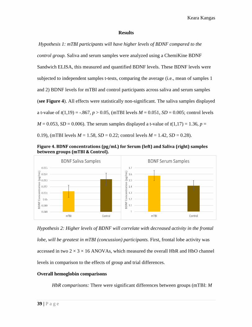
Hypothesis 1: mTBI participants will have higher levels of BDNF compared to the
control group. Saliva and serum samples were analyzed using a ChemiKine BDNF
Sandwich ELISA, this measured and quantified BDNF levels. These BDNF levels were
subjected to independent samples t-tests, comparing the average (i.e., mean of samples 1
and 2) BDNF levels for mTBI and control participants across saliva and serum samples
(see Figure 4). All effects were statistically non-significant. The saliva samples displayed
a t-value of t(1,19) = -.867, p > 0.05, (mTBI levels M = 0.051, SD = 0.005; control levels
M = 0.053, SD = 0.006). The serum samples displayed a t-value of t(1,17) = 1.36, p =
0.19), (mTBI levels M = 1.58, SD = 0.22; control levels M = 1.42, SD = 0.28).
Figure 4. BDNF concentrations (pg/mL) for Serum (left) and Saliva (right) samples between groups (mTBI & Control).
Hypothesis 2: Higher levels of BDNF will correlate with decreased activity in the frontal
lobe, will be greatest in mTBI (concussion) participants. First, frontal lobe activity was
accessed in two 2 × 3 × 16 ANOVAs, which measured the overall HbR and HbO channel
levels in comparison to the effects of group and trial differences.
Overall hemoglobin comparisons
HbR comparisons: There were significant differences between groups (mTBI: M

Keara Kangas
40 | P a g e
= -0.001, S.E. = 0.002; control: M = 0.005, S.E. = 0.002), F(1, 48) = 4.92, p = .031 (see
Figure 5). There were no significant differences (p > .05) between channels or any
interaction effects within channel × group, channel × trialtype, and channel × group ×
trialtype. There was not a significant difference between trialtype (p = 0.767) or an
interaction effect between group × trialtype (p = .109). Although looking at the pairwise
comparison for group × trialtype, there was a significant difference only when comparing
incongruent trials between groups (mTBI: M = -0.003, S.E. = 0.003; control: M = 0.007,
S.E. = 0.002), p = .006 (see Figure 5).
Figure 5. Comparison of HbR levels for overall group and incongruent trial
differences.
HbO comparisons: There was only a significant difference for the interaction within
channel × group F(1, 15) = 1.70, p = .046. There were no other significant effects (p >
.05) within channels (F(1, 15) = 1.01) or the interaction effects within channel × trialtype
(F(2, 15) =0.71) or channel × group × trialtype (F(2, 30) = 0.68). Looking between group
(F(1,48) = 0.54), trialtype (F(2, 48) = 0.781), and the interaction effect between group ×
trialtype (F(2, 48) = 0.031) there were also no significant effects, p > .05. The interaction
effects between channel × group revealed a significant difference at channel 14 (see
Figure 2; see Figure 6) (mTBI: M = 0.005, control: M = -0.029), p = .045. Some other
channels that showed moderate differences were Channel 1 (mTBI: M = -.015, control: M

Keara Kangas
41 | P a g e
= 0.015) p = .087, Channel 9 (mTBI: M = -.005, control: M = -0.029) p = .109, and
channel 10 (mTBI: M = -0.008, control: M = -0.036) p = .051; (see Figure 6).
Figure 6. Interaction effect within channel 14 and groups differences for HbO
BDNF and HbR Levels
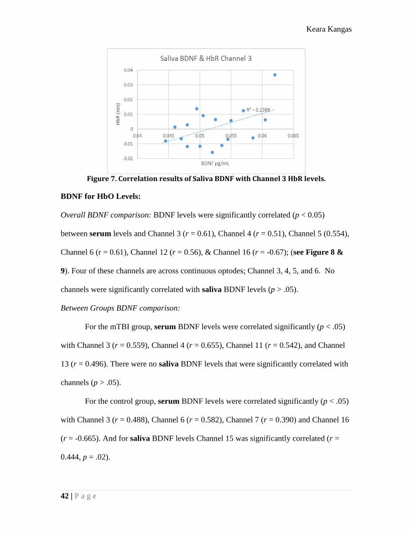
Overall BDNF comparison: Channel 3 (see Figure 2) and saliva levels were significantly
correlated together (see Figure 7); r = 0.489, p = 0.047.
Between Groups BDNF comparison:
For the mTBI group, serum BDNF levels were significantly correlated (p < .05)
with Channel 7 (r = -0.586), Channel 8 (r = -0.505), and Channel 15 (r = 0.586). There
were no significant channels compared to saliva BDNF levels (p > .05).
For the control group, serum BDNF levels that were significantly correlated (p <
.05) were with Channel 1 (r = 0.391), Channel 12 (r = -0.536), and Channel 16 (r =
0.534). As for significant saliva comparisons (p < .05) were with Channel 6 (r = 0.462),
Channel 7 (r = 0.502), and Channel 8 (r = 0.458).
Keara Kangas
42 | P a g e
Figure 7. Correlation results of Saliva BDNF with Channel 3 HbR levels.
BDNF for HbO Levels:
Overall BDNF comparison: BDNF levels were significantly correlated (p < 0.05)
between serum levels and Channel 3 (r = 0.61), Channel 4 (r = 0.51), Channel 5 (0.554),
Channel 6 (r = 0.61), Channel 12 (r = 0.56), & Channel 16 (r = -0.67); (see Figure 8 &
9). Four of these channels are across continuous optodes; Channel 3, 4, 5, and 6. No
channels were significantly correlated with saliva BDNF levels (p > .05).
Between Groups BDNF comparison:
For the mTBI group, serum BDNF levels were correlated significantly (p < .05)
with Channel 3 (r = 0.559), Channel 4 (r = 0.655), Channel 11 (r = 0.542), and Channel
13 (r = 0.496). There were no saliva BDNF levels that were significantly correlated with
channels (p > .05).
For the control group, serum BDNF levels were correlated significantly (p < .05)
with Channel 3 (r = 0.488), Channel 6 (r = 0.582), Channel 7 (r = 0.390) and Channel 16
(r = -0.665). And for saliva BDNF levels Channel 15 was significantly correlated (r =
0.444, p = .02).

Keara Kangas
43 | P a g e
Figure 8. Channels in the left PFC display a (+) correlation with serum BDNF levels
Figure 9. Channels in the right PFC display correlations with serum BDNF levels
Hypothesis 3: mTBI participants will have slower reaction times overall and in particular
during trials assessing attentional disengagement (i.e., incongruent trials). A 2 × 3
ANOVA was used to compare reaction time between groups and each trialtype. Utilizing
all trials, overall there was a significant reaction time difference between groups
(F(1,1754) = 72.55, p < 0.001); mTBI reaction time M = 365.16, SD = 97.18; control
reaction time M = 331.5, SD = 65.09) (see Figure 10). There was no signifcant difference

Keara Kangas
44 | P a g e
between trialtype F(2, 1754) = 0.89, p = .411 and no interaction effect between group and
trialtype. F(2,1754) = 0.251, p = .778.
Figure 10. The overall reaction time for all three conditions during the dot-probe task
between mTBI and control groups.
Hypothesis 4: Slower reaction times in participants with mTBI (concussion) will be
related to higher levels of BDNF and decreased activity in the frontal lobe using NIRS.
There were no significant differences (p > 0.05) when comparing reaction times to levels
of BDNF in serum (r = 0.22) or saliva (r = 0.15). There were significant results for HbO
levels in comparison to overall reaction times during all trials. Channel 9 (r = 0.67, p =
0.002) and Channel 10 (r = 0.67, p = 0.003) were the two channels that showed these
significant results for overall reaction time for all conditions (see Figure 11). There were
no significant results for HbR levels, including channel 9 (r = -0.32, p = 0.19) and
channel 10 (r = -0.42, p = 0.08).

Keara Kangas
45 | P a g e
Figure 11. Comparing overall reaction time for all conditions
with HbO levels for channel 9 & 10.

Keara Kangas
46 | P a g e
Discussion
Summary of Results
Implementing an assessment after a blow to the head will help clinicians and
athletic trainers interpret and understand concussions more quickly. There are many
factors that are involved in evaluating a return to play decision, and our study measured
mTBI participants after they were cleared to return to training and competition. Clinical
examination assesses cognitive factors and function (Broglio et al., 2014). These
assessment results can show improvements in neuropsychological performance after 2
weeks, and after 3 months, these performances can return to baseline. NIRS is an ideal
neuroimaging instrument to measure abnormal activity for mTBI athletes.
Research should continue to expand on NIRS’s ability to measure frontal lobe
activity differences in mTBI populations during attention tasks. Kontos et al. (2014) is
one of few researchers to use NIRS in mTBI populations, but this team only had a small
sample of six and did not use an attentional bias task (e.g., the dot-probe task). This
imaging technique’s functionality can measure hemoglobin levels within the first 15
minutes after a brain injury; where there is normally a dip in neuropsychological
performance (Eierud et al., 2014). The frontal lobe is one of the brain regions that is
effected by concussions (Eierud et al., 2014), and NIRS can measure the activity in this
area (Cui et al., 2011).
In comparison to our results, previous research has similarities and differences;
individual differences based on when the mTBI occurred may have played a part in our
study. NIRS appears to measure frontal lobe and behavioral differences in mTBI athletes
during the dot-probe task. However, there does not seem to be a difference between

Keara Kangas
47 | P a g e
groups for BDNF measured in serum and saliva samples for our study. During the dot-
probe task, the mTBI group had slower reaction time. This behavioral deficit in reaction
time during attention tasks has been shown in previous research, which observed slower
reaction times in individuals with a brain injury (Eierud et al., 2014). Attentional bias is
not well explored in individuals with mTBI. Although much research has concluded that
the dot-probe task measures attentional bias for threating stimuli (MacLeod et al., 1986;
MacLeod et al., 2002; Carlson et al., 2009; Carlson et al., 2013; Carlson et al., 2014).
Previous studies have repeatedly found an attentional bias to threating stimuli, where
congruent reaction times are faster than incongruent (MacLeod et al., 1986; MacLeod et
al., 2002; Carlson et al., 2009; Carlson et al., 2013; Carlson et al., 2014). Our results
show no behavioral difference between trial type, even though some studies confirmed a
bias for affective stimuli in mTBI (Croker & McDonald, 2005).
A limiting factor could be from our small sample size or since the p-value was not
approaching significance; perhaps athletes do not have an attentional bias towards threat,
which could have effected these results. Athletes may have an advantage in regulating
their attentional biases relative to the average population. This possibility merits
additional exploration, as there is limited to no research exploring the potential advantage
that athletes have on attentional bias tasks over the general population. Research has
measured attention deficits in brain injuries obtained through motor accidents, falls,
assaults, some sports, and work related injuries; in which these types of injuries resulted
in contusions, hemorrhages, and penetrating injuries (Croker & McDonald, 2005). These
differences in TBI populations could play a role in the reaction time, frontal lobe activity,
and BDNF differences.

Keara Kangas
48 | P a g e
Croker and Mcdonald (2005) noticed that TBI had an exaggerated recognition
deficit for negative emotions (i.e., Fear). This supports previous studies showing slow
processing of information post-concussion (Eierud et al., 2014). In the current study,
cognitive processing appears to be slower in mTBI: mTBI reaction times were slower in
the dot-probe task compared to controls. Beaupre et al. (2012) had similar results with
mTBI participants responding slower and less consistent than controls. Limiting factors
could have had an effect on attentional bias, but clear deficits in mTBI individuals’
reaction time during an attentional task continue to be supported.
During the dot-probe task of attentional bias, group differences in frontal lobe
activity were observed. In particular, the mTBI group had less deviation from baseline
overall in comparison to the control group; especially during incongruent trials. This
difference is an interesting factor as there were no behavioral differences between groups
for incongruent trials (where attentional bias is most pronounced), but a difference in
hemoglobin levels in the frontal lobe during this condition. Although these results does
not necessarily describe which group have greater activation, the control group activation
levels did deviate farther from baseline (zero) during this task. Previous research reports
overall decreases in frontal lobe activity in individuals with brain injuries (Whyte et al.,
1995); our results could relate and support this decrease in the frontal lobe by linking it to
lower levels of hemoglobin in the mTBI group.
In addition, meta-analytic data of fMRI studies indicate that overall there is lower
activation in the frontal lobe for mTBI individuals. Researchers accompanied this lower
activation with six regions that were more activated, including the right inferior frontal
gyrus (Eierud et al., 2014). Our results displayed the dot-probe task eliciting a larger

Keara Kangas
49 | P a g e
deflection of HbR and HbO from baseline in controls, that does not appear to be as strong
in the mTBI group. This could support how the PFC is not as responsive to task demands
for mTBI individuals, although increases and decreases in activation for task specifics
may need to be studied further.
Both goal-oriented sustained (Whyte et al., 1995) and affective (Browning et al.,
2010) attention are thought to be supported by the PFC. The dot-probe task has been
found to generally elicit greater (fMRI) activity in the PFC (Price et al., 2014; Carlson et
al., 2013; Armony & Dolan, 2002; Monk et al., 2008), whether this activity difference
should be greater for the mTBI or control group and PFC localization is unexplored.
Based on the current findings, one possibility is that the dot-probe does produces greater
PFC hemoglobin differences in the control group, and the mTBI group shows deficits in
this area.
Given that the frontal lobe is vulnerable to brain injuries, BDNF is a good
additional factor to consider when examining at concussions and BDNF’s link to
affective processing. BDNF and other biomarkers can be used as a moderator in
neuroimaging studies or could replace neuroimaging (Jeter et al., 2013). Overall, the
current results semi support previous research indicating approaching greater BDNF
levels (Kaplan et al., 2010) and less deviation from baseline in the PFC for mTBI
individuals (Whyte et al., 1995; Eierud et al., 2014). On the other hand, the results did
show greater oxygenated hemoglobin deviation in the left PFC and mPFC, which
correlated with higher serum BDNF levels.
There was also some support showing that less oxygenated hemoglobin deviation
from baseline for right PFC activity was correlated with higher levels of serum BDNF

Keara Kangas
50 | P a g e
levels. In deoxygenated hemoglobin, we found the opposite effect; the mPFC showed that
less responsive deoxygenated hemoglobin levels correlated with higher serum BDNF
levels, and the left and right PFC’s increased response correlated with higher serum
BDNF levels. Research that discusses less activity in the PFC (Eierud et al., 2014) is
related to higher levels of BDNF (Korley et al., 2016; Kaplan et al., 2010; Yang et al.,
1996) could be related to our BDNF results. The areas we described as being less
responsive and correlated with higher BDNF were present in the right PFC for
oxygenated and left and mPFC for deoxygenated hemoglobin.
Overall BDNF levels measured through serum and saliva samples do not appear
to indicate any relation to group differences when comparing mTBI and control groups;
BDNF levels did not differ between groups. However, serum BDNF levels followed the
pattern reported in previous studies (Korley et al., 2016; Kaplan et al., 2010; Yang et al.,
1996); increased BDNF in the mTBI group compared to the control group. Serum BDNF
samples may gain more significance when more samples are included. Saliva samples
were unreliable and barely registered on the standard curve, and were far from being
different between groups. To obtain more reliable results, another ELISA or analysis
needs to be performed to measure BDNF in these types of samples. Another factor in
BDNF levels was the limited research examining when BDNF increases after a brain
injury and when levels return to baseline.
Timeline for increases in BDNF varies, some research shows BDNF levels
decrease as soon as day one post-injury (Korley et al., 2016), and other studies found
higher levels of BDNF ranging from a couple hours post-injury to several days, even
months, after the injury (Kaplan et al., 2010; Yang et al., 1996). BDNF is a promising

Keara Kangas
51 | P a g e
biomarker associated with brain injuries, according to previous research (Korley et al.,
2016; Kaplan et al., 2010). The current BDNF results should not discourage further
exploration of BDNF levels in comparison to mTBI in athletes. This is because saliva
samples are easier to collect over serum samples, and past saliva BDNF studies have
shown reliable results when compared to serum BDNF levels by using the passive drool
method (Mandel et al., 2011). Our results and implications for future research will be
elaborated on in the following sections.
Effects of BDNF
BDNF levels have increased in popularity as a promising biomarker associated
with brain injuries. Concussion symptoms and affective abnormalities have been linked
to increased BDNF levels (Korley et al., 2016; Kaplan et al., 2010). The timeline for
these increases vary, as some researchers describe BDNF levels being lower day one after
a brain injury (Korely et al., 2016), but other studies notice higher levels anywhere from a
few hours to months after injury (Kaplan et al., 2010; Yang et al., 1996). Having no
concrete understanding of when BDNF levels increase overtime after injury and the time
individuals received the injury to when they participated in our study could have affected
the amount of BDNF present in the samples. Increased BDNF levels are linked to
neuroprotection, restoring connectivity, and repairing cognitive function after receiving a
brain injury (Korley et al., 2016; Kaplan et al., 2010). Higher BDNF levels also reduce
the impact of secondary brain injuries (Kaplan et al., 2010). mTBI and BDNF also have
associations with anxiety and it is possible that elevated BDNF levels post-concussion
can mediate affective symptoms following a concussion. These reasons made BDNF a
favorable measure for mTBI for our study.

Keara Kangas
52 | P a g e
In our study, BDNF levels did not appear to be significantly increased in either
saliva or serum for mTBI individuals in relation to the control group. The duplicated
samples moderately correlated with the original sample, but saliva and serum samples did
not correlate with each other—suggesting that, using the current methodology, saliva
BDNF is not a reliable replacement for serum BDNF. BDNF can be measured in
different sample types, and exists in most human tissues (Mandelet al., 2011; Mori et al.,
2003). Saliva samples are easier to collect from individuals instead of getting trained
phlebotomists to draw blood from participants. This is mostly helpful when on the field
right after a mTBI. Mandel et al. (2011) suggested saliva produced the same reliable
levels for BDNF in serum and using the passive drool method would provide higher
BDNF levels than other methods when collecting saliva samples. However, as mentioned
above, this result was not the case in our findings, when the BDNF levels were unrelated
between the sample types. A big difference between Mandel et al’s (2001) study and ours
was these researchers developed their own ELISA to measure BDNF levels in saliva
because commercial ELISA’s did not provide reliable results. Our study resulted in
barely detectable BDNF levels in saliva samples and had no relation to increased BDNF
levels for mTBI individuals compared to the control group. Saliva BDNF levels hardly
registered on the standard curve, suggesting very unreliable results. Serum sample levels
detected BDNF at quantifiable levels and followed the expected BDNF increase in mTBI
participants but these numbers were not significant. Further research using different
assays and/or different assay procedures is needed to determine the practicality and
reliability of salivary BDNF samples in mTBI populations.
Differences in mean serum samples followed the hypothesis, but did not reach

Keara Kangas
53 | P a g e
statistical significance. Increased BDNF was observed in the mTBI group compared to
the control group. With a greater sample size, the difference between serum BDNF
levels between mTBI and control groups may reach statistical significance. In addition to
increasing the sample size, a change in the assay and methods used could change the
amount of detectable BDNF in our samples.
Effects on Attentional Bias
Brain injured participants have difficulty with sustaining attention during
cognitive tasks (Whyte et al., 1995). mTBI-related affective abnormities have been linked
to abnormal frontal lobe activity (Whyte et al., 1995), but attentional bias in these
individuals are not well explored. Affective stimuli capture observers’ attention, and
interferes with goal-directed attention. Right after injury, within the first 15 minutes,
there is an extreme dip in neuropsychological performance which lingers for a week or
longer (Eierud et al., 2014). Post-concussion symptoms include slow processing of
information and limited executive functioning. Although these symptoms can resolve
after 1 to 3 months, this is not always the case (Eierud et al., 2014). The dot-probe task
was selected for our study because it is one of the most commonly used attentional bias
assessing task; this task also elicits greater PFC activity (Browing et al., 2010).
Browning et al. (2010) determined the PFC is involved in affective attention. The
PFC directs attention towards affective stimuli and helps sustain attention (Price et al.,
2014; Carlson et al., 2013; Armony & Dolan, 2022; Monk et al., 2008). Attentional bias
is measured from participants’ reaction times between conditions (i.e., congruent,
incongruent, and baseline). Faster reaction times on congruent compared to incongruent
trials is reflective of an attentional bias to threating stimuli (MacLeod et al., 1986;

Keara Kangas
54 | P a g e
MacLeod et al., 2002; Carlson et al., 2009; Carlson et al., 2013; Carlson et al., 2014).
Attentional function deficits are greater in mTBI participants. Overall, these deficits are
observable in reaction times, which are generally slower and less consistent when
compared to controls (Beaupre et al., 2012).
Our results supported previous research on the effects of brain injuries on
attention, where athletes with mTBI had slower reaction times during the dot-probe task
relative to the control group. Participants with brain injuries find difficulty with tasks that
require sustained attention (Whyte et al., 1995). Previous research found participants with
moderate to low post-concussive symptoms (determined by self-reported results on a
post-concussive symptom scale) would responded slower on matching tasks, and
participants with moderate symptoms were even slower on these tasks than the low
symptoms post-concussive groups (Chen et al., 2007). A previous study identified a
recognition bias for affective stimuli in participants with mTBI (Croker & McDonald,
2005). However, our results suggest no behavioral differences in attentional bias (i.e., no
differences in reaction time between each condition).
Overall, individuals with mTBI had much slower reaction times than their
matched controls. Athletes did not show any reaction time difference between conditions
for the dot-probe task, suggesting this sample group does not display an attentional bias
towards threating stimuli. This difference could be due to the type of sample group or
from the limited number of participants in our study. There has been little to no studies
done examining athletes’ potential advantage in regulating affective attentional biases
compared to the general population. This is a direction for future research. In particular,
athletes’ attentional bias and overall reaction time should be compared with an age and

Keara Kangas
55 | P a g e
gender matched sample to assess whether or not different patterns of attentional bias are
present in this population and the degree to which these potential differences are related
to differences in hand-eye coordination.
Effects on NIRS Measures
Overall Hemoglobin effects
Assessments should be implemented immediately after a concussion, and
neuroimaging helps gain insight to understanding concussions and concussion recovery.
NIRS is portable, quick, and easy to use. Only a few validated instruments can diagnose a
mTBI, as sport related mTBIs do not typically leave a structural lesion (Chen et al.,
2007). As a sport related mTBI can be severe and may be hard to move the athlete, an
instrument should be noninvasive and easy to implicate. NIRS would be an ideal
technique to use to measure abnormal activity within mTBI individuals. This imaging
tool has been used on bedside critically ill brain-injured patients that couldn’t be
transported easily (Taussky et al., 2012). Loss of consciousness is not always a symptom
of concussion (Broglio et al., 2014), to help diagnosis and assess these injuries
neuropsychological performance should be checked within the first 15 minutes after a
suspected brain injury.
A meta-analysis of fMRI studies described the frontal lobe being related to
concussion-related abnormalities, showing lower activation (Eierud et al., 2014). During
different cognitive tasks, measuring the frontal and parietal lobes, there was a strong
correlation between BOLD and oxy- and deoxy-Hb for all tasks (Cui et al., 2011). BOLD
deficits were across the frontal lobe (Chen et al., 2007), concluding this frontal brain
region is vulnerable to concussion by using functional and structural neuroimaging data

Keara Kangas
56 | P a g e
(Eierud et al., 2014). NIRS signals have been shown to correlate with fMRI
measurements (Cui et al., 2011). Boas and Francechini (2009) described brain activation
for NIRS measurements results in an increase in oxygenated hemoglobin and a decrease
in deoxygenated hemoglobin. NIRS can be used to measure visual, auditory, cognitive,
and language areas (Wantanabe et al., 1996). The PFC processes emotional stimuli
during tasks such as the dot-probe task (Bishop et al., 2004; Browning et al, 2010).
A clinical examination post-injury assesses motor control and cognitive function
(i.e., reaction time, working memory, and delayed recall; Broglio et al., 2014). As for
reaction time, our study found that the mTBI group had slower reaction times overall, but
no attentional bias differences. Macleod et al. (1986) first found that incongruent trials
show the biggest attentional bias and the current study found that incongruent trials
produced differences in deoxygenated hemoglobin between the two groups. Overall, for
the incongruent trials, the mTBI group deviated the least from baseline when compared to
the control group. The control showed a greater responsive in the PFC over the mTBI
group. As for the oxygenated hemoglobin, in the right PFC is where the mTBI group still
showed less responsiveness in the PFC when compared to the control group. This could
suggest the idea of the mTBI group showing less activity overall in the PFC verse the
control group. This holds true that the dot-probe produces greater PFC responsiveness in
a control population, continuing to support that the PFC is involved in affective attention.
A notable comparison in our results to previous research is that the mTBI group showed
less PFC responsiveness compared to the control group, as previous studies found
decreased activity in the frontal lobe for these individuals (Kontos et al., 2014).
Dot-probe studies found the PFC directs attention toward affective stimuli and

Keara Kangas
57 | P a g e
sustains attention in the PFC (Price et al., 2014; Carlson et al., 2013; Armony & Dolan,
2002; Monk et al., 2008). MacDonald et al. (2000) found that the left dorsolateral PFC
was activated during an attention task, claiming this area is used to implement control,
hold stimuli in working memory, or perform two tasks at once. mTBI also had slower
reaction times overall to the dot-probe task in our results, perhaps this type of impairment
is shown in the left PFC.
A previous NIRS study used a neurocognitive performance task and found that
the frontal cortex had reduced activity in mTBI participants (Kontos et al., 2014).
Although, the decreased activity in Eierud et al.’s (2014) study were in 6 frontal areas
which were mostly on the right side of the brain; this included right middle frontal gyrus,
right anterior cingulate, right middle temporal gyrus, right precentral gyrus, and right
dorsal lateral prefrontal cortex. This can be looked at a few ways because our findings
showed increased activity in the PFC overall, but both oxygenated and deoxygenated
hemoglobin levels did not deviate from baseline as strongly as the control group’s did.
The dot-probe does elicit greater activity in the PFC overall (Price et al., 2014; Carlson et
al., 2013; Armony & Dolan, 2002; Monk et al., 2008), and this result could be a factor in
our study. Showing how the mTBI group may respond to this type of task, just not as
strongly as the control group.
Affective attention produces greater activity in the PFC (Eierud et al., 2014), but
there are noticeable deficits in mTBI groups for our study. Eierud et al. (2014) meta-
analysis described limited frontal activity overall in individuals with mTBI, which can be
compared to our results because these mTBI individuals hemoglobin levels did not
deviate as greatly as the control group. This meta-analysis described the limitations in

Keara Kangas
58 | P a g e
most the studies involved being that for the fMRI studies there was not enough
information on post-injury. Along with this, they did not do any attentional bias with
affective stimuli between mTBI and control groups. Some factors that could also be
involved is sample size. Kontos et al.’s (2014) study only had a sample size of 9
participants, so our results may contradict this data due to their or our sample size, along
with our study using an attentional bias task in compares to their memory and attention
tasks used. Along with sample size, symptoms of concussion can resolve within 7-10
days, even though there are lingering post-concussion symptoms that include trouble
concentrating, memory impairments, slowed processing of information, and limited
executive functioning (Kaplan et al., 2010). These post-concussion symptoms can return
to baseline after about 1 – 3 months post-injury, even though these symptoms can take
longer (Kaplan et al., 2010). Our participants had different timelines of post-concussion
before partaking in the study; this could also be a factor in the different in results.
BDNF w/ Frontal lobe
Summary: Certain biological markers can serve as moderators in neuroimaging
studies of concussion or potentially even in place of neuroimaging measures (Jeter et al.,
2013). As previously mentioned, functional and structural neuroimaging data implicates
the frontal lobe as being vulnerable to concussion (Eierud et al., 2014). The frontal lobe
and BDNF levels are independently linked to affective processing and concussion.
Kaplan et al. (2010) found increased neurotrophins like BDNF enhances neural survival
and connections, which reduces factors associated with TBI. Given the relationship
between mTBI, anxiety, and BDNF (Montage et al., 2008) discussed in the introduction,
it is possible that elevated BDNF post-concussion mediate the symptoms of anxiety and

Keara Kangas
59 | P a g e
depression after concussion.
Overall, higher levels of BDNF serum were related to increased activity in the left
PFC and mPFC as well as decreased activity in the right PFC. After day one following a
brain injury, BDNF levels in all types of brain damage are lower than those in healthy
individuals, but mTBI may have higher BDNF when compared to moderate to severe
brain injuries (Korley et al., 2016); after several days after injury, BDNF levels increase
in the cortex and hippocampus (Kaplan et al., 2010). The findings for the right PFC is in
line with previous research, where there is less activity in this area accompanied with
higher levels of BDNF in serum.
HbR: Higher BDNF levels in saliva were related to decreased activity in the left
PFC. This could further support previous research suggesting that if mTBI have
decreased activity in the PFC (Eierud et al., 2014) their BDNF levels should be increased
(Kaplan et al., 2010). Although with the saliva barely being detectable and not
significantly different between groups; this data does not appear to be reliable and does
not match the pattern for BDNF levels in the serum samples.
HbO: Higher BDNF in serum samples correlated with greater HbO levels in the
left PFC. This explained by the overall oxygenated hemoglobin levels in the PFC
between groups, as mTBI had more oxygenated hemoglobin in the right (rather than left)
PFC. Although, previous research suggests individuals with brain injuries have decreased
frontal lobe activity and increased BDNF levels. Only one channel in the right PFC had
showed decreased activity related to higher levels of BDNF. This would support previous
research that claims decreased frontal activity (Eierud et al., 2014) and increased BDNF
(Kaplan et al., 2010) in brain injury individuals. This can be broken down further into

Keara Kangas
60 | P a g e
group types.
mTBI group: The mTBI group showed a relationship where higher serum BDNF
levels associated with greater mPFC HbR activity. On the other hand, in the right PFC,
higher serum BDNF was related to less HbR activity in the right PFC. As for the HbO,
higher levels of BDNF in serum were related to greater activity in the left and right PFC.
Only HbR levels in the right PFC were consistent with the hypothesis, stating increased
levels (decreased activity) was consistent with higher levels of BDNF. Further results
from each hemoglobin levels are not consistent with the hypothesis; decreased HbR
levels (greater activation) supported higher levels of BDNF in the mPFC and increased
HbO levels (greater activation) supported higher levels of BDNF in the left and mPFC.
There was no saliva BDNF data that was correlated to PFC activation.
Control group: For the control group, HbR positively correlated with serum
BDNF levels in the left and right PFC. In support to our hypothesis, increased HbR levels
(decreased activity) within the lPFC and decreased HbO levels (decreased activity)
within the left and mPFC were related to higher levels of BDNF. Hemoglobin levels that
were not consistent with the hypothesis were in the mPFC for decreased HbR levels
(greater activity), and right PFC for HbO increased levels (greater activity) were
correlated with higher levels of BDNF. There was some saliva BDNF correlations related
to PFC activation, but the unreliability of the saliva samples, these correlations should be
taken lightly. Increased HbR (decreased activity) in the mPFC and increased HbO
(greater activity) in the right PFC was related to higher saliva BDNF levels, contradicting
serum sample results.

Keara Kangas
61 | P a g e
Limitations
For this study, there are notable limitations to be considered. First, this study is
part of an ongoing project in which more participants will be recruited and the addition of
these participants could increase the power of the results. Second, another ELISA could
be considered when measuring BDNF levels in saliva and serum. Mandel et al. (2011)
created their own ELISA to measure BDNF in saliva and found reliable results
comparable to serum samples. Perhaps using another ELISA and a different
concentration of the saliva samples could produce results that are more reliable. Some
studies also added a protease inhibitor to their samples, which could help prevent
breakdown of BDNF after collection.
Furthermore, this thesis is limited by the timeline of increased BDNF levels after
a mTBI, ranging anywhere from a couple hours after injury up to a few months, and the
post return to play assessment period used in the current project (Korley et al., 2016;
Kaplan et al., 2010; Yang et al., 1996). There should be more consideration on
concussion duration besides return to play. Effort should be taken to get participants with
the same concussion strength and duration, which would allow for a more controlled
experiment. Not only does this effect BDNF samples, this can also change neurological
and behavioral results. Improvements in neuropsychological performance can improve
after about 2 weeks, and these deficits can return to baseline after 3 months. These types
of individual differences would be less variable if getting participants in the same post-
concussion state.
Directions for Future Research
Studies concentrating on finding new ways to improve effective post-concussion

Keara Kangas
62 | P a g e
assessments should still consider using BDNF, NIRS, and attentional bias to understand
concussion diagnosis and recovery. Diagnostic and assessment tools should be quick and
easy to use, and implemented immediately after a concussion to get the best results
(Eastman, 2011). Future research should focus on concussion duration and a return to
play timeline of concussion symptoms for BDNF. There is little research focusing on
how long BDNF is increased after a mTBI. Most research looks short-term and is not
longitudinal in nature. Yet questions remain: When do levels start to decrease? When are
they at their peak lower levels? When do they return to baseline?
There was no detectable attentional bias measured in our sample of college
athletes. Would the power to detect this effect increase, with a larger sample? Perhaps,
but there is little to no research looking at athletes’ attention compared to a control (non-
athlete) population. Is it perhaps the case that athletes have a different attentional bias
when compared to the general population? Lastly, future research should include
individuals with brain injuries that are not athletes to run as another control measure to
see if they have this same pattern of attentional bias. Additionally, including a different
task that focuses more specifically on attentional control, rather than attentional bias, to
compare activity levels in the PFC between participants. This will help eliminate or
confirm that the dot-probe task may elicit greater PFC activity in mTBI over controls.
Conclusion
Individuals may not be aware of certain concussion symptoms, and not knowing
these symptoms can be harmful to the individual and can further agitate the individual.
Having a quick assessment would help first responders like athletic trainers to determine
an athlete’s ability to return to play (Broglio et al., 2014). Brain injuries severity can

Keara Kangas
63 | P a g e
range and the symptoms that accompanying them can resolve within 7-10 days, but post-
concussion symptoms can linger for 1 – 3 months and even longer (Kaplan, Vasterling, &
Vedak, 2010). Previous studies describes BDNF being increased after a brain injury and
plays a major role in the survival of basal forebrain cholinergic neurons (Holsinger et al.,
2000); and the frontal lobe has shown vulnerabilities after a brain injury (Eierud et al.,
2014), in which the dot-probe task increases activity in the PFC (Carlson et al., 2013).
Our results did not fully support past research. Although the mTBI group overall
responded slower during the dot-probe task, they showed less of a response in the PFC
overall during this task. The dot-probe task did not elicit a noticeable attentional bias for
either group, suggesting that athletes may overall have less of an attentional bias.
Although behaviorally mTBI individuals had no attentional bias, this task still may have
demanded more oxygen for this brain region and increased activity in the mTBI group.
BDNF in saliva samples were negligible. Although serum samples were not significant,
they followed the pattern of past research and showed higher levels of BDNF in the
mTBI group. Higher BDNF levels were correlated with decreased activity for the right
PFC. This area may be the most vulnerable during this task for mTBI individuals; the left
PFC and mPFC may demand increased activity for this task where higher BDNF
correlated with these PFC regions. All these areas should be further explored.
Overall, more participants and controls are needed to further understand
attentional bias deficits in individuals with mTBI. Exploring mTBIs from non-sport
related events and including another attention task will further support or challenge
attentional bias in athletes and the general population. This includes developing a better
collection and analysis protocols for saliva samples to get an easier method to measure

Keara Kangas
64 | P a g e
BDNF differences between groups. All these factors and findings will contribute to
further research in understanding post-concussion symptoms that could lead to better
assessment and diagnosis techniques.

Keara Kangas
65 | P a g e
References
Armony, J. L., & Dolan, R. J. (2002). Modulation of spatial attention by fear-conditioned
stimuli: an event-related fMRI study. Neuropsychologia, 40(7), 817-826.
Barde, Y. A., Edgar, D., & Thoenen, H. (1982). Purification of a new neurotrophic factor
from mammalian brain. The EMBO Journal, 1(5), 549.
Beaupré, M., De Guise, E., & McKerral, M. (2012). The association between pain-related
variables, emotional factors, and attentional functioning following mild traumatic
brain injury. Rehabilitation Research and Practice, 2012.
Boas, D., & Franceschini, M. A. (2009). Near infrared imaging. Scholarpedia, 4(4),
6997.
Brazy, J. E., Lewis, D. V., Mitnick, M. H., & vander Vliet, F. F. J. (1985). Noninvasive
monitoring of cerebral oxygenation in preterm infants: Preliminary
observations. Pediatrics, 75(2), 217-225.
Broglio, S. P., Cantu, R. C., Gioia, G. A., Guskiewicz, K. M., Kutcher, J., Palm, M., &
McLeod, T. C. V. (2014). National Athletic Trainers' Association position
statement: Management of sport concussion. Journal of Athletic Training, 49(2),
245.
Browning, M., Holmes, E. A., Murphy, S. E., Goodwin, G. M., & Harmer, C. J. (2010).
Lateral prefrontal cortex mediates the cognitive modification of attentional
bias. Biological Psychiatry, 67(10), 919-925.
Carlson, J. M., Cha, J., Harmon-Jones, E., Mujica-Parodi L. R., & Hajcak, G. (2014).
Influence of the BDNF genotype on amygdalo-prefrontal white matter

Keara Kangas
66 | P a g e
microstructure is linked to nonconscious attention bias to threat. Cerebral Cortex,
24(9), 2249-2258.
Carlson, J. M., Cha, J., & Mujica-Parodi L. R. (2013). Functional and structural
amygdala – anterior cingulate connectivity correlates with attentional bias to
masked fearful faces. Cortex, 49, 2596-2600.
Carlson, J. M., Reinke, K. S., & Habib, R. (2009). A left amygdala mediated network for
rapid orienting to masked fearful faces. Neuropsychologia, 47(5), 1386-1389.
Chen, J. K., Johnston, K. M., Collie, A., McCrory, P., & Ptito, A. (2007). A validation of
the post concussion symptom scale in the assessment of complex concussion
using cognitive testing and functional MRI. Journal of Neurology, Neurosurgery
& Psychiatry, 78(11), 1231-1238.
Chiaretti, A., Antonelli, A., Riccardi, R., Genovese, O., Pezzotti, P., Di Rocco, C., ... &
Piedimonte, G. (2008). Nerve growth factor expression correlates with severity
and outcome of traumatic brain injury in children. European Journal of
Paediatric Neurology, 12(3), 195-204.
Croker, V., & McDonald, S. (2005). Recognition of emotion from facial expression
following traumatic brain injury. Brain Injury, 19(10), 787-799.
Earnshaw, W. C., Martins, L. M., & Kaufmann, S. H. (1999). Mammalian caspases:
structure, activation, substrates, and functions during apoptosis. Annual Review of
Biochemistry, 68(1), 383-424.
Eastman, P. (2011). FDA Workshop: Guidance Offered for Evaluating Devices for
Seizure Detection, TBI/Concussion and Cognitive Function. Neurology
Today, 11(13), 42-43.

Keara Kangas
67 | P a g e
Eierud, C., Craddock, R. C., Fletcher, S., Aulakh, M., King-Casas, B., Kuehl, D., &
LaConte, S. M. (2014). Neuroimaging after mild traumatic brain injury: Review
and meta-analysis. NeuroImage: Clinical, 4, 283-294.
Engvall, E. & Perlmann, P. (1971). Enzyme-linked immunosorbent assay (ELISA)
quantitative assay of immunoglobulin G. Immunochemistry, 8(9), 871-874
Greenberg, T., Carlson, J. M., Cha, J., Hajcak, G., & Mujica-Parodi L. R. (2013).
Ventromedial prefrontal cortex reactivity is altered in generalized anxiety disorder
during fear generalization. Depression & Anxiety, 30, 242-250.
Griesbach, G. S., Hovda, D. A., Molteni, R., & Gomez-Pinilla, F. (2002). Alterations in
BDNF and synapsin I within the occipital cortex and hippocampus after mild
traumatic brain injury in the developing rat: Reflections of injury-induced
neuroplasticity. Journal of neurotrauma, 19(7), 803-814.
Grundy, P. L., Patel, N., Harbuz, M. S., Lightman, S. L., & Sharples, P. M. (2000).
Glucocorticoids modulate BDNF mRNA expression in the rat hippocampus after
traumatic brain injury. Neuroreport, 11(15), 3381-3384.
Gurunluoglu, R., & Gurunluoglu, A. (2003). Paul of Aegina: landmark in surgical
progress. World journal of surgery, 27(1), 18-25.
Holsinger, R. D., Schnarr, J., Henry, P., Castelo, V. T., & Fahnestock, M. (2000).
Quantitation of BDNF mRNA in human parietal cortex by competitive reverse
transcription-polymerase chain reaction: decreased levels in Alzheimer's
disease. Molecular Brain Research, 76(2), 347-354.

Keara Kangas
68 | P a g e
Imray, C. H. E., Brearey, S., Clarke, T., Hale, D., Morgan, J., Walsh, S., & Wright, A. D.
(2000). Cerebral oxygenation at high altitude and the response to carbon dioxide,
hyperventilation and oxygen. Clinical Science, 98(2), 159-164.
Jeter, C. B., Hergenroeder, G. W., Hylin, M. J., Redell, J. B., Moore, A. N., & Dash, P.
K. (2013). Biomarkers for the diagnosis and prognosis of mild traumatic brain
injury/concussion. Journal of Neurotrauma, 30(8), 657-670.
Jobsis, F. F. (1977). Noninvasive, infrared monitoring of cerebral and myocardial oxygen
sufficiency and circulatory parameters. Science, 198(4323), 1264-1267.
Kahn, C. R., & Roth, J. (2004). Berson, Yalow, and the JCI: The agony and the
ecstasy. The Journal of Clinical Investigation, 114(8), 1051-1054.
Kang, H., & Schuman, E. M. (1995). Long-lasting neurotrophin-induced enhancement of
synaptic transmission in the adult hippocampus. Science, 267(5204), 1658.
Kaplan, G. B., Vasterling, J. J., & Vedak, P. C. (2010). Brain-derived neurotrophic factor
in traumatic brain injury, post-traumatic stress disorder, and their comorbid
conditions: role in pathogenesis and treatment. Behavioural Pharmacology, 21(5-
6), 427-437.
Kontos, A. P., Huppert, T. J., Beluk, N. H., Elbin, R. J., Henry, L. C., French, J., ... &
Collins, M. W. (2014). Brain activation during neurocognitive testing using
functional near-infrared spectroscopy in patients following concussion compared
to healthy controls. Brain Imaging and Behavior, 8(4), 621-634.
Korley, F. K., Diaz-Arrastia, R., Wu, A. H., Yue, J. K., Manley, G. T., Sair, H. I., ... &
Gordon, W. A. (2016). Circulating brain-derived neurotrophic factor has

Keara Kangas
69 | P a g e
diagnostic and prognostic value in traumatic brain injury. Journal of
Neurotrauma, 33(2), 215-225.
Lang, U. E., Hellweg, R., Kalus, P., Bajbouj, M., Lenzen, K. P., Sander, T., ... & Gallinat,
J. (2005). Association of a functional BDNF polymorphism and anxiety-related
personality traits. Psychopharmacology, 180(1), 95-99.
Larson-Dupuis, C., Chamard, É., Falardeau, V., Frasnelli, J., Beaulieu, C., Poirier, J., ...
& De Beaumont, L. (2015). Impact of BDNF Val66Met polymorphism on
olfactory functions of female concussed athletes. Brain Injury, 29(7-8), 963-970.
Lequin, R. M. (2005). Enzyme immunoassay (EIA)/enzyme-linked immunosorbent assay
(ELISA). Clinical Chemistry, 51(12), 2415-2418.
Levi‐Montalcini, R., & Hamburger, V. (1951). Selective growth stimulating effects of
mouse sarcoma on the sensory and sympathetic nervous system of the chick
embryo. Journal of Experimental Zoology, 116(2), 321-361.
MacDonald, A. W., Cohen, J. D., Stenger, V. A., & Carter, C. S. (2000). Dissociating the
role of the dorsolateral prefrontal and anterior cingulate cortex in cognitive
control. Science, 288(5472), 1835-1838.
MacLeod, C., Mathews, A., & Tata, P. (1986). Attentional bias in emotional
disorders. Journal of Abnormal Psychology, 95(1), 15.
McGeown, J., Sanzo, P., Zerpa, C., Lees, S., & Niccoli, S. (2016). The effects of exercise
on cognitive function, balance, and salivary brain derived neurotrophic factor in
healthy individuals–a pilot study. Research in Neuroscience, 5(1), 15-23.
Monk, C. S., Telzer, E. H., Mogg, K., Bradley, B. P., Mai, X., Louro, H. M., ... & Pine,
D. S. (2008). Amygdala and ventrolateral prefrontal cortex activation to masked

Keara Kangas
70 | P a g e
angry faces in children and adolescents with generalized anxiety
disorder. Archives of General Psychiatry, 65(5), 568-576.
Montag, C., Reuter, M., Newport, B., Elger, C., & Weber, B. (2008). The BDNF
Val66Met polymorphism affects amygdala activity in response to emotional
stimuli: evidence from a genetic imaging study. Neuroimage,42(4), 1554-1559.
Mori, T., Shimizu, K., & Hayashi, M. (2003). Levels of serum brain-derived neurotrophic
factor in primates. Primates, 44(2), 167-169.
Niechwiej-Szwedo, E., Gonzalez, D., Tapper, A., Mardian, E., Roy, E., & Duncan, R.
(2015). The BDNF Val66Met polymorphism is associated with improved
performance on a visual-auditory working memory task in varsity
athletes. Journal of Vision, 15(12), 676-676.
Ogawa, S., Tank, D. W., Menon, R., Ellermann, J. M., Kim, S. G., Merkle, H., &
Ugurbil, K. (1992). Intrinsic signal changes accompanying sensory stimulation:
Functional brain mapping with magnetic resonance imaging. Proceedings of the
National Academy of Sciences, 89(13), 5951-5955.
Pauling, L., Corey, R. B., & Branson, H. R. (1951). The structure of proteins: Two
hydrogen-bonded helical configurations of the polypeptide chain. Proceedings of
the National Academy of Sciences, 37(4), 205-211.
Pessoa, L., Padmala, S., & Morland, T. (2005). Fate of unattended fearful faces in the
amygdala is determined by both attentional resources and cognitive
modulation. Neuroimage, 28(1), 249-255.
Polacchini, A., Metelli, G., Francavilla, R., Baj, G., Florean, M., Mascaretti, L. G., &
Tongiorgi, E. (2015). A method for reproducible measurements of serum BDNF:
Keara Kangas
71 | P a g e
Comparison of the performance of six commercial assays. Scientific Reports, 5,
17989.
Price, R. B., Siegle, G. J., Silk, J. S., Ladouceur, C. D., McFarland, A., Dahl, R. E., &
Ryan, N. D. (2014). Looking under the hood of the dot‐probe task: An fMRI
study in anxious youth. Depression and Anxiety, 31(3), 178-187.
Schuurs, A. H., & Van Weemen, B. K. (1982). U.S. Patent No. RE31,006. Washington,
DC: U.S. Patent and Trademark Office.
Skaper, S. D. (2012). The neurotrophin family of neurotrophic factors: An
overview. Neurotrophic Factors: Methods and Protocols, 1-12.
Stone, J. L., Patel, V., & Bailes, J. E. (2014). The history of neurosurgical treatment of
sports concussion. Neurosurgery, 75, S3-S23.
Stroop, J. R. (1935). Studies of interference in serial verbal reactions. Journal of
experimental psychology, 18(6), 643.
Tanford, C., & Reynolds, J. (2003). Nature's robots: a history of proteins. OUP Oxford.
Taussky, P., O'Neal, B., Daugherty, W. P., Luke, S., Thorpe, D., Pooley, R. A., ... &
Freeman, W. D. (2012). Validation of frontal near-infrared spectroscopy as
noninvasive bedside monitoring for regional cerebral blood flow in brain-injured
patients. Neurosurgical focus, 32(2), E2.
Theophile Theophanides (2012). Introduction to Infrared Spectroscopy, Infrared
Spectroscopy - Materials Science, Engineering and Technology, Prof.
Theophanides Theophile (Ed.), ISBN: 978-953-51-0537-4, InTech,
Vagnozzi, R., Signoretti, S., Cristofori, L., Alessandrini, F., Floris, R., Isgrò, E., ... & Del
Bolgia, F. (2010). Assessment of metabolic brain damage and recovery following

Keara Kangas
72 | P a g e
mild traumatic brain injury: A multicentre, proton magnetic resonance
spectroscopic study in concussed patients. Brain, 133(11), 3232-3242.
Vickery, H. B. (1950). The origin of the word protein. The Yale Journal of Biology and
Medicine, 22(5), 387.
Watanabe, E., Yamashita, Y., Maki, A., Ito, Y., & Koizumi, H. (1996). Non-invasive
functional mapping with multi-channel near infra-red spectroscopic topography in
humans. Neuroscience Letters, 205(1), 41-44.
Whyte, J., Polansky, M., Fleming, M., Coslett, H. B., & Cavallucci, C. (1995). Sustained
arousal and attention after traumatic brain injury. Neuropsychologia, 33(7), 797-
813.
Williams, P. C., Norris, K. H., & Sobering, D. C. (1985). Determination of protein and
moisture in wheat and barley by near-infrared transmission. Journal of
Agricultural and Food Chemistry, 33(2), 239-244.
Yalow, R. S., & Berson, S. A. (1996). Immunoassay of endogenous plasma insulin in
man. Obesity Research, 4(6), 583-600.
Yang, K., Perez‐Polo, J. R., Mu, X. S., Yan, H. Q., Xue, J. J., Iwamoto, Y., ... & Hayes,
R. L. (1996). Increased expression of brain‐derived neurotrophic factor but not
neurotrophin‐3 mRNA in rat brain after cortical impact injury. Journal of
Neuroscience Research, 44(2), 157-164.