An antibody-drug conjugate that targets tissue...
36
1 An antibody-drug conjugate that targets tissue factor exhibits potent therapeutic activity against a broad range of solid tumors Running title: Potent anti-tumor activity of an ADC targeting tissue factor Esther C.W. Breij 1 , Bart E.C.G. de Goeij 1 , Sandra Verploegen 1 , Danita H. Schuurhuis 1 , Ali Amirkhosravi 3 , John Francis 3 , Vibeke Breinholt Miller 2 , Mischa Houtkamp 1 , Wim K. Bleeker 1 , David Satijn 1 & Paul W.H.I. Parren 1 1 Genmab, Utrecht, The Netherlands 2 Genmab, Copenhagen, Denmark 3 Center for Thrombosis Research, Florida Hospital, Orlando FL, United States Corresponding author: Esther Breij Genmab B.V. Yalelaan 60 3584 CM Utrecht The Netherlands E-mail: [email protected] Tel: +31 (0) 30 2 123 391 Fax: +31 (0) 30 2 123 110 Conflict of interest statement: ECWB, BECGG, SV, DHS, VBM, WKB, MH, DS and PWHI are Genmab employees and own Genmab warrants and/or stock. AA and JF received Genmab funding. Word count: 4994 Total number of figures and tables: 7 Research. on November 20, 2018. © 2013 American Association for Cancer cancerres.aacrjournals.org Downloaded from Author manuscripts have been peer reviewed and accepted for publication but have not yet been edited. Author Manuscript Published OnlineFirst on December 26, 2013; DOI: 10.1158/0008-5472.CAN-13-2440
Transcript of An antibody-drug conjugate that targets tissue...
1
An antibody-drug conjugate that targets tissue factor exhibits potent
therapeutic activity against a broad range of solid tumors
Running title: Potent anti-tumor activity of an ADC targeting tissue factor
Esther C.W. Breij1, Bart E.C.G. de Goeij1, Sandra Verploegen1, Danita H. Schuurhuis1, Ali Amirkhosravi3,
John Francis3, Vibeke Breinholt Miller2, Mischa Houtkamp1, Wim K. Bleeker1, David Satijn1 & Paul W.H.I.
Parren1
1Genmab, Utrecht, The Netherlands
2Genmab, Copenhagen, Denmark
3Center for Thrombosis Research, Florida Hospital, Orlando FL, United States
Corresponding author:
Esther Breij
Genmab B.V.
Yalelaan 60
3584 CM Utrecht
The Netherlands
E-mail: [email protected]
Tel: +31 (0) 30 2 123 391
Fax: +31 (0) 30 2 123 110
Conflict of interest statement:
ECWB, BECGG, SV, DHS, VBM, WKB, MH, DS and PWHI are Genmab employees and own Genmab
warrants and/or stock. AA and JF received Genmab funding.
Word count: 4994
Total number of figures and tables: 7
Research. on November 20, 2018. © 2013 American Association for Cancercancerres.aacrjournals.org Downloaded from
Author manuscripts have been peer reviewed and accepted for publication but have not yet been edited. Author Manuscript Published OnlineFirst on December 26, 2013; DOI: 10.1158/0008-5472.CAN-13-2440
2
Abstract
Tissue factor (TF) is aberrantly expressed in solid cancers and is thought to contribute to disease
progression through its pro-coagulant activity and its capacity to induce intracellular signaling in complex
with factor VIIa (FVIIa). To explore the possibility of using TF as target for an antibody-drug conjugate
(ADC), a panel of human TF-specific antibodies (TF HuMab) was generated. Three TF HuMab, that
induced efficient inhibition of TF:FVIIa-dependent intracellular signaling, antibody-dependent cell-
mediated cytotoxicity and rapid target internalization, but had minimal impact on TF pro-coagulant activity
in vitro, were conjugated with the cytotoxic agents monomethyl auristatin E (MMAE) or monomethyl
auristatin F (MMAF). TF-specific ADCs showed potent cytotoxicity in vitro and in vivo, which was
dependent on TF expression. TF-011-MMAE (HuMax-TF-ADC) was the most potent ADC and the
dominant mechanism of action in vivo was auristatin-mediated tumor cell killing. Importantly, TF-011-
MMAE showed excellent anti-tumor activity in patient-derived xenograft (PDX) models with variable levels
of target TF expression, derived from seven different solid cancers. Complete tumor regression was
observed in all PDX models, including models that showed TF expression in only 25-50% of the tumor
cells. In conclusion, TF-011-MMAE is a promising novel anti-tumor agent with potent activity in xenograft
models that represent the heterogeneity of human tumors, including heterogeneous target expression.
Research. on November 20, 2018. © 2013 American Association for Cancercancerres.aacrjournals.org Downloaded from
Author manuscripts have been peer reviewed and accepted for publication but have not yet been edited. Author Manuscript Published OnlineFirst on December 26, 2013; DOI: 10.1158/0008-5472.CAN-13-2440

3
Introduction
Antibody-drug conjugates (ADCs), which combine the tumor-targeting capacity of monoclonal
antibodies with the anti-tumor activity of cytotoxic agents, received renewed attention in recent years.
Trastuzumab emtansine (T-DM1), an ADC composed of the HER2-specific antibody trastuzumab and the
cytotoxic agent DM1, increased progression-free survival in patients that had received prior treatment
with unconjugated trastuzumab (1), demonstrating the added value of toxin conjugation to a monoclonal
antibody. In addition, brentuximab vedotin, a CD30-specific antibody coupled to the microtubule
disrupting agent monomethyl auristatin E (MMAE), was approved for the treatment of relapsed Hodgkin’s
lymphoma and relapsed systemic anaplastic large cell lymphoma (2). With at least thirty products in
clinical development, ADCs represent an exciting new class of anti-cancer drugs.
Tissue factor (TF), also called thromboplastin, factor III or CD142, is aberrantly expressed in
many solid cancers including pancreatic, lung, cervical, prostate, bladder, ovarian, breast and colon
cancer. Expression has been described on tumor cells and the tumor vasculature, and has been
associated with poor disease prognosis and increased metastatic properties (reviewed in (3)). This, in
combination with the known internalizing capacity of TF (4), led us to explore the possibility of using TF as
a novel target for an ADC.
TF is the main physiological initiator of the extrinsic coagulation pathway. Proteolytic cleavage of
factor VII (FVII), the physiological ligand of TF, generates activated FVII (FVIIa), which associates with TF
to form the TF:FVIIa complex. This complex proteolytically activates coagulation factor X (FX) to generate
FXa, eventually leading to thrombin generation and clot formation (5). TF is expressed in a wide range of
organs, including brain, heart, intestine, kidney, lung, placenta, uterus and testes (6). Under physiological
conditions, TF expression is mostly restricted to the cells of the sub-endothelial vessel wall, such as
smooth muscle cells, pericytes and fibroblasts, that are not in direct contact with the blood (6). In healthy
individuals, blood leukocytes do not express TF on the cell surface, although TF expression has been
described on 1-2% of monocytes (7, 8). Activation of the coagulation cascade occurs when membrane-
bound TF is exposed to circulating FVII(a), for example after disruption of the vessel wall by injury or after
up-regulation of TF on monocytes under inflammatory conditions (9).
Research. on November 20, 2018. © 2013 American Association for Cancercancerres.aacrjournals.org Downloaded from
Author manuscripts have been peer reviewed and accepted for publication but have not yet been edited. Author Manuscript Published OnlineFirst on December 26, 2013; DOI: 10.1158/0008-5472.CAN-13-2440

4
In addition to initiation of coagulation, formation of the TF:FVIIa complex on the cell membrane
induces an intracellular signaling cascade by activation of protease-activated receptor 2 (PAR-2),
resulting in the production of pro-angiogenic factors, cytokines and adhesion molecules (10). This
signaling cascade is further amplified by coagulation factors generated downstream of the TF:FVIIa
complex, such as FXa and thrombin, all of which recognize one or more receptors of the PAR family (10).
TF-expressing tumor cells are thought to exploit both TF pro-coagulant activity and TF:FVIIa-
mediated intracellular signaling. Experimental tumor models showed that interference with TF using
siRNA or monoclonal antibodies reduced tumor outgrowth, tumor-associated angiogenesis and
metastatic potential in vivo (11-13). Previous studies demonstrated that it is possible to generate TF-
specific antibodies that have minimal impact on TF pro-coagulant capacity (13, 14), potentially allowing
specific targeting of TF-positive tumors without a major impact on hemostasis.
Here, we report the development of TF-011-MMAE, an ADC composed of a human TF-specific
monoclonal antibody, a protease-cleavable linker and the potent cytotoxic agent MMAE. By carefully
selecting TF-specific antibodies that interfere with TF:FVIIa-dependent intracellular signaling, but not with
TF pro-coagulant activity, and that show efficient internalization and lysosomal targeting, we developed
an ADC that efficiently kills tumor cells in vivo with only minimal effect on parameters of coagulation. TF-
011-MMAE was extensively tested in pre-clinical efficacy studies, including studies in patient-derived
xenograft (PDX) models that showed heterogeneous target expression.
Research. on November 20, 2018. © 2013 American Association for Cancercancerres.aacrjournals.org Downloaded from
Author manuscripts have been peer reviewed and accepted for publication but have not yet been edited. Author Manuscript Published OnlineFirst on December 26, 2013; DOI: 10.1158/0008-5472.CAN-13-2440

5
Methods
Cells
Human tumor cell lines AsPC-1 (pancreas adenocarcinoma; 100,000-300,000 TF molecules/cell), BxPC-3
(pancreas adenocarcinoma; >350,000 TF molecules/cell), HCT-116 (colorectal carcinoma; <15,000 TF
molecules/cell), HPAF-II (pancreas adenocarcinoma; >350,000 TF molecules/cell), MDA-MB-231 (breast
adenocarcinoma; >350,000 TF molecules/cell), SK-OV-3 (ovarian adenocarcinoma; 50,000-175,000 TF
molecules/cell) and TOV-21G (ovarian adenocarcinoma; <7,000 TF molecules/cell) were obtained from
the American Type Culture Collection. The epidermoid adenocarcinoma cell line A431 (>300,000 TF
molecules/cell) was obtained from the Deutsche Sammlung von Mikroorganismen und Zellkulturen
GmbH, and HaCaT human keratinocytes (150,000-200,000 TF molecules/cell) were a kind gift from Dr.
Wiiger (Biotechnology Center of Oslo, Norway). To guarantee cell line authenticity, cell lines were
aliquoted and banked, and cultures were grown and used for a limited number of passages before
starting a new culture from stock. Cell lines were routinely tested for mycoplasma contamination. TF cell
surface expression was quantified by QIFIKIT analysis (DAKO) according to the manufacturer’s
guidelines, using a mouse anti-human TF antibody (R&D systems).
Recombinant expression of full-length TF or the TF extracellular domain
A codon-optimized construct was generated for the expression of full-length TF (Genbank accession
NP001984), cloned into the mammalian expression vector pEE13.4 (Lonza Biologics) and transfected
into Freestyle™ 293-F cells (HEK-293F, Invitrogen) or NSO cells as described (15). To generate
recombinant His-tagged soluble TF, PCR was used to amplify the part encoding the extracellular domain
(aa 1-251) of TF from the construct, adding a C-terminal His tag containing 6 His residues (TF-ECDHis).
The construct was cloned in pEE13.4 and expressed in HEK-293F cells. TF-ECDHis was purified from
cell supernatant using immobilized metal affinity chromatography.
Generation of human TF-specific antibodies and ADCs
Human IgG1� TF-specific antibodies (TF HuMab) were generated by immunization of HuMAb mice
(Medarex) (16) with TF-ECDHis and/or TF-expressing NSO cells. Hybridomas were generated from mice
Research. on November 20, 2018. © 2013 American Association for Cancercancerres.aacrjournals.org Downloaded from
Author manuscripts have been peer reviewed and accepted for publication but have not yet been edited. Author Manuscript Published OnlineFirst on December 26, 2013; DOI: 10.1158/0008-5472.CAN-13-2440

6
that showed TF-specific antibodies in serum, as assessed by binding to TF-transfected HEK293F or A431
cells, or to bead-coupled TF-ECDHis using Fluorimetric Microvolume Assay Technology (Applied
Biosystems). TF-specific hybridomas were identified by screening supernatants for TF-specific antibodies
as described above. To determine the antibody variable region sequences of TF-specific hybridomas,
mRNA was extracted and the immunoglobulin variable heavy and light chain regions were amplified,
cloned and sequenced. Recombinant antibodies were generated as described (17), and the recombinant
IgG1� was used for further characterization of the TF HuMab. Fab fragments were generated as
described (17). The IgG1� antibodies IgG1-b12 (18) and HuMab-KLH (19) were included as isotype
control antibodies.
Antibodies TF-011, -098 and -111, as well as IgG1-b12, were conjugated with MMAE through a
protease-cleavable valine-citrulline (vc) dipeptide and a maleimidocaproyl-containing (mc) linker, or with
monomethyl auristatin F (MMAF) through an mc linker as described (20, 21). The average drug-antibody
ratio was 4:1.
Flow cytometry
Binding of TF HuMab and TF-ADCs to membrane-bound TF was analyzed by flow cytometry as
described (22), using phycoerythrin (PE)-conjugated goat anti-human IgG (Jackson ImmunoResearch
Laboratories) to detect binding of TF HuMab or ADCs.
Biacore analysis
The affinity of TF HuMab for TF was measured by surface plasmon resonance in a Biacore 3000 (GE
Healthcare). TF HuMab were immobilized on a CM-5 sensor chip (GE Healthcare), according to the
manufacturer’s guidelines, and a concentration series of TF-ECDHis was injected over the HuMab (30
μL/min; 180 seconds). The HuMab surface was regenerated using 10 mM glycine-HCl pH 2.0. Kinetic
analysis was performed using double reference subtraction and model 1:1 (langmuir) binding analysis.
FVIIa ELISA
Research. on November 20, 2018. © 2013 American Association for Cancercancerres.aacrjournals.org Downloaded from
Author manuscripts have been peer reviewed and accepted for publication but have not yet been edited. Author Manuscript Published OnlineFirst on December 26, 2013; DOI: 10.1158/0008-5472.CAN-13-2440

7
TF-ECDHis (0.5 μg/mL) was immobilized and incubated with recombinant FVIIa (100 nM, Novo Nordisk)
in the presence of TF HuMab (1 hour (h), room temperature (RT)). Plates were washed and incubated
with rabbit-anti-FVIIa (2.5 μg/mL; Abcam), followed by incubation with swine-anti-rabbit IgG-HRP
(1:2,500; DAKO). Binding was visualized as described (17).
Phosphorylation inhibition assay – Western Blot
BxPC-3 or HaCaT cells were cultured in serum-free medium for 1.5 h, prior to pre-incubation with TF
HuMab (30 min, 37°C). Next, cells were stimulated with 10 nM FVIIa (10 min, 37°C) and lysed.
Phosphorylated ERK1/2 (p-ERK1/2) and total ERK1/2 were detected in cell lysates by Western Blot using
standard procedures, using rabbit anti-p-ERK1/2 and rabbit-anti-ERK1/2 (Cell Signaling technology) as
primary antibodies, and donkey-anti-rabbit-IgG-HRP (Jackson Immunoresearch) as detection antibody.
IL-8 release assay
MDA-MB-231 cells were cultured in serum-free medium for 105 min, prior to incubation with TF HuMab
(15 min). FVIIa (10 nM) was added and after 5 h (37°C), IL-8 production was measured in culture
supernatant by ELISA (Sanquin), according to the manufacturer's protocol.
FXa generation assay
Recombinant lipidated full-length TF (Innovin; Dade Behring) was incubated with TF HuMab in HEPES
buffer containing 3 mM CaCl2 (30 min, RT). FXa generation was initiated by adding 1 nM recombinant
FVIIa and 200 nM FX (Enzyme Research Laboratories). After 30 min (37°C), the reaction was stopped by
adding 5 mM EDTA in HEPES buffer, and FXa was detected by measuring conversion of the FXa
substrate Chromogenix-2765 (Instrumation Laboratory Company) according to the manufacturer’s
guidelines.
Thromboelastography
Citrated human whole blood was obtained from healthy volunteers with the donor’s consent and approval
from the Ethical committee of the Florida Hospital Center. Whole blood was incubated with 10 μg/mL
Research. on November 20, 2018. © 2013 American Association for Cancercancerres.aacrjournals.org Downloaded from
Author manuscripts have been peer reviewed and accepted for publication but have not yet been edited. Author Manuscript Published OnlineFirst on December 26, 2013; DOI: 10.1158/0008-5472.CAN-13-2440
8
lipopolysaccharide (LPS) or PBS without Ca2+ and Mg2+ (4 h, 37ºC), followed by incubation with TF
HuMab (10 min, RT). Thromboelastography was performed as described (23). In this system, the LPS-
induced decrease in clotting lag time (R) represents a measure for TF activity. Antibody-mediated
inhibition of TF activity was calculated as follows: % inhibition of TF activity = 100 - ([RNo-LPS – Rtest item+LPS]
/ [RNo-LPS – Risotype-mAb+LPS] x 100).
Immunofluorescent confocal microscopy
SK-OV-3 and A431 cells were grown on glass coverslips (Thermo Fisher Scientific) at 37°C for 16 h.
Cells were incubated with 50 �g/mL leupeptin (Sigma) for 1 h to block lysosomal activity, followed by
incubation with 1 �g/mL TF HuMab (1, 3 or 24 h, 37°C). Cells were fixed with 4% formaldehyde (30 min,
RT) and stained with fluorescein isothiocyanate (FITC)-labeled goat anti-human IgG (Jackson
Immunoresearch) to identify TF HuMab, and mouse anti-human CD107a (LAMP-1)-allophycocyanin
(APC) (BD Pharmingen) to identify lysosomes. Staining was analyzed with a Leica SPE-II confocal
microscope and LAS-AF software.
Fab-TAMRA/QSY7 internalization and degradation assay
Goat-anti-human IgG Fab-fragments (Jackson Immunoresearch) were conjugated with the fluorophore
and quencher pair TAMRA/QSY7 (Fab-TAMRA/QSY7) as described (24). TF HuMab (1 μg/mL) were pre-
incubated with Fab-TAMRA/QSY7 (2 μg/mL; 30 min, 4ºC) and the complex was added to SK-OV-3 or
A431 cells while shaking (200 rpm, 37ºC). After 24 h, TAMRA-fluorescence was measured on a FACS
Canto-II (BD Biosciences).
Cytotoxicity assay in vitro
Cells were seeded in 96-well plates (2,500-5,000 cells/well) and incubated for 6 h (37ºC), before adding
ADCs. After 3-5 days (37ºC), the viability of the culture was assessed using Alamar Blue (Biosource
International), according to the manufacturer’s guidelines. Staurosporine (Sigma, 10 μg/mL) was used a
positive control (100% cell death) and untreated cells were used as a negative control. The percentage of
Research. on November 20, 2018. © 2013 American Association for Cancercancerres.aacrjournals.org Downloaded from
Author manuscripts have been peer reviewed and accepted for publication but have not yet been edited. Author Manuscript Published OnlineFirst on December 26, 2013; DOI: 10.1158/0008-5472.CAN-13-2440

9
viable cells was calculated as follows: % viable cells = [(fluorescence test sample – fluorescence
staurosporine)/(fluorescence untreated cells-fluorescence staurosporine)]*100.
Antibody-dependent cell-mediated cytotoxicity (ADCC) assay
Lysis of tumor cells by ADCC was measured in a 51Cr release assay as described (25), using A431,
BxPC-3 and MDA-MB-231 cells as target cells and human peripheral blood mononuclear cells (PBMC),
isolated from healthy donors (Sanquin), as effector cells.
Immunohistochemical analysis of TF expression in PDX models
A tissue microarray (TMA) containing formalin-fixed, paraffin-embedded (FFPE) PDX tissue (Oncotest
GmbH) was incubated with FITC-labeled TF-011 or mouse anti-human cytokeratin antibody (Cell Marque)
(1 h, RT), after antigen retrieval (citrate/EDTA buffer, pH8, in a pressure cooker for 5 min for TF-011-FITC
and citrate buffer, pH6, for mouse anti-cytokeratin). Endogenous peroxidase (PO) activity was exhausted
by incubation with H2O2 and non-specific antibody binding was blocked using chicken serum or normal
human serum. Binding of TF-011-FITC was detected using rabbit anti-FITC (Zymed) and Powervision
(anti-rabbit IgG1)-PO (Leica Biosystems). Mouse-anti-cytokeratin binding was detected using Ultravision-
PO (Thermo Scientific). PO was visualized with amino-ethyl-carbazole, resulting in a red color. Nuclei
were visualized using hematoxylin. Immunostaining was scored manually, by estimating the TF-positive
tumor area in relation to the total tumor area as identified by human cytokeratin staining. The TF-positive
tumor area was scored according to the following intervals: 0 (no TF-positive cells), 0-25%, 25-50%, 50-
75% or >75% TF-positive cells.
Xenograft models
Cell line-derived xenograft models were established in female SCID mice by subcutaneous (s.c.) injection
of 2-10*106 (HPAF-II), 5*106 (A431, AsPC-1 and BxPC-3) or 0.5*106 (HCT-116) tumor cells as described
(22). TF HuMab were injected intraperitoneally 1 h after tumor injection (prophylactic treatment) or when
tumors had reached a size of 100-400 mm3 (therapeutic treatment, starting between day 8-13). All
experiments were approved by the Utrecht University Animal Ethics Committee.
Research. on November 20, 2018. © 2013 American Association for Cancercancerres.aacrjournals.org Downloaded from
Author manuscripts have been peer reviewed and accepted for publication but have not yet been edited. Author Manuscript Published OnlineFirst on December 26, 2013; DOI: 10.1158/0008-5472.CAN-13-2440
10
PDX models were initiated by s.c. implantation of human tumor fragments in the flanks of NMRI nu/nu
mice at Oncotest GmbH (Freiburg, Germany). When tumors had reached a size of 80-200 mm3, mice
were treated intravenously with 4 mg/kg ADC or 20 mg/kg paclitaxel (Teva-Gry Pharma). Tumor volume
was determined as described above. All experiments were conducted according to the guidelines of the
German Animal Welfare Act (Tierschutzgesetz).
Statistical analyses
Data were analyzed using GraphPad Prism 5 software. For mouse xenograft studies, differences in tumor
size between treatment groups were analyzed by one-way ANOVA, using mean tumor sizes from the last
day that all groups were complete (i.e. before mice in isotype control groups had to be sacrificed due to
large tumor burden).
Research. on November 20, 2018. © 2013 American Association for Cancercancerres.aacrjournals.org Downloaded from
Author manuscripts have been peer reviewed and accepted for publication but have not yet been edited. Author Manuscript Published OnlineFirst on December 26, 2013; DOI: 10.1158/0008-5472.CAN-13-2440

11
Results
Target binding characteristics of TF HuMab
From a large panel of human TF-specific IgG1� antibodies (TF HuMab), eight clones were selected for
extensive functional characterization in vitro and in vivo. All TF HuMab showed dose-dependent binding
to TF-positive MDA-MB-231 breast cancer cells (Figure 1A). EC50 values ranged from 0.07 μg/mL for TF-
011 to 0.49 μg/mL for TF-109 (sub-nanomolar to nanomolar range) (Supplementary Table S1). Similar
results were obtained using BxPC-3 pancreas adenocarcinoma and A431 epidermoid carcinoma cells
(data not shown). Biacore analysis demonstrated that TF HuMab bound TF with affinities ranging from 1.8
nM for TF-025 to 307 nM for TF-098 (Supplementary Table S1).
TF-specific antibodies interfere with TF:FVIIa-mediated intracellular signaling
TF-specific antibodies may interfere with the interaction between TF and FVIIa, possibly resulting in
inhibition of TF:FVIIa-dependent intracellular signaling. To measure competition between TF HuMab and
FVIIa for TF binding, FVIIa was incubated with TF-ECDHis in the presence of TF HuMab and binding of
FVIIa was detected by ELISA. Except for TF-044 and TF-013, TF HuMab efficiently inhibited binding of
FVIIa to TF, with only 9-21% of FVIIa binding remaining at the highest antibody concentration tested (30
μg/mL) (Figure 1B, Table 1).
Next, the capacity of TF HuMab to interfere with TF:FVIIa-dependent PAR-2 signaling was
assessed by measuring antibody-mediated inhibition of ERK1/2 phosphorylation and IL-8 production,
both of which have been implicated in tumor cell proliferation, migration and metastatic potential (26, 27).
When pre-incubated with BxPC-3 or HaCaT cells, all TF HuMab, except TF-013, inhibited TF:FVIIa-
induced ERK phosphorylation, as shown by Western Blot analysis (Figure 1C and data not shown).
Inhibition of TF:FVIIa-induced ERK phosphorylation was confirmed in A431 cells using Alphascreen,
which allowed more quantitative detection of p-ERK1/2 (Table 1, Supplementary Figure S1). The TF
HuMab that showed efficient inhibition of ERK1/2 phosphorylation, also inhibited TF:FVIIa-dependent
production of IL-8 by MDA-MB-231 cells when antibodies were allowed to bind the cells before stimulation
with FVIIa (Figure 1D). In the reverse experiment, where the tumor cells were incubated with FVIIa prior
to adding TF HuMab, inhibition of TF:FVIIa-induced IL-8 production was clearly less efficient, confirming
Research. on November 20, 2018. © 2013 American Association for Cancercancerres.aacrjournals.org Downloaded from
Author manuscripts have been peer reviewed and accepted for publication but have not yet been edited. Author Manuscript Published OnlineFirst on December 26, 2013; DOI: 10.1158/0008-5472.CAN-13-2440
12
competition between FVIIa and TF HuMab for TF binding (Supplementary Figure S2). In agreement with
poor inhibition of FVIIa binding, TF-044 only moderately inhibited TF:FVIIa-dependent intracellular
signaling, whereas TF-013 showed almost no inhibition.
These results suggest that TF HuMab recognize distinct functional epitopes in the TF
extracellular domain. This was confirmed in a cross-competition study, which indicated that TF-011, -025,
-098, -111, -109 and -114 bind different, but overlapping, epitopes, whereas TF-013 and TF-044
recognize a non-overlapping epitope (Supplementary Table S2).
TF HuMab show minor interference with FXa generation and coagulation in vitro
Proteolytic activation of FX by the TF:FVIIa complex, generating FXa, is an important step in the extrinsic
coagulation pathway. Depending on the binding domain, TF-specific antibodies may interfere with binding
of FX to the catalytic domain of TF:FVIIa, thereby impairing FXa generation and coagulation (14). None of
the TF HuMab in our panel substantially inhibited FXa generation as shown in a chromogenic FXa
generation assay (Table 1). TF-013 induced the highest inhibition of FXa generation, but even for this
antibody, the reduction in FXa activity was maximally 22%.
The impact of TF HuMab (TF-011, TF-013 and TF-098) on whole blood coagulation was
assessed by thromboelastography (TEG). Citrated whole blood, obtained from healthy donors, was
incubated with LPS to induce TF expression on monocytes and release of monocyte-derived TF-positive
microparticles (8, 28). LPS treatment induced a decrease in clotting lag time compared to untreated blood
(Supplementary Table S3), which, in this system, represents a measure for TF activity. TF-011 and -098
showed minor interference with TF activity, as shown by a small delay in LPS-induced clotting. At a
concentration of 50 μg/mL, the average inhibition of TF activity was 22% for TF-011 and 31% for TF-098.
TF-013 showed a somewhat stronger inhibition of TF pro-coagulant activity (66%). Results obtained at
10, 20 and 50 μg/mL TF HuMab were comparable (Figure 1E). TF HuMab did not have impact on other
parameters of clot formation such as the clot kinetics (K-value and � angle) or clot strength (maximal
amplitude), as shown by the similar shape of the TEG trace in the presence or absence of TF HuMab
(Figure 1F, Supplementary Table S3). This was as expected, as TF is thought to be important for the
initiation but not the amplification or propagation of coagulation (9).
Research. on November 20, 2018. © 2013 American Association for Cancercancerres.aacrjournals.org Downloaded from
Author manuscripts have been peer reviewed and accepted for publication but have not yet been edited. Author Manuscript Published OnlineFirst on December 26, 2013; DOI: 10.1158/0008-5472.CAN-13-2440
13
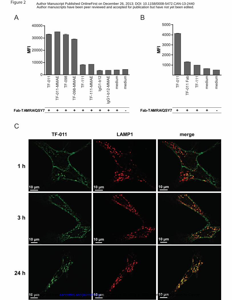
TF HuMab are rapidly internalized after target binding
Since ADCs generally rely on internalization for release of the payload, we characterized the
internalization characteristics of TF HuMab in the TAMRA/QSY7 assay. This assay uses a fluorophore
(TAMRA) and quencher (QSY7) pair. In close proximity, e.g. upon conjugation to the same protein,
TAMRA fluorescence is quenched by QSY7. TF HuMab were complexed with TAMRA/QSY7-conjugated
anti-human IgG Fab fragments (Fab-TAMRA/QSY7), and the complex was incubated with A431 or SK-
OV-3 cells. After 6 h, TAMRA fluorescence was detected in cells that had been incubated with TF-011,
TF-098 or TF-111 (Figure 2A,B), indicating internalization of the HuMab-Fab-TAMRA/QSY7 complex and
degradation in the reducing environment of the endosomes and lysosomes. Internalization was most
efficient for TF-011 and TF-098. Interestingly, internalization of TF-011 was reduced when Fab fragments
were used instead of the intact antibody (Figure 2B), suggesting that internalization of TF-011 is
stimulated by bivalent target binding.
Efficient internalization of TF-011 was confirmed by confocal microscopy. 1 h after incubation with
SK-OV-3 cells, TF-011 was already detectable in intracellular vesicles, some of which co-localized with
the lysosomal marker LAMP-1, indicating internalization and lysosomal targeting. Co-localization of TF-
011 and LAMP-1 was enhanced after 3 h, and after 24 h, most TF-011 co-localized with LAMP-1,
indicating efficient accumulation in the lysosomal compartment (Figure 2C). Similar results were obtained
with A431 cells (data not shown).
Our results demonstrate that TF HuMab are rapidly internalized and degraded upon target
binding, indicating that TF may be a very suitable ADC target.
Generation of TF-specific ADCs and cytotoxicity in vitro
TF-specific ADCs were generated by conjugation of TF-011, -098 and -111 with the dolastatin analogs
MMAE or MMAF. Auristatins are potent cytotoxic agents that induce cell death by disrupting microtubules
(20, 21). MMAE was conjugated through the protease-cleavable vc linker, and can therefore be released
from the antibody by lysosomal proteases, such as cathepsin B (21). MMAF was conjugated through a
non-cleavable linker, and relies on intracellular degradation of the ADC for release (20). TF HuMab were
Research. on November 20, 2018. © 2013 American Association for Cancercancerres.aacrjournals.org Downloaded from
Author manuscripts have been peer reviewed and accepted for publication but have not yet been edited. Author Manuscript Published OnlineFirst on December 26, 2013; DOI: 10.1158/0008-5472.CAN-13-2440

14
conjugated with an average of four auristatins per antibody, a ratio that was shown to provide the optimal
therapeutic index for brentuximab vedotin (29). Direct comparison of TF HuMab and TF-specific ADCs
(TF-ADCs) in vitro, confirmed that target binding and internalization characteristics were preserved in the
ADCs (Figure 2A, Supplementary Figure S3).
TF-ADCs showed excellent cytotoxicity in vitro. TF-ADCs efficiently and dose-dependently killed
A431 and HPAF-II cells (Figure 3A,B), which express high levels of TF on the plasma membrane
(>300,000 molecules per cell). The EC50 for TF-ADC-mediated tumor cell killing in A431 and HPAF-II cells
ranged from 4-10 ng/mL, for TF-011-MMAE and TF-098-MMAE, to 5-80 ng/mL, for TF-111-MMAF. In cell
lines that showed low (HCT-116; <15,000 molecules per cell) or very low (TOV-21G; <7,000 molecules
per cell) TF expression, TF-ADCs showed very limited or no cytotoxic activity (Figure 3C,D). Similarly, TF-
ADCs did not show cytotoxic activity in TF negative tumor cells (data not shown). This, in addition to the
lack of cytotoxic activity of the non-binding control ADCs IgG1-b12-MMAE and IgG1-b12-MMAF, indicates
that the efficacy of TF-ADCs is dependent on target expression.
Unconjugated TF HuMab did not induce direct cytotoxicity in vitro (data not shown), indicating
that the cytotoxicity of TF-ADCs was auristatin-dependent.
TF HuMab and TF-ADCs efficiently induce ADCC in vitro
Monoclonal antibodies of the IgG1 isotype may exert cytotoxicity through Fc-mediated effector functions
such as ADCC, and these effector mechanisms may be preserved upon conjugation with cytotoxic agents
(30). Unconjugated TF-011, TF-098 and TF-111 potently induced killing of A431 cells by ADCC (Figure
3E), with median EC50 values of 15 ng/mL (range 0.5-19 ng/mL), 18 ng/mL (range 5.0-57 ng/mL) and 76
ng/mL (range 15-102 ng/mL), respectively. Similar results were observed with BxPC-3 and MDA-MB-231
cells (data not shown). Importantly, the capacity to induce ADCC was preserved after conjugation with
MMAE (Figure 3E).
Anti-tumor activity of TF-specific ADCs in cell line-derived xenograft models
All TF-ADCs potently inhibited tumor growth in established xenografts derived from HPAF-II and A431
cells, at a dose of 3 mg/kg (four injections in two weeks; Figure 4A and Supplementary Figure S4A).
Research. on November 20, 2018. © 2013 American Association for Cancercancerres.aacrjournals.org Downloaded from
Author manuscripts have been peer reviewed and accepted for publication but have not yet been edited. Author Manuscript Published OnlineFirst on December 26, 2013; DOI: 10.1158/0008-5472.CAN-13-2440

15
MMAE-conjugates showed significantly better efficacy than their MMAF-conjugated counterparts (Figure
4B, Supplementary Figure S4B). Complete tumor regression, i.e. no measurable tumor remaining at 20-
30 days after the last treatment, was observed for most mice in the TF-098-MMAE and TF-111-MMAE
treatment groups, and for all mice in the TF-011-MMAE group. Strikingly, 3 out of 7 mice that had been
treated with TF-011-MMAE, remained tumor-free until the end of the study (139 days after discontinuation
of treatment). In mice that did show tumor recurrence after treatment with TF-011-MMAE (4 out of 7 mice
in the HPAF-II model), measurable tumors were not detected until 56-70 days after discontinuation of
treatment. Recurring tumors could successfully be re-treated with TF-011-MMAE, TF-098-MMAE or TF-
111-MMAE (Figure 4C; Supplementary Figure S4C), indicating that TF expression was maintained in
tumor cells that showed outgrowth after completion of the first treatment cycle.
The isotype control ADCs IgG1-b12-MMAE and IgG1-b12-MMAF did not inhibit tumor growth,
indicating that the efficacy of TF-ADCs was dependent on target binding. This was supported by the lack
of activity of TF-ADCs in the low TF expressing HCT-116 xenograft model (Supplementary Figure S4D).
To study the potential contribution of ADCC and inhibition of TF:FVIIa-dependent intracellular
signaling to the anti-tumor activity of TF-ADCs in vivo, xenograft studies were performed using
unconjugated TF-011, TF-098 or TF-111. Prophylactic treatment with TF-011, TF-098 or TF-111
significantly reduced outgrowth of BxPC-3, HPAF-II, AsPC-1 and A431 xenografts (Supplementary Figure
S5A-D and data not shown). When treatment was initiated after the tumors had established (therapeutic
treatment), TF-098 and TF-111 induced a small, albeit significant, reduction of tumor growth in the BxPC-
3 model (Supplementary Figure S5E,F). However, therapeutic treatment with unconjugated TF HuMab
was unable to inhibit tumor growth in the A431, AsPC-1 and HPAF-II xenograft models (Supplementary
Figure S5G,H and data not shown). This suggests that the anti-tumor activity of TF-ADCs in the
established xenografts tested here is to a large extent mediated by the cytotoxic activity of MMAE or
MMAF.
The anti-tumor efficacy of the most potent ADC, TF-011-MMAE, was assessed at different dose
levels and dosing schedules. At weekly dosing (four doses), treatment with 1 mg/kg TF-011-MMAE was
sufficient to induce tumor regression (Figure 4D). When administered as a single dose, 2 and 4 mg/kg TF-
011-MMAE induced tumor regression, whereas inhibition of tumor growth was observed after treatment
Research. on November 20, 2018. © 2013 American Association for Cancercancerres.aacrjournals.org Downloaded from
Author manuscripts have been peer reviewed and accepted for publication but have not yet been edited. Author Manuscript Published OnlineFirst on December 26, 2013; DOI: 10.1158/0008-5472.CAN-13-2440

16
with 0.5 or 1 mg/kg (Figure 4E). Interestingly, comparison of tumor volumes on day 24, when mice had
received either one or two doses of TF-011-MMAE, demonstrated that a single dose of 1 mg/kg TF-011-
MMAE was more effective than two doses of 0.5 mg/kg, although the cumulative dose was the same in
both treatment groups. Similarly, treatment with a single dose of 2 mg/kg was more effective than two
doses of 1 mg/kg (Figure 4F). This suggests that, at the same cumulative dose (exposure), dosing
schedules giving a higher peak plasma level (Cmax) are more effective. For treatment with a cumulative
dose of 4 mg/kg, no difference in efficacy was observed between a single dose of 4 mg/kg and two doses
of 2 mg/kg, because tumor regression was complete in both groups.
In summary, TF-ADCs showed potent anti-tumor activity in vivo, which was dependent on both
TF targeting and conjugation with auristatins. TF-011-MMAE was selected for further preclinical studies
and clinical development (designated as HuMax-TF-ADC).
TF-011-MMAE induces efficient tumor cell killing in PDX models with heterogeneous TF
expression
Like most solid tumor targets, expression of TF in cancer is heterogeneous between patients, within
patients and even within tumors (3). We addressed the capacity of TF-011-MMAE to inhibit the growth of
tumors with heterogeneous target expression using PDX models, which are thought to represent the
heterogeneity that exists between human tumors (31-33). Immunohistochemical (IHC) analysis of
xenografted primary human tumor biopsies confirmed heterogeneity of TF expression, and seven PDX
models were selected based on variable levels of TF expression. TF expression was observed in >75% of
tumor cells in the PDX models for lung and pancreas adenocarcinoma (Figure 5A,B), and in 50-75% of
tumor cells in the models for bladder carcinoma, prostate carcinoma and lung squamous cell carcinoma
(Figure 5C-E). In PDX models for cervix squamous cell carcinoma and ovarian adenocarcinoma, only 25-
50% of tumor cells were TF-positive (Figure 5F,G).
PDX tumors were implanted s.c. in nude mice, and when tumors had established, mice were
treated with TF-011-MMAE (4 mg/kg, two doses). In models where the sensitivity to microtubule-targeting
agents was not known, a paclitaxel treatment group (20 mg/kg, 3-4 doses) was included.
Research. on November 20, 2018. © 2013 American Association for Cancercancerres.aacrjournals.org Downloaded from
Author manuscripts have been peer reviewed and accepted for publication but have not yet been edited. Author Manuscript Published OnlineFirst on December 26, 2013; DOI: 10.1158/0008-5472.CAN-13-2440

17
In the models for pancreas and lung adenocarcinoma, that showed >75% TF-positive tumor cells,
TF-011-MMAE induced complete tumor regression in all mice (Figure 6A). Similarly, TF-011-MMAE
induced efficient tumor regression in PDX models that expressed TF in 50-75% of tumor cells (bladder
cancer, prostate cancer and lung squamous cell carcinoma; Figure 6B). Importantly, tumor regression
was also observed in models that showed TF expression in only 25-50% of the tumor cells (ovarian and
cervical cancer; Figure 6C).
Treatment with two doses of HuMax-TF-ADC was at least as efficient as treatment with three or
four doses of paclitaxel (Figure 6). Moreover, TF-011-MMAE induced tumor regression in mice that
showed tumor recurrence after paclitaxel treatment, even in mice with relatively large tumors
(Supplementary Figure S6). This demonstrates that prior treatment with paclitaxel did not affect the
sensitivity of the tumors to treatment with TF-011-MMAE, indicating that TF expression and sensitivity to
MMAE-mediated tumor cell killing was retained in tumors that showed outgrowth after paclitaxel
treatment.
Research. on November 20, 2018. © 2013 American Association for Cancercancerres.aacrjournals.org Downloaded from
Author manuscripts have been peer reviewed and accepted for publication but have not yet been edited. Author Manuscript Published OnlineFirst on December 26, 2013; DOI: 10.1158/0008-5472.CAN-13-2440
18
Discussion
TF-011-MMAE was selected from a panel of six TF-specific ADCs, consisting of three different TF
HuMab conjugated with vcMMAE or mcMMAF. TF-011-MMAE showed excellent anti-tumor activity in
vivo, with auristatin-mediated tumor cell killing as the dominant mechanism of action. Furthermore, TF-
011-MMAE and unconjugated TF-011 induced inhibition of TF:FVIIa-mediated intracellular signaling and
ADCC in vitro, although it is unclear to what extent these mechanisms may contribute to the inhibition of
tumor growth in cancer patients. To our knowledge, TF-011-MMAE is the first ADC that uses a TF-
specific antibody to deliver a cytotoxic agent to tumor cells.
Importantly, TF-011-MMAE induced complete tumor regression in PDX models, even if only a
sub-population of the tumor cells expressed TF. PDX models are thought to represent the genetic and
histological heterogeneity in human tumors, and efficacy of treatment in such models was shown to have
predictive value for the clinic (31-33). The high potency of TF-011-MMAE in tumors with non-
homogeneous target expression, may be related to the capacity of MMAE to cause a bystander effect by
diffusion across cell membranes after intracellular release (34). Especially in solid tumors, where antibody
penetration may be limited (35, 36), this may be a major advantage. As opposed to uncharged MMAE,
the negative charge of MMAF is thought to prevent diffusion across membranes (20). This difference in
membrane permeability probably underlies the difference in efficacy observed between MMAE- and
MMAF-conjugates.
Although TF plays a crucial role in coagulation and hemostasis, TF-011 showed minimal impact
on coagulation in vitro. Previous studies suggested that TF-specific antibodies can roughly be divided into
two categories: those that inhibit FVIIa binding and/or TF:FVIIa-induced intracellular signaling with minor
impact on TF pro-coagulant activity, and those that interfere with FXa activation and coagulation without
impacting on TF:FVIIa-induced intracellular signaling (14, 37). Our in vitro studies suggest that TF-011
belongs to the first category. This notion is supported by non-clinical toxicology studies in cynomolgus
monkeys. TF-011 and TF-011-MMAE, which show comparable binding to cynomolgus monkey and
human TF, did not significantly impact on functional bleeding time or systemic parameters of coagulation
in cynomolgus monkeys at doses up to 100 mg/kg or 5-6 mg/kg, respectively (Genmab, data on file). It
Research. on November 20, 2018. © 2013 American Association for Cancercancerres.aacrjournals.org Downloaded from
Author manuscripts have been peer reviewed and accepted for publication but have not yet been edited. Author Manuscript Published OnlineFirst on December 26, 2013; DOI: 10.1158/0008-5472.CAN-13-2440

19
may seem counterintuitive that TF:FVIIa binding, the first step in the coagulation cascade, can be
inhibited without impacting on hemostasis or clotting. This apparent paradox is most likely explained by
the many amplification steps in the coagulation cascade downstream of TF:FVIIa (38). As a result, only
little TF:FVIIa binding is required to maintain hemostasis. The work of Parry and colleagues, who
demonstrated that transgenic mice expressing only 1% of normal TF activity were viable and had
relatively normal hemostasis (39), supports this.
In summary, TF-011-MMAE is a promising new ADC that is being developed for the treatment of
solid tumors. TF is thought to be an excellent ADC target, due to its broad expression profile across solid
cancer types and rapid internalization and degradation after antibody binding. TF-011-MMAE induced
complete tumor regression in PDX models derived from a broad range of solid tumors, demonstrating the
high potency of TF-011-MMAE for treatment of cancer.
Research. on November 20, 2018. © 2013 American Association for Cancercancerres.aacrjournals.org Downloaded from
Author manuscripts have been peer reviewed and accepted for publication but have not yet been edited. Author Manuscript Published OnlineFirst on December 26, 2013; DOI: 10.1158/0008-5472.CAN-13-2440
20
Acknowledgements
We thank Elke Gresnigt-van den Heuvel, Imke Lodewijks, Gemma Rigter, Agnes de Goffau, Marije
Overdijk, Patrick Engelberts and Antonio Ortiz-Buijsse for technical support, Dr. Tom Vink for help with
the manuscript, Dr. Joost Bakker for help with the graphics and Dr. Wiiger for providing HaCaT cells.
Research. on November 20, 2018. © 2013 American Association for Cancercancerres.aacrjournals.org Downloaded from
Author manuscripts have been peer reviewed and accepted for publication but have not yet been edited. Author Manuscript Published OnlineFirst on December 26, 2013; DOI: 10.1158/0008-5472.CAN-13-2440

21
References
1. Verma S, Miles D, Gianni L, Krop IE, Welslau M, Baselga J, et al. Trastuzumab Emtansine for
HER2-Positive Advanced Breast Cancer. N Eng J Med 2012;367(19):1783-1791.
2. Senter PD, Sievers EL. The discovery and development of brentuximab vedotin for use in
relapsed Hodgkin lymphoma and systemic anaplastic large cell lymphoma. Nat Biotech
2012;30(7):631-637.
3. Förster Y, Meye A, Albrecht S, Schwenzer B. Tissue factor and tumor: Clinical and laboratory
aspects. Clinica Chimica Acta 2006;364(1-2):12-21.
4. Mandal SK, Pendurthi UR, Rao LV. Cellular localization and trafficking of tissue factor. Blood
2006;107(12):4746-4753.
5. Mackman N, Tilley RE, Key NS. Role of the extrinsic pathway of blood coagulation in hemostasis
and thrombosis. Arterioscler Thromb Vasc Biol 2007;27(8):1687-1693.
6. Drake TA, Morrissey JH, Edgington TS. Selective cellular expression of tissue factor in human
tissues. Implications for disorders of hemostasis and thrombosis. Am J Pathol 1989;134(5):1087-
1097.
7. Egorina EM, Sovershaev MA, Bjorkoy G, Gruber FX, Olsen JO, Parhami-Seren B, et al.
Intracellular and surface distribution of monocyte tissue factor: application to intersubject
variability. Arterioscler Thromb Vasc Biol 2005;25(7):1493-1498.
8. Amirkhosravi A, Alexander M, May K, Francis DA, Warnes G, Biggerstaff J, et al. The importance
of platelets in the expression of monocyte tissue factor antigen measured by a new whole blood
flow cytometric assay. Thromb Haemost 1996;75(1):87-95.
9. Vine AK. Recent advances in haemostasis and thrombosis. Retina 2009;29(1):1-7.
10. Chu AJ. Tissue factor, blood coagulation, and beyond: an overview [published online September
24, 2011]. Int J Inflam. doi:10.4061/2011/367284.
11. Yu JL, May L, Lhotak V, Shahrzad S, Shirasawa S, Weitz JI, et al. Oncogenic events regulate
tissue factor expression in colorectal cancer cells: implications for tumor progression and
angiogenesis. Blood 2005;105(4):1734-1741.
Research. on November 20, 2018. © 2013 American Association for Cancercancerres.aacrjournals.org Downloaded from
Author manuscripts have been peer reviewed and accepted for publication but have not yet been edited. Author Manuscript Published OnlineFirst on December 26, 2013; DOI: 10.1158/0008-5472.CAN-13-2440

22
12. Ngo CV, Picha K, McCabe F, Millar H, Tawadros R, Tam SH, et al. CNTO 859, a humanized anti-
tissue factor monoclonal antibody, is a potent inhibitor of breast cancer metastasis and tumor
growth in xenograft models. Int J Cancer 2007;120(6):1261-1267.
13. Versteeg HH, Schaffner F, Kerver M, Petersen HH, Ahamed J, Felding-Habermann B, et al.
Inhibition of tissue factor signaling suppresses tumor growth. Blood 2008;111(1):190-199.
14. Kirchhofer D, Moran P, Chiang N, Kim J, Riederer MA, Eigenbrot C, et al. Epitope location on
tissue factor determines the anticoagulant potency of monoclonal anti-tissue factor antibodies.
Thromb Haemost 2000;84(6):1072-1081.
15. Vink T, Oudshoorn-Dickmann M, Roza M, Reitsma J-J, de Jong RN. A simple, robust and highly
efficient transient expression system for producing antibodies [published online July 16, 2013].
Methods. doi:10.1016/j.ymeth.2013.07.018.
16. Fishwild DM, O'Donnell SL, Bengoechea T, Hudson DV, Harding F, Bernhard SL, et al. High-
avidity human IgG kappa monoclonal antibodies from a novel strain of minilocus transgenic mice.
Nat Biotechnol 1996;14(7):845-851.
17. Labrijn AF, Meesters JI, de Goeij BECG, van den Bremer ETJ, Neijssen J, van Kampen MD, et
al. Efficient generation of stable bispecific IgG1 by controlled Fab-arm exchange. Proceedings of
the National Academy of Sciences 2013;110(13):5145-5150.
18. Parren PW, Ditzel HJ, Gulizia RJ, Binley JM, Barbas CF, 3rd, Burton DR, et al. Protection against
HIV-1 infection in hu-PBL-SCID mice by passive immunization with a neutralizing human
monoclonal antibody against the gp120 CD4-binding site. AIDS 1995;9(6):F1-6.
19. Lammerts van Bueren JJ, Bleeker WK, Bogh HO, Houtkamp M, Schuurman J, van de Winkel JG,
et al. Effect of target dynamics on pharmacokinetics of a novel therapeutic antibody against the
epidermal growth factor receptor: implications for the mechanisms of action. Cancer Res
2006;66(15):7630-7638.
20. Doronina SO, Mendelsohn BA, Bovee TD, Cerveny CG, Alley SC, Meyer DL, et al. Enhanced
activity of monomethylauristatin F through monoclonal antibody delivery: effects of linker
technology on efficacy and toxicity. Bioconjug Chem 2006;17(1):114-124.
Research. on November 20, 2018. © 2013 American Association for Cancercancerres.aacrjournals.org Downloaded from
Author manuscripts have been peer reviewed and accepted for publication but have not yet been edited. Author Manuscript Published OnlineFirst on December 26, 2013; DOI: 10.1158/0008-5472.CAN-13-2440
23
21. Doronina SO, Toki BE, Torgov MY, Mendelsohn BA, Cerveny CG, Chace DF, et al. Development
of potent monoclonal antibody auristatin conjugates for cancer therapy. Nat Biotechnol
2003;21(7):778-784.
22. Overdijk MB, Verploegen S, van den Brakel JH, Lammerts van Bueren JJ, Vink T, van de Winkel
JG, et al. Epidermal growth factor receptor (EGFR) antibody-induced antibody-dependent cellular
cytotoxicity plays a prominent role in inhibiting tumorigenesis, even of tumor cells insensitive to
EGFR signaling inhibition. J Immunol 2011;187(6):3383-3390.
23. Amirkhosravi A, Bigsby G, Desai H, Rivera-Amaya M, Coll E, Robles-Carrillo L, et al. Blood
clotting activation analysis for preoperative differentiation of benign versus malignant ovarian
masses. Blood Coagulation & Fibrinolysis 2013;24(5):510-517.
24. Ogawa M, Kosaka N, Longmire MR, Urano Y, Choyke PL, Kobayashi H. Fluorophore-quencher
based activatable targeted optical probes for detecting in vivo cancer metastases. Mol Pharm
2009;6(2):386-395.
25. Bleeker WK, Lammerts van Bueren JJ, van Ojik HH, Gerritsen AF, Pluyter M, Houtkamp M, et al.
Dual Mode of Action of a Human Anti-Epidermal Growth Factor Receptor Monoclonal Antibody
for Cancer Therapy. The Journal of Immunology 2004;173(7):4699-4707.
26. Hjortoe GM, Petersen LC, Albrektsen T, Sorensen BB, Norby PL, Mandal SK, et al. Tissue factor-
factor VIIa–specific up-regulation of IL-8 expression in MDA-MB-231 cells is mediated by PAR-2
and results in increased cell migration. Blood 2004;103(8):3029-3037.
27. Gessler F, Voss V, Dützmann S, Seifert V, Gerlach R, Kögel D. Inhibition of tissue
factor/protease-activated receptor-2 signaling limits proliferation, migration and invasion of
malignant glioma cells. Neuroscience 2010;165(4):1312-1322.
28. Aras O. Induction of microparticle- and cell-associated intravascular tissue factor in human
endotoxemia. Blood 2004;103(12):4545-4553.
29. Hamblett KJ, Senter PD, Chace DF, Sun MM, Lenox J, Cerveny CG, et al. Effects of drug loading
on the antitumor activity of a monoclonal antibody drug conjugate. Clin Cancer Res
2004;10(20):7063-7070.
Research. on November 20, 2018. © 2013 American Association for Cancercancerres.aacrjournals.org Downloaded from
Author manuscripts have been peer reviewed and accepted for publication but have not yet been edited. Author Manuscript Published OnlineFirst on December 26, 2013; DOI: 10.1158/0008-5472.CAN-13-2440

24
30. Junttila TT, Li G, Parsons K, Phillips GL, Sliwkowski MX. Trastuzumab-DM1 (T-DM1) retains all
the mechanisms of action of trastuzumab and efficiently inhibits growth of lapatinib insensitive
breast cancer. Breast Cancer Res Treat 2011;128(2):347-356.
31. Moro M, Bertolini G, Tortoreto M, Pastorino U, Sozzi G, Roz L. Patient-derived xenografts of non
small cell lung cancer: resurgence of an old model for investigation of modern concepts of
tailored therapy and cancer stem cells [published online May 2, 2012]. J Biomed Biotechnol.
doi:10.1155/2012/568567.
32. Tentler JJ, Tan AC, Weekes CD, Jimeno A, Leong S, Pitts TM, et al. Patient-derived tumour
xenografts as models for oncology drug development. Nat Rev Clin Oncol 2012;9(6):338-350.
33. Hidalgo M, Bruckheimer E, Rajeshkumar NV, Garrido-Laguna I, De Oliveira E, Rubio-Viqueira B,
et al. A pilot clinical study of treatment guided by personalized tumorgrafts in patients with
advanced cancer. Mol Cancer Ther 2011;10(8):1311-1316.
34. Okeley NM, Miyamoto JB, Zhang X, Sanderson RJ, Benjamin DR, Sievers EL, et al. Intracellular
activation of SGN-35, a potent anti-CD30 antibody-drug conjugate. Clin Cancer Res
2010;16(3):888-897.
35. Jain RK. Physiological Barriers to Delivery of Monoclonal Antibodies and Other Macromolecules
in Tumors. Cancer Research 1990;50(3 Supplement):814s-819s.
36. Rudnick SI, Lou J, Shaller CC, Tang Y, Klein-Szanto AJP, Weiner LM, et al. Influence of Affinity
and Antigen Internalization on the Uptake and Penetration of Anti-HER2 Antibodies in Solid
Tumors. Cancer Research 2011;71(6):2250-2259.
37. Ahamed J, Belting M, Ruf W. Regulation of tissue factor–induced signaling by endogenous and
recombinant tissue factor pathway inhibitor 1. Blood 2005;105(6):2384-2391.
38. Jesty J, Beltrami E. Positive Feedbacks of Coagulation: Their Role in Threshold Regulation.
Arteriosclerosis, Thrombosis, and Vascular Biology 2005;25(12):2463-2469.
39. Parry GC, Erlich JH, Carmeliet P, Luther T, Mackman N. Low levels of tissue factor are
compatible with development and hemostasis in mice. J Clin Invest 1998;101(3):560-569.
Research. on November 20, 2018. © 2013 American Association for Cancercancerres.aacrjournals.org Downloaded from
Author manuscripts have been peer reviewed and accepted for publication but have not yet been edited. Author Manuscript Published OnlineFirst on December 26, 2013; DOI: 10.1158/0008-5472.CAN-13-2440

25
Figure legends
Figure 1. Functional characteristics of TF HuMab in vitro. (A) Binding of TF HuMab to MDA-MB-231
cells as assessed by flow cytometry. Results from a representative experiment are shown (n=3). (B)
Competition between TF HuMab and FVIIa for TF binding. FVIIa was incubated with TF-ECDHis in the
presence of TF HuMab, and binding of FVIIa was measured by ELISA. Results from a representative
experiment are shown (n=3), error bars indicate SEM. (C) TF:FVIIa-induced ERK phosphorylation in the
presence of TF HuMab. BxPC-3 cells were incubated with FVIIa after pre-incubation with TF HuMab, and
p-ERK1/2 and total ERK1/2 were detected in cell lysates by Western Blot. Full-length blots are presented
in Supplementary Figure S7. (D) TF:FVIIa-induced IL-8 production in MDA-MB-231 cells in the presence
of TF HuMab. Cells were incubated with TF HuMab prior to stimulation with FVIIa. IL-8 production was
measured in cell culture supernatants using ELISA. Results from a representative experiment are shown
(n=3), error bars indicate SEM. (E, F) Effect of TF HuMab on whole blood coagulation as assessed by
thromboelastography. Citrated whole blood was incubated with LPS to induce TF expression, followed by
incubation with TF HuMab. Coagulation was initiated by re-calcification. (E) Inhibition of TF activity in the
presence of TF HuMab. TF activity was defined as the difference in clotting lag time (R) between
unstimulated and LPS-stimulated whole blood. TF HuMab-mediated inhibition of TF activity was
expressed the percentage change in TF activity. Data represent the average of 3 donors, error bars
represent SEM. (F) TEG trace overlays of coagulation in the presence of 20 μg/mL TF-011, TF-013, TF-
098 or an isotype control IgG. Results from a representative donor are shown (n=3).
Figure 2. TF HuMab are rapidly internalized and trafficked to the lysosomes. (A, B) TF HuMab or
TF-ADCs were complexed with anti-human Fab-fragments that had been conjugated with the fluorophore
and quencher pair TAMRA/QSY7 (Fab-TAMRA/QSY7), and the complex was added to A431 (A) or SK-
OV-3 (B) cells. Upon internalization and degradation of the complex, dissociation of TAMRA and QSY
results in de-quenching of TAMRA. The resulting fluorescent signal was measured by flow cytometry.
Error bars indicate SEM of duplicates. (C) SK-OV-3 cells were incubated with TF-011 in the presence of
an inhibitor of lysosomal degradation. After 1, 3 or 24 h, cells were fixed and TF-011 and the lysosomal
Research. on November 20, 2018. © 2013 American Association for Cancercancerres.aacrjournals.org Downloaded from
Author manuscripts have been peer reviewed and accepted for publication but have not yet been edited. Author Manuscript Published OnlineFirst on December 26, 2013; DOI: 10.1158/0008-5472.CAN-13-2440

26
protein LAMP-1 were detected using confocal microscopy. Left panel = TF-011 (green), middle panel =
LAMP-1 (red), right panel = merge (yellow).
Figure 3. Cytotoxicity of TF-specific ADCs in vitro. (A-D) Cells were incubated in the presence of TF-
ADCs and the viability of the cultures was assessed after 3-5 days using the Alamar Blue assay. IgG1-
b12-MMAE and IgG1-b12-MMAF were included as isotype control ADCs. Curves represent dose-
dependent cytotoxicity of TF-ADCs in the different cell lines. Results are representative of at least two
experiments, error bars represent SEM. The inserts show TF expression in the different cell lines as
assessed by flow cytometry, using 1 μg/mL mouse-anti-TF antibody (black lines) or an isotype control IgG
(filled grey histograms). (A-B) Cytotoxicity of TF-ADCs in (A) A431 and (B) HPAF-II cells, which show high
TF expression on the cell surface (>300,000 TF molecules/cell). (C-D) Cytotoxicity of TF-ADCs in cell
lines with low (C; HCT-116, <15,000 molecules/cell) or very low (D; TOV-21G, <7,000 molecules/cell) TF
expression. (E) TF HuMab and TF-ADCs induce ADCC. 51Cr-labeled A431 cells were incubated with TF
HuMab or TF-ADCs in the presence of freshly isolated PBMC, as a source of effector cells. 51Cr release
was measured to assess cytotoxicity, and the percentage kill was calculated. Results are representative
of experiments performed with PBMC from six (TF HuMab) or two (TF-ADC) different donors. Error bars
indicate SEM.
Figure 4. TF-ADCs show potent anti-tumor activity in vivo. HPAF-II xenografts were established by
s.c. injection in SCID mice, and treatment with TF-ADCs was initiated at day 13 after tumor inoculation.
(A-C) Treatment with 3 mg/kg TF-ADCs. (A) Tumor growth in the different treatment groups. IgG1-b12-
MMAE and IgG1-b12-MMAF were included as isotype control ADCs, IgG1-b12 was included as isotype
control IgG. Curves represent average tumor size per treatment group (7 mice per group), error bars
indicate SEM. The number of mice that showed complete tumor regression (i.e. no measurable tumor
remaining) in each of the treatment groups is indicated between brackets. (B) Tumor volumes in the
different treatment groups at day 27 after tumor inoculation. Differences in average tumor size between
treatment groups were analyzed by one-way ANOVA, ** P<0.01, *** P<0.001. (C) HPAF-II xenografts that
showed outgrowth after completion of the first treatment cycle were re-treated with 3 mg/kg TF-ADC (four
Research. on November 20, 2018. © 2013 American Association for Cancercancerres.aacrjournals.org Downloaded from
Author manuscripts have been peer reviewed and accepted for publication but have not yet been edited. Author Manuscript Published OnlineFirst on December 26, 2013; DOI: 10.1158/0008-5472.CAN-13-2440

27
doses, at the indicated time points). Mice were re-treated with the same TF-ADC as they had received in
the first treatment cycle. Curves represent individual mice. (D-F) Anti-tumor activity of TF-011-MMAE in
the HPAF-II xenograft model at different dose levels and dosing frequencies. (D,E) Average tumor size
after treatment with 0.5, 1, 2 or 4 mg/kg TF-011-MMAE (7 mice per group), at weekly dosing (D) or as a
single dose (E). IgG1-b12 was included as an isotype control antibody. Error bars indicate SEM. (F)
Tumor volume per treatment group at day 24, when mice had received either one or two doses of TF-
011-MMAE. Differences in average tumor size between groups that had received the same cumulative
dose of TF-011-MMAE were compared by one-way ANOVA (* P<0.05, *** P<0.001).
Figure 5. Heterogeneous expression of TF in PDX models. IHC analysis was performed to assess TF
expression in PDX models. The percentage of TF-positive tumor cells was estimated by comparing
human cytokeratin staining (which identifies human tumor cells) with TF staining (indicating TF-positive
tumor cells). In PDX models for (A) lung adenocarcinoma and (B) pancreatic adenocarcinoma, >75% of
the tumor cells showed TF expression. In PDX models for (C) bladder (urothelial) adenocarcinoma, (D)
prostate adenocarcinoma and (E) lung squamous cell carcinoma, TF expression was observed in 50-75%
of the tumor cells. In PDX models for (F) squamous cell carcinoma of the cervix and (G) ovarian
adenocarcinoma, 25-50% of the tumor cells showed TF expression. (H) Representative pictures showing
immunostaining with the isotype control antibody IgG1-b12.
Figure 6. Anti-tumor activity of TF-011-MMAE in PDX models with heterogeneous target
expression. PDX models were established by s.c. implantation of tumor fragments in mice. When tumors
had reached a size of 80-200 mm3, mice were randomized and treatment was initiated. Mice were treated
with TF-011-MMAE or paclitaxel at the indicated doses and time points. IgG1-b12-MMAE was included as
an isotype control ADC, IgG1-b12 was included as an isotype control IgG. (A) Tumor growth in PDX
models showing >75% TF-positive tumor cells (lung adenocarcinoma and pancreatic adenocarcinoma),
(B) Tumor growth in PDX models showing 50-75% TF-positive tumor cells (bladder adenocarcinoma,
prostate adenocarcinoma and lung squamous cell carcinoma) and (C) Tumor growth in PDX tumors with
Research. on November 20, 2018. © 2013 American Association for Cancercancerres.aacrjournals.org Downloaded from
Author manuscripts have been peer reviewed and accepted for publication but have not yet been edited. Author Manuscript Published OnlineFirst on December 26, 2013; DOI: 10.1158/0008-5472.CAN-13-2440

28
25-50% TF-positive cells (cervical squamous cell carcinoma and ovarian adenocarcinoma). Data points
represent average tumor size per group (8 mice per group). Error bars represent SEM.
Research. on November 20, 2018. © 2013 American Association for Cancercancerres.aacrjournals.org Downloaded from
Author manuscripts have been peer reviewed and accepted for publication but have not yet been edited. Author Manuscript Published OnlineFirst on December 26, 2013; DOI: 10.1158/0008-5472.CAN-13-2440

1
Table 1
TF HuMab: functional characteristics in vitro.
HuMab FVIIa bindinga ERK phosphorylationb IL-8 releasec FXa generation
IC50 (SD)
μg/mL
Maximal
inhibitiond(SD)
%
IC50 (SD)
μg/mL
Maximal
inhibitione (SD)
%
IC50 (SD)
μg/mL
Maximal
inhibitionf (SD)
%
Maximal
inhibitiong (SD)
%
TF-011 0.19 (0.07) 91 (3) 0.12 (0.03) 69 (4) 1.4 (0.4) 62 (6) 19 (9)
TF-013 2.9 (4.2) 27 (10) 1.37 (0.31) 26 (6) NA 0 (14) 22 (8)
TF-025 0.33 (0.01) 90 (2) 0.33 (0.06) 66 (0) 3.5 (2.7) 76 (5) 9 (2)
TF-044 0.21 (0.04) 54 (10) 60 (NA) 45 (5) 11.2 (4.8) 17 (19) 0 (3)
TF-098 0.16 (0.04) 85 (4) 0.28 (0.06) 64 (5) 1.4 (0.4) 59 (20) 14 (1)
TF-109 0.23 (0.10) 90 (2) 0.36 (0.08) 72 (4) 2.0 (0.8) 70 (14) 4 (1)
TF-111 0.33 (0.14) 79 (7) >10,000 52 (1) >10,000 40 (39) 0 (5)
TF-114 0.20 (0.05) 90 (4) 0.16 (0.05) 68 (0) 1.4 (0.7) 72 (6) 10 (4)
aFVIIa ELISA, average of 3 experiments.
bAlphascreen Surefire ERK assay, A431 cells, average of 2 experiments.
cMDA-MB-231 cells, average of 3 experiments.
d-fInhibition measured at plateau of dose-response curve, at 30 μg/mL (d), 10 μg/mL (e) or 120 μg/mL (f).
gInhibition (percentage) measured at plateau of dose-response curve (at 0.9 μg/mL IgG); average of two
experiments.
Research. on November 20, 2018. © 2013 American Association for Cancercancerres.aacrjournals.org Downloaded from
Author manuscripts have been peer reviewed and accepted for publication but have not yet been edited. Author Manuscript Published OnlineFirst on December 26, 2013; DOI: 10.1158/0008-5472.CAN-13-2440

MFI
0.0
01
0.0
10
.11
10
10
00
10
00
20
00
30
00
TF
-013
TF
-114
TF
-011
TF
-025
TF
-109
TF
-044
TF
-098
TF
-111
HuM
ab-K
LH
0.0
01
0.0
10
.11
10
10
00
.0
0.5
1.0
1.5
TF
-013
TF
-114
TF
-011
TF
-025
TF
-109
TF
-044
TF
-098
TF
-111
HuM
ab-K
LH
OD405 nm IL-8 [pg/mL]
0.0
10
.11
10
10
00
20
0
40
0
60
0T
F-0
11
TF
-098
TF
-109
TF
-111
TF
-114
TF
-025
TF
-013
TF
-044
HuM
ab-K
LH
% inhibition of TF activity
1 1
0 2
05
01
1
0 2
05
01
1
0 2
05
01
1
0 2
05
00
20
40
60
80
10
0T
F-0
11
TF
-013
TF
-098
isoty
pe c
trl I
gG
Fig
ure
1
p-E
RK
1/2
ER
K 1/2
++
++
FV
IIa
(10 n
M)
-+
011
09
811
41
09
TF
Hu
Ma
b-
-
+111
+04
4
+01
3
+02
5
FV
IIa
(10 n
M)
TF
Hu
Ma
b
--
+-
p-E
RK
1/2
ER
K 1/2
-30
-20
-10010
20
30
05
10
15
20
25
30
35
40
45
50
isoty
pe c
ontr
ol Ig
G1
TF
-011
TF
-013
TF
-098
Do
no
r A
: 2
0 μ
g/m
Lamplitude (mm)
clo
ttin
g lag
tim
e (
min
)
A CD
EFB
IgG
[μ
g/m
L]
IgG
[μ
g/m
L]
IgG
[μ
g/m
L]
IgG
[μ
g/m
L]
Research. on November 20, 2018. © 2013 American Association for Cancercancerres.aacrjournals.org Downloaded from
Author manuscripts have been peer reviewed and accepted for publication but have not yet been edited. Author Manuscript Published OnlineFirst on December 26, 2013; DOI: 10.1158/0008-5472.CAN-13-2440
TF
-01
1
TF
-01
1-M
MA
E
TF
-09
8
TF
-09
8-M
MA
E
TF
-11
1
TF
-11
1-M
MA
E
IgG
1-b
12
IgG
1-b
12
-MM
AE
me
diu
m
me
diu
m
0
10000
20000
30000
40000M
FI
-++ + +Fab-TAMRA/QSY7 + + + + +
TF
-01
1
TF
-01
1 F
ab
TF
-11
1
me
diu
m
me
diu
m
1000
2000
3000
4000
5000
MF
I
-++ + +Fab-TAMRA/QSY7
1 h
LAMP1 merge
3 h
24 h
TF-011
10 μm10 μm10 μm
10 μm10 μm10 μm
10 μm 10 μm10 μm
A
C
B
Figure 2
Research. on November 20, 2018. © 2013 American Association for Cancercancerres.aacrjournals.org Downloaded from
Author manuscripts have been peer reviewed and accepted for publication but have not yet been edited. Author Manuscript Published OnlineFirst on December 26, 2013; DOI: 10.1158/0008-5472.CAN-13-2440

2 3 4 5log-FITC
2 3 4 5log-FITC
2 3 4 5log-FITC
2 3 4 5log-FITC
0.001 0.01 0.1 1 10 100 1000 100000
50
100
ADC [ng/mL]
via
ble
cells (
%)
0.01 0.1 1 10 100 1000 100000
50
100
ADC [ng/mL]
via
ble
cells (
%)
0.001 0.01 0.1 1 10 100 1000 100000
50
100
ADC [ng/mL]
via
ble
cells (
%)
0.001 0.01 0.1 1 10 100 1000 100000
50
100
ADC [ng/mL]
via
ble
cells (
%)
0.1 1 10 100 1000 100000
10
20
30
40
50
IgG (ng/mL)
% c
yto
toxic
ity
A431 HPAF-II
HCT-116 TOV-21G
TF-098-MMAE
TF-011-MMAE
IgG1-b12-MMAE
TF-111-MMAE
TF-098-MMAF
TF-011-MMAF
IgG1-b12-MMAF
TF-111-MMAF
TF-098-MMAE
TF-011-MMAE
IgG1-b12-MMAE
TF-111-MMAE
TF-098-MMAF
TF-011-MMAF
IgG1-b12-MMAF
TF-111-MMAF
TF-011
IgG1-b12
TF-098
TF-111
TF-011-MMAE
IgG1-b12-MMAE
TF-098-MMAE
TF-111-MMAE
A B
C
E
D
Figure 3
Research. on November 20, 2018. © 2013 American Association for Cancercancerres.aacrjournals.org Downloaded from
Author manuscripts have been peer reviewed and accepted for publication but have not yet been edited. Author Manuscript Published OnlineFirst on December 26, 2013; DOI: 10.1158/0008-5472.CAN-13-2440

0 10 20 30 400
250
500
750
1000
� � � �
treatment 3 mg/kg�
IgG1-b12 (0/7)
IgG1-b12-MMAF (0/7)
IgG1-b12-MMAE (0/7)
TF-111-MMAF (0/7)
TF-011-MMAF (0/7)
TF-098-MMAF (0/7)
TF-011-MMAE (7/7)
TF-098-MMAE (5/7)
TF-111-MMAE (6/7)
days after tumor inoculation
tum
or
siz
e (
mm
3)
Treatment groups (# mice withcomplete tumor regression/n):
TF
-01
1-M
MA
E
TF
-01
1-M
MA
F
TF
-09
8-M
MA
E
TF
-09
8-M
MA
F
TF
-11
1-M
MA
E
TF
-11
1-M
MA
F
IgG
1-b
12
-MM
AE
IgG
1-b
12
-MM
AF
IgG
1-b
12
0
250
500
750
1000
tum
or
siz
e (
mm
3)
** *** ***
0 20 40 60 80 100 1200
250
500
750
1000
1250
1500
���� ����
days after tumor inoculation
days after tumor inoculation
days after tumor inoculation
tum
or
siz
e (
mm
3)
tum
or
siz
e (
mm
3)
tum
or
siz
e (
mm
3)
tum
or
siz
e (
mm
3)
TF-011-MMAE - m731
TF-011-MMAE - m753
TF-098-MMAE - m769
TF-111-MMAE - m738
TF-111-MMAE - m741
treatment 3 mg/kg�
weekly dosing
0 10 20 30 400
200
400
600
800
1000
1200↓ ↓ ↓ ↓
IgG1-b12 4 mg/kg
TF-011-MMAE 0.5 mg/kg
TF-011-MMAE 1 mg/kg
TF-011-MMAE 4 mg/kg
TF-011-MMAE 2 mg/kg
treatment↓
0 10 20 30 400
200
400
600
800
1000
1200↓
single dose
IgG1-b12 4 mg/kg
TF-011-MMAE 0.5 mg/kg
TF-011-MMAE 1 mg/kg
TF-011-MMAE 4 mg/kg
TF-011-MMAE 2 mg/kg
treatment↓
IgG
1-b
12
- 4
mg
/kg
0.5
mg
/kg
- s
ing
le d
os
e
0.5
mg
/kg
- t
wo
do
se
s
1 m
g/k
g -
sin
gle
do
se
1 m
g/k
g -
tw
o d
os
es
2 m
g/k
g -
sin
gle
do
se
2 m
g/k
g -
tw
o d
os
es
4 m
g/k
g -
sin
gle
do
se
4 m
g/k
g -
tw
o d
os
es
100
1000
1.0 2.0
day 24
*** *
4.0 8.00.5 mg/kg
A B
C D
E F
Figure 4
Research. on November 20, 2018. © 2013 American Association for Cancercancerres.aacrjournals.org Downloaded from
Author manuscripts have been peer reviewed and accepted for publication but have not yet been edited. Author Manuscript Published OnlineFirst on December 26, 2013; DOI: 10.1158/0008-5472.CAN-13-2440

A B C
07
14
21
28
35
42
0
50
0
10
00
15
00
��
days a
fter
firs
t tr
eatm
en
t
tumor size (mm3)
Lu
ng
ad
en
oc
arc
ino
ma
07
14
21
0
50
0
10
00
15
00
��
days a
fter
firs
t tr
eatm
en
t
tumor size (mm3)
Pa
nc
rea
s a
de
no
ca
rcin
om
a
07
14
21
28
35
42
0
50
0
10
00
15
00�
�
days a
fter
firs
t tr
eatm
en
t
tumor size (mm3)
Bla
dd
er
(uro
the
lia
l) a
de
no
ca
rcin
om
a
07
14
21
28
35
0
50
0
10
00
15
00�
� days a
fter
firs
t tr
eatm
en
t
tumor size (mm3)
Pro
sta
te a
de
no
ca
rcin
om
a
07
14
21
28
35
0
50
0
10
00
15
00
�� days a
fter
firs
t tr
eatm
en
t
tumor size (mm3)
Lu
ng
sq
ua
mo
us
ce
ll c
arc
ino
ma
07
14
21
28
35
42
0
50
0
10
00
15
00�
� days a
fter
firs
t tr
eatm
en
t
tumor size (mm3)
Ce
rvic
al s
qu
am
ou
s c
ell c
arc
ino
ma
07
14
21
28
35
42
0
50
0
10
00
15
00
�� d
ays a
fter
firs
t tr
eatm
en
t
tumor size (mm3)
Ov
ari
an
ad
en
oc
arc
ino
ma
IgG
1-b
12
IgG
1-b
121-M
MA
E
TF
-011-M
MA
E
paclit
axe
l
treatm
ent A
DC
(4 m
g/k
g)
�
treatm
ent paclit
axe
l (20 m
g/k
g)
Fig
ure
6
Research. on November 20, 2018. © 2013 American Association for Cancercancerres.aacrjournals.org Downloaded from
Author manuscripts have been peer reviewed and accepted for publication but have not yet been edited. Author Manuscript Published OnlineFirst on December 26, 2013; DOI: 10.1158/0008-5472.CAN-13-2440

Published OnlineFirst December 26, 2013.Cancer Res Esther C.W. Breij, Bart E.C.G. de Goeij, Sandra Verploegen, et al. tumors.potent therapeutic activity against a broad range of solid An antibody-drug conjugate that targets tissue factor exhibits
Updated version
10.1158/0008-5472.CAN-13-2440doi:
Access the most recent version of this article at:
Material
Supplementary
http://cancerres.aacrjournals.org/content/suppl/2013/12/30/0008-5472.CAN-13-2440.DC1
Access the most recent supplemental material at:
Manuscript
Authoredited. Author manuscripts have been peer reviewed and accepted for publication but have not yet been
E-mail alerts related to this article or journal.Sign up to receive free email-alerts
Subscriptions
Reprints and
To order reprints of this article or to subscribe to the journal, contact the AACR Publications
Permissions
Rightslink site. Click on "Request Permissions" which will take you to the Copyright Clearance Center's (CCC)
.http://cancerres.aacrjournals.org/content/early/2013/12/24/0008-5472.CAN-13-2440To request permission to re-use all or part of this article, use this link
Research. on November 20, 2018. © 2013 American Association for Cancercancerres.aacrjournals.org Downloaded from
Author manuscripts have been peer reviewed and accepted for publication but have not yet been edited. Author Manuscript Published OnlineFirst on December 26, 2013; DOI: 10.1158/0008-5472.CAN-13-2440