An animal cell. Spermatozoa heart (cryosection) ELECTRON MICROSCOPY IMAGES OF VARIOUS MIT0CHONDRIA....
14
An animal cell
-
Upload
paula-little -
Category
Documents
-
view
213 -
download
0
Transcript of An animal cell. Spermatozoa heart (cryosection) ELECTRON MICROSCOPY IMAGES OF VARIOUS MIT0CHONDRIA....

An animal cell
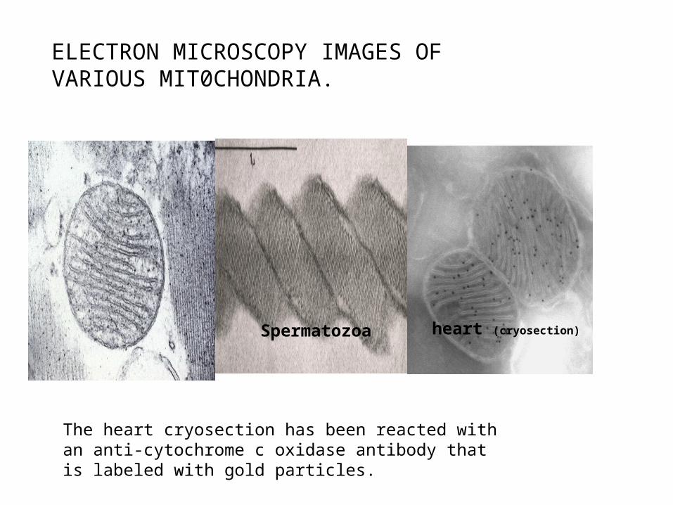
Spermatozoa heart (cryosection)
ELECTRON MICROSCOPY IMAGES OF VARIOUS MIT0CHONDRIA.
The heart cryosection has been reacted with an anti-cytochrome c oxidase antibody that is labeled with gold particles.

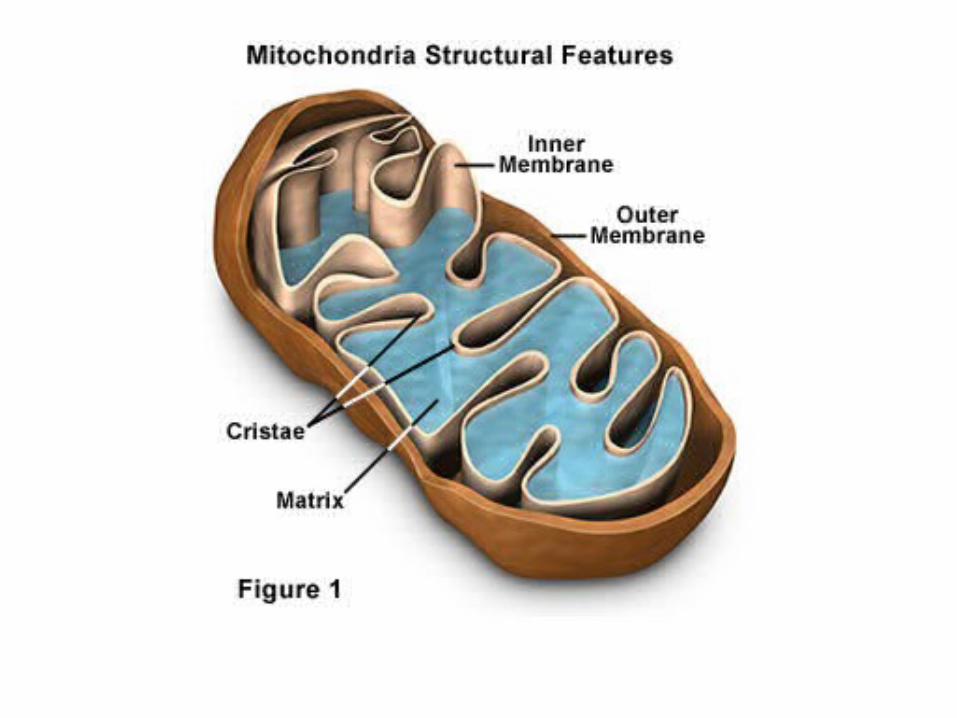
The internal structure of mitochondria [Review]Terrence G. Frey and Carmen A. Mannella Trends in Biochemical Sciences, 2000, 25:7:319-324
outer membrane
inner boundary membrane
cristae
crista junction
3D reconstructed electrontomogram of mitochondriaDr.Terry Frey; San Diego State University

Thin section of heart tissue showing the reticulum structure of mitochondria visualized by EM.

3) 3D view of reticulum
143B osteosarcomaGFP-pH transfectedConfocal microscope3D projection

2) Different states (fragmented, intermediate, reticulum)
A B Cfragmented intermediate reticular
10 m
- MitoTracker Red staining- osteosarcoma cells (143B)

-Osteosarcoma cells were arrested in G0 by FCS starvation for 60-72 h- Released by FCS addition- Stained with MitoTracker Red
Fragmented Reticular G0 G1 S G2/M
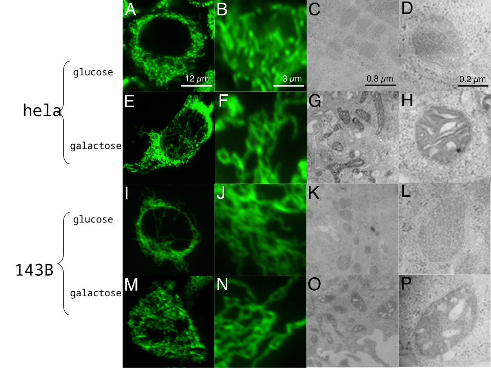
glucose
galactose
glucose
galactose
hela
143B

EM images of mitochondria from fibroblast cell lines. ABCD, different segments of an image of normal fibroblasts. C. Mitochondria from rh0 fibroblastcell.

A B
C D10 m 2.5 m
GF
P-E
1
Fre
e -
RF
P
Mer
ged
imag
e
24 hours post transfection
Distribution of Pyruvate dehydrogenase complex that has been tagged with GFP in fibroblast cells that show the mitochondrialr eticulum

Localization of GFP-tagged PDH after various times of incubation of cells. The complexes are essentially fixed with respect to the reticulum.

Ultra thin cryo section - bovine heartCORE II labeled with 10 nm gold particles Rob Gilkerson
Green: anti COX I antibodyRed: anti E2 antibody
Relative distribution of Cytochrome oxidase and PDH in a mitochondrial reticulum.
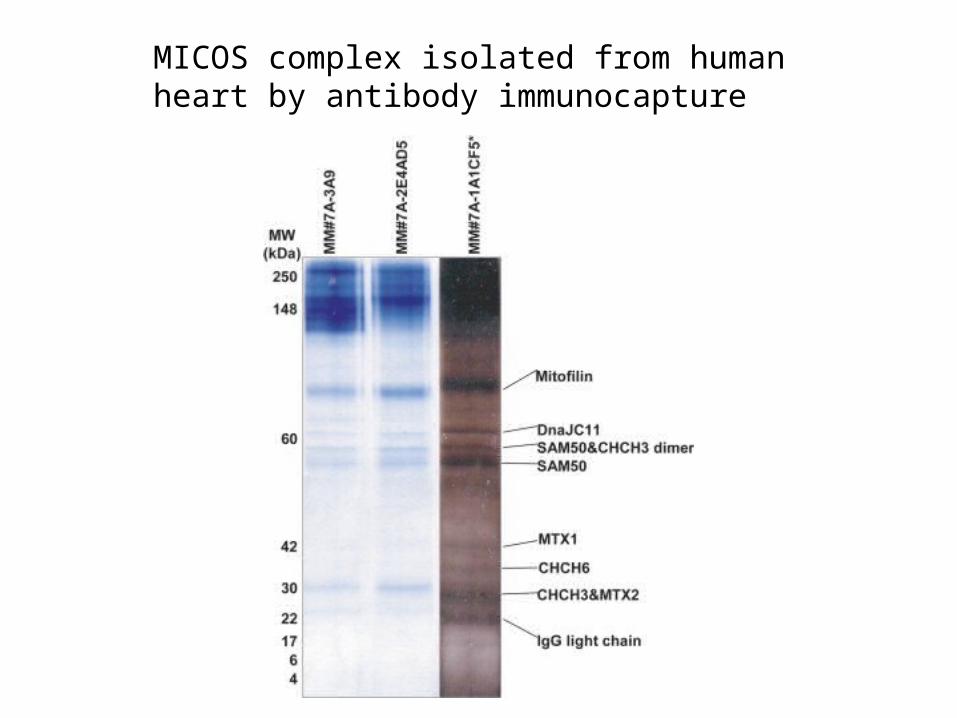
MICOS complex isolated from human heart by antibody immunocapture