
Amplified insert assembly: an optimized approach to ...
11
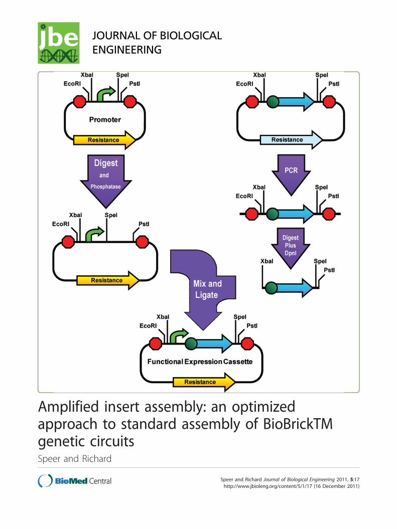
Amplified insert assembly: an optimized approach to standard assembly of BioBrickTM genetic circuits Speer and Richard Speer and Richard Journal of Biological Engineering 2011, 5:17 http://www.jbioleng.org/content/5/1/17 (16 December 2011)
Transcript of Amplified insert assembly: an optimized approach to ...
Amplified insert assembly: an optimizedapproach to standard assembly of BioBrickTMgenetic circuitsSpeer and Richard
Speer and Richard Journal of Biological Engineering 2011, 5:17http://www.jbioleng.org/content/5/1/17 (16 December 2011)

METHODOLOGY Open Access
Amplified insert assembly: an optimizedapproach to standard assembly of BioBrickTM
genetic circuitsMichael A Speer and Tom L Richard*
Abstract
A modified BioBrick™ assembly method was developed with higher fidelity than current protocols. The methodutilizes a PCR reaction with a standard primer set to amplify the inserted part. Background colonies are reduced bya combination of dephosphorylation and digestion with DpnI restriction endonuclease to reduce vector and insertbackground respectively. The molar ratio of the insert to vector in the ligation was also optimized, with theaccuracy of the transformed construct approaching 100%.
Keywords: BioBrick, Synthetic biology, PCR, amplification, insert, vector, assembly, DpnI
BackgroundThe methods used for controlled modification of geneticmaterial have experienced several major improvementssince the initial development of recombinant DNA techni-ques in the early 1970s [1-4]. While there have been manyefforts to simplify and standardize the genetic assemblyprocess [5-7], one format that has gained widespreadacceptance is the BioBrick™[8]. BioBrick assembly is com-monly applied within a synthetic biology conceptual fra-mework that abstracts and classifies basic functional unitsof genetic material (promoters, ribosome binding sites,protein coding sequences like reporters, terminators, etc.)as “parts” that can be assembled into “devices” that createnew functionality, and higher level “systems” to accom-plish complex tasks [9]. While the BioBrick standard hasbeen revised several times [10-12], the basic format hasremained the same. Each “BioBrick part” is contained on aplasmid and is flanked by four unique restriction sites,(two 5’ and two 3’) with the inner restriction sites that canbe cut by two endonucleases yielding compatible ends.Using this format, individual parts may be selectivelydigested and then ligated together to form new compositeparts or devices while preserving the format, connectingthe individual parts by a benign mixed restriction site
known as a “scar” (Figure 1). The chief utility of this tech-nology is that all parts using the same format are inter-changeable and composite parts can be recombined in thesame way as individual parts [8]. This has allowed entirecatalogs of compatible biological parts to be developed[9,13]. The major limitation of this format is that compo-site parts must be assembled piece by piece. To makethis system useful and affordable, easy and reliable stan-dard methods of performing these assemblies have beendeveloped [14].The original method used to perform “bio-bricking” is
called the standard assembly method. In using thismethod, one part is designated the insert and is digestedout of its plasmid using enzymes EcoRI and SpeI or XbaIand PstI [8]. The other part remains in its plasmid, butthe plasmid is opened up using compatible enzymes:EcoRI and XbaI or SpeI and PstI respectively (Figure 2).Upon successful double ligation the inner XbaI and SpeIsticky ends will form a benign scar and the two parts willbe adjacent to one another in the final circular construct.This protocol requires the following steps: 1) extractionof plasmids containing the two parts to be assembled; 2)digestion of the plasmids to create compatible ends onDNA fragments; 3) separation of the digested DNA byagarose gel electrophoresis; 4) extraction of the selectedfragments from the gel; 5) ligation of the fragmentstogether; and 6) transformation of the ligated plasmidproduct into cells. This process has many advantages: the
* Correspondence: [email protected] of Agricultural and Biological Engineering, The PennsylvaniaState University, 249 Agricultural Engineering Bldg., University Park, PA 16802,USA
Speer and Richard Journal of Biological Engineering 2011, 5:17http://www.jbioleng.org/content/5/1/17
© 2011 Speer and Richard; licensee BioMed Central Ltd. This is an Open Access article distributed under the terms of the CreativeCommons Attribution License (http://creativecommons.org/licenses/by/2.0), which permits unrestricted use, distribution, andreproduction in any medium, provided the original work is properly cited.

parts can all be on plasmids with the same antibioticresistance; thermostable enzymes (e.g. BglII and BamHI)can be used; and only two successful ligations are neces-sary to achieve a circularized plasmid. However, there isa key disadvantage: the gel electrophoresis and extractionare difficult to automate and result in poor yields of puri-fied DNA, especially for smaller parts.To address many of these problems, the three antibio-
tic (3A) assembly method was developed [15]. Thismethod successfully eliminates the necessity for gel elec-trophoresis and the associated costs/problems/time andenables the automated assembly of small parts. Usingthis method the two plasmids containing the parts to beassembled are extracted as before, but both parts (desig-nated “prefix” and “suffix”) are digested out of theirrespective plasmids and then inserted into a third plas-mid in a three way ligation. This third plasmid is calledthe “construction plasmid” and has a different antibioticresistance than the part plasmids (Figure 3). Variousmeans are employed to eliminate background transfor-mation of the construction plasmid including the inser-tion of a specific “cell death” gene [16-18] and the useof PCR-linearized plasmid backbone [14]. While this 3AAssembly greatly simplifies the assembly of biologicalparts, it has some disadvantages as well. The three-wayligations are less efficient and produce comparativelyfewer circularized products and fewer transformants. Itis also possible for these backbones to be ligated to theconstruction plasmid in place of the desired partbecause the digested prefix and suffix plasmid back-bones are included in the ligation. Both of these pro-blems lead to a loss of accuracy and the need to screenand sequence a number of colonies.
We proposed and critically tested an alternate BioBrickassembly method called Amplified Insert Assembly toeliminate many of these problems. This method com-bines the functional simplicity of the standard assemblymethod with the ease and flexibility of 3A Assembly andis compatible with any BioBrick standard in which therestriction endonucleases are able to be heat inactivated.It is based on the original standard assembly method inthat it uses a double ligation to insert one part, desig-nated the “insert”, adjacent to another part that remainsin its plasmid, and thus is the designated “vector”. Theneed for gel electrophoresis is eliminated because theinsert is amplified from its original plasmid using high-fidelity PCR, and background transformation is elimi-nated by simple enzymatic treatments. Very small partscan also be assembled with ease because this PCR stepadds sufficient length to allow purification using a DNAbinding column. While these combined treatments elimi-nate the need for gel electrophoresis and enable theassembly of small parts, they also substantially decreasethe required starting quantity of DNA, generally makinguse of the BioBrick repository more affordable andversatile.Several aspects of the amplified insert (AI) assembly
protocol will be familiar to those using standard cloningtechniques, as they are very similar to the processesinvolved in typical cloning of DNA [19]. This newassembly protocol is differentiated by 1) the use of com-mon flanking primers which preserve, during amplifica-tion, the existing restriction sites flanking the BioBrickpart, 2) these primers also add sufficient length of DNA(~300 bp) to the amplified part to allow even very smallparts (e.g. RBSs) to be purified easily using a DNA
Figure 1 Assembly of Parts Using the BioBrick Standard Format. The promoter and the protein coding sequence, initially existing as partson separate plasmids, are digested and ligated to form a composite part on one plasmid. Because XbaI (TCTAGA) and SpeI (ACTAGT) formcompatible ends (CTAG), the two individual parts are connected by a mixed restriction site (ACTAGA) that is not recognized by any restrictionendonuclease and is called the “scar”. The individual parts may exist on plasmids with different resistances and the final composite parts may ormay not be on a plasmid with the same resistance as its constituent parts.
Speer and Richard Journal of Biological Engineering 2011, 5:17http://www.jbioleng.org/content/5/1/17
Page 2 of 10

binding column, 3) clean-up of the amplified insert byDpnI digestion, and 4) the elimination of backgroundvector by the dephosphorylation of the vector DNA.
ResultsThe protocol for utilizing the AI assembly method issimple and fast. First, the plasmid DNA for both parts ispurified from an overnight culture. The insert DNA isthen amplified from its plasmid using 25 cycles of high-fidelity PCR. This amplification uses standard primersthat anneal to all BioBrick plasmids and eliminate the
need to order custom oligos for each assembly, asrequired by other PCR based assembly methods [20-22].While this PCR is running, the vector plasmid isdigested with either EcoRI and XbaI or SpeI and PstI.After two hours of digestion, the vector is also treatedwith Antarctic Phosphatase to remove the terminalphosphates in the cut vector and prevent self-ligation.When the PCR product is finished it is purified using aDNA binding column and then digested for one hourwith either EcoRI and SpeI or XbaI and PstI to comple-ment the vector. DpnI is also added to this digest as a
Figure 2 Standard Assembly. This figure illustrates a typical standard assembly. The plasmid containing the promoter is digested with SpeI andPstI, and the plasmid containing the coding sequence is digested with XbaI and PstI. Both of these digests are separated using agarose gelelectrophoresis and the relevant parts are recovered from the gel. The gel extracts are then ligated together and the circular product istransformed into cells. Because of the necessity for gel electrophoresis, performing this same assembly using the small-sized promoter as theinsert instead of the protein coding sequence would be extremely difficult.
Speer and Richard Journal of Biological Engineering 2011, 5:17http://www.jbioleng.org/content/5/1/17
Page 3 of 10

third restriction endonuclease. Once all digests areheated to inactivate the endonucleases (80°C for 20min), they are ligated using T4 Ligase at a molar ratioof 4:1 (insert:vector), transformed into competent cells,and plated onto media of appropriate resistance. Thismethod is illustrated in Figure 4.The high accuracy of this assembly is enabled by the
use of DpnI restriction endonuclease to eliminate insertbackground and Antarctic Phosphatase to eliminate vec-tor background. This additional processing is an enzy-matic substitute for the mechanical separation of gelelectrophoresis, and can be performed concurrently to
the restriction digest without requiring any additionaltime. DpnI restriction endonuclease is a frequent bluntcutter (recognition site GATC) but only cuts sites witha methylated adenine residue [23]. Many cloning strainsof E. coli are positive for Dam methyltransferase whichspecifically methylates this GATC site [24], making itsusceptible to DpnI cleavage. Because the syntheticDNA created during PCR amplification is not methy-lated, the DpnI only cuts the template DNA. Thus thePCR followed by digestion with DpnI provides an enzy-matic amplification and clean-up of the BioBrick insert.Similarly, the use of a phosphatase prohibits the cut
Figure 3 Three Antibiotic (3A) Assembly. Using this method of assembly, the promoter is digested with EcoRI and SpeI and the codingsequence is digested with XbaI and PstI. A PCR linearized “construction plasmid” with a different antibiotic resistance than the others is alsodigested with EcoRI and PstI and may or may not be treated with DpnI and phosphatase to remove some of the background plasmids. Thesethree digests are then ligated together and the circular product of that ligation is transformed into cells.
Speer and Richard Journal of Biological Engineering 2011, 5:17http://www.jbioleng.org/content/5/1/17
Page 4 of 10

vector DNA from re-ligating by removing the terminalphosphate groups necessary to bond DNA bases. Toverify and quantify the accuracy and efficiency of AIassembly, promoters and protein coding sequences ofvarious sizes were inserted to create functional BioBrickexpression cassettes for the lacZ a fragment [25] andmRFP[26], allowing the constructs to be screened by
color. In addition to the traditional additive assemblyprocedure, AI assembly was also used for codingsequence replacement, which is commonly used for pro-moters that are contained in testing devices (Figure 5).The molar ratios of insert to vector were adjusted todetermine the optimum for this protocol, as this ratiohas been shown to have a substantial effect on ligation
Figure 4 Amplified insert Assembly. The insert part is amplified from its plasmid using high-fidelity PCR and the purified PCR product isdigested with XbaI, PstI, and DpnI. Meanwhile the vector part’s plasmid is digested with SpeI and PstI and phosphatased. The products of thesetwo digestions are then ligated and the circular product is transformed into cells. This assembly can easily be done in reverse manner using thepromoter as the insert and leaving the coding sequence in its vector, thus providing additional flexibility.
Speer and Richard Journal of Biological Engineering 2011, 5:17http://www.jbioleng.org/content/5/1/17
Page 5 of 10

Figure 5 Experimental Amplified Insert Assemblies. 1) The Ptet promoter is amplified from its plasmid and inserted 5’ of lacZ a with RBS.The amplified fragment is 350 bp and the insert size is 50 bp. 2) The J23100 constitutive promoter is amplified from its testing device andinserted 5’ of lacZ a. The amplified fragment is 1200 bp and the insert size is 40 bp. 3) The lacZ a is amplified from its plasmid and is inserted 3’of the J23100 promoter while replacing the existing coding sequence in the testing device and restoring the standard BioBrick format. Theamplified fragment is 830 bp and the insert size is 530 bp. 4) The entire promoter testing device is amplified from its plasmid and inserted 3’ oflacZ a creating a composite mRFP generator and testing device. The amplified fragment is 1200 bp and the insert size is 900 bp.
Speer and Richard Journal of Biological Engineering 2011, 5:17http://www.jbioleng.org/content/5/1/17
Page 6 of 10

success [27]. As a comparison, three antibiotic (3A)assembly was also performed as instructed in the Bio-Brick™ assembly kit (Ginkgo BioWorks, Boston MA,USA). For all assemblies the insert parts were initiallycontained on ampicillin resistant BioBrick plasmids, andall composite parts were contained on chloramphenicolresistant plasmids. This allowed the separate assessmentof insert background in AI assembly by plating thetransformation product on ampicillin plates. This back-ground number allows us to quantify the additionalnumber of incorrect colonies if the vector and insertplasmids both had the same resistance. This backgroundassessment was not done for 3A assembly, as thismethod requires that the construction plasmid have adifferent resistance than the prefix or suffix and there-fore the insert background is theoretically insignificantas it would not appear on plates of the correctresistance.The results of these tests are summarized in Table 1.
Non-colored chloramphenicol resistant colonies areenumerated as “incorrect” and colonies that appear onampicillin plates are enumerated as “background”. The3A assemblies were each transformed twice (columnpurified/concentrated and unpurified) to ensure suffi-cient colonies for enumeration, as the unpurified 3A
ligation produced only a few colonies. The unpurifiedtransformation data are given first. All AI assembly liga-tions were transformed unpurified. For the AI assemblytests, various combinations of placing the insert 5’ (liga-tions #1 and #2) or 3’ (ligations #3 and #4) of the vectorpart are represented. Sequences of the assembled partsare available in [Additional File 1].
ConclusionsBased on these results shown in Table 1 it can be seenthat the amplified insert assembly method produces thedesired composite part with greater accuracy when com-pared to 3A assembly. There is also a greater trendtoward success at the higher molar ratios, with the 4:1(insert:vector) ratio giving greater accuracy in three outof the four assemblies tested. Furthermore, it can beseen that the amplified insert assembly is equally adeptat performing the standard additive assembly (ligations#1 and #4) as well as coding sequence replacement (liga-tions #2 and #3). These results also show a trend towarddecreased accuracy when the insert is placed behind thevector part (ligations #3 and #4). This could be due tothe lower activity of PstI in the reaction buffer used(75% compared to 100% for all other enzymes accordingthe NEB’s buffer chart). While this can be slightly
Table 1 Transformation Results
Amplified Insert Assembly
# Ligation Inputs Results
Insert Vector Ratio Color Correct Incorrect Background % Accuracy
Ptet lacZ a 1:1 Blue 819 6 1 99.6 ± 0.9%
1 Ptet lacZ a 2:1 Blue 868 13 1 98.4 ± 0.7%
Ptet lacZ a 4:1 Blue 793 0 1 99.9 ± 0.2%
PJ23100 lacZ a 1:1 Blue 778 3 3 99.2 ± 0.6%
2 PJ23100 lacZ a 2:1 Blue 1454 5 1 99.6 ± 0.8%
PJ23100 lacZ a 4:1 Blue 607 0 1 99.8 ± 0.3%
lacZ a PJ23100 1:1 Blue 391 12 6 95.6 ± 2.2%
3 lacZ a PJ23100 2:1 Blue 547 2 3 99.1 ± 0.7%
lacZ a PJ23100 4:1 Blue 465 5 1 98.7 ± 3.3%
mRFP Driver lacZ a 1:1 Red 676 34 5 94.6 ± 3.4%
4 mRFP Driver lacZ a 2:1 Red 1329 24 1 98.2 ± 0.9%
mRFP Driver lacZ a 4:1 Red 1761 22 6 98.4 ± 1.0%
Three-Antibiotic Assembly
# Ligation Inputs Results
Prefix Suffix Vector Color Correct Incorrect % Accuracy
5 PJ23100 lacZ a pSB1C3 Blue 8 1 89%
5* PJ23100 lacZ a pSB1C3 Blue 418 43 90.7 ± 0.6%
6 lacZ a mRFP Driver pSB1C3 Red 3 1 75%
6* lacZ a mRFP Driver pSB1C3 Red 174 29 85.7 ± 4.6%
The cumulative number of colonies after performing assemblies in triplicate on three separate days are shown. 3A ligations marked with an asterisk (*) werepurified and concentrated before transformation. The highest % accuracy is bolded for each ligation reaction.
Speer and Richard Journal of Biological Engineering 2011, 5:17http://www.jbioleng.org/content/5/1/17
Page 7 of 10

frustrating, it also illuminates the fact that the amplifiedinsert assembly method allows an added level of choicein determining which enzymes to use. In contrast, with3A assembly all enzymes must be used for every assem-bly. While it should be noted that the extremely lownumbers of colonies given by the unpurified 3A ligationis a substantial limitation of this method, this limitationis readily eliminated by a quick purification step. Similarassemblies were also performed using the standardassembly method with gel electrophoresis. These assem-blies had an accuracy of 94%, but only yielded an aver-age of three colonies per plate, and none of theassemblies involving small inserts yielded any colonies.These results show that the amplified insert assembly
method preserves the functional simplicity (and thereforeaccuracy) of the original standard assembly method whilepossessing several positive characteristics of 3A assembly,including the elimination of gel electrophoresis and theability to assemble small parts. While the PCR step cansometimes add up to two hours to the total assemblytime, the total time from cultures to plating is usuallyunder six hours, and the hands-on time is only slightlyincreased when compared to 3A assembly. Furthermore,because this protocol allows the amplification of insertfrom a small amount of DNA, plasmid preps of commonparts (e.g. promoters, terminators, and RBSs) can be keptas a stock and used repeatedly as insert template. If thePCR is performed in advance from a plasmid stock, thetotal time required for AI assembly is four hours. A com-parison of the various BioBricking methods can be foundin Table 2.Because this procedure uses PCR to amplify the insert,
there is an inherent risk of causing mutations in thepart sequence. To mitigate this risk a few steps shouldalways be taken: 1) a high fidelity polymerase shouldalways be used; 2) the number of PCR cycles should bekept at or below 30; and 3) the final construct shouldalways be sequenced to detect any mutations. It alsooccasionally happens that PCR can unexpectedly fail fora number of reasons, but because our reaction uses the
same polymerase and primers for each assembly, the useof a standard 2× stock can drastically reduce the usererror involved in the amplification process. Despitethese risks we have yet to observe a mutated plasmid inover 100 successful sequenced assemblies, and havefound this method to be a quick, effective, and reliableway to perform all types of BioBrick assembly.
MethodsFor all experiments E. coli strain DH5a was grown inSOB liquid medium [28] supplemented with appropriateantibiotic (100 μg/ml Ampicillin; 50 μg/ml Kanamycin;35 μg/ml Chloramphenicol) or on 2% agar plates at 37°Csupplemented with X-gal (200 mg/L) and the appropriateantibiotic. All parts and plasmids used in this experimentwere obtained from the Registry of Standard BiologicalParts http://partsregistry.org/Main_Page, and a list of theparts and plasmids used is provided in Table 3. Plasmidswere extracted using the E.Z.N.A Plasmid Mini Kit(Omega Bio-Tek, Norcross, GA, USA). DNA was quanti-fied using a NanoDrop 2000 spectrophotometer (ThermoScientific, Wilmington, DE, USA).
Amplified insert assemblyPCR
Parts were amplified via PCR using the high fidelityVent® DNA Polymerase (New England BioLabs, Ips-wich, MA, USA) and run according to the suppliersprotocol (2011) in 1× Thermopol Buffer (2 mM Mg2+) for 25 cycles (25 sec@94°C, 25 sec@58°C, exten-sion (1 min/kb)@72°C). Primers VF2 (tgccacct-gacgtctaagaa) and VR (attaccgcctttgagtgagc) wereused that flank the restriction sites by approximately125 bp and have a Tm of 60°C. PCR cleanups wereperformed using the E.Z.N.A. Cycle-Pure Kit(Omega Bio-Tek, Norcross, GA, USA).
DigestionAll digests were performed in 50 μl volumes con-taining 1x NEBuffer 2 (New England BioLabs,
Table 2 Comparison of Alternative Assembly Methods
Standard Assembly Three Antibiotic Assembly Amplified Insert Assembly
Plasmid Extraction X X X
Restriction Digest X X X
Phosphatase Treatment X X
DpnI Digest X
Gel Electrophoresis X
PCR X X
Ligation X X X
Purification After Gel Before Transformation Before Digestion
Accuracy Depends on size: Gel extraction difficult for small inserts 89% 99%
Speer and Richard Journal of Biological Engineering 2011, 5:17http://www.jbioleng.org/content/5/1/17
Page 8 of 10

Ipswich, MA, USA) and either 0.5 pmol of purifiedPCR product or 0.25 pmol of purified plasmid.Twenty units of each enzyme were used for thedigests, except for SpeI and Antarctic Phosphatasewhich were used in 10 unit and 5 unit quantitiesrespectively. Digests were performed according tothe following scheme:
To place the insert part in 5’ of the vector part. Thevector was digested with EcoRI and XbaI for two hours(while the insert PCR was taking place) and then treatedwith Antarctic Phosphatase (supplemented with the spe-cified buffer) for one hour while the purified insert wasdigested with DpnI, EcoRI, and SpeI;To place the insert 3’of the vector part. The vector was
digested with SpeI and PstI for two hours (while theinsert PCR was taking place) and then treated with Ant-arctic Phosphatase (supplemented with the specifiedbuffer) for one hour while the insert was digested withDpnI, XbaI, and PstI.
All digests were heat inactivated for 20 min at 80°C.All enzymes were purchased from New England Bio-Labs (Ipswich, MA, USA).
LigationLigations were performed in 20 μl volumes using T4Ligase (New England BioLabs, Ipswich, MA, USA)according to the manufacturers protocol (2011) forsticky ends. A total of 6 μl of unpurified digests wasused for each ligation, with the molar ratios variedfrom 1:1 to 4:1 (insert:vector).
Three-antibiotic (3A) assemblyvector construction
The backbone of the plasmid pSB1C3 was amplifiedand linearized via PCR using Phusion® High-FidelityDNA Polymerase (New England BioLabs, Ipswich,MA, USA). Phusion® was used here because it hashigh processivity (4 kb/minute), a higher fidelitythan Vent®, and works well for large parts or, in this
case, a 2.5 kb plasmid backbone. However, becauseof its high processivity Phusion® is not ideal for theamplification of small parts, which is why Vent® wasused elsewhere. Primers which bind in the BioBricksites and have a Tm of 61°C were used for 35 cyclesaccording to the manufacturer’s (2011) specifica-tions. Constructs were purified using the E.Z.N.A.Cycle-Pure Kit and used immediately.
Digestion0.25 pmol of DNA were used for all 50 μl digests.The prefix parts were digested with EcoRI and SpeIand the suffix parts were digested with XbaI andPstI. The linear construct plasmid was digested withEcoRI and PstI and also supplemented with DpnIand Antarctic Phosphatase to eliminate backgroundas described above. During the phosphatase step,vector digests were supplemented with 6 μl of Ant-arctic Phosphatase buffer. All reactions were heatinactivated for 20 min at 80°C after three hours ofdigestion.
Ligation2 μl of each digest (6 μl total) was added to the liga-tion mix as described above. The ligations weredone in duplicate with one ligation being trans-formed in its unpurified state, while the other liga-tion was purified using an E.Z.N.A MicroEluteCycle-Pure Kit (Omega Bio-Tek, Norcross, GA,USA) and concentrated in a volume of 5 μl, whichwas used in its entirety to transform cells.
Transformation and selection5 μl of ligation product was added to 100 μl DH5acompetent cells (1 × 10⁷ cfu/μg DNA) and transformedusing an Eppendorf 2510 electroporator (Hamburg, Ger-many) at 15,000 V/cm. Transformed cells were dilutedinto 900 μl prewarmed SOC medium [28] and letrecover for 30-60 min at 37°C. 100 μl of this recoverysolution was then plated in triplicate on chlorampheni-col plates and singly on an ampicillin plate to assess
Table 3 Parts and Plasmids
Part# Description Size (bp) Use Plasmid Resistance
BBa_I732018 lacZ a with a strong ribosome binding site 534 Insert pSB1AK3 Ampicillin, Kanamycin
BBa_J23100 Strong constitutive promoter in mRFP testing device 35 + 890 Insert pBca1020 Ampicillin
BBa_R0040 TetR repressible promoter 54 Insert pSB1A2 Ampicillin
BBa_I732018 lacZ a with a strong ribosome binding site 534 Vector pSB1C3 Chloramphenicol
BBa_J23100 Strong constitutive promoter in mRFP testing device 35 + 890 Vector pSB1C3 Chloramphenicol
The plasmids used are described according to the parts registry number www.partsregistry.org and the size of the BioBrick part contained on the plasmid. Partsgiven two sizes are promoters contained on promoter testing devices that have an mRFP coding sequence between the SpeI and PstI sites. The sizes are givenas the promoter and mRFP gene respectively.
Speer and Richard Journal of Biological Engineering 2011, 5:17http://www.jbioleng.org/content/5/1/17
Page 9 of 10
background insert plasmid (for AI assembly only). Theplates were developed for two days before color countswere made. Strains containing only lacZa (without apromoter), as well as the mRFP driver were also testedto ensure that they did not produce a false positive forthe X-gal screen.
Additional material
Additional file 1: Gene Sequences. This file contains the genesequences of the parts used as well as the final constructs created byassembly.
List of abbreviations usedRBS: ribosome binding site; ORF: open reading frame; mRFP: monomeric redfluorescent protein; PCR: polymerase chain reaction; SOB super optimalbroth; bp: base pairs.
AcknowledgementsFunding was provided by the Pennsylvania Agricultural Experiment Stationand Penn State University. Laboratory assistance was provided by ErikMcCann and Jason Hegedus.
Authors’ contributionsMAS conceived of and developed the idea of using amplified insertassembly for standard assembly of BioBrick parts. MAS and TLR designed theexperiments and drafted the manuscript. Both authors read and approvedthe final manuscript.
Authors’ informationMAS is a Ph.D. candidate, graduate research assistant, and mentor for thePenn State iGEM team. TLR is a professor of agricultural and biologicalengineering and director of the Penn State Institutes of Energy and theEnvironment, a past-president and Fellow of the Institute of BiologicalEngineering, and also a mentor of the Penn State iGEM team.
Competing interestsThe authors declare that they have no competing interests.
Received: 3 May 2011 Accepted: 16 December 2011Published: 16 December 2011
References1. Lobban PE, Kaiser AD: Enzymatic End-to-End Joining of DNA Molecules.
Journal of Molecular Biology 1973, 78:453.2. Mertz JE, Davis RW: Cleavage of DNA by Ri Restriction Endonuclease
Generates Cohesive Ends. Proceedings of the National Academy of Sciencesof the United States of America 1972, 69:3370.
3. Jackson DA, Berg P, Symons RH: Biochemical Method for Inserting NewGenetic Information into DNA of Simian Virus 40-Circular Sv40 DNAMolecules Containing Lambda Phage Genes and Galactose Operon ofEscherichia-Coli. Proceedings of the National Academy of Sciences of theUnited States of America 1972, 69:2904.
4. Cohen SN, Chang ACY, Boyer HW, Helling RB: Construction of BiologicallyFunctional Bacterial Plasmids in-Vitro. Proceedings of the National Academyof Sciences of the United States of America 1973, 70:3240-3244.
5. Gayle RB, Auger EA, Gough GR, Gilham PT, Bennett GN: Formation ofMboii Vectors and Cassettes Using Asymmetric Mboii Linkers. Gene 1987,54:221-228.
6. Rebatchouk D, Daraselia N, Narita JO: NOMAD: A versatile strategy for invitro DNA manipulation applied to promoter analysis and vector design.Proceedings of the National Academy of Sciences of the United States ofAmerica 1996, 93:10891-10896.
7. Stahl S, Sjolander A, Hansson M, Nygren PA, Uhlen M: A General Strategyfor Polymerization, Assembly and Expression of Epitope-Carrying
Peptides Applied to the Plasmodium-Falciparum Antigen Pf155/Resa.Gene 1990, 89:187-193.
8. Knight TF: Idempotent Vector Design for Standard Assembly of BioBricks.MIT Synthetic Biology Working Group Technical Reports 2003 .
9. Endy D: Foundations for engineering biology. Nature 2005, 438:449-453.10. Che A: BioBricks++ Assembly Scheme. IT Synthetic Biology Working Group
Technical Reports 2004 2004.11. Phillips IE, Silver PA: A New Biobrick Assembly Strategy Designed for
Facile Protein Engineering. Dspace 2006.12. Anderson JC, Dueber J, Leguia M, Wu G, Goler J, Arkin A, Keasling J:
BglBricks: A flexible standard for biological part assembly.4:1:1.13. Canton B, Labno A, Endy D: Refinement and standardization of synthetic
biological parts and devices. Nature Biotechnology 2008, 26:787-793.14. Shetty R, Lizarazo M, Rettberg R, Knight TF: Assembly of BioBrick standard
biological parts using three antibiotic assembly. Methods in Enzymology2011, 498:311-326.
15. Shetty RP, Endy D, Knight TF: Engineering BioBrick vectors from BioBrickparts. Journal of Biological Engineering 2008, 2:5.
16. Van Reeth T, Dreze PL, Szpirer J, Szpirer C, Gabant P: Positive selectionvectors to generate fused genes for the expression of His-taggedproteins. Biotechniques 1998, 25:898-904.
17. Bernard P: Positive selection of recombinant DNA by CcdB. Biotechniques1996, 21:320-323.
18. Quandt J, Hynes MF: Versatile Suicide Vectors Which Allow DirectSelection for Gene Replacement in Gram-Negative Bacteria. Gene 1993,127:15-21.
19. Sambrook J, Russell DW, Cold Spring Harbor L: Molecular cloning: alaboratory manual/Joseph Sambrook, David W. Russell Cold Spring Harbor, N.Y.: Cold Spring Harbor Laboratory; 2001.
20. Sleight SC, Bartley BA, Lieviant JA, Sauro HM: In-Fusion BioBrick assemblyand re-engineering. Nucleic Acids Research 2010, 38:2624-2636.
21. Gibson DG, Young L, Chuang RY, Venter JC, Hutchison CA, Smith HO:Enzymatic assembly of DNA molecules up to several hundred kilobases.Nature Methods 2009, 6:343-U341.
22. Horton RM, Hunt HD, Ho SN, Pullen JK, Pease LR: Engineering HybridGenes without the Use of Restriction Enzymes-Gene-Splicing by OverlapExtension. Gene 1989, 77:61-68.
23. Geier GE, Modrich P: Recognition Sequence of the Dam Methylase ofEscherichia-Coli-K12 and Mode of Cleavage of Dpn-I Endonuclease.Journal of Biological Chemistry 1979, 254:1408-1413.
24. Bergerat A, Kriebardis A, Guschlbauer W: Preferential Site-SpecificHemimethylation of Gatc Sites in Pbr322 DNA by DamMethyltransferase from Escherichia-Coli. Journal of Biological Chemistry1989, 264:4064-4070.
25. Zabin I: Beta-Galactosidase Alpha-Complementation-a Model of Protein-Protein Interaction. Molecular and Cellular Biochemistry 1982, 49:87-96.
26. Campbell RE, Tour O, Palmer AE, Steinbach PA, Baird GS, Zacharias DA,Tsien RY: A monomeric red fluorescent protein. Proceedings of theNational Academy of Sciences of the United States of America 2002,99:7877-7882.
27. Topcu Z: An optimized recipe for cloning of the polymerase chainreaction-amplified DNA inserts into plasmid vectors. Acta BiochimicaPolonica 2000, 47:841-846.
28. Hanahan D: Studies on Transformation of Escherichia-Coli with Plasmids.Journal of Molecular Biology 1983, 166:557-580.
doi:10.1186/1754-1611-5-17Cite this article as: Speer and Richard: Amplified insert assembly: anoptimized approach to standard assembly of BioBrickTM genetic circuits.Journal of Biological Engineering 2011 5:17.
Speer and Richard Journal of Biological Engineering 2011, 5:17http://www.jbioleng.org/content/5/1/17
Page 10 of 10


![Artificial Swarm Intelligence · 2019-04-10 · optimized solutions as a unified amplified intelligence [1-8]. Inspired by nature, the algorithms used by ASI systems are modeled on](https://static.fdocuments.net/doc/165x107/5f48efbb31f7c203394f05a7/artificial-swarm-intelligence-2019-04-10-optimized-solutions-as-a-unified-amplified.jpg)


