Ammonium-Dependent Shortening of CLS in Yeast...
11
Hindawi Publishing Corporation Oxidative Medicine and Cellular Longevity Volume 2013, Article ID 161986, 10 pages http://dx.doi.org/10.1155/2013/161986 Research Article Ammonium-Dependent Shortening of CLS in Yeast Cells Starved for Essential Amino Acids Is Determined by the Specific Amino Acid Deprived, through Different Signaling Pathways Júlia Santos, 1,2 Cecília Leão, 1,2 and Maria João Sousa 3 1 Life and Health Sciences Research Institute (ICVS), School of Health Sciences, University of Minho, 4710-057 Braga, Portugal 2 ICVS/3B’s-PT Government Associate Laboratory, Braga/Guimar˜ aes, Portugal 3 Molecular and Environmental Biology Centre (CBMA), Department of Biology, University of Minho, 4710-057 Braga, Portugal Correspondence should be addressed to Maria Jo˜ ao Sousa; [email protected] Received 16 May 2013; Revised 9 July 2013; Accepted 16 July 2013 Academic Editor: Sergio Giannattasio Copyright © 2013 J´ ulia Santos et al. is is an open access article distributed under the Creative Commons Attribution License, which permits unrestricted use, distribution, and reproduction in any medium, provided the original work is properly cited. Ammonium (NH 4 + ) leads to chronological life span (CLS) shortening in Saccharomyces cerevisiae BY4742 cells, particularly evident in cells starved for auxotrophy-complementing amino acids (leucine, lysine, and histidine) simultaneously. Here, we report that the effect of NH 4 + on aging yeast depends on the specific amino acid they are deprived of. Compared with no amino acid starvation, starvation for leucine alone or in combination with histidine resulted in the most pronounced NH 4 + -induced CLS shortening, whereas starvation for lysine, alone or in combination with histidine resulted in the least sensitivity to NH 4 + . We also show that NH 4 + -induced CLS shortening is mainly mediated by Tor1p in cells starved for leucine or histidine but by Ras2p in cells starved for lysine, and in nonstarved cells. Sch9p protected cells from the effect of NH 4 + under all conditions tested (starved or nonstarved cells), which was associated with Sch9p-dependent Hog1p phosphorylation. Our data show that NH 4 + toxicity can be modulated through manipulation of the specific essential amino acid supplied to cells and of the conserved Ras2p, Tor1p, and Sch9p regulators, thus providing new clues to the development of environmental interventions for CLS extension and to the identification of new therapeutic targets for diseases associated with hyperammonemia. 1. Introduction In all living organisms, cell survival is mediated by metabolic regulation in response to environmental conditions. is regulation is conserved from yeasts to mammals and is medi- ated by complex nutrient signaling pathways that control the necessary metabolic changes that take place when envi- ronmental conditions change [1]. In yeast, when nutrients are depleted, cells undergo a growth arrest phase character- ized by downregulation of growth signaling pathways and upregulation of several processes, such as accumulation of carbohydrates, autophagy, and stress resistance [2, 3]. e length of time these nondividing yeast cells remain viable for is defined as the chronological life span (CLS) of the population [4]. e composition of the culture medium can modulate CLS, and therefore, culturing cells in different media leads to differences in CLS [5]. Manipulation of several single components of the culture medium is known to extend CLS, such as reducing glucose concentration (known as caloric restriction-CR) or manipulating the supply of amino acids [5–9]. Several studies in the literature report different effects of amino acids on life span regulation, depending on which amino acid is deprived [6–10]. In this context, it is known that starvation for nonessential amino acids (strains without auxotrophies) used as preferred nitrogen sources can extend CLS [11–13], while starvation for auxotrophy- complementing amino acids (essential amino acids) reduces CLS [7, 10]. However, not all essential amino acids contribute equally to the effects on CLS. In fact, it has been described that leucine plays a more important role in CLS extension in auxotrophic strains [6, 10] and that extra supplementation of leucine promotes CLS extension in standard 2% glucose medium [6]. Recently, it has also been shown that leucine influences autophagy and extension of CLS during CR [14].
Transcript of Ammonium-Dependent Shortening of CLS in Yeast...

Hindawi Publishing CorporationOxidative Medicine and Cellular LongevityVolume 2013, Article ID 161986, 10 pageshttp://dx.doi.org/10.1155/2013/161986
Research ArticleAmmonium-Dependent Shortening of CLS in Yeast CellsStarved for Essential Amino Acids Is Determined by the SpecificAmino Acid Deprived, through Different Signaling Pathways
Júlia Santos,1,2 Cecília Leão,1,2 and Maria João Sousa3
1 Life and Health Sciences Research Institute (ICVS), School of Health Sciences, University of Minho, 4710-057 Braga, Portugal2 ICVS/3B’s-PT Government Associate Laboratory, Braga/Guimaraes, Portugal3Molecular and Environmental Biology Centre (CBMA), Department of Biology, University of Minho, 4710-057 Braga, Portugal
Correspondence should be addressed to Maria Joao Sousa; [email protected]
Received 16 May 2013; Revised 9 July 2013; Accepted 16 July 2013
Academic Editor: Sergio Giannattasio
Copyright © 2013 Julia Santos et al. This is an open access article distributed under the Creative Commons Attribution License,which permits unrestricted use, distribution, and reproduction in any medium, provided the original work is properly cited.
Ammonium (NH4
+) leads to chronological life span (CLS) shortening in Saccharomyces cerevisiaeBY4742 cells, particularly evidentin cells starved for auxotrophy-complementing amino acids (leucine, lysine, and histidine) simultaneously. Here, we report that theeffect of NH
4
+ on aging yeast depends on the specific amino acid they are deprived of. Compared with no amino acid starvation,starvation for leucine alone or in combination with histidine resulted in the most pronounced NH
4
+-induced CLS shortening,whereas starvation for lysine, alone or in combination with histidine resulted in the least sensitivity to NH
4
+. We also show thatNH4
+-induced CLS shortening is mainly mediated by Tor1p in cells starved for leucine or histidine but by Ras2p in cells starvedfor lysine, and in nonstarved cells. Sch9p protected cells from the effect of NH
4
+ under all conditions tested (starved or nonstarvedcells), which was associated with Sch9p-dependent Hog1p phosphorylation. Our data show that NH
4
+ toxicity can be modulatedthroughmanipulation of the specific essential amino acid supplied to cells and of the conserved Ras2p, Tor1p, and Sch9p regulators,thus providing new clues to the development of environmental interventions for CLS extension and to the identification of newtherapeutic targets for diseases associated with hyperammonemia.
1. Introduction
In all living organisms, cell survival is mediated by metabolicregulation in response to environmental conditions. Thisregulation is conserved from yeasts to mammals and is medi-ated by complex nutrient signaling pathways that controlthe necessary metabolic changes that take place when envi-ronmental conditions change [1]. In yeast, when nutrientsare depleted, cells undergo a growth arrest phase character-ized by downregulation of growth signaling pathways andupregulation of several processes, such as accumulation ofcarbohydrates, autophagy, and stress resistance [2, 3]. Thelength of time these nondividing yeast cells remain viablefor is defined as the chronological life span (CLS) of thepopulation [4]. The composition of the culture medium canmodulate CLS, and therefore, culturing cells in differentmedia leads to differences in CLS [5].Manipulation of several
single components of the culture medium is known to extendCLS, such as reducing glucose concentration (known ascaloric restriction-CR) or manipulating the supply of aminoacids [5–9]. Several studies in the literature report differenteffects of amino acids on life span regulation, depending onwhich amino acid is deprived [6–10]. In this context, it isknown that starvation for nonessential amino acids (strainswithout auxotrophies) used as preferred nitrogen sourcescan extend CLS [11–13], while starvation for auxotrophy-complementing amino acids (essential amino acids) reducesCLS [7, 10]. However, not all essential amino acids contributeequally to the effects on CLS. In fact, it has been describedthat leucine plays a more important role in CLS extensionin auxotrophic strains [6, 10] and that extra supplementationof leucine promotes CLS extension in standard 2% glucosemedium [6]. Recently, it has also been shown that leucineinfluences autophagy and extension of CLS during CR [14].

2 Oxidative Medicine and Cellular Longevity
The target of rapamycin complex 1 (TORC1) controls cellgrowth in response to the availability of nutrients, includingamino acids [15, 16]. The TOR pathway responds to nitrogenby regulating processes such as the transcription of genesinvolved in nitrogen metabolism: nitrogen catabolite repres-sion (NCR) sensitive genes, amino acid biosynthesis genes(general amino acid control pathway-GAAC), retrograderesponse genes (RTG-Pathway), and genes involved in thestability of amino acid permeases and autophagy [17, 18]. Inmammalian cells, amino acids, predominantly leucine, regu-late mTOR by controlling the ability of the positive regulatorRheb-GTP to activate mTORC1. The fundamental role ofleucine in TORC1 regulation has been demonstrated throughthe observation that withdrawal of leucine alone is almostas effective in downregulating TORC1 as withdrawal of allamino acids combined [19]. In yeast, the EGO complex is anupstream regulator of TORC1, thought to be responsible foramino acid signaling to TORC1. During leucine starvation,TORC1 activation by this complex is disrupted, which resultsin a reduction in Sch9p phosphorylation and slow growth[20, 21].
The protein kinase A (PKA) pathway is involved in theregulation of metabolism, stress response, and proliferation,responding to the presence of a rapidly fermentable sugar andother essential nutrients sustaining growth, such as aminoacids and phosphate [16, 22]. Readdition of nitrogen (aminoacids or ammonium) to cells starved for nitrogen activatesthe PKA pathway through plasma membrane sensors knownas transceptors. Sch9p is a protein kinase that shares manytargets with PKA and TORC1, and different interactionsbetween these pathways, either cooperating or antagonizingtheir effects, have been described [23]. It was shown thatSch9p mediates PKA activation in the fermentable growthmedium induced (FGM) pathway, in response to amino acidand ammonium, but not in phosphate-induced activation[24].
We have previously shown that decreasing the ammo-nium (NH
4
+) concentration in the culture medium extendsthe CLS of Saccharomyces cerevisiae BY4742 cells [25]. NH
4
+
reduced the CLS of cells cultured to stationary phase underboth standard amino acid supplementation and amino acidrestriction conditions in a concentration-dependent manner,and a significant increase in cell survival was observedwhen the starting NH
4
+ concentration in the medium wasdecreased. We also showed that when stationary phase cellswere transferred to water, the CLS was also significantlyshortened by addition of NH
4
+, indicating that NH4
+ alonecould induce the loss of cell viability observed in culturemedia. The negative effects of NH
4
+ were particularly evi-dent in cells cultured or incubated under restriction ofauxotrophy-complementing amino acid markers (leucine,lysine, and histidine). These negative effects of NH
4
+ donot appear to require its metabolization. The PKA and TORpathways were involved in NH
4
+-induced CLS shortening,but deleting SCH9 did not revert the decrease in cell viabilitydespite abolishing PKA activation in response to NH
4
+,suggesting Sch9p plays an independent role in cell survival[25].
Here, we show that NH4
+ toxicity during yeast agingin water depends on the specific starved auxotrophy-com-plementing amino acid. Sch9p, contrary to Tor1p and Ras2p,mediates cell survival in response to NH
4
+ in all starva-tion conditions through the phosphorylation of Hog1p. Ourresults provide new insights in the modulation of CLS byNH4
+, linking NH4
+ toxicity to amino acid limitation. Thisscenario of enhanced NH
4
+ toxicity in amino acid starvationconditions is present in hyperammonemic patients, who areoften on dietary protein restriction [26]. The use of a simplermodel like yeast can help elucidate the underlying mech-anism involved in the modulation of conserved signalingpathways, in response to NH
4
+.
2. Materials and Methods
2.1. Strains and Growth Conditions. Saccharomyces cere-visiae strain BY4742 (MATa his3Δ1 leu2Δ0 lys2Δ0 ura3Δ0)(EUROSCARF, Frankfurt, Germany) and the respectiveknockouts inHOG1,RAS2, SCH9, andTOR1 genes were used.For experiments with nonstarved and amino-acid-starvedcells, the strains were first cultured at 26∘C, 150 rpm, indefined minimal medium (SC medium) containing 0.17%yeast nitrogen base without amino acids and withoutammonium sulphate (Difco, BD), supplemented with 0.5%(NH4)2SO4, with appropriate amino acids and nucleotide
base (50mg/L histidine, 50mg/L lysine, 300mg/L leucine,and 100mg/L uracil) and 2% D-glucose, to exponentialphase (OD
600= 1.0–1.5). These cells were harvested and
resuspended in (A) SCmedium containing 4% glucose (non-starved cells) or in (B) SC medium containing 4% glucoseand lacking (1) amino acids (aa-starved cells); (2) leucine(Leu-starved cells); (3) histidine (His-starved cells); (4) lysine(Lys-starved cells); (5) histidine and lysine (His-Lys-starvedcells); (6) leucine and lysine (Leu-Lys-starved cells), and(7) leucine and histidine (Leu-His-starved cells). After 24hours, cells were collected by centrifugation, washed threetimes with water, and resuspended at a cell density of about3.8× 107cells/mL in water (pH 7.0), or water with (NH
4)2SO4
(0.5%, pH 7.0). Viability of 24-hour-starved cultures wasconsidered to be 100% of survival, and this was consideredday 0 of the experiment. pH 7.0 was maintained throughoutthe experiment in cultures with adjusted pH. Cell viability ofculture aliquots was assessed by CFU at day 0 (100% viability)and in subsequent days, as indicated. ForCFUdetermination,diluted samples were incubated for 2 days at 30∘C on YEPDagar plates.
2.2. Trehalase Activity. Trehalase activity was determinedaccording to [27]. Briefly, crude enzyme extracts wereobtained by resuspending the cell pellet in ice-cold 50mMMES/KOH buffer (pH 7.0) containing 50 𝜇M CaCl
2and
adding a roughly equal volume of 0.5mm diameter glassbeads, followed by vigorous mixing during 1 minute intervalsinterspersed with periods of cooling on ice. The extractswere then dialyzed overnight at 4∘C in a dialysis cellulosemembrane (Cellu Sep H1, Orange). The dialyzed extractwas then used to assess trehalase activity by measuring thereleased glucose using a glucose oxidase assay (GOD, Roche).

Oxidative Medicine and Cellular Longevity 3
Protein concentration was determined using the Bradfordassay (Bio-Rad, Germany) according to the manufacturer’sinstructions.
2.3. Western Blot Analysis. Western blot analysis was per-formed according to [28]. Briefly, protein lysates were sepa-rated on 12.5% SDS-PAGE gels and transferred to polyvinyli-dene fluoride membranes (Hybond-P; Amersham). Themembranes were blocked with 5% bovine serum albumin(BSA) in Tris-buffered saline (TBS, 50mM Tris, 150mMNaCl, and pH 7.6) containing 0.05% Tween 20 for 1 h at roomtemperature. Membranes were then incubated overnight at4∘C with primary antibodies directed against Hog1p (rabbitanti-Hog1pMAPK; Santa Cruz Biotechnology, Inc., USA) at a1 : 1000 dilution or rabbit anti-phospho-p38 MAPK (Cell Sig-naling Technology, Beverly, MA, USA) at a 1 : 50000 dilutionand against Pgk1p (mousemonoclonal anti-PGK1; MolecularProbes) at a 1 : 5000 dilution. This was followed by a one-hour incubation at room temperature with secondary anti-body Peroxidase-AffiniPure Goat AntiRabbit IgG (1 : 10000;Jackson ImmunoResearch) or Peroxidase-AffiniPure GoatAntiMouse IgG (1 : 10000; Jackson ImmunoResearch).
3. Results and Discussion
3.1. 𝑁𝐻4
+-Induced Cell Death during Yeast Aging in WaterDepends on the Specific Auxotrophy-Complementing AminoAcid Deprived from the Starvation Medium. In Saccha-romyces cerevisiae BY4742, NH
4
+ leads to chronological lifespan (CLS) shortening, particularly relevant in cells starvedfor the auxotrophy-complementing amino acids simulta-neously. The effect of NH
4
+ has been observed both incells aged in spent culture medium limited for the essentialamino acids and in cells aged in water after a 24-hourincubation in amino-acid-deprived medium [25]. We nowsought to evaluate how the absence of specific auxotrophy-complementing amino acids affects NH
4
+ toxicity duringyeast CLS. For this purpose, cells were first grown toexponential phase in SC medium and then starved for eachof the three essential amino acids of the BY4742 strain(leucine, lysine, and histidine) alone or in combinationsof two, as well as in their absence (aa-starved cells). Asa control, we used the same medium, but without aminoacid deprivation, therefore adding the three auxotrophy-complementing amino acids (nonstarved cells). Cells werethen transferred to water with and without NH
4
+, and cellviability was evaluated over time. The protocol followed issystematized in Figure S1 in Supplementarymaterial availableonline at http://dx.doi.org/10.1155/2013/161986.
The results presented in Figure 1(a) revealed that aa-,lysine- (Lys-), or nonstarved cells displayed a longer CLSin water without NH
4
+ than leucine- (Leu-) or histidine-(His-) starved cells. Furthermore, absence of any of the threeamino acids in the medium, individually or at the sametime, decreased CLS upon transfer of cells to water withNH4
+, in comparison with the CLS of cells incubaed withoutamino acid restriction, though this effect was much lessaccentuatedwhen only lysinewas removed (Figure 1(b)). Twoof the amino acids were then removed at the same time in
different combinations (Figures 1(c) and 1(d)). Simultaneousabsence of lysine and histidine (Lys-His-starved cells) had theleast effect on NH
4
+-induced CLS shortening (Figure 1(d)),whereas NH
4
+ was most toxic to cells starved both forleucine and histidine (Leu-His starved cells). Comparingthese results with those from Figure 1(b) (removal of oneamino acid at a time from the medium), it can be observedthat survival of Leu- or His-starved cells in water withNH4
+ was much lower than that of cells that were alsostarved for lysine (Leu-Lys- or His-Lys-starved cells). Onthe other hand, the opposite effect was observed if Lys-or His-starved cells were simultaneously starved for leucine(Lys-Leu- or His-Leu-starved cells), where NH
4
+-inducedCLS shortening was more severe. Additionally, histidinestarvation in combination with one of the other two aminoacids does not appear to have a major role in regulating CLSin response to NH
4
+, since the cell death profiles under thoseconditions were similar to those exhibited by Lys- or Leu-starved cells.
Taken together, the results suggest that from the threeauxotrophy-complementing amino acids tested, starvationfor leucine alone or in combination with histidine resultedin the most severe effects on NH
4
+-induced CLS shortening,while starvation for lysine, alone or in combination withhistidine, resulted in the less sensitive NH
4
+ phenotype.
3.2. Ras2p, Tor1p, and Sch9p Differently Mediate 𝑁𝐻4
+-Induced Cell Death during Yeast Aging in Water. The toxiceffects of NH
4
+ in aa-starved BY4742 cells are the result ofactivation of the PKA and TOR pathways and are negativelyregulated by Sch9p [25]. In addition, the results shown in theprevious section demonstrated that ammonium affects CLSshortening depending on the specific essential amino aciddeprived from the medium. We therefore sought to elucidatethe role of Ras2/PKA, Tor1p, and Sch9p signaling pathwaysin CLS shortening induced by NH
4
+ under the differentstarvation conditions. For this, we first tested the effect ofstarving 𝑡𝑜𝑟1Δ, 𝑟𝑎𝑠2Δ, and 𝑠𝑐ℎ9Δ cells for each of the threeauxotrophy-complementing amino acids individually. As acontrol, we used the samemedium in the absence or presenceof all three essential amino acids. Similarly to what wedescribed above, cells were first grown to exponential phasein SC medium, then incubated in the different starvationmedia, and next transferred to water with or without NH
4
+
(For schematic representation of the protocol please seeFigure S1).
The 𝑡𝑜𝑟1Δ strain displayed a lower NH4
+-induced celldeath than the wild-type strain in all starvation conditionstested (Figures 1(b) and 2(b)). Furthermore, Lys-starved cellsdisplayed almost the same loss of cell viability as nonstarvedcells in the presence of NH
4
+, showing that starvation for thisamino acid does not induce sensitivity toNH
4
+ in the absenceof TOR1. For nonstarved cells, there was no difference in theeffect of NH
4
+ between wild-type and 𝑡𝑜𝑟1Δ strains. On theother hand, deletion of TOR1 also rescued the CLS of His-starved cells in water without NH
4
+ (Figures 1(a) and 2(a)).In the 𝑟𝑎𝑠2Δ strain, the loss of cell viability induced
by NH4
+ in nonstarved cells or Lys-starved cells wassignificantly reduced when compared with the wild-type

4 Oxidative Medicine and Cellular Longevity
0 1 2 3 4 5 60
20
40
60
80
100
120CF
U co
unts
(%)
Time (days)
Non-starv. H2Oaa-starv. H2OLeu-starv. H2OHis-starv. H2OLys-starv. H2O
(a)
0 1 2 3 4 5 60
20
40
60
80
100
120
CFU
coun
ts (%
)
Time (days)
Non-starv. H2O + NH4+
aa-starv. H2O + NH4+
Leu-starv. H2O + NH4+
His-starv. H2O + NH4+
Lys-starv. H2O + NH4+
(b)
0 1 2 3 4 5 6 7Time (days)
0
20
40
60
80
100
120
CFU
coun
ts (%
)
Non-starv. H2Oaa-starv. H2OHis-Lys-starv. H2OLeu-Lys-starv. H2OLeu-His-starv. H2O
(c)
0 1 2 3 4 5 6Time (days)
0
20
40
60
80
100
120
CFU
coun
ts (%
)
Non-starv. H2O + NH4+
aa-starv. H2O + NH4+
His-Lys-starv. H2O + NH4+
Leu-Lys-starv. H2O + NH4+
Leu-His-starv. H2O + NH4+
(d)
Figure 1: Ammonium-induced cell death during yeast aging in water is dependent on the specific auxotrophy-complementing amino aciddeprived from the starvationmedium. Survival of wild-type S. cerevisiae (BY4742) cells, nonstarved or starved for leucine, histidine, or lysine,in different combinations upon ((a) and (c)) transfer to water (open symbol) or ((b) and (d)) water with (NH
4)2SO4, 0.5% (dark symbol). In
all the cultures, starting cell density was about 3.8 × 10⁷ cells/mL, and the initial pH was adjusted to 7.0. Values are means ± SEM (𝑛 = 3).(b) ∗∗𝑃 < 0.01 (aa-starved H
2O + NH
4
+ versus Lys-starved H2O + NH
4
+), ∗∗𝑃 < 0.01 (aa-starved H2O + NH
4
+ versus Leu-starved H2O
+ NH4
+), and ∗∗∗𝑃 < 0.001 (nonstarved H2O + NH
4
+ versus Lys-starved H2O + NH
4
+); (d) ∗𝑃 < 0.01 (nonstarved H2O + NH
4
+ versusHis-Lys-starved H
2O + NH
4
+), ∗∗𝑃 < 0.01 (aa-starved H2O + NH
4
+ versus Leu-His-starved H2O + NH
4
+), and ∗∗∗𝑃 < 0.001 (aa-starvedH2O + NH
4
+ versus His-Lys-starved H2O + NH
4
+). Statistical analysis was performed by two-way ANOVA. All time points have error bars;however, for time points with reduced standard error, they are not visible.

Oxidative Medicine and Cellular Longevity 5
0 1 2 3Time (days)
4 5 60
20
40
60
80
100
120
CFU
coun
ts (%
)
tor1Δ
(a)
0 1 2 3Time (days)
4 5 60
20
40
60
80
100
120
CFU
coun
ts (%
)
tor1Δ
(b)
0 1 2 3Time (days)
4 5 60
20
40
60
80
100
120
CFU
coun
ts (%
)
ras2Δ
(c)
0 1 2 3Time (days)
4 5 60
20
40
60
80
100
120
CFU
coun
ts (%
)
ras2Δ
(d)
0 1 2 3Time (days)
4 5 6
Non-starv. H2Oaa-starv. H2OLeu-starv. H2OHis-starv. H2OLys-starv. H2O
0
20
40
60
80
100
120
CFU
coun
ts (%
)
sch9Δ
(e)
0 1 2 3Time (days)
4 5 6
Non-starv. H2O + NH4+
aa-starv. H2O + NH4+
Leu-starv. H2O + NH4+
His-starv. H2O + NH4+
Lys-starv. H2O + NH4+
0
20
40
60
80
100
120
CFU
coun
ts (%
)
sch9Δ
(f)
Figure 2: Tor1p regulates ammonium CLS shortening in response to amino acid starvation. Survival of ((a) and (b)) tor1Δ, ((c) and (d))ras2Δ, and ((e) and (f)) sch9Δ cells, nonstarved or starved for leucine, histidine, or lysine, individually or all at the same time, upon transferto water (open symbol) or water with (NH
4)2SO4, 0.5% (dark symbols). In all the cultures, starting cell density was about 3.8 × 10⁷ cells/mL,
and the initial pH was adjusted to 7.0. Values are means ± SEM (𝑛 = 3). All time points have error bars; however, for time points with reducedstandard error, they are not visible.

6 Oxidative Medicine and Cellular Longevity
strain. In contrast, deletion of RAS2 had only a slight effecton the sensitivity of His- or Leu-starved cells to NH
4
+, as wellas of cells starved for all three amino acids (aa-starved cells).Furthermore, for the last two starvation conditions, deletionof RAS2 induced a strong shortening of CLS in water withoutNH4
+, indicating that Ras2p is important to ensure longevityunder these conditions (Figures 1(a) and 2(c)).
Absence of Sch9p reduced survival after cells were trans-ferred to water with or without NH
4
+ for all conditions tested(starved or nonstarved).Data fromFigures 2(e) and 2(f) showthat Leu- or His-starved cells of the 𝑠𝑐ℎ9Δ strain behaved asaa-starved cells when transferred to water with or withoutNH4
+. In non- or Lys-starved cells, the loss of cell viabilityin water, with or without NH
4
+, was much less pronouncedthan in the other starvation conditions, as observed for wild-type cells.
3.3. Ras2p, Tor1p, and Sch9p Mediate PKA Activation inResponse to 𝑁𝐻
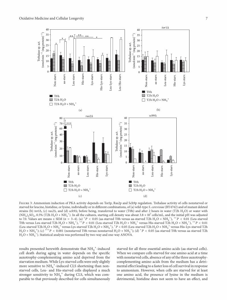
4
+ during Yeast Aging in Water. To furtherevaluate the role of PKA in NH
4
+-induced CLS shorteningand the potential effects of Tor1p, Ras2p, and Sch9p as PKAupstream regulators, we assessed PKA activation in BY4742(wild-type), 𝑡𝑜𝑟1Δ, 𝑟𝑎𝑠2Δ, and 𝑠𝑐ℎ9Δ strains starved foreach of the three essential amino acids, individually or incombination. Trehalase is a target of PKA regulation, and itsactivity has been extensively used to monitor PKA activation[22, 29]. In order to evaluate PKA activation, we thereforemeasured trehalase activity in cells grown and incubatedas described above in material and methods section. (Forschematic representation of the protocol please see FigureS1). We observed that in wild-type cells, leucine starvationresulted in the highest trehalase activity after 2 h of incubationwith NH
4
+, whereas its presence alone led to the lowesttrehalase activity. In contrast, and under the same conditions,starvation for lysine or histidine alone gave rise to thelowest trehalase activity, whereas their presence alone ledto the highest trehalase activity (Figure 3(a)). The resultsalso showed that in the presence of NH
4
+, aa-starved cellsexhibited a PKA activation pattern similar to nonstarvedcells, with values that are between those obtained for Leu-and His- or Lys-starved cells. This suggests that in aa-starved cells, the higher contribution expected from PKAactivation due to the absence of leucine is probably balancedby the decrease of PKA activity induced by the absence ofhistidine and lysine. PKA activation by NH
4
+ was decreasedin 𝑡𝑜𝑟1Δ, 𝑟𝑎𝑠2Δ, and 𝑠𝑐ℎ9Δ mutants in comparison withthe wild-type strain, both for nonstarved cells and underall amino acid starvation conditions (Figures 3(b), 3(c), and3(d)). The observed reduction in PKA activation correlateswith the decrease in NH
4
+-induced CLS shortening in the𝑡𝑜𝑟1Δ strain under all the conditions tested. In addition,the decrease in PKA activation induced by NH
4
+ in theras2Δ strain was accompanied by an increase in cell survivalfor non- or lysine-starved cells, but not for cells under theremaining starvation conditions (Leu-, His-, or aa-starvedcells). Conversely, for nonstarved cells and for cells starvedin the presence of leucine (His-starved and Lys-starved cells)before transfer to water (T0), there was a significant increasein PKA activation in the 𝑟𝑎𝑠2Δ strain, indicating that Ras2p
seems to downregulate PKA activity in the presence ofleucine. On the other hand, the decrease in PKA activationinduced by NH
4
+ in the sch9Δ strain was not associatedwith an extended CLS in water with NH
4
+ in nonstarvedcells or under any of the starvation conditions tested, whichis in accordance with previous results described for aa-starved cells [25]. Together, the results suggest that NH
4
+
induces PKA activation through Tor1p, Ras2p, and Sch9p.However, absence of Ras2p, although able to decrease PKAactivation, did not revert NH
4
+-induced CLS shorteningin Leu-, His-, and aa-starved cells, indicating that in theabsence of this protein, other pathways, independent of PKAand possibly mediated by Tor1p, are still activated and caninduce cell death. Furthermore, Ras2p, at least under someconditions, appears to also activate other pathways relevantto cell survival, since its absence leads to a shorter CLS inwater. A prosurvival role was also observed for Sch9p underall conditions, either in the absence or presence of NH
4
+.
3.4. Sch9p Protects Cells from 𝑁𝐻4
+-Induced Cell Deaththrough Hog1p Activation. Hog1p is a kinase that regulatesand is regulated by Sch9p and mediates stress responseindependently of the PKA and TOR pathways [30]. It waspreviously shown that Hog1p is involved in the resistance ofaa-starved cells to the toxic effects of NH
4
+ during CLS inwater [25]. In order to access if the protective role of Sch9pin response to NH
4
+ under the different amino acid starva-tion conditions described in the previous sections could bemediated through a Sch9p-dependent Hog1p activation, weexamined Hog1p phosphorylation during CLS in water withand without NH
4
+ in BY4742 (wild-type) and 𝑠𝑐ℎ9Δ cells. Asshown in Figure 4, Hog1p phosphorylation in wild-type cellsincreased in the presence of NH
4
+ in all starvation conditionstested (aa-, Leu-, His-, and Lys-starved cells), being higher inHis- andLys-starved cells. Deletion of SCH9 almost abolishedHog1p phosphorylation in aa-, Leu-, and His-starved cells,whereas some residual phosphorylation was still detected inLys-starved cells.The lowerHop1p phosphorylation observedfor cells starved for aa- and Leu-starved cells is in agreementwith previous results, showing that the presence of leucineis required for Sch9p phosphorylation via TORC1 [21]. Also,Hog1p phosphorylation in Lys-starved 𝑠𝑐ℎ9Δ cells is in agree-ment with the activation of pathways other than the PKA inthe absence of Ras2p, suggested by the rescue of the loss ofviability found for Lys-starved 𝑟𝑎𝑠2Δ cells (Figure 2(d)).
Taken together, results show that Sch9p is involved inHog1p activation in response to NH
4
+ under all starvationconditions, indicating that the increased resistance affordedby Sch9p could, actually, be mediated through Hog1p activa-tion.
4. Conclusions
It has been previously shown that the CLS of stationary phasecells of Saccharomyces cerevisiae BY4742 transferred to waterwas significantly shortened by the addition of NH
4
+ andthat the negative effects of NH
4
+ were particularly evidentfor cells under restriction of auxotrophy-complementingamino acid markers (leucine, lysine, and histidine) [25]. The
Oxidative Medicine and Cellular Longevity 7
0
5
10
15
20
25
30
35
40
T0 hT2 h H2OT2 h H2O + NH4
+
Treh
alas
e sp.
act.
(nm
ol m
in−1
(mg
prot
ein)
−1)
∗∗∗∗
∗∗∗∗
Non
-sta
rv.
aa-s
tarv
.
Leu-
starv
.
His-
star
v.
Lys-
star
v.
Leu-
Lys-
star
v.
His-
Lys-
star
v.
Leu-
His-
star
v.
(a)
0
5
10
15
20
25
30
35
40
Non
-sta
rv.
aa-s
tarv
.
Leu-
starv
.
His-
starv
.
Lys-
star
v.
tor1Δ
T0 hT2 h H2OT2 h H2O + NH4
+
Treh
alas
e sp.
act.
(nm
ol m
in−1
(mg
prot
ein)
−1)
(b)
0
10
20
30
40
6070
80
Non
-sta
rv.
aa-s
tarv
.
Leu-
starv
.
His-
star
v.
Lys-
star
v.
∗∗∗
ras2Δ
T0 hT2 h H2OT2 h H2O + NH4
+
Treh
alas
e sp.
act.
(nm
ol m
in−1
(mg
prot
ein)
−1)
(c)
T0 hT2 h H2OT2 h H2O + NH4
+
0
5
10
15
20
25
30
35
40Tr
ehal
ase s
p. ac
t.(n
mol
min−1
(mg
prot
ein)
−1)
Non
-sta
rv.
aa-s
tarv
.
Leu-
starv
.
His-
starv
.
Lys-
star
v.
∗
sch9Δ
(d)
Figure 3: Ammonium induction of PKA activity depends on Tor1p, Ras2p and Sch9p regulation. Trehalase activity of cells nonstarved orstarved for leucine, histidine, or lysine, individually or in different combinations, of (a) wild-type S. cerevisiae (BY4742) and of mutant deletedstrains (b) tor1Δ, (c) ras2Δ, and (d) sch9Δ; before being, transferred to water (T0h) and after 2 hours in water (T2h H
2O) or water with
(NH4)2SO4, 0.5% (T2h H
2O + NH
4
+). In all the cultures, starting cell density was about 3.8 × 10⁷ cells/mL, and the initial pH was adjustedto 7.0. Values are means ± SEM (𝑛 = 3–4). (a) ∗𝑃 < 0.05 (aa-starved T0h versus aa-starved T2h H
2O + NH
4
+), ∗∗𝑃 < 0.01 (Leu-starvedT0h versus Leu-starved T2h H
2O + NH
4
+), ∗∗𝑃 < 0.01 (Leu-starved T2h H2O + NH
4
+ versus His-starved T2h H2O + NH
4
+), ∗∗𝑃 < 0.01(Leu-starved T2h H
2O +NH
4
+ versus Lys-starved T2h H2O +NH
4
+), ∗𝑃 < 0.05 (Leu-starved T2h H2O +NH
4
+ versus His-Lys-starved T2hH2O + NH
4
+); (c) ∗∗∗𝑃 < 0.001 (nonstarved T0h versus nonstarved H2O + NH
4
+); (d) ∗𝑃 < 0.05 (aa-starved T0h versus aa-starved T2hH2O + NH
4
+). Statistical analysis was performed by two-way and one-way ANOVA.
results presented herewith demonstrate that NH4
+-inducedcell death during aging in water depends on the specificauxotrophy-complementing amino acid deprived from thestarvationmedium.While Lys-starved cells were only slightlymore sensitive to NH
4
+-induced CLS shortening than non-starved cells, Leu- and His-starved cells displayed a muchstronger sensitivity to NH
4
+ during CLS, which was com-parable to that previously described for cells simultaneously
starved for all three essential amino acids (aa-starved cells).When we compare cells starved for one amino acid at a timewith nonstarved cells, absence of any of the three auxotrophy-complementing amino acids from the medium has a detri-mental effect leading to a faster loss of cell survival in responseto ammonium. However, when cells are starved for at leastone amino acid, the presence of lysine in the medium isdetrimental, histidine does not seem to have an effect, and

8 Oxidative Medicine and Cellular Longevity
aa-starvation Leu-starvation
WT WT
C NaCl T0 T0H2O NH4+ H2O NH4
+ T0 H2O NH4+ T0 H2O NH4
+
Pi-Hog1p
Hog1p
Pgk1p
sch9Δ sch9Δ
(a)
His-starvation Lys-starvation
WT WT
C NaCl T0 T0H2O NH4+ H2O NH4
+ T0 H2O NH4+ T0 H2O NH4
+
Pi-Hog1p
Hog1p
Pgk1p
sch9Δ sch9Δ
(b)
Figure 4: Ammonium induces Sch9p-dependent Hog1p phosphorylation in starvation conditions. Westernblot analysis of Pi-Hog1p levelspresent in S. cerevisiae (BY4742) wild-type (WT) and sch9Δ cells starved for (a) all three amino acids or leucine and (b) starved for histidineor lysine, before (T0) and after 20 minutes upon transfer to water (H
2O) or water with (NH
4)2SO4, 0.5% (NH
4
+). In all the cultures, startingcell density was about 3.8 × 10⁷ cells/mL, and the initial pH was adjusted to 7.0. Control cells were grown on YPD medium (Control-C) andincubated for 5 minutes in YPD medium supplemented with 1M NaCl.
leucine has a protective effect on ammonium-induced CLSshortening. The results regarding leucine are in accordancewith the literature since it has been described that leucineplays a more important role in CLS extension in auxotrophicstrains [6, 10]. In a recent study, supplementation of extraleucine to SC medium or transformation of auxotrophicleucine strain into a prototrophic leucine strain resulted inCLS extension. The importance of leucine was attributedto the regulation of the branched side chain amino acidssynthesis that appears to be misregulated in a leu2Δ strain.In agreement, supplemental levels of the branch side aminoacids isoleucine, threonine, and valine also extended CLS ina leu2Δ strain [6]. The negative effect observed for lysinein cell survival during ammonium-induced cell death canpossibly be due to the fact that autophagy is inhibited inthe presence of ammonium [25], and the lack of autophagymight be responsible for this effect since lysine seems to actin an autophagy-dependentmanner on the regulation of CLS.Autophagy-deficient strains showed no improvement in CLSextension after regaining LYS prototrophy in contrast to wild-type autophagy competent cells that increased CLS extensionwith LYS prototrophy [6].
Both Ras2p and Tor1p are involved in NH4
+-inducedCLS shortening in aa-starved cells [25]. We now further
established that Ras2p involvement on NH4
+-induced CLSshortening was present under all conditions tested, and didnot depend on starvation. In turn, Tor1p function in thedecrease of CLS by NH
4
+ was relevant only under amino acidstarvation, being differently modulated by the specific aminoacid deprived from the medium. Starvation for leucine andhistidine, which induced a strong shortening of CLS in thepresence of NH
4
+, had a high impact in the regulation ofTor1p function, whereas starvation for lysine, whichwas asso-ciated with only a small NH
4
+-induced CLS shortening, hada considerably less significant impact on Tor1p regulation.These results are in agreement with previous results showingthat leucine has an important impact in the regulation ofTORC1 [20, 21]. Our results suggest that the presence ofNH
4
+
in the medium (commonly present as the nitrogen source)may be at least partly responsible for the reported decrease inCLS in leucine-starved cells [6, 14].
PKA activation has been described to be associatedwith the NH
4
+-induced CLS shortening of aa-starved cellsin water [25]. From the results now obtained, and whenwe compare values from nonstarved and starved wild-type cells, it appears that leucine starvation (alone or incombination with starvation for another amino acid) is themain factor responsible for PKA activation in response to

Oxidative Medicine and Cellular Longevity 9
NH4
+, correlating with its stronger effect on CLS shortening.This activation is dependent on Ras2p, Tor1p, and Sch9p,as deficiency in any of these proteins leads to its decrease.However, since the decrease in PKA activation resulted indistinct cell fate outcomes in the differentmutants, the resultssuggest that these proteins activate PKA by independentpathways and/or also regulate other pathways that they donot share and that have different impacts on NH
4
+-inducedCLS shortening. Also, we cannot exclude the possibility thatthe observed effects on trehalase activity may result from apotential effect of Sch9p, Ras2p, or Tor1p on the activity ofother proteins also involved in trehalase regulation such asBmh1/2p or Dcs1p [29].
Opposite to our results, Sch9p has been described toinhibit PKA activity when glucose is added to glycerol-growncells. However, these authors observed that the inhibitionwas mediated through the regulation of Tpk2p localization[31], an isoform that does not seem to have a relevant rolein response to ammonium under our conditions. In fact,we have previously observed that Tpk1p is the main PKAisoform involved in ammonium effects [25]. In addition to itsinvolvement in PKA activation, Sch9p also increases Hog1pphosphorylation, extending CLS in water with or withoutNH4
+.In summary, herewith we show that the toxic effects
of NH4
+ on CLS shortening are regulated by a starvation-dependent and a starvation-independent component and aremediated essentially by Tor1p in the first case and by Ras2pin the second. We also provide evidence that when cellsare starved for amino acids, the presence of leucine canameliorate NH
4
+-induced CLS shortening, while lysine hasthe opposite effect, and the presence of histidine has no effect.Together, our data add new knowledge on CLS regulation,indicating that the modulation of nitrogen sources suppliedto cells can drastically modulate CLS and providing newclues for the development of environmental interventionsfor chronological life span extension. Additionally, and sinceNH4
+-induced cell death is involved in different humandisorders that are accompanied by hyperammonemia [32],our results, showing that NH
4
+ toxicity can be modulated byamino acids through different pathways, may also afford newinsights into the understanding of the cell molecular basestriggering cell death in such pathologies.
Authors’ Contribution
Maria Joao Sousa and Cecılia Leao contributed equally to thiswork.
Acknowledgment
This work was supported by FCT, Portugal, Grant PTDC/AGR-ALI/102608/2008.
References
[1] J. Santos, C. Leao, and M. J. Sousa, “Growth culture condi-tions and nutrient signaling modulating yeast chronological
longevity,”Oxidative Medicine and Cellular Longevity, vol. 2012,Article ID 680304, 10 pages, 2012.
[2] J. V. Gray, G. A. Petsko, G. C. Johnston, D. Ringe, R. A. Singer,and M. Werner-Washburne, “Sleeping beauty’: quiescence inSaccharomyces cerevisiae,” Microbiology and Molecular BiologyReviews, vol. 68, no. 2, pp. 187–206, 2004.
[3] M. Rubio-Texeira, G. Van Zeebroeck, K. Voordeckers, andJ. M. Thevelein, “Saccharomyces cerevisiae plasma membranenutrient sensors and their role in PKA signaling,” FEMS YeastResearch, vol. 10, no. 2, pp. 134–149, 2010.
[4] V. D. Longo, E. B. Gralla, and J. S. Valentine, “Superoxidedismutase activity is essential for stationary phase survival inSaccharomyces cerevisiae: mitochondrial production of toxicoxygen species in vivo,” Journal of Biological Chemistry, vol. 271,no. 21, pp. 12275–12280, 1996.
[5] P. Fabrizio and V. D. Longo, “The chronological life span ofSaccharomyces cerevisiae,” Aging Cell, vol. 2, no. 2, pp. 73–81,2003.
[6] A. L. Alvers, L. K. Fishwick, M. S. Wood et al., “Autophagyand amino acid homeostasis are required for chronologicallongevity in Saccharomyces cerevisiae,” Aging Cell, vol. 8, no. 4,pp. 353–369, 2009.
[7] P. Gomes, B. Sampaio-Marques, P. Ludovico, F. Rodrigues, andC. Leao, “Low auxotrophy-complementing amino acid concen-trations reduce yeast chronological life span,” Mechanisms ofAgeing and Development, vol. 128, no. 5-6, pp. 383–391, 2007.
[8] C. J. Murakami, C. R. Burtner, B. K. Kennedy, and M. Kae-berlein, “Amethod for high-throughput quantitative analysis ofyeast chronological life span,” Journals of Gerontology A, vol. 63,no. 2, pp. 113–121, 2008.
[9] D. L. Smith Jr., J. M. McClure, M. Matecic, and J. S. Smith,“Calorie restriction extends the chronological lifespan of Sac-charomyces cerevisiae independently of the Sirtuins,”Aging Cell,vol. 6, no. 5, pp. 649–662, 2007.
[10] V. M. Boer, S. Amini, and D. Botstein, “Influence of genotypeand nutrition on survival and metabolism of starving yeast,”Proceedings of the National Academy of Sciences of the UnitedStates of America, vol. 105, no. 19, pp. 6930–6935, 2008.
[11] J. C. Jiang, E. Jaruga, M. V. Repnevskaya, and S. M. Jazwinski,“An intervention resembling caloric restriction prolongs lifespan and retards aging in yeast,” FASEB Journal, vol. 14, no. 14,pp. 2135–2137, 2000.
[12] S.-J. Lin, P.-A. Defossez, and L. Guarente, “Requirement ofNAD and SIR2 for life-span extension by calorie restriction inSaccharomyces cerevisiae,” Science, vol. 289, no. 5487, pp. 2126–2128, 2000.
[13] R. W. Powers III, M. Kaeberlein, S. D. Caldwell, B. K. Kennedy,and S. Fields, “Extension of chronological life span in yeast bydecreased TOR pathway signaling,” Genes and Development,vol. 20, no. 2, pp. 174–184, 2006.
[14] J. P. Aris, A. L. Alvers, R. A. Ferraiuolo et al., “Autophagyand leucine promote chronological longevity and respirationproficiency during calorie restriction in yeast,” ExperimentalGerontology, 2013.
[15] R. Loewith and M. N. Hall, “Target of rapamycin (TOR) innutrient signaling and growth control,” Genetics, vol. 189, no. 4,pp. 1177–1201, 2011.
[16] B. Smets, R. Ghillebert, P. De Snijder et al., “Life in the midstof scarcity: adaptations to nutrient availability in Saccharomycescerevisiae,” Current Genetics, vol. 56, no. 1, pp. 1–32, 2010.

10 Oxidative Medicine and Cellular Longevity
[17] J. L. Crespo and M. N. Hall, “Elucidating TOR signalingand rapamycin action: lessons from Saccharomyces cerevisiae,”Microbiology and Molecular Biology Reviews, vol. 66, no. 4, pp.579–591, 2002.
[18] C. De Virgilio and R. Loewith, “The TOR signalling networkfrom yeast to man,” International Journal of Biochemistry andCell Biology, vol. 38, no. 9, pp. 1476–1481, 2006.
[19] J. Avruch, X. Long, S. Ortiz-Vega, J. Rapley, A. Papageorgiou,and N. Dai, “Amino acid regulation of TOR complex 1,”American Journal of Physiology. Endocrinology and Metabolism,vol. 296, no. 4, pp. E592–E602, 2009.
[20] M. Binda, M.-P. Peli-Gulli, G. Bonfils et al., “The Vam6 GEFcontrols TORC1 by activating the EGO complex,” MolecularCell, vol. 35, no. 5, pp. 563–573, 2009.
[21] G. Bonfils, M. Jaquenoud, S. Bontron, C. Ostrowicz, C. Unger-mann, and C. De Virgilio, “Leucyl-tRNA synthetase controlsTORC1 via the EGO complex,”Molecular Cell, vol. 46, no. 1, pp.105–110, 2012.
[22] J. M. Thevelein, L. Cauwenberg, S. Colombo et al., “Nutrient-induced signal transduction through the protein kinase A path-way and its role in the control of metabolism, stress resistance,and growth in yeast,” Enzyme andMicrobial Technology, vol. 26,no. 9-10, pp. 819–825, 2000.
[23] B. Smets, P. De Snijder, K. Engelen et al., “Genome-wide expres-sion analysis reveals TORC1-dependent and -independentfunctions of Sch9,” FEMS Yeast Research, vol. 8, no. 8, pp. 1276–1288, 2008.
[24] M. Crauwels, M. C. V. Donaton, M. B. Pernambuco, J. Wind-erickx, J. H. De Winde, and J. M. Thevelein, “The Sch9protein kinase in the yeast Saccharomyces cerevisiae con-trols cAPK activity and is required for nitrogen activationof the fermentable-growth-medium-induced (FGM) pathway,”Microbiology, vol. 143, no. 8, pp. 2627–2637, 1997.
[25] J. Santos, M. J. Sousa, and C. Leao, “Ammonium is toxicfor aging yeast cells, inducing death and shortening of thechronological lifespan,” PLoS One, vol. 7, Article ID e37090,2012.
[26] O. Braissant, “Current concepts in the pathogenesis of ureacycle disorders,” Molecular Genetics and Metabolism, vol. 100,supplement 1, pp. S3–S12, 2010.
[27] M. B. Pernambuco, J. Winderickx, M. Crauwels, G. Griffioen,W. H.Mager, and J. M.Thevelein, “Glucose-triggered signallingin Saccharomyces cerevisiae: different requirements for sugarphosphorylation between cells grown on glucose and thosegrown on non-fermentable carbon sources,” Microbiology, vol.142, part 7, pp. 1775–1782, 1996.
[28] N. Camougrand, I. Kissova, B. Salin, and R. J. Devenish, “Mon-itoringmitophagy in yeast,”Methods in enzymology, vol. 451, pp.89–107, 2008.
[29] W. Schepers, G. Van Zeebroeck, M. Pinkse, P. Verhaert, andJ. M. Thevelein, “In vivo phosphorylation of Ser21 and Ser83during nutrient-induced activation of the yeast protein kinaseA, (PKA) target trehalase,” The Journal of Biological Chemistry,vol. 287, pp. 44130–44142, 2012.
[30] A. Pascual-Ahuir andM. Proft, “TheSch9 kinase is a chromatin-associated transcriptional activator of osmostress-responsivegenes,” EMBO Journal, vol. 26, no. 13, pp. 3098–3108, 2007.
[31] A. Zhang, Y. Shen, W. Gao, and J. Dong, “Role of Sch9 in reg-ulating Ras-cAMP signal pathway in Saccharomyces cerevisiae,”FEBS Letters, vol. 585, no. 19, pp. 3026–3032, 2011.
[32] M. D. Norenberg, K. V. R. Rao, and A. R. Jayakumar, “Signalingfactors in the mechanism of ammonia neurotoxicity,”MetabolicBrain Disease, vol. 24, no. 1, pp. 103–117, 2009.

Submit your manuscripts athttp://www.hindawi.com
Hindawi Publishing Corporationhttp://www.hindawi.com Volume 2013
Oxidative Medicine and Cellular Longevity
Hindawi Publishing Corporation http://www.hindawi.com Volume 2013Hindawi Publishing Corporation http://www.hindawi.com Volume 2013
The Scientific World Journal
International Journal of
EndocrinologyHindawi Publishing Corporationhttp://www.hindawi.com
Volume 2013
ISRN Anesthesiology
Hindawi Publishing Corporationhttp://www.hindawi.com Volume 2013
Hindawi Publishing Corporationhttp://www.hindawi.com
OncologyJournal of
Volume 2013
PPARRe sea rch
Hindawi Publishing Corporationhttp://www.hindawi.com Volume 2013
OphthalmologyJournal of
Hindawi Publishing Corporationhttp://www.hindawi.com Volume 2013
ISRN Allergy
Hindawi Publishing Corporationhttp://www.hindawi.com Volume 2013
BioMed Research International
Hindawi Publishing Corporationhttp://www.hindawi.com Volume 2013
Hindawi Publishing Corporationhttp://www.hindawi.com Volume 2013
ObesityJournal of
ISRN Addiction
Hindawi Publishing Corporationhttp://www.hindawi.com Volume 2013
Hindawi Publishing Corporationhttp://www.hindawi.com Volume 2013
Computational and Mathematical Methods in Medicine
ISRN AIDS
Hindawi Publishing Corporationhttp://www.hindawi.com Volume 2013
Clinical &DevelopmentalImmunology
Hindawi Publishing Corporationhttp://www.hindawi.com
Volume 2013
Diabetes ResearchJournal of
Hindawi Publishing Corporationhttp://www.hindawi.com Volume 2013
Evidence-Based Complementary and Alternative Medicine
Volume 2013Hindawi Publishing Corporationhttp://www.hindawi.com
Hindawi Publishing Corporationhttp://www.hindawi.com Volume 2013
Gastroenterology Research and Practice
Hindawi Publishing Corporationhttp://www.hindawi.com Volume 2013
ISRN Biomarkers
Hindawi Publishing Corporationhttp://www.hindawi.com Volume 2013
MEDIATORSINFLAMMATION
of