Amersham Typhoon Biomolecular Imager - Cytiva
5
Combine 4 instruments into 1 without compromising sensitivity and dynamic range Amersham Typhoon Biomolecular Imager
Transcript of Amersham Typhoon Biomolecular Imager - Cytiva

Combine 4 instruments into 1 without compromising sensitivity and dynamic range
Amersham
Typhoon Biomolecular Imager

Phosphor Imaging
Densitometry (OD)
RGB Fluorescence
IR1 & IR2 Fluorescence
Amersham Typhoon RGB
º º º
Amersham Typhoon RGB
º
Amersham Typhoon 5
Three preconfigured models available:
Phosphorylase B
2.8 1.4 0.7 0.36 0.18 0.09 0.045 0.02 0.01 0.006 (ng)
Sample Phosphorylase B in LMW markerGel Amersham WB 8 -18% SDS -PAGEImaging Excitation Emission filter
635 nm Cy5 670BP30LOD 5.6 pgDynamic range 4.8 orders of magnitudeLinearity R2=0.9997 and k=1.01
(trendline in log -log plot)
2
3
4
5
6
7
8
0 1 2 3 4 5 6
log
(inte
grat
ed in
tens
ity)
log (pg phosphorylase B)
Fig. X Phosphorylase B was labeled with CyDye DIGE fluor Cy5 minimal dye and separated using the Amersham WB electrophoresis gel. The gel was imaged with Amersham Typhoon using normal scan speed. A selection of a dilution series is shown in the image, the arrow indicates the limit of detection (LOD ). The detection limit was 5.6 pg and the linear dynamic range (DR) was 4.8 orders of magnitude.
Fig 1. Phosphorylase B was labeled with CyDye™ DIGE fluor Cy5 minimal dye and separated using the Amersham WB electrophoresis gel. The gel was imaged with Amersham Typhoon using normal scan speed. The detection limit was 5.6 pg and the linear dynamic range (DR) was 4.8 orders of magnitude.
Cy3 Figure
Phosphorylase B
2.8 1.4 0.7 0.36 0.18 0.09 0.045 0.02 (ng)
Sample Phosphorylase B in LMW markerGel Amersham WB 8 -18% SDS -PAGEImaging Excitation Emission filter
532 nm Cy3 570BP20LOD 22 pgDynamic range 4.2 orders of magnitudeLinearity R2=0.9998 and k=1.02
(trendline in log -log plot)
Fig. X Phosphorylase B was labeled with CyDye DIGE fluor Cy3 minimal dye and separated using the Amersham WB electrophoresis gel. The gel was imaged with Amersham Typhoon using normal scan speed. A selection of a dilution series is shown in the image, the arrow indicates the limit of detection (LOD ). The detection limit was 22 pg and the linear dynamic range (DR) was 4.2 orders of magnitude.
2
3
4
5
6
7
8
1 2 3 4 5 6
log
(inte
grat
ed in
tens
ity)
log (pg phosphorylase B)
Fig 2. Phosphorylase B was labeled with CyDye DIGE fluor Cy3 minimal dye and separated using the Amersham WB electrophoresis gel. The gel was imaged with Amersham Typhoon using normal scan speed. The detection limit was 22 pg and the linear dynamic range (DR) was 4.2 orders of magnitude.
Cy™ 5 Cy3
log (pg phosphorylase B) log (pg phosphorylase B)
Detect low levels of proteinsThe ability to distinguish subtle differences in expression among low amounts of biomolecules in complex mixtures adds power to many areas of biological research. The highly versatile Amersham™ Typhoon™ laser scanner enables you to generate data of the highest quality through linearity of signal response, quantitative accuracy, and extremely low limits of detection. The completely customizable Typhoon supports phosphorimaging, 2-D DIGE imaging, red/green/blue (RGB) and near IR fluorescence as well as sensitive and accurate quantitation of proteins.
Enhance your sensitivity• Improved PMTs with 5 Laser Configuration Option
• Multiplex detection of proteins with minimal crosstalk
Enhance your dynamic range• Broad linear dynamic range greater than 5 orders of magnitude
2

4 Instruments into 1The Amersham Typhoon is an upgradeable laser scanner, completely customizable for your specific research needs. The system combines 4 instruments into 1, which allows for several imaging modes at any time, including near infrared (NIR) fluorescence, RGB fluorescence, Phosphor imaging, and densitometry of colorimetrically-stained samples (e.g., Coomassie™ blue and silver stain).
Phosphor imaging
Fig. X Scanned image of a 14C autoradiographic standard using the Amersham Typhoon. A selection of the standard is shown in the image, the arrow indicates the limit of detection (LOD). The linear dynamic range (DR) was 5.3 orders of magnitude.
Sample 14C autoradiographic standard ( CFQ12000)3 hour exposure to BAS -SR Imaging Plate
Imaging Excitation Emission filter532 nm IP BP390
LOD 0.00518 µCi/gDynamic range 5.3 orders of magnitudeLinearity R2=0.9998 and k=1.03
(trendline in log -log plot)
-2
-1
0
1
2
3
4
5
0 1 2 3 4 5 6 7
log
(si
gn
al in
ten
sity
)
log (nCi/g)
59.3 50.9 41.3 31.0 20.7 10.1 5.18 nCi/gFig 6. Scanned image of a 14C autoradiographic standard using Amersham Typhoon. The linear dynamic range (DR) was 5.3 orders of magnitude.
Fig 5. Autoradiography images of rat injected with 14C glucose. The magnified area shows part of the spine.
NIR 680 Figure
Antibody heavy chain
Sample IRDye® 680 goat anti -rabbit antibodyGel Amersham WB 13.5% SDS -PAGEImaging Excitation Emission filter
685 nm 720BP20 (IRshort)LOD 3 pgDynamic range 5.2 orders of magnitudeLinearity R2=0.9988 and k=1.00
(trendline in log -log plot)
Fig. X Antibody conjugated with IRDye 680 was separated using the Amersham WB electrophoresis gel. To reduce noise, the gel was imaged with Amersham Typhoon using slow scan speed. A selection of a dilution series is shown in the image, the arrow indicates the limit of detection (LOD). The detection limit was 3 pg and the linear dynamic range (DR) was 5.2 orders of magnitude.
386 192 96 48 24 12 6 3 (pg)
2
3
4
5
6
7
8
0 1 2 3 4 5 6
log
(inte
grat
ed in
tens
ity)
log (pg antibody)
Fig 3. Antibody conjugated with IRDye™ 680 was separated using the Amersham WB electrophoresis gel. To reduce noise, the gel was imaged with Amersham Typhoon using slow scan speed. The detection limit was 3 pg and the linear dynamic range (DR) was 5.2 orders of magnitude.
NIR 800 Figure
Antibody heavy chain
Sample IRDye® 800 goat anti -rabbit antibodyGel Amersham WB 13.5% SDS -PAGEImaging Excitation Emission filter
785 nm 825BP30 (IRlong)LOD 3 pgDynamic range 5.2 orders of magnitudeLinearity R2=0.9988 and k=1.00
(trendline in log -log plot)
Fig. X Antibody conjugated with IRDye 800 was separated using the Amersham WB electrophoresis gel. To reduce noise, the gel was imaged with Amersham Typhoon using slow scan speed. A selection of a dilution series is shown in the image, the arrow indicates the limit of detection (LOD ). The detection limit was 3 pg and the linear dynamic range (DR) was 5.2 orders of magnitude.
386 192 96 48 24 12 6 3 (pg)
2
3
4
5
6
7
8
0 1 2 3 4 5 6
log
(inte
grat
ed in
tens
ity)
log (pg antibody)
Fig 4. Antibody conjugated with IRDye™ 800 was separated using the Amersham WB electrophoresis gel. To reduce noise, the gel was imaged with Amersham Typhoon using slow scan speed. The detection limit was 3 pg and the linear dynamic range (DR) was 5.2 orders of magnitude.
Enhance your flexibility• Select the lasers you need for blue, green, red, or NIR detection
• Phosphorimaging for radiolabeled target detection and enhanced sensitivity
NIR 680
Phosphor imagingNIR 800
log (pg antibody)
log (pg antibody) log (nCi/g)
3
System Quantity Product code
Amersham Typhoon 5 1 29187191
Amersham Typhoon RGB 1 29187193
Amersham Typhoon IP 1 29187194
Amersham Eraser 1 29187190
Accessory Cabinet AmTyphoon 1 29191637
Ordering information
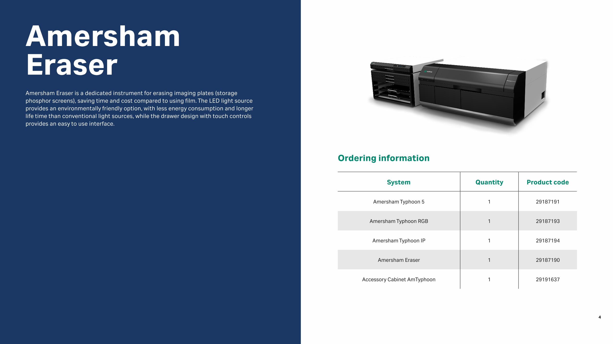
Amersham EraserAmersham Eraser is a dedicated instrument for erasing imaging plates (storage phosphor screens), saving time and cost compared to using film. The LED light source provides an environmentally friendly option, with less energy consumption and longer life time than conventional light sources, while the drawer design with touch controls provides an easy to use interface.
4

cytiva.com
Cytiva and the Drop logo are trademarks of Global Life Sciences IP Holdco LLC or an affiliate. Amersham, Cy, CyDye, and Typhoon of Global Life Sciences Solutions USA LLC or an affiliate doing business as Cytiva.
IRDye is a tradmark of LI-Cor Biosciences, Inc. Coomassie is a trademark of Thermo Fisher Scientific, Inc. All other third-party trademarks are the property of their respective owners
©2020 Cytiva
For local office contact information, visit cytiva.com/contact
CY12888-14Jul20